DOCT OR AL DISSERT A TION IN ODONT OL OG Y S AS AN N AR A GHI MALMÖ UNIVERSIT S TABILIT Y OF ALIGNED MAXILL AR Y ANTERIOR TEETH AFTER ORTHODONTIC TREA TMENT
SASAN NARAGHI
STABILITY OF ALIGNED
MAXILLARY ANTERIOR
TEETH AFTER ORTHODONTIC
TREATMENT
Amount of changes with different retention methods
and without retention
S T A B I L I T Y O F A L I G N E D M A X I L L A R Y A N T E R I O R T E E T H A F T E R O R T H O D O N T I C T R E A T M E N T
© Copyright Sasan Naraghi 2021 Photo and illustration Sasan Naraghi ISBN 978-91-7877-151-6 (print) ISBN 978-91-7877-152-3 (pdf) DOI 10.24834/isbn.9789178771523 Tryck: Holmbergs, Malmö 2021
Malmö University
SASAN NARAGHI
STABILITY OF ALIGNED
MAXILLARY ANTERIOR
TEETH AFTER
ORTHODONTIC TREATMENT
Amount of changes with different retention methods
and without retention
Malmö University
Faculty of Odontology
This publication is also available in electronic format at: http://mau.diva-portal.org/
“Retention is one of the most difficult problems in orthodontia; in fact, it is the problem.”
CONTENTS
PREFACE ... 13 ABSTRACT ... 14 POPULÄRVETENSKAPLIG SAMMANFATTNING ... 17 THESIS AT A GLANCE ... 19 INTRODUCTION ... 20 Background ...20Factors influencing stability and relapse ...21
Dental arch changes in untreated subjects ...21
Dental arch changes in treated subjects ...22
Occlusion ...24
Well-aligned teeth ...24
Contact point displacement ...24
Effect of rotations on contact point displacement ...25
Tooth shape ...26
Root position ...27
The alveolar bone ...27
Gingival and periodontal fibres ...27
Muscles and soft tissue ...29
Common retention devices in the maxillary arch ...30
Adherence ...33 Significance ...34 AIMS ... 35 Paper I ...35 Paper II ...35 Paper III ...35
HYPOTHESES ... 36
Paper I ...36
Paper II ...36
Paper III ...36
Paper IV ...36
MATERIALS AND METHODS ... 37
Paper I and II ...37
Paper III ...38
Paper IV ...40
Ethical considerations ...43
STATISTICAL ANALYSIS ... 44
Sample size calculation ...44
Group comparisons ...44
Measurement error analysis ...45
Paper I ...45 Paper II ...45 Paper III ...45 Paper IV ...46 RESULTS ... 47 Paper I ...47 Paper II ...48 Paper III ...49 Paper IV ...53 Additional follow-up ...56 DISCUSSION ... 58 Paper I ...58 Paper II ...59 Paper III ...60 Paper IV ...61 Additional follow-up ...62 Limitations ...65 Strengths ...66 Generalizability ...66 Future research...66
CONCLUSIONS ... 68 Paper I and II ...68 Paper III ...68 Paper IV ...69 Clinical implications ...69 ACKNOWLEDGEMENTS ... 70 REFERENCES ... 72
ABBREVIATIONS AND DEFINITIONS ... 82
PREFACE
This thesis is based on the following four papers, which are referred to as Paper I to IV.
Paper I. Naraghi S, Andrén A, Kjellberg H, Mohlin B. Relapse tendency after orthodontic correction of upper front teeth retained with a bonded retainer. The Angle Orthodontist, 2005; 76:570–576.
Paper II. Andrén A, Naraghi S, Mohlin B, Kjellberg H. Pattern and amount of change after orthodontic correction of upper front teeth seven years post-retention. The Angle Orthodontist, 2010; 80:620–625.
Paper III. Naraghi S, Ganzer S, Bondemark L, Sonesson M. Stability of maxillary anterior teeth after two years of retention in adolescents - A randomised controlled trial comparing two bonded and a vacuum-formed retainer. European Journal of Orthodontics. Accepted for publication No-vember 2020.
Paper IV. Naraghi S, Ganzer S, Bondemark L, Sonesson M. Compari-son of post-treatment changes with and without retention in adolescents treated for maxillary impacted canines—A randomised controlled trial. E-published ahead to print March 5, 2020, European Journal of Ortho-dontics, cjaa010.
These papers are reprinted with kind permission from the copyright hol-ders.
ABSTRACT
Well-aligned anterior teeth are the major reason for the patients seeking orthodontic treatment, and keeping teeth aligned and stable afterwards is a goal for the orthodontist and the patient. Relapse after treatment is a common problem, and it is defined as when teeth go back to their pre-vious positions. Removable or fixed retainers have been used to avoid relapse after treatment. It has been common practice to use removable retention to retain anterior teeth in the maxilla. However, in recent deca-des, it has become increasingly common to retain with bonded retainers. Almost all previous studies on retention devices in the maxilla were based on removable retainers. Consequently, there existed knowledge gaps and lack of short-term and long-term studies on the capability to maintain the stability of the maxillary anterior teeth with bonded retainers. Hence, the reason for the papers in this study. In addition, it is not known if retention is needed in all orthodontic patients or if there are patients, based on their initial malocclusion and individual variations, who may not need reten-tion after treatment.
The research questions addressed in this thesis thus originate from know-ledge gaps and clinical needs concerning retention strategies after ortho-dontic treatments. To provide strong clinical evidence, randomised con-trolled trials (RCT) as well as intention to treat (ITT) methodology has been assessed. The results are expected to be beneficial for the patients who will be offered the most effective retention strategy for maxillary anterior teeth based on patients’ preferences.
In Paper I and II, 45 and 27 adolescents’ patients were collected from the Orthodontic Clinic in Mariestad, Sweden. At the time when Paper I and II were conducted, there were no studies that had evaluated the long-term effect of bonded retainers in the maxilla. In two RCTs, Paper III and IV, 90 and 63 adolescents’ patients were collected from the Orthodontic Clinic in Växjö, Region Kronoberg, Sweden.
Paper I: The aim was to investigate the amount and pattern of relapse of
maxillary anterior teeth previously retained with a bonded retainer.
Paper II: The aim was to investigate the amount and pattern of changes
of maxillary anterior teeth seven years post-retention, which previously were retained with a bonded retainer.
Paper III: The aim was to evaluate post-treatment changes in the
irre-gularity of the maxillary six anterior teeth and single tooth Contact Point Discrepancy (CPD) of three different retention methods.
Paper IV: The aim was to evaluate whether retention is needed after
ort-hodontic treatment for impacted maxillary canines and with moderate pre-treatment irregularity in the maxilla.
Key findings in Paper I
• The contact relationship between the laterals and the centrals is the most unstable contact. Canines are the most stable teeth.
• There was no difference in the relapse pattern between rotational displacements and labiolingual displacement.
Key findings in Paper II
• There was a strong correlation between irregularity at one- and seven-years post-retention. Stable cases one-year post-retention were stable and unstable cases deteriorated with time.
Key findings in Paper III
• All three retention methods showed equally effective retention ca-pacity and all the changes found in the three groups were small and considered clinically insignificant. Thus, the null hypothesis was confirmed. All three methods can be recommended.
Key findings in Paper IV
• Changes between the retention and the non-retention group were statistically but not clinically significant. Since satisfactory clinical results one-year post-treatment were found in the non-retention group, retention does not appear always to be needed.
• Most of the changes occur within the first 10-week period after treatment with no retention.
Key conclusions and clinical implications
Both removable and bonded retainers are effective for holding teeth in position and can be used for preventing the relapse. It can be enough to retain with bonded retainer 12-22 instead of 13-23. It might be possible to avoid retention in selected cases in the short-term, but a longer eva-luation period is needed.
POPULÄRVETENSKAPLIG
SAMMANFATTNING
Efter en tandregleringsbehandling måste tänderna fixeras för att de inte ska gå tillbaka mot sina ursprungspositioner, det vill säga tänderna har gått i recidiv. För att undvika recidiv efter ortodontisk behandling an-vänds avtagbara plastskenor eller fastlimmade metalltrådar, så kallade retentionstrådar, som fästs på insidan av de sex framtänderna. Retention används från minst ett år till obegränsad tid.
Syftet med studie I och II var att undersöka storleken och mönstret på reci-divet efter ortodontisk korrektion av överkäkens framtänder som har varit fixerade under ca tre år med en retentionstråd. Förändringar på kort och långsikt studerades. Studien gjordes på avgjutningar av patienters tänder som framställts i gips (studiemodeller). Kontaktpunktsförskjutningarna mellan de sex framtänderna mättes med hjälp av ett digitalt skjutmått. Modellerna skannades och eventuella rotationer av överkäkständerna från hörntand till hörntand analyserades. Alla värden jämfördes med si-tuationen före behandling, direkt efter behandling samt ett år respektive sju år efter det att retentionstråden togs bort.
Syftet med studie III var att analysera tre metoder för att retinera och stabilisera överkäksframtänderna efter tandregleringsbehandling. Jämfö-relser gjordes mellan retention under två år med en limmad tråd på de fyra framtänderna (12–22), en limmad tråd på sex tänder (13–23, de fyra framtänderna och de två hörntänderna) och en avtagbar nattandställning som omfattade samtliga tänder i överkäken.
Syftet med studie IV var att utvärdera om det var möjligt att undvika retention i fall där överkäkshörntänderna blivit kvar i käken och måste dras fram med fast tandställning. Patienterna lottades i två grupper. I den ena gruppen retinerades tänderna och i den andra gruppen var de utan retention.
Utvärdering av studie III och IV gjordes också på gipsmodeller. Model-lerna 3D-skannades och mätningar av kontaktpunktsförskjutningarna mellan de sex framtänderna genomfördes med hjälp av ett datorprogram. Rotationer av överkäkständerna från hörntand till hörntand mättes mot en referenslinje.
Studie I visade små men signifikanta förändringar mellan tandkontak-terna ett år efter att retentionen togs bort. Särskilt förändringsbenägna var laterala incisiver, därefter centrala incisiver och minst förändringsbe-nägna var hörntänder.
Studie II visade ett starkt samband mellan förändringar ett och sju år efter retentionen. De tandkontakter som redan hade börjat recidivera efter ett år, förvärrades ytterligare vid långtidsuppföljningen efter sju år.
Studie III visade att alla tre former av retention, limmad tråd 13–23, 12– 22 samt avtagbar vakumpressad plastskena (Essix) var effektiva i att hålla kvar framtändernas positioner i överkäken.
Resultaten från studie IV, att inte använda retention över huvud taget, var kliniskt godtagbara ett år efter att tandställningen togs bort. Den största förändringen i gruppen utan retention skedde inom tio veckor efter bort-tagning av tandställning.
Klinisk betydelse
Både avtagbar och bondade retainrar är effektiva för att stabilisera över-käksframtänder. För retention av överkäksframtänder kan det vara till-räckligt med en limmad tråd mellan fyra framtänder (12–22). Efter tand-reglering av retinerade hörntänder i överkäken kan det vara möjligt att undvika retention på kort sikt, men längre utvärderingsperiod behövs.
A
T
A
GLANCE
AT
A
G
LAN
C
E
Pu rp os e St ud y d es ig n Sa m pl e M ai n ou tc om e pa ra m et er M ai n find in gs Inv es tig at e pa tte rn a nd a m oun t o f re la ps e of m ax ill ar y an te rio r t ee th re ta in ed w ith bond ed re ta in er on e-yea r a fte r r et ai ne r r em ov al . C ase se rie s 45 p ati en ts . R et ro sp ec tiv e and s ing le ce nt re . Littl e’ s i rr egu la rit y i nd ex . C on tac t po in t di sp lace m en t b et w ee n tee th . R ot ati on s o f s ix an te rio r t ee th to ra ph e. Th e con tac t r el at ion sh ip b et w ee n th e la te ra ls a nd ce nt ra ls a re th e m os t un st ab le c on tac t. C an in es a re th e m os t s ta bl e t ee th . Ther e w as no d iff er en ce in th e re la pse p at te rn b et w ee n ro ta tion al di sp lace m en ts a nd la bi oli ngu al di sp lace m en t. To e va lu at e l ong te rm e ff ec ts o f th e fir st st udy se ve n-yea rs po st -re te nti on Fo llo w -up o f ca se se rie s 27 p ati en ts fr om th e fir st st ud y pa rti ci pa te d. Littl e’ s i rr egu la rit y i nd ex . C on tac t po in t di sp lace m en t b et w ee n tee th . R ot ati on s o f s ix an te rio r t ee th to ra ph e. Th er e w as a st rong c orr el ati on b et w ee n Littl e’ s irr egu la rit y ind ex a nd c on tac t po in t d is pl ace m en t on e-and se ve n-yea rs po st -r et en tion . To c om pa re d iff er en ces b et w ee n th ree re te nti on m et hod s i n kee pi ng po siti on o f a lign ed tee th a fte r or thodon tic tr ea tm en t du ring tw o yea rs in re te nti on . R ando m ise d con tro lle d t ria l 90 p ati en ts ra ndo m is ed in to th re e gr oup s: G roup A ) bon de d re ta in er 1 3-23 . G roup B ) bond ed re ta in er 1 2-22 . G roup C ) vac uu m -f or m ed re ta ine r cov er ing a ll er up te d tee th . Littl e’ s i rr egu la rit y i nd ex . C on tac t po in t di sp lace m en t b et w ee n tee th . R ot ati on s o f s ix an te rio r t ee th to sa gitt al pl an e. A ll th re e re te nti on m et hod s s ho w ed e qu all y eff ec tiv e re te nti on ca pac ity a nd a ll t he c ha ng es found in th e t hr ee gr oup s w er e sm all and c on si de re d cli ni ca lly in si gn ifi ca nt . All th ree m et hod s can be reco mm ended . To inv es tig at e ch ang es a fte r or thodon tic tr ea tm en t o f i m pac te d ca ni ne s b et w ee n st udy g roup w ith no re te nti on a nd c on tro l g roup w ith re ten tion on e-yea r po st -re te nti on . R ando m ise d con tro lle d tri al 63 p ati en ts ra ndo m is ed in to tw o gr oup s, st udy gr oup w ith n o re te nti on af te r a n i nt er im p er iod o f 10 w ee ks a nd c on tro l gr oup w ith re te nti on . Littl e’ s i rre gu la rit y ind ex . Con tac t po in t d isp lace m en t be tw ee n t ee th . R ot ati on s o f six a nt er io r t ee th to sa gitt al pl an e. C ha ng es b et w ee n t he re te nt ion a nd th e no n-re te nti on gr oup w er e st ati sti ca lly bu t no t c lin ica lly si gn ifi ca nt and si nce sa tis fac to ry c lin ica l r es ult s on e-yea r po st -tr ea tm en t w er e found in the no n-re te nti on g roup re te nti on do es no t app ea r t o be n ee de d. Mos t o f t he re la pses in th e no n-re te nti on g roup o cc urr ed du ring th e 10 -w eek po st -tr ea tm en t i nt er im p er iod , a nd , thu s, t hi s p er iod w as u se fu l i n i de nti fy ing p ati en ts w ho m igh t h av e a re la ps e i mm ed ia te ly a fte r t rea tm en t.INTRODUCTION
Background
The goal of orthodontic treatment is to create a normal or so-called ideal occlusion that is morphologically stable and aesthetically and functio-nally well-balanced (1). There is an association between ideal occlusion, function, and aesthetics and quality of life (2, 3), however, their relation is still in many aspects unclear.
An ideal occlusion is supposed to go hand in hand with optimal oral function and health and, not least, acceptable aesthetics. There is, howe-ver, limited evidence supporting the belief that an ideal occlusion impro-ves chewing ability and speech and reduces the risk for development of temporomandibular disorders (TMD) (4). Nevertheless, orthodontic treatment reduces occlusal interferences (5).
Young individuals show more of their maxillary anterior teeth, but with ageing, they show less because of the lengthening of the nose and the upper lip covering more of the maxillary anterior teeth (6). However, as adults, they may show more of the lower incisors (7).
Irregularity, in general, has not shown significant correlations to caries and periodontitis (8, 9). Studies have shown that aesthetics, no doubt, is the major motivating factor for orthodontic treatment both in adolescents and adults. Visible teeth irregularities are the most important treatment motivating conditions (10-13). In a qualitative study of teenagers on a waiting list for orthodontic treatment, the authors found that the treatment decision was forced on the individual (14). Factors like being as others, influence from media and, not least, dentists were important.
Even if the concern for aesthetics seems to be most significant in young individuals (15), there is a considerable concern for aesthetics in older subjects (16-19). These studies show that patients are interested in
well-aligned anterior teeth and do not care so much about malocclusions in molar areas. Therefore, from the patient’s point of view, aesthetics and stability of the maxillary anterior teeth is of considerable importance for treatment and maintenance of the treatment results (11, 17). After the ort-hodontic treatment and retention period, when relapse may occur, it is mainly the visibility of anterior teeth irregularity that causes a lack of satisfaction (20) and calls for new treatment (10, 14, 18). Some studies in-dicate that malposition of anterior teeth may contribute to low self-esteem and a general feeling of dissatisfaction (11, 14, 16) and have negative ef-fect on quality of life (21-24).
However, despite the importance of aesthetics in the maxillary anterior region for the patients, most of the studies on retention has been in the mandible. In Sweden, 30-35 per cent of children and adolescents, and young adults up to 22 years of age are offered free orthodontic treat-ment. In other words, at least every fourth young individual is wearing some type of retention device after their orthodontic treatment to sustain the results. Maintenance of retainers for a very long time or permanently costs society and patients time and money, including some side-effects on stability for the patients, especially for the retainer failure (25). Some of these patients probably do not need to wear a retainer for such a long time because their teeth may be stable. Unfortunately, we do not know which patients have stable occlusion and who does not.
Factors influencing stability and relapse
Dental arch changes in untreated subjects
“There is nothing permanent, except change”, said Heraclitus, a Greek philosopher and it is true when looking at dental arches. The results of changes with age could include: decreases in arch length and intercanine width; minimal overall changes in intermolar width, overjet, and over-bite; and increases in incisor irregularity which occurs, regardless of ort-hodontic treatment or not (26-30). From adolescence to adulthood, there is a significant increase of intermolar distance in the mandible in male subjects and a significantly more rounded lower dental arch form and reduction in arch depth in both sexes, leading to the increased irregularity of the lower incisors (31). In a cast analysis study, arch size and form were measured in a longitudinal survey of untreated adults, at 20 years
of age and again at 55 years of age (32). During this period, arch lengths decreased significantly with time. The arch widths increased, especially in the molar area, and even some small expansion occurred in the canine region in the maxilla. These slow changes did not affect the inter-arch re-lationship. In a longitudinal study from 25 to 45 years of age, they found changes that were, on average, of small magnitude but statistically signi-ficant (p < 0.05) (33). Both genders experienced a signisigni-ficant increase in dental crowding in both arches and it was more pronounced in the ante-rior segments and more severe in the lower front.
Thilander, in a longitudinal study of a population with normal occlu-sion between the ages of 5 and 31 years, found anterior crowding, especi-ally in the mandible (34). The author explains it as the natural migration of teeth even in the absence of third molars. The conclusion from these studies is that dental arches change over time and the natural path is the reduction of the arch length and migration of teeth, leading to crowding in anterior regions, especially in the lower arch, until 55 years of age. Stability or minimal changes were observed for subjects between 47-58 years of age (35).
Dental arch changes in treated subjects
Changes to the dental arch are mostly similar to the untreated population (36, 37). In a 20-year longitudinal study, changes in arch width of the maxillary and mandibular canine and molar in 60 subjects, older than 20 years of age, were examined (38). Half of the subjects were treated orthodontically. When comparing these two groups, the arch width and length decreased in both groups. Some intercanine expansion occurred in the maxillary arch but only in the treated group. It seems that expansion in the maxilla is more stable and do not affect the irregularity of maxillary incisors (27, 39, 40) as it does in the mandible (27, 41).
Orthodontic relapses are usually described as changes toward the pre-treatment status. These changes occur very fast, and in animal studies, it is less than one day to 21 days (42, 43) and ranges from 30 to 85 per cent of total active tooth movement if the teeth are not kept in their new posi-tion (42). This is the reason why, after orthodontic treatment, the result must be stabilised by some type of retention device to prevent relapse. However, after this first period of remodelling of periodontal structures (44), comes the later period of changes (45). These changes gene-rally involve the natural ageing process, which contributes to the total crowding. In a recent study, the authors concluded that 25 per cent of
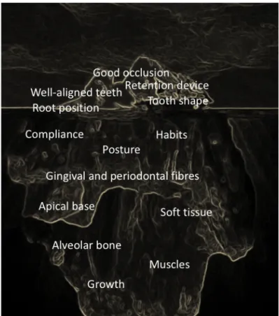
post-treatment changes are because of normal age changes (46). For these reasons, many orthodontics advocate lifelong retention to even prevent these ageing mechanisms, especially in the anterior region in the mandi-ble (47-53). As previously mentioned, malalignment of maxillary anterior teeth is often the foremost reason why patients seek orthodontic treat-ment. The focus from now on will therefore be on the maxillary anterior teeth. There are many reasons for relapse after orthodontic treatment and its effect on maxillary anterior teeth. It can be compared to an iceberg, as illustrated in Figure 1. The part an orthodontist can see, and influ-ence is above the surface. The part which is below the surface, the ort-hodontist cannot influence, but is well-aware of its presence and impact. Some factors depend on the patient’s cooperation like habits and posture, and some factors are based on genetics like growth, which are out of the orthodontist´s control.
Figure 1. Reasons to relapse illustrated as an iceberg. Below the water surface, the
orthodontist has no influence but on the surface are the entities which can be con-trolled by the orthodontist.
Occlusion
Stable occlusion is a well-known parameter to achieve stable results and reduce the risks of relapse (10, 54-57). In a prospective and longitudinal study, the authors concluded “Settling of the occluding teeth seems not to improve the functional occlusion after fixed appliance removal. Thus, it is necessary to check the functional occlusion before the appliance is remo-ved to eliminate unsatisfying functional occlusal contacts” (58). Hence, if we do not have good or preferably excellent occlusion before debonding, there is a risk that settling does not compensate for treatment shortco-mings. We know that settling occurs after removing the fixed appliances, no matter what kind of retention we use (59-63). However, it may take longer in cases with Vacuum-formed retainer (VFR) in both jaws, especi-ally in females (64).
Well-aligned teeth
Several studies emphasise on well-aligned teeth to reach better stability. Surbeck et al. found that the pattern of pre-treatment rotational displace-ment of maxillary anterior teeth tended to repeat itself post-retention (65). This finding was confirmed in later studies (66, 67). It has been suggested that incomplete alignment during treatment is a risk factor for relapse and slight over-correction during active treatment of severely rotated teeth, is recommended (65). The high percentage of teeth tending to return to their original positions (68).
In another study, they found that the maxillary incisors irregularity increased significantly (1.52 mm) during long-term post-treatment (69). None of the studied clinical factors, Little’s irregularity index, interca-nine, interpremolar and intermolar widths, arch length and perimeter, de-monstrated to be predictive of the maxillary crowding relapse.
Contact point displacement
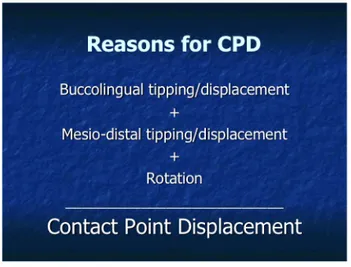
Buccolingual- and mesiodistal tipping together with the rotation of the tooth are reasons to contact point displacement (CPD) between adjacent teeth. These changes solely or in combination cause irregularity and mis-alignment between teeth shown in Figure 2.
Figure 2. Reasons for contact point displacement
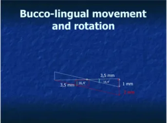
Effect of rotations on contact point displacement
The CPD changes caused by rotation is depending on the tooth width and amount of the rotation. When a central incisor is 9 mm, a rotation of 12.8 degrees round its long axis gives 1 mm of CPD, see Figure 3.
A lateral incisor of 7 mm, a rotation of 16.4 degrees gives 1 mm of CPD, as illustrated in Figure 4.
Figure 4. Effect of rotation on lateral incisor causing CPD
When tipping is involved together with the same amount of rotation, the CPD increases more which is illustrated in Figure 5.
Figure 5. Combination of tipping together with rotation
Tooth shape
It is known that triangle-shaped mandible incisors may cause crowding and are more prone to relapse than more square-shaped teeth (70). Crea-ting contact surfaces instead of contact points in triangle-shaped teeth and reducing tooth width by interproximal reduction (IPR), have been used
with relatively good results with better stability in orthodontics especially in mandibular anterior teeth (71-73).
Root position
In maxillary front, the laterals and canines erupt sometimes in palatal po-sition because of the eruption direction or/and in combination with space deficiency. Uprighting the root is a demanding and time-consuming pro-cess. There must be enough space for the root of the malpositioned tooth to be able for the orthodontist to move the root in proper position by ade-quate biomechanics. If the root is not uprighted with adeade-quate root torque, there is a great risk of relapse of the tipped crown to the previous position (74). Excellent finish and proper position of the crown and the root are important for the end result and the stability of achieved alignment (57).
The alveolar bone
The cortical thickness reflects overall bone mass and density. Studies show that women with osteoporosis have thinner cortex in the mandible (75). Animal studies showed that tooth movement was higher in affected rats with osteoporosis than normal individuals (76). It is hypothesised that persons with thinner mandibular cortices experience greater levels of dental relapse after orthodontic treatment—a reflection of decreased bone density with less bone support for thinner mandibular cortex.
In a retrospective study of relapse of lower incisors and cortical bone thickness, the authors found that subjects with thinner mandibular cort-exes had more dental relapse after orthodontic treatment (77). They con-cluded that greater relapse occurs in patients with decreased bone support based on cortical thickness.
During orthodontic tooth movement in adults, the alveolar bone loses at mean 24 per cent of its density around the six maxillary anterior teeth after seven months of orthodontic treatment (78).
Gingival and periodontal fibres
The periodontal space (distance between the root cementum and the al-veolar bone) is called the periodontal ligament (PDL) (79). It is about 0.25 mm and occupied by cells, vessels, fluid, nerves, and fibres. These fibres consist of collagen fibres that are arranged into fibre bundles. The portion of these fibres that are embedded into either cementum or bone is called Sharpey’s fibres. These fibres occasionally pass through the bone
of the alveolar process to continue as principal fibres of an adjacent PDL. They run buccally and lingually to connect with the fibres of the perioste-um. Other fibres are: Circular (runs around the tooth in the free gingiva), dento-gingival, dento-periostal, alveolo-gingival, and trans-septal fibres, see Figure 6 illustrating different fibres.
Figure 6. Different fibres surrounding teeth. Periodontal ligament (PDL), circular
fibres (CF), dento-gingival fibres (DGF), dento-periostal fibres (DPF), trans-septal fibres (TSF) and alveolo-gingival fibres (AGF)
When we move teeth orthodontically, resorption of alveolar bone at the pressure side occurs rapidly and allows the movement of teeth through the bone. The periodontal ligaments and Sharpey’s fibres act as an an-chorage zone for new bone and new cementum.
The middle and most apical part of the root is more stable to relapse, whereas the marginal third of the root is unstable (74). After movement is completed, the new site for the tooth and the periodontal ligament is re-established on the pressure side, similar to an unmoved tooth. On the ten-sion side, where the formation of new bone is required, re-establishment
of the bone occurs more slowly, and the periodontal ligament maintains tension on the bone, seeking to draw the tooth back to its original position causing relapse (48).
The challenge is to establish conditions preserving the new position of the tooth. Reitan described the relapse that occurred after tipping of the teeth in dogs without retention (44). He noticed that some relapse already occurred after two hours, partly caused by the up righting of the tooth. Relapse continued to occur during the following four days. After that, this process stopped because of the hyalinised zone (cell-free zone) this time on the tension side (44). A similar pattern was observed in children after tipping teeth without subsequent retention.
The periodontal ligament remodels fast, but the gingival fibres have a slow turnover rate and take as long as 232 days to remodel after experi-mental tooth rotation (44). Therefore, some authors suggest fiberotomy when teeth are derotated by orthodontic forces (80, 81) to eliminate the tension caused by stretching the supra crestal fibres (82).
Muscles and soft tissue
Teeth and alveolar bone are affected by the forces from muscles and soft tissues around them. The equilibrium theory has been discussed for deca-des (83, 84). Influential factors are, intrinsic factors like tongue and lips, extrinsic factors like habits (85), forces from occlusion and forces from the periodontal membrane (84). Forces from the tongue during swallo-wing are 2.5 times higher than forces from the lips and 1.5 times higher during the resting position. Two aspects, amount and duration of the for-ces are important. Forfor-ces during swallowing and occlusion are high but their duration is low.
On the other hand, forces during the rest and speech are low but much longer (83). A healthy periodontium stands against this unbalance. When it is compromised in patients with periodontitis, it can be noted that ante-rior teeth are flared outward in the maxilla and the mandible. Also, habits influence the balance negatively and may induce different forces, based on the magnitude and duration of the habits, to the teeth during the reten-tion period.
Common retention devices in the maxillary arch
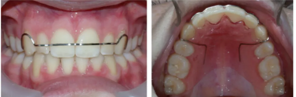
Several different fixed and removable retainers, solitary or in combina-tion, have been used to retain maxillary anterior teeth after orthodontic treatment (Figures 7-11). Fixed retainers in the maxilla may involve two teeth as in median diastema to eight teeth in cases of extraction of first premolars. The most common length is, however, 13-23 and 12-22 in which in the first one the wire is attached with composite to all six max-illary anterior teeth and in the latter from one lateral incisor to the other lateral incisor involving all four incisors. It has been advocated in the literature that 12-22 failure is less than 13-23 over time (86).
Zachrisson introduced the bonded retainers with twisted stainless steel wires in 1977 (87). Bonded retainers have become more and more popu-lar among patients (88), and the current shift is from removable retainers toward bonded retainers in different countries (89-94). The common use of flexible stainless steel twisted (three or more wires are twisted around each other) or multi-strand wire (several wires are twisted around a cen-tral wire) like 5-stranded or 7-stranded wires. The size varies from 0.016 to 0.022 in the maxilla. Braided or plain flexible square wires, glass-fibre reinforced and lately the CAD/CAM produced titanium wire have also been used (95). It is recommended using plain 0.016x0.016-inch or brai-ded 0.016x0.022-inch stainless steel wires to increase torque control (96). The use of V-loop shape retainer instead of straight wire retainer has been used to facilitate interproximal cleaning. The results showed a slightly higher rate of bond failure without any clinically or statistically signifi-cance (97).
Various materials of light-cured composite resin, polyacid-modified resin composite (compomer) and resin-modified glass ionomer have been used in direct or indirect bonding technique. The placement of the retainer usually occurs directly after or before debonding. Some prefer to bond the retainer several weeks prior to debonding to assure stability of the retai-ner and the possibility to repair the retairetai-ner in case of bond failure when braces and arch wire still are in place.
Vacuum-formed retainer (VFR) was introduced in 1993 (98). This aligner was called Essix which is an acronym for S-six which stands for ‘Sheridan’s Simple System for Stabilising the Social Six’ – the ESSIX-System was conceived initially to stabilise the six anterior teeth but, later on, extended to involve even molars. There are several different possibi-lities to also use it for achieving small tooth movements.
There is a different thickness, 0.75, 1.0 and 1,5 mm and different ma-terial. VFRs are made of Polyethylene, Polyurethane, and Polypropylene. Polyethylene is recommended for long-term wear (99). The problem with VFR and other removable retainers is that compliance reduces with time (100, 101). Some, like the Jensen retainer and the Hawley retainer, are designed to allow vertical tooth movements. They are even suitable for preventing transversal relapse.
The choice of retention device may depend on the initial malocclusion, expected growth, occlusal development, and the expected adherence. Bonded retainers seem to be popular as they effectively prevent tipping and rotation of the teeth (102, 103). They are rather independent of coope-ration and can be used for long periods (65, 87, 104, 105), although there are few studies on real long-term use. Bonded retainers are, on the other hand, operative sensitive and retainer failures are common (86, 101, 106, 107) especially in the maxilla (106, 108). Hawley retainers, Jensen retai-ners, and VFRs allow prosthetic pontics for missing teeth in the aesthetic zone. Some appliances, such as positioners and spring retainers, are used rarely but can also be useful for minor tooth movements.
Figure 7. To the left, a retainer bonded to six teeth. To the right, a retainer bonded to
Figure 8. Vacuum-formed retainer 17-27
Figure 9. Jensen retainer
Figure 11. Positioner to the left and spring retainer to the right
Adherence
Patient adherence is very important when it comes to removable retai-ners. It appears that compliance with removable orthodontic appliances is suboptimal, with patients wearing appliances considerably less than stipulated and routinely overreporting the duration of wear (109). Many orthodontists believe that a lack of awareness of dental problems, a lack of motivation to cooperate, and personal problems are the main reasons for non-compliance (110). To increase adherence, giving information to the patient in different ways is important (109). Although there is insuf-ficient evidence to allow clinicians to choose a single method, the results demonstrate the value of spending time with patients to illustrate the im-portance of adherence (111). Therelationship between orthodontist and patient seems to play a key role in patient adherence (112). It is well-known that adherence reduces with time (101).
There is a difference between compliance and adherence, even though the terms have been used as synonyms. Adherence is an active choice of patients to follow through with the prescribed treatment while taking re-sponsibility for their own well-being. Compliance is a passive behaviour in which a patient is following a list of instructions from the doctor. Dif-ferent kinds of sensors have been used together with removable applian-ces to monitor and increase patients’ compliance (112-115). The overall results are good, and an objective measurement of wear time allows a more realistic view of compliance by patient and orthodontist. Some of these studies showed knowing that wear time is recorded, does not neces-sarily increase the amount of time the patient wears removable appliances (113, 114).
Significance
In the maxilla, the removable retainers have been used routinely. The bonded retainers have become popular because they have good retention qualities and are not dependent on the patient’s cooperation (89). Ho-wever, there was a lack of scientific studies on maxillary bonded retai-ners (116, 117). Papers I and II were aimed to examine the effectiveness and durability of bonded retainers in maintaining the results both short-term and long-short-term after orthodontic treatment. The orthodontists want to know which kind of retention is more effective to use, removable or bonded retainer. Do they need to extend the bonded retainer to involve canines or is it enough to involve only four incisors in the maxilla? It has been a lack of knowledge in these areas and Paper III was mending to fill this gap with a randomised clinical trial (RCT) study design between three commonly used retention devices. Retainers have been used routi-nely in decays, but no scientific study has investigated what happens if no retainer is used in the maxilla. In previous studies with no retention in the mandible, interproximal reduction (IPR) was carried out (47, 71, 118, 119) with satisfying results. If the results are similar in the maxilla remains to be investigated. Paper IV was aimed to answer this question in a highly scientific manner with its RCT study design.
To add more to this framework, a one-year post-retention evaluation was carried out in Paper IV to find out what happens when we take out the retainer in the group with retention.
AIMS
Paper I
To investigate the amount and pattern of relapse of maxillary anterior teeth previously retained with a bonded retainer.
Paper II
To investigate the amount and pattern of changes of maxillary anterior teeth seven years post-retention, which previously were retained with a bonded retainer.
Paper III
To evaluate post-treatment changes in the irregularity of the maxillary, six anterior teeth and single tooth contact point discrepancy (CPD) of three different retention methods after two years in retention.
Paper IV
To evaluate whether retention is needed after orthodontic treatment for impacted maxillary canines and with moderate pre-treatment irregularity in the maxilla.
HYPOTHESES
Paper I
The bonded retainer in the maxilla is effective to stabilise teeth in position one year after removal.
Paper II
There are no major changes seven years after removal of the bonded re-tainer.
Paper III
There would be no difference in retention capacity between any of the th-ree maxillary retention methods (bonded retainer 13-23, bonded retainer 12-22, and vacuum-formed retainer) during two years of retention.
Paper IV
No statistically or clinically significant difference will occur in the posi-tion of the maxillary anterior teeth of patients with and without retenposi-tion, one-year post-treatment.
MATERIALS AND METHODS
Paper I and II
The study group originally consisted of 45 patients in Paper I and 27 patients on recall for Paper II. Recordings from study models before treat-ment (T1), at debonding (T2), one year after removal of the retainer (T3), and seven years post-retention (T4) were present. All patients had been treated with fixed edgewise appliances by the same orthodontist. The pa-tients were selected from the Orthodontic Clinic in Mariestad, Sweden, when their maxillary bonded retainer was removed.
The wire used was 0.0195-inch Wildcat (GAC International Inc., Cen-tral Islip, NY). All six maxillary permanent anterior teeth had to be pre-sent before treatment and having irregularity. Spaced dentitions in the maxillary anterior teeth and treatments started as adults were excluded. Labiolingual displacements of the anatomic contact points from the me-sial of the right canine through to the meme-sial of the left canine were mea-sured with a digital calliper on the casts from T1, T2, T3, and T4, with 0.1 mm accuracy. CPD less than 0.5 mm were judged to be zero.
An Agfa DuoScan F40 (Agfa-Gevaert N.V., Mortsel, Belgium) scan-ner was used to scan the casts at 300 DPI (dots per inch) resolutions. All 45 x 3 casts were scanned in 300 DPI and then placed on the upper third part of the glass, with almost the same size of the scanned area. All ante-rior teeth were optimally in contact with the glass surface of the scanner to avoid distortions.
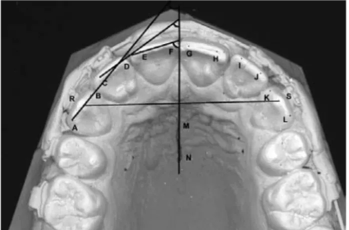
A free computer program, Scion Image, from Research Services Branch (RSB), National Institutes of Health (NIH), USA was used to mark points on the pictures of the scanned casts to measure rotation changes and inter-canine distance. The rotations were measured as the angle between a line through the mesial and distal points on the incisal edge of the teeth and
the raphe line. The intercanine distance was measured between the cusp tips of the maxillary canines as shown in Figure 12.
The Little’s irregularity index (LII) (120), which is the sum of five CPDs, and rotations of anterior teeth with the raphe line and intercanine distance, were calculated at T1, T2, T3, and T4 (Figure 13).
Figure 12. Showing the tooth angles on the right side to raphe line and intercanine
distance
Figure 13. Little’s irregularity index
Paper III
In Paper III, 90 patients were recruited to the study. All participants were recruited from patients with ongoing orthodontic treatment at the Ort-hodontic Clinic in Växjö, Public Dental Service, Region Kronoberg, Sweden. They were all treated by three experienced orthodontists. The inclusion criteria were adolescents treated with the fixed appliance in the maxilla or in both maxilla and mandible. The exclusion criteria were pa-tients with syndromes, agenesis or extracted maxillary anterior teeth, or in need of orthognathic surgery.
Three months before the estimated removal of the fixed appliances, the patients were invited to take part in the trial. After gaining informed con-sent from the patient and their custodians, the patients were randomly al-located to one of the three retention groups as follow: A) bonded retainer 13-23, B) bonded retainer 12-22, and C) removable vacuum-formed retai-ner (VFR) covering the maxillary teeth including the second molars. The material in the bonded retainers was Penta-One 0.0195 (Masel, Carlsbad, California, USA) with bonding agent Transbond Supreme LV (TSLV-3M, Unitek, Monrovia, California), and in VFR (EssixTM, Erkodur, 1.5 mm 120ø, Erkodent® Erich Kopp GmbH, Pfalzgrafenweiler, Germany).
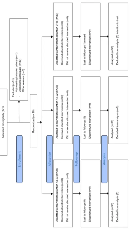
The retainers were bonded the same day directly after debonding. The VFRs were produced within one day after debonding. The patients fol-lowed a standard protocol for VFR-wear: 22–24 hours per day during the first four weeks, then every night. One year after debonding, the wearing time was reduced to every other night. Retention check-ups were perfor-med one month, one year, and two years after insertion of the retainers. The randomisation process was prepared by an independent person and carried out by three staff members not involved in the trial. The randomi-sation used blocks of 30. The randomirandomi-sation notes were delivered in a sealed opaque envelope each. Every new participant picked an envelope and revealed the group assignment by opening the envelope. Recruitment went on until the total number of participants met the estimated sample size.
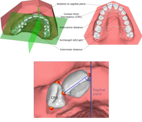
The primary outcomes were changes in single CPD and LII measured on digitalised three-dimensional study casts before and after two years in retention. The secondary outcomes were: changes in arch length defi-ned as the sum of the distances between the mesial contact point of the first permanent molar and the central incisal contact point in the maxilla; changes in intercanine width, defined as the distance between the cani-nes’ cusp tips in the maxilla; changes in intermolar width, defined as the distance between the mesiobuccal cusp tips of the first permanent molars in the maxilla; maximum single tooth rotation of one of the six maxillary anterior teeth, defined as the angle between the line from distal to the mesial contact point and the sagittal plane; and overjet and overbite were also measured.
Data on all participants were evaluated on an intention to treat (ITT) basis. Consequently, all randomised patients remained in the allocated
group. Subsequently, patients lost in follow-up were included in the final analysis by assessing the group’s maximum value for primary outcome change (irregularity of the maxillary six anterior teeth and single tooth discrepancy) and the secondary variables representing the change in arch length, intercanine width and the intermolar width, overjet, and overbite. Measurements were made on digitalised 3D study casts for all the three retention groups before treatment (T0), at the end of active treatment/start of retention (T1), and the two-year follow-up (T2). Before assessment of measurements, the study casts were digitised with a stationary 3D scanner (D3, 3Shape, Copenhagen, Denmark). On the digital models, the mea-surement points were located using a validated computer program, the OnyxCeph3TM (121) (software (v3.2.142), Image Instruments, Chemnitz,
Germany) with semi-automatic segmentation.
The measurement points were then manually adjusted in order to im-prove consistency. The measurement points for CPD were defined as the mesial and distal point of the broadest mesiodistal diameter of the tooth. Further, the CPD was measured as a projection on the occlusal plane, shown in Figure 14, and the flow chart of the patients, Figure 15.
Paper IV
In Paper IV, 63 adolescent patients were recruited to the study. All parti-cipants were recruited from patients with ongoing orthodontic treatment at the Orthodontic Clinic in Växjö, Public Dental Service, Region Kro-noberg, Sweden. They were all treated by one experienced orthodontist. After gaining informed consent from the patient and their custodians, the patients were randomised to one of two groups, a non-retention group as study group (31 subjects) and a retention group with VFR as the control group (32 subjects). The randomisation process was prepared and carried out by an independent person not involved in the trial. The randomisation used blocks of 20 (10 + 10). Opaque envelopes contained 20 sealed no-tes each (ten nono-tes signed retention, and ten nono-tes signed non-retention). Every new participant in the trial took a note from the first envelope. When the first envelope was empty, the next envelope was opened, and so on, until the number of participants met the estimated sample size, see the flow chart in Figure 16.
Data on all participants were evaluated on an ITT basis. Consequently, all randomised patients remained in the allocated group. Subsequently, patients with discontinued observation or lost to follow-up were still
in-cluded in the final analysis by assessing the group’s maximum value for change in the primary outcomes as well as for the secondary variables. The trial period lasted between the removal of the fixed orthodontic app-liances (T1) and one year after debonding (T2). The measurement met-hods, primary and secondary outcomes were the same as Paper III.
Figur
e 15.
Figur
e 15.
Flow chart three retention methods
Figure 16. Flow chart retention or no retention
Ethical considerations
In Paper I and II Ethical Committee of Västra Götaland, Sweden, classi-fied them as quality control and thereby, no ethical approval was needed. In Paper III and IV, the Regional Ethical Research Board, Linköping, Sweden, approved the trials (Dnr 2013/130-131). The participants recei-ved the information that participation was of free will, and they could refuse participation or discontinue with the study at any time. The patients and their custodians signed a consent form.
STATISTICAL ANALYSIS
Sample size calculation
In Paper, III and IV the sample size calculations were assessed and based on a clinically relevant difference in LII of 3 mm and standard deviation (SD) 3 mm between the groups. The level of significance was set to 5 per cent and the power to 90 per cent. The calculation resulted in a sample size of 23 patients in each group. To compensate for dropouts, at least 30 patients were planned to be enrolled in each group.
Group comparisons
In all papers, arithmetic means and standard deviations (SD) were cal-culated at the group level for each variable.
In Paper I and II, paired t-tests were applied to test differences in CPD, rotations, and intercanine distance between T1, T2, T3, and T4. Pearson’s product-moment correlation test was applied to test correlations between CPD and rotations at T1 and changes during treatment and the follow-up period. The data were analysed using the statistical package SAS ® v8.2 program (SAS Institute Inc, Cary, NC, USA). For all statistical analyses, the statistical significance level was set to 5 per cent.
In Paper III and IV, the statistical analysis was conducted using the pro-gramming language ‘R’ (v. 3.60) and (v. 4.0.2) (122). In Paper III, the out-come measures were tested for normality in distribution with histograms and Shapiro-Wilk test. Variance homogeneity was tested with Levene’s test. Statistical significance was then tested with One-Way ANOVA follo-wed by pairwise t-test for variables with a normal distribution. Variables with non-normal distribution were tested with the Kruskal–Wallis test, followed by Dunn’s test. When p-values were calculated, Holm–Bonfer-roni correction was applied to compensate for multiple comparisons.
Dif-ferences with a P-value less than 5 per cent (P <0.05) were considered statistically significant.
In Paper IV, the Shapiro–Wilk test was used for normality testing. Homogeneity of variance was tested with Levene’s modified test. Hypo-thesis testing was conducted with t-test for independent variables with a normal distribution. Independent variables that were not normally distri-buted were evaluated with the Mann–Whitney U-test. Correlations bet-ween post-treatment increase of irregularity and possible predictors were assessed with scatterplots and Spearman’s rank correlation coefficient.
Measurement error analysis
Paper I
The reproducibility of the measurements for rotations and intercanine distance was determined by double measurements of 45 scanned models from 15 patients at T1, T2, and T3. The error of the method was calcula-ted using Dahlberg’s equation (123).
Where D is the difference between repeated measurements and N is the number of measurements. The errors were 3.1 degrees for canines, 2.8 degrees for laterals, and 2.4 degrees for centrals. The error of measuring the intercanine distance was 1.1 mm.
Paper II
The measurement error was calculated from double measurements of 27 models (T4), using Dahlberg’s formula. The error for CPD measurements was 0.2 mm. The measurement errors for rotation were 3.1 degrees for canines, 2.8 degrees for laterals, and 2.4 degrees for centrals.
Paper III
The same observer conducted repeated segmentation and measurements on T1, and T2 models of 15 randomly selected cases after at least two weeks. The two-measurement series were compared with Bland–Altman method (124). All variables showed sufficient agreement and low bias. Further, paired t-tests could not reveal any statistically significant dif-ferences/bias between the two-measurement series. The absolute mean
measurement error for CPDs was 0.1 mm, for LII 0.2 mm, for overjet, overbite, arch length, intercanine and intermolar width 0.1 mm each, and 2.4 degrees for tooth rotations.
Paper IV
Forty-five randomly selected study casts were repeatedly measured on two separate occasions with at least two weeks’ interval by the same exa-miner. A paired t-test revealed no significant mean differences between the two series of record occasions. The size of the method error was de-termined using Dahlberg’s formula. The mean measurement error for the CPDs was 0.1 mm, for LII 0.2 mm, for intercanine and intermolar width 0.2 mm each, and 2.6 degrees for tooth rotations.
RESULTS
Paper I
Before treatment (T1), the mean irregularity index was 10.1 (range 3.0 – 29.9). The largest displacements were recorded between laterals and centrals followed by the displacement between laterals and canines. The smallest deviations were found between the centrals as shown in Figure 17.
After treatment (T2), the mean irregularity index was 0.7 (range 0.0 – 2.1). There was a significant difference in the index between T1 and T2 (P<.0001).
One-year post-retention (T3), the mean irregularity index was 1.4 (range 0 – 5.1). There was a significant difference in the index between T2 and T3 (P<.0001).
Figure 17. Contact point displacements before treatment (T1), after
Paper II
Results from the second study showed that there were no statistically sig-nificant differences between the change in mean CPDs for the contacts canines/laterals, laterals/centrals, or centrals/centrals, as shown in Figure 18. The mean irregularity index for 27 patients examined in the second study was 10.3 (range 3.7 – 29.9) at T1, 0.9 (range 0.0 – 2.1) at T2, 1.3 (range 0.0 – 3.5) at T3 and 2.0 (range 0.0 – 5.8) at T4. No correlations were found between the pre-treatment and post-retention irregularity T1/ T3 and T1/T4. There was a significant association between the irregula-rity index at T3 and T4 (R = 0.938, P < 0.0001).
The irregularity index of the maxillary anterior teeth changed very litt-le or not at all during the one-year post-retention. Further changes long-term resulted in an irregularity index of mean 2.0 mm (range 0.0 – 5.8). The contact relationship between the laterals and centrals seems to be the most critical. Forty rotated teeth in 21 patients were corrected more than 20 degrees. Mean relapse during the one-year post-retention was 6.7 degrees (range 0.0 – 14.7). Mean changes under seven years were 8.2 degrees (range 0.0 – 19.3)
Figure 18. Contact point displacement (CPD) before treatment (T1), after treatment
Fourteen cases out of 27 were stable one-and seven-years post-retention showing individual differences illustrated in Figure 19.
Figure 19. Stable and non-stable cases one and seven-years post-retention
Paper III
There were no significant differences in gender, age, irregularity, treat-ment duration, and treattreat-ment outcome were found between the groups see Table 1. 0 1 2 3 4 5 6 7 1 3 5 7 9 11 13 15 17 19 21 23 25 27 Patients
Irregularity Index 1 and 7 years post retention
T3 T4
Fourteen cases out of 27 were stable one- and seven-years post-retention showing individual differences illustrated in Figure 19.
Figure 19. Stable and non-stable cases one- and seven-years post-retention
Paper III
There were no significant differences in gender, age, irregularity, treat-ment duration, and treattreat-ment outcome between the groups see Table 1.
Table 1.
Baseline
demographic
data
considering
the number of subjects, gender distribution,
maximum contact point discrepancies (max CPD), maxi -mum rotations of anterior teeth, extractions, bimaxillary treatment and treatment duration in months. P-values calculated with the Kruskal-W allis test
for continuous variables and Chi-square test for categorical variables.
G ro up Pre -tr eat m en t num be r of pa tie nt s a nd gen der dis tr ib ut io n Pre - tr eat m en t age , ye ar s m ea n (S D ) Pre - tr eat m en t m ax C PD m ea n (S D ) m ax C PD (T 1) m ea n (S D ) M ax r ot at ion T1 -T 0 m ea n (S D ) N um ber of pa tie nt s w ho ha d ext ra ct io ns N um ber of pa tie nt s w ho ha d bim ax illa ry tr eat m en t Trea tm en t du ra tio n in m on th s m ea n (S D ) A - B onde d r et ai ne r 13 -23 30 ( ♀ 17; ♂ 13) 13 .8 ( 1. 5) 3. 3 m m (2. 0) 0. 5 m m (0. 3) 11. 8° ( 8. 0) 12 23 24. 3 ( 10. 4) B - B onde d r et ai ne r 12 -22 30 ( ♀ 20; ♂ 10) 14 .0 (1 .8 ) 3. 3 m m (1. 7) 0. 4 m m (0. 3) 14. 1° (10. 6) 11 25 22. 3 ( 8. 3) C - V acu um -f or m ed ret ai ner 30 ( ♀ 17; ♂ 13) 13. 9 ( 1. 9) 2. 7 m m (1. 2) 0. 4 m m (0. 2) 14. 3° ( 9. 5) 5 17 24. 6 ( 12. 9) P va lue 3 -gr oup -c om pa ris on 0. 659 0. 893 0. 451 0. 797 0. 520 0. 108 0. 056 0. 852 Tot al popul at ion 90 ( ♀ 54; ♂ 36) 13. 9 ( 1. 7) 3. 1 m m (1. 7) 0. 4 m m (0. 3) 13. 4° (9. 4) 28 65 23. 7 ( 10. 6)
During the two years of retention, Little’s irregularity index and maxi-mum single tooth discrepancies increased slightly in all three groups wit-hout any statistically significant group differences. The null-hypothesis was thereby confirmed, as shown in Table 2 and Figure 20.
The group with bonded retainer 13-23 showed statistically significant less rotational changes during retention compared to the group with bon-ded retainer 12-22 (p = 0.014) and the group with VFR (p = 0.036) (Table 2). Further, the intercanine width increased in the group with VFR while it diminished in the group with bonded retainer 13-23 and remained un-changed in the group with bonded retainer 12-22. The difference in inter-canine width was statistically significant between bonded retainer 13-23 and VFR (p = 0.023) (Table 2).
The intercanine width was smaller in Group A (bonded retainer 13-23), than in Group C (VFR) (0.5 mm), but clinically insignificant. Regarding overjet, overbite, intermolar width, and arch length, minor changes wit-hout any significant inter-group differences were found.
Table 2.
Changes (T2-T1) during the retenti
on period (T1= start of retention
and
T2=
Two years of retention).
Results on the intention
to treat basis represented by means and 95% confidence interval (CI). Test conducted with Kruskal – W allis test P
a for 3-group comparison
followed
by Dunn’
s test
P
b for between-group comparison. P-values adjusted for multiple compa
risons with Holm
– Bonferroni correction. Table 2. Changes (T2 -T1) during the retention period (T1 = start of retention and T2 = two years of retention) . Results on intention -to -treat basis represented by
means and 95% confidence intervals (CI). Tests conducted with Kruskal
-Wallis test
P
a for 3
-group comparison followed by Dunn´s test
P
b for between
-group
comparison. P
-values adjusted for multiple comparisons with Holm
-Bonferroni correction. Group A B on d ed ret ai n er 13 -23 (n = 30) 13 -23 vs. 12 -22 Group B B on d ed ret ai n er 12 -22 (n = 30) 12 -22 vs. V F R Group C V ac u u m -f or m ed r et ai n er V F R (n =3 0 ) V F R vs. 13 -23 3- gr ou p co m p ari so n M ea n 95% C I M ea n 95% C I M ea n 95% C I P a l ow er uppe r P b lo w er uppe r P b lo w er uppe r P b Chan ge T 2-T1 L ittl e ir re gu la ri ty i n d ex 0 .134 0. 3 m m 0 .1 0 .5 0 .397 0. 6 m m 0 .4 0 .8 0 .500 1. 0 m m 0 .4 1 .6 0 .138 A rch l en gth 0 .239 0. 1 m m -0 .1 0 .3 0 .283 -0. 1 m m -0 .3 0 .1 0 .583 0. 0 m m -0 .2 0 .2 0 .537 Int er ca ni ne w idt h 0 .019 -0. 3 m m -0 .5 -0 .1 0 .066 0. 0 m m -0 .4 0 .4 0 .593 0. 2 m m 0 .0 0 .4 0 .023 Int er m ol ar w idt h O ve rj et O ve rb it e M ax im u m r ot at ion 0 .775 0.951 0.938 0.010 -0. 3 m m 0. 1 m m 0. 2 m m 2 .9 ° -0 .7 -0 .1 0 .0 1 .7 0 .1 0.3 0.4 4.1 0 .999 0.931 0.999 0.014 -0. 4 m m 0. 1 m m 0. 3 m m 5 .2 ° -0 .8 -0 .3 0.1 4.2 0 .0 0 .5 0 .5 6 .2 0 .999 0.999 0.945 0.649 -0. 4 m m 0. 0 m m 0. 3 m m 6 .6 ° -0 .6 -0 .4 -0 .1 3 .9 -0 .2 0 .4 0 .7 9 .3 0 .898 0.999 0.999 0.036
53
Figure 20. Changes in LII during the two years follow-up
Paper IV
No significant differences in age and treatment duration were found bet-ween the groups or betbet-ween genders. Surgical exposure was carried out in 15 patients in the retention group and 19 patients in the non-retention group see Table 3.
Table 3. Baseline demographic data, standard deviation (SD)
Group Gender n Age, yrs mean (SD) Impacted palatal canines, n Impacted buccal canines, n Retention Female 24 13.1 (1.7) 17 10 Male 8 12.1 (1.4) 6 2 Total 32 12.8 (1.7) 23 12 No retention Female 15 12.9 (1.1) 14 3 Male 16 13.0 (1.4) 15 4 Total 31 12.9 (1.2) 29 7 Both groups Total 63 12.9 (1.5) 52 19
The difference in irregularity change between the retention and non-reten-tion groups was statistically significant (p < 0.0001, 0.4 versus 1.3 mm) thereby the null-hypothesis was not confirmed, see Figure 21. The maxi-mum increase in irregularity was 2.5 mm in the retention and 3.2 mm in the non-retention group (p < 0.001) (Table 4). Moreover, the mean maxi-mum single contact point discrepancy was lower in the retention group compared to the non-retention group, but not significant (Table 4). Small but not statistically significant changes between the groups were found in arch length, intercanine and intermolar width. No significant correlations between post-treatment changes in irregularity and duration of treatment, pre-treatment contact point discrepancies or tooth derotations were detec-ted, as shown in Figure 22, which follows the earlier studies (69). In the non-retention group, including the 10-week interim period, most changes occurred during the ten weeks, see Figure 23. In the retention group, 93.5 per cent had a LII less than 3 mm, and in the non-retention group, 80 per cent had a Little’s irregularity index less than 3 mm after the one-year observation period.
Figure 21. The changes in the Little’s irregularity index in the two groups between
Figure 22. Correlations between changes in irregularity and (A) duration of treatment
(rho = 0.28); (B) maximum CPD (rho = 0.15) and (C) maximum derotation (rho = 0.15)
Table 4. Mean changes in mm CPD during the retention period (T1= post-treatment,
to T2= one-year follow-up) in the retention group and non-retention group. Results (ITT) represented by means and 95% confidence intervals (CI).
Additional follow-up
In the retention group the retention device was removed after one-year post-retention. It was minor and non-statistical changes after removal of the retainer the following year post-retention. The results were statisti-cally significant, looking at total changes during these two years’ post-treatment. However, the LII under 3 mm was maintained with clinically good results as shown in Table 5 and Figure 24.
Table 5. Post-treatment changes in irregularity, arch length, intercanine width, and
intermolar width.
Retention group
(n=32) ←P→ Non-retention group (n=31)
Mean 95% CI Mean 95% CI lower upper lower upper
C han ge T2 -T1 Littles Index 0.4 mm 0.2 0.6 <.001 1.3 mm 0.9 1.7 Arch length 0.0 mm -0.2 0.2 .145 -0.3 mm -0.5 -0.1 Intercanine width 0.1 mm -0.1 0.3 .279 0.4 mm 0.0 0.8 Intermolar width -0.1 mm -0.3 0.1 .492 0.0 mm -0.4 0.4 Maximum CPD T1 Maximum CPD T2 0.5 mm 0.7 mm 0.3 0.5 0.7 0.9 .330 .002 0.5 mm 1.1 mm 0.3 0.9 0.7 1.3 Maximum derotation T2-T1 4.7° 3.5 5.9 .034 6.4° 5.4 7.4
N=31 0-‐1 year 1-‐2 years 0-‐2 years
LII–mean (SD) – min/max 0.4 mm (0.7) -‐0.5/2.5 0.4 mm (0.7) -‐0.8/2.3 0.9 mm (0.8) -‐0.5/2.8 Arch length – mean (SD) 0.0 mm (0.6) 0.0 mm (0.4) -‐0.1 mm (0.7) Intercanine width – mean (SD) 0.1 mm (0.7) 0.2 mm (0.6) 0.3 mm (1.1) Intermolar width – mean (SD) -‐0.1 mm (0.8) 0.0 mm (0.5) -‐0.1 mm (1.1)