ANNA-PAULINA WIEDEL
FIXED OR REMOVABLE APPLIANCE FOR
EARLY ORTHODONTIC TREATMENT OF
FUNCTIONAL ANTERIOR CROSSBITE
Evidence-based evaluations of success rate of interventions,
treatment stability, cost-effectiveness and patients perceptions
ANN A -P A ULIN A WIEDEL MALMÖ UNIVERSIT FIXED OR REMO V ABLE APPLIAN CE FOR EARL Y ORTHODONTIC TREA TMENT OF FUN CTION AL ANTERIOR CR OSSBITE
Swedish Dental Journal, Supplement 238, 2015
SWEDISH DENT AL JOURN AL, SUPPLEMENT 238, 20 1 5. DOCT OR AL DISSERT A TION IN ODONT OL OG Y
F I X E D O R R E M O V A B L E A P P L I A N C E F O R E A R L Y O R T H O D O N T I C T R E A T M E N T O F F U N C T I O N A L A N T E R I O R C R O S S B I T E
Swedish Dental Journal, Supplement 238, 2015
© Copyright Anna-Paulina Wiedel 2015
Foto: Hans Herrlander och illustration: Anna-Paulina Wiedel ISBN 978-91-7104-643-7 (print)
ISBN 978-91-7104-644-4 (pdf) ISSN 0348-6672
ANNA-PAULINA WIEDEL
FIXED OR REMOVABLE
APPLIANCE FOR EARLY
ORTHODONTIC TREATMENT
OF FUNCTIONAL ANTERIOR
CROSSBITE
Evidence-based evaluations of success rate of
interventions, treatment stability, cost-effectiveness
and patients perceptions
This publication is also available at, www.mah.se/muep
CONTENTS
PREFACE ... 9 ABSTRACT ... 10 Paper I ...11 Paper II ...11 Paper III ...12 Paper IV ...12Key conclusions and clinical implications ...12
POPULÄRVETENSKAPLIG SAMMANFATTNING ... 13 INTRODUCTION ... 16 Anterior crossbite ...16 Stability ...23 Economic evaluation ...24 Patients´ perceptions ...25 Evidence-based evaluation ...25 Significance ...28 AIMS ... 30 HYPOTHESES ... 31
SUBJECTS AND METHODS ... 32
Subjects ...32 Methods ...35 Papers I and II ...35 Paper III ...39 Paper IV ...41 Side effects ...42
RESULTS ... 44
Paper I – Treatment effects ...44
Paper II – Stability ...46
Paper III – Cost-minimization ...47
Paper IV – Pain and discomfort ...49
DISCUSSION ... 54
Methodological aspects...55
Treatment effects of anterior crossbite correction ...56
Stability of anterior crossbite correction ...57
Cost-minimization analysis ...58
Analysis of pain, discomfort and impairment of jaw function ...59
Ethical considerations ...61
Future research...61
CONCLUSIONS ... 63
Key conclusions and clinical implications ...64
ACKNOWLEDGEMENTS ... 65
REFERENCES ... 68
PREFACE
This thesis is based on the following papers, which are referred to in the text by their Roman numerals I-IV:
I. Wiedel AP, Bondemark L. Fixed versus removable
orthodontic appliances to correct anterior crossbite in the mixed dentition -a randomized controlled trial. Eur J Orthod. 2015;37:123-7.
II. Wiedel AP, Bondemark L. Stability of anterior crossbite
correction: A randomized controlled trial with a 2-year follow-up. Angle Orthod. 2015;85:189-95.
III. Wiedel AP, Norlund A, Petrén S, Bondemark L. A cost
minimization analysis of early correction of anterior crossbite – a randomized controlled trial. Eur J Orthod. 2015 May 4. (E-published ahead of print, PMID 25940585).
IV. Wiedel AP, Bondemark L. An RCT of self-perceived pain,
discomfort and impairment of jaw function in children undergoing orthodontic treatment with fixed or removable appliances. Angle Orthod. 2015 July 17. (E-published ahead of print, PMID 26185899).
The papers are reprinted with kind permission from the copyright holders.
ABSTRACT
Anterior crossbite with functional shift also called pseudo Class III is a malocclusion in which the incisal edges of one or more maxillary incisors occlude with the incisal edges of the mandibular incisors in centric relationship: the mandible and mandibular incisors are then guided anteriorly in central occlusion resulting in an anterior crossbite.
Early correction, at the mixed dentition stage, is recommended, in order to avoid a compromising dentofacial condition which could result in the development of a true Class III malocclusion and temporomandibular symptoms. Various treatment options are available. The method of choice for orthodontic correction of this condition should not only be clinically effective, with long-term stability, but also cost-effective and have high patient acceptance, i.e. minimal perceived pain and discomfort. At the mixed dentition stage, the condition may be treated by fixed (FA) or removable appliance (RA). To date there is insufficient evidence to determine the preferred method.
The overall aim of this thesis was therefore to compare and evaluate the use of FA and RA for correcting anterior crossbite with functional shift in the mixed dentition, with special reference to clinical effectiveness, stability, cost-effectiveness and patient perceptions. Evidence-based, randomized controlled trial (RCT) methodology was used, in order to generate a high level of evidence.
The thesis is based on the following studies:
The material comprised 64 patients, consecutively recruited from the Department of Orthodontics, Faculty of Odontology, Malmö
University, Sweden and from one Public Dental Health Service Clinic in Malmö, Skane County Council, Sweden. The patients were no syndrome and no cleft patients. The following inclusion criteria were applied: early to late mixed dentition, anterior crossbite affecting one or more incisors with functional shift, moderate space deficiency in the maxilla, no inherent skeletal Class III discrepancy, ANB angle> 0º, and no previous orthodontic treatment. Sixty-two patients agreed to participate and were randomly allocated for treatment either with FA with brackets and wires, or RA, comprising acrylic plates with protruding springs.
Paper I compared and evaluated the efficiency of the two different treatment strategies to correct the anterior crossbite with anterior shift in mixed dentition. Paper II compared and evaluated the stability of the results of the two treatment methods two years after the appliances were removed. In Paper III, the cost-effectiveness of the two treatment methods was compared and evaluated by cost-minimization analysis. Paper IV evaluated and compared the patient´s perceptions of the two treatment methods, in terms of perceived pain, discomfort and impairment of jaw function.
The following conclusions were drawn from the results:
Paper I
• Anterior crossbite with functional shift in the mixed dentition can be successfully corrected by either fixed or removable appliance therapy in a short-term perspective.
• Treatment time for correction of anterior crossbite with functional shift was significantly shorter for FA compared to RA but the difference had minor clinical relevance.
Paper II
• In the mixed dentition, anterior crossbite affecting one or more incisors can be successfully corrected by either fixed or removable appliances, with similarly stable outcomes and equally favourable prognoses.
Paper III
• Correction of anterior crossbite with functional shift using fixed appliance offers significant economic benefits over removable appliances, including lower direct costs for
materials and lower indirect costs. Even when only successful outcomes are considered, treatment with removable appliance is more expensive.
Paper IV
• The general levels of pain intensity and discomfort were low to moderate in both groups.
• The level of pain and discomfort intensity was higher for the first three days in the fixed appliance group, and peaked on day two for both appliances.
• Adverse effects on school and leisure activities as well as speech difficulties were more pronounced in the removable than in the fixed appliance group, whereas in the fixed appliance group, patients reported more difficulty eating different kinds of hard food.
• Thus, while there were some statistically significant differences between patients´ perceptions of fixed and removable
appliances but these differences were only minor and seems to have minor clinical relevance. As fixed and removable appliances were generally well accepted by the patients, both methods of treatment can be recommended.
Key conclusions and clinical implications
Four outcome measures were evaluated: -success rate of treatment, treatment stability, cost-effectiveness and patient acceptance, which is important from both patient and care giver perspectives. It is concluded that both methods have high success rates, demonstrate good long-term stability and are well accepted by the patients. Treatment by removable appliance is the more expensive alternative. Thus, in the studies on which this thesis is based, fixed appliance emerges as the preferred approach to correction of anterior crossbite with functional shift in the mixed dentition.
POPULÄRVETENSKAPLIG
SAMMANFATTNING
Frontal invertering med tvångsföring av underkäken framåt benämns även som pseudo klass III och innebär att en eller flera överkäksframtänder kan bita i kontakt mot underkäksframtänderna men vid sammanbitning förs underkäken framåt till ett underbett för att få maximala tandkontakter mellan käkarna.
Behandling av frontal invertering med tvångsföring rekommenderas oftast i växelbettet d.v.s. vid ca 8-10 års ålder när barnets mjölktänder byts ut till permanenta tänder. Behandling utförs för att undvika tuggmuskel eller käkledsbesvär eller för att undvika att ett verkligt underbett ska utvecklas. En rad olika behandlingsmetoder har prövats men evidensen för vilken behandlingsmetod som fungerar bäst är ofullständig.
Syftet med avhandlingen var att i växelbettet utvärdera och jämföra två vanliga behandlingsmetoder för att korrigera frontal invertering med tvångsföring avseende lyckande frekvens, behandlingseffektivitet, behandlingsstabilitet på längre sikt, kostnadseffektivitet samt patientupplevd smärta och obehag. För att få så högt bevisvärde som möjligt valdes randomiserad kontrollerad studiedesign vilket innebar att patienterna lottades till antingen fast eller avtagbar tandställning. Avhandlingen är baserad på följande 4 studier:
Alla studierna baseras på ett patientmaterial om 62 patienter som lottats till två grupper med 31 patienter i vardera gruppen. Delarbete I utvärderar och jämför behandlingseffektiviteten mellan fast och avtagbar tandställning i överkäken. Den fasta tandställningen bestod
av metallfästen som fastsatts till 8-10 tänder i överkäken och en tunn metallbåge som sammankopplar tänderna. Den avtagbara tandställningen utgjordes av en plastplatta i gommen med metallfjädrar som tryckte överkäkens framtänder framåt/utåt. I delarbete II utvärderades och jämfördes stabiliteten av behandlingsresultaten två år efter avslutad tandställningsbehandling. I delarbete III utvärderades och jämfördes med en kostnads-minimeringsanalys kostnadseffektiviteten mellan de två olika tandställningarna. Delarbete IV utvärderade och jämförde patienternas upplevda smärta och obehag av fast och avtagbar tandställning.
Konklusioner
Delarbete I
• I ett korttidsperspektiv visade båda behandlingsmetoderna hög lyckande frekvens (>90%) vid behandling av frontal invertering med anterior tvångsföring i växelbettet.
• Behandlingstiden för korrigering av frontal invertering med anterior tvångsföring, var signifikant kortare för fast apparatur jämfört med avtagbar apparatur men skillnaden bedöms ha liten klinisk relevans.
Delarbete II
• I växelbettet kunde frontal invertering med anterior tvångsföring korrigeras med fast eller med avtagbar tandställning med hög och likartad stabilitet två år efter avslutad behandling.
Delarbete III
• Fast tandställning är mer kostnadseffektiv än avtagbar vid korrigering av frontal invertering med anterior tvångsföring. • Den fasta tandställningen hade mindre direkta och indirekta
kostnader.
• Även när enbart lyckade behandlingar inräknades var behandling med avtagbar tandställning dyrare än fast tandställning.
Delarbete IV
• Generellt var smärt och obehagsnivåerna låga till måttliga i bägge grupperna och bägge grupperna hade högst nivåer dag två.
• Smärt och obehagsintensitet var något högre de första tre behandlingsdagarna i fast apparatur gruppen.
• Påverkan på skolaktiviteter, fritidsaktiviteter och tal var mer uttalad i avtagbar apparaturgruppen medan fast
apparaturgruppen upplevde mer svårigheter att äta, speciellt hård föda.
• Signifikanta skillnader fanns mellan patienternas upplevelse av fast och avtagbar apparatur men skillnaderna hade mindre klinisk relevans. Fast och avtagbar apparatur var generellt väl accepterade av patienterna och båda metoderna kan rekommenderas.
Klinisk betydelse
Utifrån de fyra utfallsmåtten, behandlingars lyckandefrekvens, behandlingsstabilitet, kostnadseffektivitet och patientacceptans, vilka är viktiga ur såväl patient- som vårdgivarperspektiv, gav båda behandlingsmetoderna bevis på hög lyckandefrekvens med god stabilitet på sikt samt behandlingarna accepterades bra av patienterna. Eftersom den avtagbara tandställningen var dyrare än den fasta rekommenderas i första hand i växelbettet fast tandställning vid behandling av frontal invertering med tvångsföring.
INTRODUCTION
Anterior crossbite
Definition
Anterior crossbite is defined as lingual positioning of one or more maxillary incisors in relationship to the mandibular anterior teeth in centric occlusion and is also defined as a reversed overjet. (1, 2) The condition may be dental or skeletal in origin. (1-3) Dental anterior crossbite can be caused by lingual positioning and/or abnormal axial inclination of the maxillary incisors. (1) It may also be due to a functional, protrusive shift of the mandible, caused by interference with the normal path of mandibular closure: this condition is referred to as pseudo Class III malocclusion or anterior crossbite with functional shift and those are skeletal Class I. (1, 3) An anterior crossbite on a skeletal Class III base may be caused by retrusion of the maxilla, protrusion of the mandible or a combination of both. (4) Cephalometrically, a skeletal Class III relationship is defined as a negative ANB angle. The dental Angle Class III malocclusion is defined as mesial positioning of the mandibular molars and canines relative to the maxillary molars and canines. (3)
Etiology
Both environmental and hereditary factors are involved. There are also other causative factors, as yet unidentified.
Dental anterior crossbite
Various circumstances have been proposed under which a dental anterior crossbite may develop. The maxillary lateral incisors may erupt to the lingual of the dental arch, or with an abnormal
inclination, and may be trapped in this position. Traumatic injuries to the primary dentition also may cause lingual displacement of the permanent tooth bud. Inadequate arch length can lead to lingual deviation of the permanent teeth during eruption. Also implicated are habits like biting the upper lip has been suggested to protrude the mandible and causing retroclination of the maxillary incisors. (1-3)
Skeletal anterior crossbite
A prognathic mandible is known to have relation to genetic inheritance. Retrognatic maxilla is more frequent in the Asian population for example and might also have some inheritance factor. A habit of constant protrusion of the mandibular condyle from the fossa or inhibited growth of the maxilla, due for example to a persisting functional anterior shift, may stimulate growth of the mandible. A large tongue might also be a growth stimulus for the mandible. (2, 3) Clefts in the maxilla between the premaxillary and lateral segment and the early surgery related to these patients can also lead to anterior crossbites, presenting as dentally retroclined and palatally dislocated maxillary incisors only or skeletal Class III malocclusions, often with a retrognathic maxilla, depending on cleft type. Finally, skeletal Class III malocclusion is also associated with various syndromes, such as Apert and Cruzon for example. (2)
Prevalence
The prevalence of all types of anterior crossbites reported in the literature varies from 2.2-12 percent, depending on the ethnic group and age of the children studied and, whether or not an edge to edge relationship is included in the data. Higher frequencies of Class III malocclusion are reported in Asian populations. (2, 5-8) In a Swedish study, 11 percent of school children had anterior crossbites, 36 percent with functional shift. (9)
Studies indicate that about one-third of children with anterior crossbites have dental Class III and two-thirds have skeletal Class III malocclusions. Of the skeletal malocclusions, about one-third has mandibular protrusion, one third maxillary retrusion and one-third a combination of both. Thus, in patients with skeletal Class III malocclusions, the prevalence of mandibular skeletal protrusion and maxillary skeletal retrusion seems to be similar. (2-4)
Treatment indications
Dental origin
Anterior crossbite can be functionally and/or esthetically disturbing. Early treatment of anterior crossbite with anterior functional shift has been recommended, to prevent adverse long-term effects on growth and development of the teeth and jaws, which might result in a compromising dentofacial condition and possibly the development of a true Class III malocclusion. It may also cause disturbance of temporal and masseter muscle activity in children, which can increase the risk of craniomandibular disorders. (1-3, 6, 8, 10, 11)
In cases where the maxillary incisors are lingually positioned, treatment of anterior crossbite might also preserve maxillary arch space and reduce the risks of future space deficiency. (12)
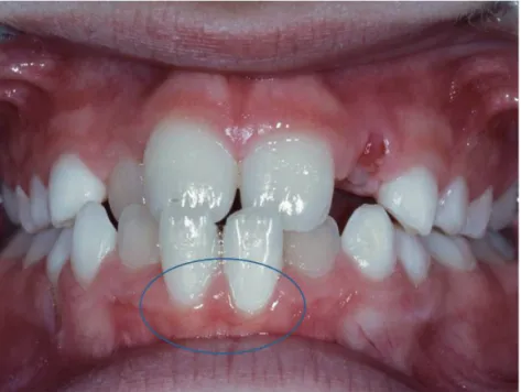
Moreover, early treatment will improve maxillary lip posture and facial appearance. (13) Lingually positioned maxillary incisors limit lateral jaw movement and they or their mandibular antagonists sometimes undergo pronounced incisal abrasion, a further indication forearly correction of the anterior crossbite. (3) In persistent anterior crossbite with functional shift, abrasion of the maxillary incisors can occur (Figure 1). This traumatic occlusion may also cause gingival irritation, recession (Figure 2) and increased mobility of both the maxillary and mandibular incisors affected. (11)
Figure 1. Abrasion of the enamel on the labioincisal edges of 11, 21, 22. These teeth had previously been in anterior crossbite with functional shift.
Figure 2. Initial gingival recession 31,41.
Skeletal origin
Patients with severe malocclusions, including those with skeletal Class III malocclusion, referred for combined orthodontic and surgical treatment have reported impaired aesthetics and chewing capacity as well as symptoms from the masticatory muscles, TMJ and headaches. (14) Clinical signs, such as pain on palpation of the TMJ and related muscles are also reported by these patients. (14)
Treatment methods
Dental origin
Various treatment options are available for anterior crossbite with functional shift. A recent systematic review disclosed a wide variety of treatment modalities, more than 12 methods, in use for correction of dental anterior crossbite without skeletal Class III malocclusion. However, there was a lack of strong evidence to support any of the techniques. This review highlighted the need for high quality clinical trials to identify the most effective intervention for correction of anterior crossbites without skeletal Class III malocclusion. (15)
on the number of incisors in anterior crossbite, the appliances used, tooth rotations and patient compliance. (3, 15)
During the planning stages of the present studies, before Paper I was conducted, all clinical orthodontists and 300 randomly selected general practitioners working in Sweden were sent questionnaires about their preferred treatment, approaches for correcting anterior crossbite with functional shift. For 80 percent of the general practitioners, the method of choice was a removable acrylic plate with protruding springs for the maxillary incisors. In contrast the preferred treatment method of 80 percent of the specialist orthodontists was a fixed appliance with brackets and wires (unpublished data). These results formed the basis for selection of the fixed and removable appliances to be evaluated and compared in this thesis.
A recent Swedish study has subsequently partly confirmed the results of the unpublished survey above, i.e. that consultant orthodontists most commonly recommended that general practitioners should use removable appliances for treatment of anterior crossbite with functional shift. (16)
Among general practitioners, the most common approach to correction of anterior crossbite with functional shift seems to be the removable appliance consisting of an acrylic plate with protruding springs. (3, 16) It comprises of acrylic plate with protrusion springs for the incisors in anterior crossbite, often bilateral occlusal coverage of the posterior teeth, stainless steel clasps on either the deciduous first molars or the first premolars (if erupted) and the permanent molars. It is recommended that the protrusion springs are activated once a month until normal incisor overjet is achieved. Lateral occlusal coverage is often used to avoid vertical interlock between the incisors in crossbite and the mandibular incisors and also to increase the retention of the appliance. This occlusal coverage can be removed as soon as the anterior crossbite is corrected. The dentist instructs the patient firmly to wear the appliance day and night, except for meals and tooth-brushing, i.e. the appliance should to be worn at least 22 hours a day. Progress is usually evaluated every four weeks. The same appliance can subsequently serve inactive as a passive retainer, often for a retention period of two-three months. (1-3, 17) An additional retrusive labial bow for the mandibular incisors might also be incorporated in the acrylic plate, in order to retrude the mandibular incisors and to make it more difficult for the patient to
achieve anterior shift of the mandible during treatment. A protruding screw is also sometimes used instead of a spring, to protrude the incisors in anterior crossbite. (2, 3)
Another type of removable appliance type is Fränkel III, with acryl material on both maxillary and mandibular teeth, buccal-anterior acrylic shields to enhance maxillary anterior growth and a labial bow on the mandibular incisors, for retrusion of the incisors and the mandible. (4)
A wooden spatula is also sometimes used to correct a single tooth in dental anterior crossbite without a deep overbite. The patient is instructed to place a wooden spatula approximately 45 degrees behind the tooth in crossbite and using the lower incisor as a fulcrum, to exert slight pressure on the tooth in a labial direction. (1, 11)
If the anterior crossbite involves a single lingually positioned maxillary incisor occluding with a single labially displaced mandibular incisor, a cross elastic may sometimes be used. A button or bracket is bonded to the maxillary incisor lingually and another button bonded to the occluding mandibular incisor buccally. The two brackets are connected by an intermaxillary elastic, correcting the incisors in anterior crossbite. Cross elastics should be used with care as there is a risk of extrusion of the affected incisors. (3)
The fixed appliance can consist of varying numbers of stainless steel brackets and wires of different dimensions and materials, sometimes with loops and bends. (1, 3, 18, 19)
Also described in the literature is a fixed appliance system with “2x4 appliance”, comprising bands on the maxillary first permanent molars and brackets on the four maxillary incisors. A flexible wire is often used for nivellation and then a steel wire with advancing loops. (3, 18) This appliance seems to be particularly effective in cases where the anterior crossbite is combined with lack of space, pronounced tipping and tooth rotation. (3)
In the permanent dentition fixed appliance with Class III elastics can be used and often the elastic in the maxilla is hooked to more distal teeth and in the mandible to more anterior teeth resulting in anterior pull on the maxilla and distal pull on the mandible and the teeth. In cases of crowding also, it is sometimes combined with extractions of the first lower premolar and second upper premolar but this only permits milder Class III discrepancy to be camouflaged
Other studies describe treatment of cases where only one incisor is in crossbite: composite material is bonded to the opposing mandibular incisor, to create an inclined bite plane. However, difficulties are reported in cases of deep bite and rotated incisors. (20, 21)
Skeletal origin
A recent systematic review disclosed various orthodontic treatment modalities for Class III malocclusion. (22) Different kinds of extraoral pull have been used to inhibit mandibular anterior growth and/or to enhance maxillary anterior growth. If the patient has a retrognathic maxilla, a protraction facemask also called reverse head-gear (Delaire or Petit mask) can be used in the early mixed dentition. The reverse headgear is applied to a fixed appliance in the maxilla by elastics pulling the maxilla forward; pillows in the masks are applied to the forehead and chin, exerting a retrusive pull on the mandible at the same time. It is often combined with lateral expansion. It is claimed that this appliance results in displacement of the maxilla anterior to processus pterygoideus at the os sphenoidea. (4, 23-25)
In a multicentre RCT of early Class III orthopedic treatment with a protraction facemask and untreated controls, successful outcomes to Class I occlusion were reported in 70 per cent of the subjects. (26)
Another appliance is the extraoral high pull in combination with a chin cup, to force the chin and mandible backward and upwards. This appliance is sometimes used for mild mandibular prognathism in the early mixed dentition but often requires lengthy treatment and is reported to have little effect. (4, 27, 28)
Recently, bone-anchored maxillary protraction (BAMP) has been used to treat Angle Class III malocclusion with maxillary hypoplasia. The maxillary miniplate is fixed by three monocortical screws at the infrazygomatic crest, and the mandibular miniplate with two screws between the lateral incisor and the canine. Elastics pulls between the upper and lower miniplate 24 h a day, will apply protrusive force to the maxilla and retrusive force to the mandible. (29)
Finally, in cases of severe skeletal Class III discrepancies in the permanent dentition, the most recommended treatment method is a combination of bimaxillary fixed appliance and orthognathic surgery in the maxilla and/or mandible. The most common orthognathic
surgery procedure in the maxilla for Class III discrepancy is forward movement with Le Fort 1 osteotomy. In the mandible, both sagittal split and ramus osteotomy are common surgical methods to set back the mandible. (3)
Stability
The fundamental goal of orthodontic treatment is to achieve a normal occlusion, which is morphologically stable in the long-term and functionally and aesthetically acceptable. Therefore, the real success rate and effectiveness of different treatment methods can be evaluated only after long-term follow-up. In general an appropriate follow-up period is five years after completion of active treatment. However this may vary, depending on the kind of outcome achieved or the aim of the treatment. (27, 30-32)
As early correction of anterior crossbite is undertaken in the growing child, it is important to evaluate long-term post-treatment changes. There are however, very few studies analysing the post-treatment effects of anterior crossbite correction and most are retrospective in design. (15, 33, 34)
Treatment Prognosis
Factors reported to influence successful treatment of anterior crossbite include the age at which the appliance is inserted, the severity of the malocclusion and heredity. (27)
If the patient can achieve an edge-to-edge incisor position, this improves the prognosis for orthodontic correction. Less favorable factors for only orthodontic treatment include a severely negative ANB angle or large gonion angle. Normal or deep overbite and a favorable growth pattern increase post-treatment stability after orthodontic correction. (3, 35)
Long-term stability, especially for early treatment of Class III malocclusion of skeletal origin, can be difficult to predict. In many cases early treatment may be successful and normal occlusion can be achieved. However, as mandibular growth continues after maxillary growth has ceased, there may be recurrence of previously corrected skeletal Class III malocclusion. (4, 27)
Economic evaluation
Economic evaluation is defined as the comparative analysis of alternative courses of action in terms of their costs (input) and consequences (output). (36)
Economic evaluation of health care interventions has assumed increasing importance and interest over the years. (37) Cost-effective health care requires assessment of the economic implications of different interventions. (38) Less cost-effective health-care may result in reduced services in other important health care areas. In future it is likely that decisions about allocation of resources for publicly funded orthodontic services will increasingly include economic evaluations: allocation will then be based not only on evidence of clinical effectiveness of treatment but also appropriate economic analysis to confirm value for money. (39)
Four main types of economic evaluations can be applied to accumulate evidence and compare the expected costs and consequences of different procedures. A cost-effectiveness analysis is characterized by analysis of both costs and outcomes, in cases where the outcomes of the different methods can differ. A cost-minimization analysis, which is a form of cost-effectiveness analysis, is used when outcomes of treatment alternatives are equivalent (e.g. anterior cross-bite will be corrected irrespective of which treatment is used) and the aim is to identify which alternative has the lower cost. In cost-utility analysis a utility-based outcome is used for instance to compare quality of the life following treatment. Biological, physical, sociological or psychological parameters are measured as to how they influence a person´s well-being. Finally, in a cost-benefit analysis the consequences (effects) are expressed in monetary units. (38)
Economic calculations are often divided into direct, indirect and societal (or total) costs.
Direct costs comprise material costs and treatment time needed for manpower all sessions for each patient. The material costs include for example orthodontic brackets, wires, and bonding, impression materials, consumables, laboratory material and fees. The costs for clinical treatment time include costs of the premises and equipment, maintenance, cleaning and staff costs.
Indirect costs are often the consequences of treatment and are defined as loss of income, incurred by the patient or the patient´s
parents, in taking time off from work to attend clinical appointments and to travel to and from the clinic.
The societal costs are the sum of direct and indirect costs, i.e. the total cost.
Patients´ perceptions
Pain and discomfort are recognized side effects of orthodontic treatment. (40, 41) Usually pain starts about 4 hours after insertion of the appliance, peaks between 12 hours and 3 days after insertion and then decreases for up to 7 days. (41-45) Almost all patients (95 percent) report pain or discomfort 24 hours after insertion of fixed appliances. Moreover, fixed appliances are reported to elicit higher pain responses than removable appliances. (46, 47) Higher pain scores are also reported for anterior than for posterior teeth. (43, 48) There is lack of consensus about gender differences, one study reporting no differences (49), and others indicating that girls are more prone to pain. (43, 48)
Experience of pain is always subjective and comprises both sensory and affective aspects, denoted as intensity and discomfort. Several studies also pointed out that pain associated with orthodontic treatment has a potential impact on daily life, primarily as psychological discomfort. (48, 50) Swallowing, speech and jaw function can be altered during orthodontic treatment. (43, 46) Chewing hard food can be difficult and reduced masticatory ability is reported 24 hours after fixed appliance insertion, with a return to baseline 4 to 6 weeks later. (43, 51)
Successful orthodontic outcomes depend on effective treatment methods, but it is also important to take into account patients´ acceptance of treatment: negative experiences such as pain and discomfort may be interpreted as potential disadvantages of various treatment approaches.
Evidence-based evaluation
To date there is no RCT comparing the effects of fixed and removable appliance therapy for correcting dental anterior crossbite with functional shift in the mixed dentition (15); hence no evidence-based conclusions can be drawn with respect to treatment of this malocclusion and these treatment methods. (30, 31)
The purpose of scientific assessment of healthcare is to identify interventions which offer the greatest benefits for patients while utilizing resources in the most effective way. Consequently, scientific assessment should be applied not only to medical innovations but also to established methods.
Evidence-based health care can be defined as the implementation of the evidence of systematic and precise studies in clinical decision-making. However, such evidence cannot be applied indiscriminately to all patients. Apart from scientific evidence, other factors are important determinants of treatment outcome, including the patient´s circumstances and, values and the clinician´s experience and preferences. When patients are informed about the treatment, both technical expertise and clinical experience are essential, but it also important that the clinician is well-informed about actual research and developments in the field. The goal of evidence-based health care is to find more effective treatment methods and to identify and avoid more ineffective methods. The evidence-based approach is also a valuable instrument for identifying knowledge gaps and clarifying the need for clinical trials. (52)
In an evidence-based approach, randomized controlled trials (RCTs) have become the golden standard design for evaluation of effectiveness. RCTs are considered to generate the highest level of evidence and provide the least biased assessment of differences in effects of two or more treatment alternatives. (53) Case series, case reports and finally expert opinion, generate low or insignificant evidence (Figure 3).
Consequently, using RCTs diminishes the influence of clinicians´ or patients´ preferences for certain treatment, and most importantly, the random allocation process ensures that confounding factors, that is factors over which we have no control during the trial, will affect the various constituent groups equally. (54)
For ethical reasons it is sometimes difficult to undertake highly evidence-based studies. It must also be remembered that lack of evidence not necessarily is synonymous with lack of effect. The study design has to be determined by the research question to be addressed, and therefore well-designed prospective or retrospective studies may also provide valuable evidence. However, such studies must then be interpreted with caution because of the limitations inherent in the study design. (55)
Basically, the following approaches can be recommended for comparing different interventions. (54)
• create a relevant question, i.e. use PICO: P = population, I = intervention, C = control, and O = outcome. The following is an example of a research question formulated according to PICO: Is fixed appliance treatment (intervention) more cost-effective (outcome) than removable appliance treatment (control) in 8-9 year-old patients with anterior crossbite with functional shift and no skeletal Class III (population)
• if possible use RCT design
• have sufficient numbers of subjects, i.e. make a sample size estimation
• use valid and reliable methods
• use the intention-to-treat (ITT) approach, i.e. all cases, successful or not, are included in the final analysis: thus, if any subjects withdraw from the trial, or do not respond to the treatment, these are still being recorded and counted as unsuccessful
Significance
Early orthodontic treatment, in the mixed dentition, versus later treatment, in the permanent dentition, is a controversial issue. Advocates of early treatment argue that it is easy to carry out, reduces the complexity of treatment in the permanent dentition, permits improved control of growth, may increase the patients’ self-esteem and reduces the risk of damage to teeth and tissues. It is clear that early treatment of anterior crossbite with functional shift is easy to carry out and allows the control of growth and improves function. Obvious, there are several important arguments to treat anterior crossbite with functional shift early. A recent systematic review (15) disclosed more than 12 methods for correcting anterior crossbite without skeletal Class III affecting one or more incisors. The best level of evidence currently available is from retrospective studies. The review emphasized the need for high quality clinical trials to identify the most effective treatment.
It is apparent that there are significant knowledge gaps with respect to treatment of anterior crossbite with functional shift in the mixed dentition. There is inadequate evidence to indicate the most effective treatment method and whether this treatment achieves long-term stability. The most cost-effective treatment has yet to be determined; moreover, patient perceptions of pain and discomfort associated with different treatment methods are poorly documented for this treatment.
The four studies on which this thesis is based were designed to address these important aspects of the most common orthodontic methods for correction of anterior crossbite with functional shift in the mixed dentition, namely short and long-term treatment effects of fixed and removable appliance, cost-effectiveness and patients´ perceptions of pain, discomfort and impairment of jaw function during treatment.
In order to generate a high level of evidence, evidence-based, randomized controlled trial (RCT) methodology was used. These studies are intended to complement current knowledge about effective treatment of anterior crossbite with functional shift without skeletal Class III in the mixed dentition. The results from the 4 studies are also expected to be beneficial for the patients who will be offered the most accepted treatment and with less pain and discomfort in
relation to efficacy. Of importance to clinicians, the conclusions are intended facilitate decision-making as to which treatment will give the best outcome. Finally, analysis of the cost-effectiveness will aid decisions by dental healthcare providers, who require evidence of high cost-effectiveness, i.e. good value for money.
AIMS
Paper I
To apply RCT methodology to assess and compare the effectiveness of fixed and removable orthodontic appliances in correcting anterior crossbite with functional shift in the mixed dentition.
Paper II
By means of a randomized controlled trial (RCT), to compare and evaluate the stability of outcome in patients who had undergone fixed or removable appliance therapy at the mixed dentition stage to correct anterior crossbite with functional shift affecting one or more incisors.
Paper III
To evaluate and compare the costs of fixed or removable appliance therapy to correct anterior crossbite with functional shift and to relate the costs to the effects using cost-minimization analysis.
Paper IV
To evaluate and compare patients´ perceptions of pain, discomfort and impairment of jaw function associated with correction of anterior crossbite with functional shift in the mixed dentition, using fixed and removable appliances.
HYPOTHESES
Paper I
Treatment of anterior crossbite with functional shift by fixed and removable appliances is equally effective.
Paper II
For correction of anterior crossbites with functional shift at the mixed dentition stage, use of fixed or removable appliances achieves similar long-term stability of outcome.
Paper III
Treatment with removable and fixed appliances to correct anterior crossbite with functional shift is equally cost-effective.
Paper IV
There will be minor differences between fixed and removable appliance therapy in terms of perceived pain intensity, discomfort and impairment of jaw function.
SUBJECTS AND METHODS
Subjects
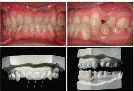
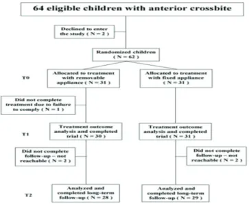
All patients participating in the four studies on which this thesis is based were consecutively recruited from the Department of Orthodontics, Faculty of Odontology, Malmö University, Malmö, Sweden and from one Public Dental Health Service Clinic in Malmö, Skåne County Council, Sweden. The following inclusion criteria applied: early to late mixed dentition, anterior crossbite affecting one or more incisors, no inherent skeletal Class III discrepancy (ANB angle > 0 degree), moderate space deficiency in the maxilla (up to 4 mm), a non-extraction treatment plan, and no previous orthodontic treatment (Figure 4).
Sixty-four patients who met the inclusion criteria were consecutively recruited. Two declined to participate; thus 62 patients were randomized into two groups, the fixed appliance (FA) or removable appliance (RA) group. The FA group comprised 12 girls and 19 boys (mean age 10.4, SD 1.52) and the RA group, 13 girls and 18 boys (mean age 9.1, SD 1.19).
One subject in the RA group withdrew from the study (Paper I) after non-compliance between T0 (before treatment) and T1 (after treatment finished). One further subject in the RA group had a relapse between T1 (after treatment finished) and T2 (2 years after treatment finished) and was retreated with a fixed appliance. Moreover, four subjects, two from each group, were excluded because they could not be contacted for the two-year follow-up. Thus at T2 in Paper II, 57 subjects remained in the study, 29 in the FA group and 28 in the RA group. The patient flow is illustrated in Figure 5.
Figure 5. Flowchart for Paper I, II and III.
In Paper III, the costs for patients in the two groups from Papers I and II were calculated, including all retreatments also (Figure 5).
Figure 6. Flowchart Paper IV.
In Paper IV, all 31 patients in each group completed the trial (Figure 6).
Ethical considerations
The informed consent form and study protocol were approved by the Ethics Committee of Lund University, Lund, Sweden, which follows the guidelines of the Declaration of Helsinki, (Dnr: 334/2004).
Consent and randomization
All patients and parents were informed of the purpose of the trial. After written consent was obtained, the patients were randomly allocated for treatment by either RA or FA. The subjects were randomized by an independent person in blocks of 10, as follows: 7 opaque envelopes were prepared with 10 sealed notes in each (5 notes for each group). Thus, for every new patient in the study, a note was extracted from the first envelope. When the envelope was empty, the second envelope was opened, and the 10 new notes were extracted successively as patients were recruited to the study. This procedure was then repeated 6 more times. The envelopes were in the care of one investigator, who randomly extracted a note and informed the clinician as to which treatment was to be used.
Methods
Papers I and II
After randomization, all patients were treated according to a pre-set standard concept. Impressions for study casts were taken on all subjects at the start (T0), after treatment finished including the retention period (T1) and 2 years post-retention (T2).
The patients were treated by a general practitioner under the supervision of two specialists in orthodontics. The two specialists were the supervising orthodontists at the two clinics where the study was conducted and the general practitioner became a postgraduate student during the trial period. Consequently, all treatments were performed in a general dentistry setting and therefore the fees and treatment codes for general practitioners were applied.
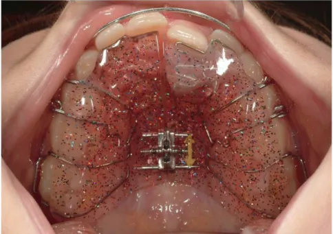
Figure 7. An example of fixed appliance.
The fixed appliance
The appliance consisted of stainless steel brackets (Victory, slot .022, APC PLUS adhesive precoated bracket system, 3M Unitek, Monrovia, California, USA). Usually, eight brackets were bonded to the maxillary incisors, deciduous canines and either to the deciduous
first molars or the first premolars, if erupted. All patients were treated according to a standard straight-wire concept designed for light forces (Figure 7). (19) The arch-wire sequence was: .016 heat-activated nickel-titanium (HANT), .019x.025 HANT, and finally .019x.025 stainless steel wire. To raise the bite, composite (Point Four 3M Unitek, US) was bonded to the occlusal surfaces of both the mandibular second deciduous molars. This prevented vertical interlock between the incisors in crossbite and the mandibular incisors. The composite was removed as soon as the anterior crossbite was corrected. Progress was evaluated every four weeks. The same fixed appliance then served as a passive retainer for a retention period of three months.
Figure 8. An example of removable appliance.
The removable appliance
The appliance comprised an acrylic plate, with protrusion springs for the incisors in anterior crossbite, bilateral occlusal coverage of the posterior teeth, stainless steel clasps on either the deciduous first molars or the first premolars (if erupted) and the permanent molars (Figure 8). The protrusion springs were activated once a month until normal incisor overjet was achieved. Lateral occlusal
coverage was used to avoid vertical interlock between the incisors in crossbite and the mandibular incisors and also to increase retention of the appliance. The occlusal coverage was removed as soon as the anterior crossbite was corrected. The patient was firmly instructed by the dentist to wear the appliance day and night, except for meals and tooth-brushing, i.e. the appliance was to be worn at least 22 hours a day. Progress was evaluated every four weeks. The same appliance then served as a passive retainer for a retention period of three months.
Outcome measures in Paper I and II
The following measures were assessed:• success rate of anterior crossbite correction (yes or no) • treatment duration in months: from insertion to date of
appliance removal
• overjet and overbite in millimetres for incisors in anterior crossbite
• arch length incisal (ALI): distance in millimetres from the incisal edge of the maxillary incisor in anterior crossbite to tangents of the mesiobuccal cusp tips of the maxillary first molar (Papers I and II) (Figure 9).
• arch length gingival (ALG): distance in millimetres from the gingival margin of the maxillary incisor in anterior crossbite to tangents of the mesiobuccal cusp tips of the maxillary first molar (Papers I and II) (Figure 9).
• maxillary dental arch length total (ALT): distance in millimetres at the alveolar crest between the mesial surface of the left and right maxillary first molars (Papers I and II) (Figure 9).
• tipping effect of maxillary incisor, i.e. (ALI minus ALG (before treatment)) divided by (ALI minus ALG (after treatment)) (Paper I).
• transverse maxillary molar distance (MD): transverse distance in millimetres between the mesiobuccal cusp tips of the maxillary first molars (Papers I and II) (Figure 9).
Figure 9. Measures on casts.
Successful treatment was defined as positive overjet (normal inter-incisal relationship) for all incisors at T1, when treatment was completed or at the latest within one year of treatment start T0 (Papers I and II) and at T2, two years post-retention (Paper II).
The duration of treatment was registered from the patient files as the time taken in months to correct the anterior crossbite with functional shift. If normal overjet was not achieved, the treatment time was recorded as one year, i.e. the pre-set maximum duration of treatment (Paper I).
Intention to treat (ITT)
Data on all patients were analysed on an ITT basis, i.e. if the anterior crossbite was not corrected within one year in Papers I and II, or if a corrected anterior crossbite relapsed during the two-year follow-up period in Paper II, the outcome was defined as unsuccessful. Thus, all cases, successful or not, were included in the final analysis.
Measurements
The overjet, overbite, arch length, and transverse maxillary molar distance were measured with a digital sliding calliper (Digital 6,
8M007906, Mauser-Messzeug GmbH, Oberndorf/Neckar, Germany). All measurements were made to the nearest 0.1 mm.
Changes in the different measurements were calculated as the difference between T1 and T0 in Papers I and II. Differences between T2 and T1, T2 and T0 were calculated in Paper II. All study cast measurements were blinded, i.e. the examiner was unaware of the group to which the patient belonged. Furthermore, the T0, T1 and T2 casts were randomized for measurements.
Paper III
The following calculations were made:
• total costs (societal costs), including both direct and indirect costs.
• success rate of anterior crossbite correction after treatment finished (T1) and 2 years post retention (T2)(yes or no) • number of appointments
The direct costs comprised material costs and treatment time for manpower for all sessions and for each patient.
Material costs, i.e. costs for impression material, orthodontic brackets, orthodontic wires, orthodontic bonding, consumables, laboratory material and fees etc. were compiled and calculated according to average commercial prices.
Treatment time costs included the costs of the premises, dental equipment, maintenance, and cleaning and were calculated according to average commercial prices in Sweden; these figures were used to establish estimated costs for each unit in the study. Similarly, staff salaries, including payroll tax, were calculated for dental assistants, general dental practitioners, and the supervising orthodontists, based on a previous economic calculation from 2010 (56) and upgraded according to the Consumer Price Index for 2013. All estimates of treatment time costs were calculated in Swedish currency, at SEK 937 (108 Euro) per hour for a general practitioner. In addition, the number of appointments, scheduled and emergency appointments, was noted.
The indirect costs were defined as loss of income (wages plus social security costs) incurred by the patients´ parents, assuming that they were absent from work to accompany the patient to the orthodontic
appointment. Data sourced from the Swedish National Bureau of Statistics (57) gave the wages of an average Swedish worker as SEK 243 or 28 Euro per hour. One parent accompanied the patient to appointments. Parental absence from work was estimated at 80-90 minutes per appointment, i.e. 20-30 minutes for the appointment and 60 minutes´ travelling time, for parent and child, to and from the dental clinic. Appointments for insertion and removal of FA were recorded as 30 minutes each and all other appointments for FA or RA were recorded at 20 minutes each.
All costs were based on 2013 prices and were expressed in Euro, SEK 100=11.56 Euro on mean currency value. (58)
The sum of direct and indirect costs was defined as “societal costs”. The cost-analysis was based on the Intention-To-Treat (ITT) principle, i.e. the analysis included data on costs for patients needing re-treatment due to non-compliance and relapse.
Cost-minimization analysis
A cost-minimization analysis (CMA) was undertaken, based on the findings in Paper II (59), that the treatment alternatives achieve equal outcomes (i.e. anterior crossbite will be corrected irrespective of which treatment alternative is applied).
Three different measurements were made for societal costs
1. Calculation and comparison of mean societal costs of successful cases only, on completion of active treatment in both groups, i.e. by dividing the social costs of the successful cases (FA group N=31 and RA group N=30) by the number of successful cases in each group (FA group N=31 and RA group N=30).
2. Calculation and comparison of all societal costs for both successful and unsuccessful treatment in the respective groups (FA group N=31 divided by N=31 and RA group N=31 divided by N=30).
3. Finally, societal costs were calculated for all patients, including the 2 year follow-up period and all re-treatment, for the total number of patients in each group. Thus, the costs of two re-treatments in the FA and two re-treatments in the RA group were added to the societal costs in each group, to calculate the mean societal costs including re-treatments. Societal costs
including re-treatment/number of patients, i.e. societal costs for 33 treatments in the FA group divided by 31 patients in this group and societal costs for 33 treatments in the RA group divided by 31 patients.
Paper IV
The measures were: • Pain intensity • Discomfort
• Impairment of jaw function
• Self-estimated disturbance to appearance
The assessment included 26 questions for self-reporting. Most of the questions were sourced from questionnaires which have previously been shown be valid and reliable. (60) Two new questions were included: “Do you have pain in your lip” and “Do you think your orthodontic appliance disturbs your appearance”.
The patients in both groups completed the questionnaires before insertion of the appliance (baseline), later on the day of insertion and then every day/evening for the following seven days. In addition, the questionnaire was filled in, at the first scheduled appointment after 4 weeks and at the second scheduled appointment 8 weeks post-insertion of the appliance.
The patients were given instructions on how to fill in the questionnaire and needed about 10 minutes to complete it. At baseline and at the first and second scheduled appointments the patients filled in the questionnaires at the clinic. During the first seven days of treatment, the patients filled in the questionnaires daily at home.
Pain and discomfort
All questions 1-10 are presented in Table 1, Paper IV. Questions 1 to 7, on pain and discomfort, were graded on a VAS scale with the end phrases ”no pain” and worst pain imaginable” or ”no tension” and ”worst tension imaginable”. (60)
Headache
Question 8 had a binary response (yes/no). For questions 9 and 10 there were multiple choice responses, whereby one answer was to be
selected from the three presented. The responses to Question 9 were “sporadic”, “frequent” or “constant” and to question 10 “1-3 times a month”, “once or twice a week”, “every other day”.
Use of analgesics
At the patients´ first revisit to the clinic, four weeks post-insertion, they were questioned verbally about whether they had taken analgesics to ease discomfort or pain associated with insertion of the appliance.
Impairment of jaw function
There were 15 questions on jaw function: three on mandibular function, five on psychosocial activities, and seven on eating specific foods (Table 2, Paper IV). Each item was assessed on a 4-point scale, with the options “not at all”, “slightly”, “very difficult” or “extremely difficult”. (60)
Self-estimated disturbance to appearance
One question related to the patient’s perception of the influence of the appliance on personal appearance: “Do you think your orthodontic appliance disturbs your appearance?” and was graded on a VAS scale with the end phrases “not at all” and “very much”. The question was answered at the second rescheduled appointment, 8 weeks after insertion of the appliance.
Side effects
Intra-oral radiographs of the maxillary incisors were taken routinely, before and after treatment. The patients in both groups showed good to acceptable oral hygiene before treatment. The presence of white spot lesions was recorded before and after treatment.
Statistical analysis
Sample size calculation
In Papers I and II, the sample size for each group was calculated
and based on a significance level of α = 0.05 and a power (1-β) of
90 percent, to detect a mean difference of 1 month (± 1 month) in treatment duration between the groups. According to the sample size calculation, each group would require 21 patients. To increase the power further and to compensate for possible drop-outs, it was decided to select a further a 20 patients, i.e. 31 patients for each group.
Descriptive statistics
SPSS software (version 20.0 and 21.0 SPSS Inc, Chicago Ill., USA) was used for statistical analysis of the data. For numerical variables, normally distributed, arithmetic means and standard deviations (SD) were calculated (Papers I-II). The 95% confidence interval for the mean was calculated for all study model measurements in Papers I and II. For numerical variables not normally distributed, median values and interquartile ranges were calculated for costs in Paper III and questionnaire responses in Paper IV.
Differences between groups
Analysis of means was made with independent sample t-test to compare active treatment duration and treatment effects between the groups in Papers I and II. A Kolmogorov-Smirnov test indicated that the numerical variables in Papers III and IV were not normally distributed and thereby Mann-Whitney U test was applied for intergroup comparisons of costs (Paper III) and questionnaire responses on pain and discomfort (Paper IV). For categorical variables, chi-2 test was used in Papers I-IV. A P value of less than 5 percent (P< 0.05) was regarded as statistically significant.
Method error analysis
In Papers I and II, ten randomly selected study casts were measured on two occasions, at an interval of at least one month. Paired t-tests disclosed no significant mean differences between the two series of records, i.e. no systematic errors were detected. The method error size
was calculated according to Dahlbergs´ formula and did not exceed
RESULTS
Paper I – Treatment effects
In all, 64 patients were invited to participate in the study, but two declined. Thus, 62 patients were randomized into the two groups. Group A (removable appliance group) comprised 13 girls and 18 boys (mean age 9.1 years, SD 1.19) and group B (fixed appliance group), 12 girls and 19 boys (mean age 10.4 years, SD 1.65). Before treatment start, no significant differences were found between the groups regarding overjet, overbite, ALI, ALG, ALT, MD, gender distribution or the number of incisors in anterior crossbite (Table 2, Paper II). However, the age difference was significant between the groups (P<0.05).
Success rate
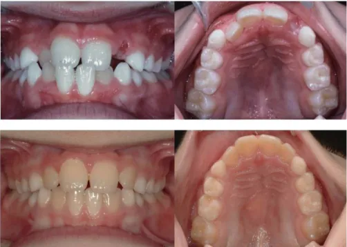
The anterior crossbites with functional shift in all patients in the fixed appliance group, and all except one in the removable appliance group, were successfully corrected. The patient who was not successfully treated was unable to accept the removable appliance and after the trial was treated with a fixed appliance. Thus the success rate in both groups was very high, and the intergroup difference was not significant (Figure 10).
Treatment time
The average duration of treatment, including the 3-month retention period, was 6.9 months (SD 2.8) in the removable appliance group and 5.5 months (SD 1.41) in the fixed appliance group. Thus, treatment duration was significantly less in the fixed appliance group (P< 0.05).
Figure 10. Before and after treatment.
Measurements (T0-T1) between the appliance groups
The increase in overjet after treatment was significantly greater in the fixed appliance group (P< 0.05) (Table 2, Paper I). The increases in incisal (ALI) and gingival arch length (ALG) after treatment were also significantly greater in the fixed than in the removable appliance group. After treatment, no significant intergroup differences were disclosed with respect to overbite, total maxillary dental arch length or transverse maxillary molar distance. The tipping effect of the incisors was relatively small, with no significant intergroup difference.
Measurements within the appliance groups (T0-T1)
Within the groups, overjet, incisal arch length (ALI) and the tipping effect of the incisors increased significantly. The fixed appliance group also showed a significant increase in gingival arch length (ALG) (Table 2, Paper I).
Untoward effects of treatment
Untoward effects necessitating emergency treatment occurred in both groups. In the fixed appliance group, loss of brackets (nine
In the removable appliance group, clasps and/or acrylic fractures occurred in four patients. A further four patients lost their removable appliances and were provided with new ones during treatment.
Paper II – Stability
One subject in the removable appliance group had a relapse between T1 (after treatment finished) and T2 (2 years after treatment finished) and was retreated with a fixed appliance. Moreover, four subjects, two from each group, were excluded because they could not be contacted for the two-year follow-up. Thus in Paper II, the number of subjects at T2 comprised 29 in the fixed appliance group (FA group) and 28 in the removable appliance group (RA group). The patient flow is illustrated in Figure 5.
Successrate
At the two-year follow-up, and altogether in the two groups relapses had occurred in three subjects. Thus 27 of 29 patients in the fixed appliance group and 27 of 28 patients in the removable appliance group had maintained normal inter-incisal relationships. It was also noted that at follow-up, transition to the permanent dentition had occurred in almost all of the subjects in both groups.
Measurements between and within the groups (T1-T2)
During T1 to T2, a small but significant increase in overbite occurred in the removable appliance group.
A small, significant inter-group difference was found with respect to overjet. There were no other significant changes in the outcome variables (Table 4, Paper II).
Measurement between and within the groups (T0-T2)
The overall changes during the study period (T0-T2) are shown in (Table 5, Paper II). Significant increases in overjet and incisal arch length (ALI) were found in both groups.
In the fixed appliance group, ALI and ALG increased significantly more than in the removable appliance group. No other significant intra- or inter-group differences were observed.
Figure 11. Casts of a patient with anterior crossbite 21, 22 before treatment, after treatment and 2 years post retention.
Paper III – Cost-minimization
Of the 31 patients in the removable appliance (RA) group, one poorly compliant patient failed to complete the study. All 31 patients in the fixed appliance (FA) group were treated successfully. During the 2-year post-treatment follow-up, relapses occurred in one subject in the RA group and in 2 subjects in the FA group. Consequently, four patients, 2 in each group, needed retreatment and this was undertaken with fixed appliances. The patients needing retreatment showed no differences in baseline characteristics from subjects who were treated successfully. The patient flow chart is presented in (Figure 5).
Societal costs (total costs)
The mean societal costs (direct and indirect costs) for patients with successful outcomes were significantly lower for the FA group than for the RA group (p<0.000), (Table 1, Paper III).
For both successful and unsuccessful outcomes, the mean societal costs were also significantly lower for the FA group than for the RA group (p<0.000).
The total mean societal costs for all 31 patients in each group, including the two retreatments in each group, were significantly lower for the FA group than for the RA group (p<0.005), (Table 1, Paper III) 678 Euro than 1031 Euro respectively i.e. treatment by RA was 52% more expensive than by FA.
Direct costs – material
For patients with successful treatment outcomes (31 FA and 30 RA) the mean material costs were significantly lower for the FA group than for the RA group (p<0.000). The mean material costs for both successful and unsuccessful outcomes (31/31 FA and 31/30 RA) were
also significantly lower for the FA group compared to the RA group (p<0.000). Finally, when re-treatments were included, the mean material costs were significantly higher for the RA group than for the FA group (p<0.000), (Table 1, Paper III).
Direct costs – treatment time
The mean total treatment time and costs for the patients with successful treatment outcomes (31 FA and 30 RA) were 179 minutes/323 Euro for fixed appliance versus 205 minutes/371 Euro for removable appliance. The mean total treatment time and costs, for both successful and unsuccessful outcomes, (31 in each group) were 179 minutes/323 Euro for fixed appliance and 212 minutes/382 Euro for removable appliance. When retreatments are included, the mean total treatment time and costs were 194 minutes/351 Euro for fixed appliance and 231 minutes/417 Euro for removable appliance. There was no significant difference in treatment time costs, between the two types of appliance (Table 1, Paper III).
Indirect costs
The mean indirect costs for successful treatments were significantly lower for the fixed appliance than for removable appliance: 275 Euro vs 346 Euro (p<0.01). For both successful and unsuccessful outcomes combined, the mean indirect costs were also significantly lower for fixed appliance than for RA: 275 Euro vs 356 Euro (p<0.01). When all retreatments were included, the indirect cost was significantly lower for fixed appliance than for RA: 293 Euro (SD 153) vs 383 euro (SD 200) (p<0.01) (Table 1, Paper III).
The indirect costs comprised 44% of the societal costs for FA therapy and 37% for RA therapy.
Number of appointments
The mean number of appointments for patients with successful treatment outcomes was 7.2 for the FA group and 9.2 for the RA group (p=0.005). For successful and unsuccessful outcomes combined, the number of appointments was 7.2 for the FA and 9.6 for the RA group (p=0.005). When re-treatments were included, the mean number of appointments was 7.8 for the FA group and 10.1 for the RA group, with no significant difference between the groups.
In each treatment group, an average of one emergency/unscheduled appointment was recorded per patient, most frequently for loss of brackets in the FA group or fractured clasps or acrylic plate edges in the RA group.
Side effects
Intra oral radiographs of the maxillary incisors were taken before and after treatment: no cases of root resorption were diagnosed in either group. Moreover, during treatment there was no case of interference to maxillary canine eruption by a lateral incisor. In both groups the patients showed good to acceptable oral hygiene before and during treatment. The presence of white spot lesions before and after treatment was also recorded: no new lesions developed in either group.
Paper IV – Pain and discomfort
All 62 randomized patients completed the trial (Figure 6). The fixed appliance (FA) group comprised 12 girls and 19 boys (mean age 10.4 years, SD 1.65) and the removable appliance (RA) group, 13 girls and 18 boys (mean age 9.1 years, SD 1.19).
The response rate for the separate questions ranged from 90 to 100%. No gender differences were found for the responses to any of the questions.
At baseline, i.e. before insertion of the appliances, there were no significant intergroup differences in responses to any of the questions.
Pain intensity
The general intensity of pain was low to moderate in both groups, although on day 2 a few children, primarily in the FA group, reported high pain levels (Table 3, Paper IV) (Figure 12, 13). The intensity of pain was significantly higher for FA on day 2, when the maxillary incisors were in contact (p=0.017) (Table 3, Paper IV) (Figure 12) and on day 1, when the maxillary incisors were not in contact (p=0.040) (Figure 13). Overall the pain intensity peaked in both groups after 2 days of treatment. After these two days of treatment no significant difference was found in pain intensity between the groups.
Although the intensity of pain was low, the patients in the RA group experienced more pain in their palate (p=0.021) after 6 days of treatment.
After 7 days, the RA group also reported more pain from the lips than the FA group (p=0.040).
At both rescheduled appointments, after 4 and 8 weeks of treatment the difference in pain intensity between RA and FA groups was non-significant for any pain-related question. Very low levels of pain were experienced in the tongue at any time for both appliances.
Overall, none of the patients reported any use of analgesics during the trial period.