Linköping University Medical Dissertations No. 1340
Influence of Genetics and
Mechanical Properties on
Large Arteries in Man
Rachel De Basso
Division of Cardiovascular Medicine Department of Medical and Health Sciences
Faculty of Health Sciences Linköping University, Sweden
Rachel De Basso, 2013
Cover picture: Ultrasound image of the popliteal artery.
Published article has been reprinted with the permission of the copyright holder.
Contents
CONTENTS
ABSTRACT ... 1 LIST OF PAPERS ... 3 ABBREVIATIONS ... 5 INTRODUCTION ... 7Cardiovascular disease and mortality ... 7
Structure of the arterial wall ... 7
Mechanical properties of arteries ... 9
Effects of aging on arteries ... 11
Aneurysmal disease ... 12 Fibrillin-1 ... 15 Angiotensin-converting enzyme ... 17 AIMS ... 19 MATERIALS ... 21 Ethical approval ... 21 Studied subjects ... 21 METHODS ... 23 Ultrasound measurements ... 23
Measuring mechanical properties of arteries ... 26
Noninvasive blood pressure measurements ... 27
Classification of cardiovascular events (Paper V) ... 28
Measurement of ACE level (Paper III) ... 29
Genotyping ACE (Paper III) ... 30
Genotyping Fibrillin-1 (Paper IV-V) ... 30
RESULTS ... 33
Mechanical properties of the popliteal artery ... 33
Angiotensin-converting enzyme and abdominal aortic stiffness ... 36
Fibrillin-1 and abdominal aortic stiffness ... 38
Fibrillin-1 and cardiovascular morbidity and mortality ... 39
DISCUSSION ... 43
Popliteal artery, an unusual muscular artery ... 43
Genetics and arterial stiffness ... 47
ACE and mechanical properties of the abdominal aorta ... 47
Fibrillin-1 and the cardiovascular system ... 49
Methodological considerations and limitations ... 53
CONCLUSIONS ... 55
POPULÄRVETENSKAPLIG SAMMANFATTNING ... 57
ACKNOWLEDGEMENTS ... 61
Abstracts
ABSTRACT
Arterial pathology is the major contributor to cardiovascular diseases and mortality. The mechanical properties of arteries are independent factors for cardiovascular disease and mortality, where genetics influence the structure of the arterial wall, which may result in change in arterial stiffness. The aims of this thesis were to study the mechanical properties of the popliteal artery (PA) in healthy subjects and the influence of angiotensin-converting enzyme (ACE) polymorphism and Fibrillin-1 (FBN1) polymorphism on large arteries. Further, the impact of FBN1 polymorphism on cardiovascular morbidity and mortality was investigated.
The PA is, after the abdominal aorta, the most common site of aneurysmal development. The PA was studied in healthy subject with ultrasound and the diameter increased and the distensibility decreased with age, with men having lower distensibility than women. This seems not to be the behavior of a true muscular artery but rather of a central elastic artery such as the aorta, and might have implications for the susceptibility to aneurysm formation, as well as the association of dilating disease between the PA and the aorta. The wall stress in the PA was low and unaffected by age, probably caused by a compensatory remodeling response with an increase in wall thickness. This indicates that other mechanisms than wall stress contribute to the process of pathological dilatation in the PA.
The ACE D allele may be associated with abdominal aortic aneurysm. Elderly men with the ACE D allele were associated with increased abdominal aortic stiffness compared to men carrying the I/I genotype. This suggests that the ACE D allele impairs arterial wall integrity, and in combination with local hemodynamic and other genetic factors it may have a roll in aneurysm formation.
The FBN1 2/3 genotype has been associated with increased systolic blood pressure. The FBN1 2/3 genotype in middle-aged men was associated with increased abdominal aortic stiffness and blood pressure which indicates an increased risk for developing cardiovascular disease. The increased presence of plaque in the carotid artery of middle-aged men with the FBN1 2/3 genotype indicates a pathological arterial wall remodeling with a more pronounced atherosclerotic burden, but did however not affect the risk of cardiovascular events and/or death in this population. This relationship needs to be studied further.
List of Papers
LIST OF PAPERS
This thesis is based on the following papers, which will be referred to by their roman numbers.
I. Debasso R, Åstrand H, Bjarnegård N, Rydén Ahlgren Å, Sandgren T and Länne T.
The popliteal artery, an unusual muscular artery with wall properties similar to the aorta – implications for the susceptibility to aneurysm formation?
Journal of Vascular Surgery 2004; 39(4): 836-842.
II. De Basso R, Åstrand H, Rydén Ahlgren Å, Sandgren T and Länne T. Low wall stress in popliteal artery – other mechanisms responsible for the predilection of aneurysmal dilatation?
Manuscript.
III. Ljungberg LU, De Basso R, Alehagen U, Björck HM, Persson K, Dahlström U and Länne T.
Impaired abdominal aortic wall integrity in elderly men carrying the angiotensin-converting enzyme D allele.
European Journal of Endovascular Surgery 2011; 42(3): 309-316.
IV. Powell JT, Turner RJ, Sian M, Debasso R and Länne T.
Influence of fibrillin-1 genotype on the aortic stiffness in men. Journal of Applied Physiology 2005; 99(3): 1036-1040.
V. De Basso R, Hedblad B, Carlson J, Persson M, Östling G and Länne T. Increased carotid plaque burden in men with the Fibrillin-1 2/3 genotype.
Manuscript.
Abbreviations
ABBREVIATIONS
AA Abdominal AortaAAA Abdominal Aortic Aneurysm ACE Angiotensin-Converting Enzyme
ACEi Angiotensin-Converting Enzyme inhibitors Ang I Angiotensin I
Ang II Angiotensin II
ARBs Angiotensin II Receptor Blockers β Beta Stiffness
BMI Body Mass Index BSA Body Surface Area BP Base Pair
CAD Coronary Artery Disease CC Compliance Coefficient CA Carotid Artery
CCA Common Carotid Artery CFA Common Femoral Artery CI Confidence Interval CV Coefficient of Variation CVD Cardiovascular Disease D Deletion
D/D Deletion/Deletion DBP Diastolic Blood Pressure DC Distensibility Coefficient EGF Epidermal Growth Factor Ep Pressure Strain Elastic Modulus FBN1 Fibrillin-1
I Insertion
I/D Insertion/Deletion I/I Insertion/Insertion IMT Intima Media Thickness LD Lumen Diameter MAP Mean Arterial Pressure MFS Marfan Syndrome MI Myocardial Infarction MMP Matrix Metalloproteinase
PA Popliteal Artery
PAA Popliteal Artery Aneurysm PCR Polymerase Chain Reaction PP Pulse Pressure
PWV Pulse Wave Velocity SBP Systolic Blood Pressure
TGF-β Transforming Growth Factor-Beta VNTR Variable Number of Tandem Repeat WS Wall Stress
Introduction
INTRODUCTION
Cardiovascular disease and mortality
Heart diseases and stroke are the leading causes of death in the western world (Lloyd-Jones et al., 2010). The mortality rate of circulatory diseases has decreased with more than 60% from 1987 to 2011. However, still in 2011 diseases of the circulatory system were the underlying cause of death in 39% of women and in 38% of men in Sweden (Socialstyrelsen, 2012). Reduction in population levels of cholesterol, blood pressure, smoking and a wider use of effective treatments among persons with existing cardiovascular disease (CVD) accounts for the decline in coronary heart disease deaths. Unfortunately, trends of increasing obesity, increasing prevalence of hypertension and type 2 diabetes mellitus in the pediatric population, will likely result in future increases of CVD and stroke in adults (Lloyd-Jones et al., 2010).
The mechanical properties of arteries are independent factors for CVD and mortality (Laurent et al., 2001, Gasecki et al., 2012). To understand the pathology of CVD it is of interest to investigate the mechanical properties of the arterial wall in healthy subjects and its development during life as well as the genetic influence on CVD and mortality. Genetic factors may act indirectly through age, blood pressure, smoking, cholesterol levels, glycemia or directly affect the structure of the arterial wall, which may result in increased arterial stiffness (Laurent et al., 2005). Arterial pathology is the major contributor to cardiovascular diseases and mortality.
Structure of the arterial wall
The elastin and collagen ratio varies in the arterial system where elastin being the dominant component in central arteries and collagen in peripheral arteries. The arterial system is divided in two major categories dependent on the composition of the arterial wall: the elastic arteries (the aorta, common carotid artery, common iliac artery and main pulmonary artery) and the muscular arteries (e.g. the common femoral artery, renal artery, popliteal artery, brachial artery, coronary artery and cerebral artery), were the central elastic arteries have a large proportion of elastin components, larger diameter and being
located closer to the heart, and the peripheral muscular arteries contain a higher proportion of collagen and smooth muscle cells than elastic fibers and have medium-sized diameters.
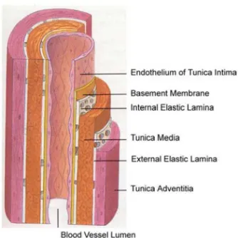
The arterial wall consists of three layers: tunica intima, tunica media and tunica adventitia (Figure 1). The tunica intima is the innermost layer to the blood flow and consists of a single layer of endothelial cells, which are arranged according to the blood flow, and a sub endothelial layer of elastin and collagen fibres that anchor it to the internal elastic lamina. The internal elastic lamina consists of a fenestrated membrane of elastin and merges the tunica intima and tunica media and is more prominent in muscular arteries than in elastic arteries.
The tunica media is the middle layer and the most important part of the wall and where the mechanical properties of the wall are determined. In elastic arteries the tunica media consists primarily of elastic laminae that
Figure 1. Schematic illustration of the various layers of the arterial wall: tunica intima,
Introduction
The outer layer, tunica adventitia, is separated from the tunica media with the external elastic lamina and consists mostly of collagen and some elastin tissue that connects with surrounding connective tissue, small blood vessels (vasa vasorum), nerves and fibroblasts (Nichols et al., 2011).
Mechanical properties of arteries
The mechanical properties of arteries have a major impact on cardiac work. The central elastic arteries accommodate blood ejected from the left ventricle and stores a part of the stroke volume during each systole and during diastole drain this stored volume, thus permitting continuous perfusion of peripheral organs and tissue. This cushioning effect is known as the Windkessel effect.
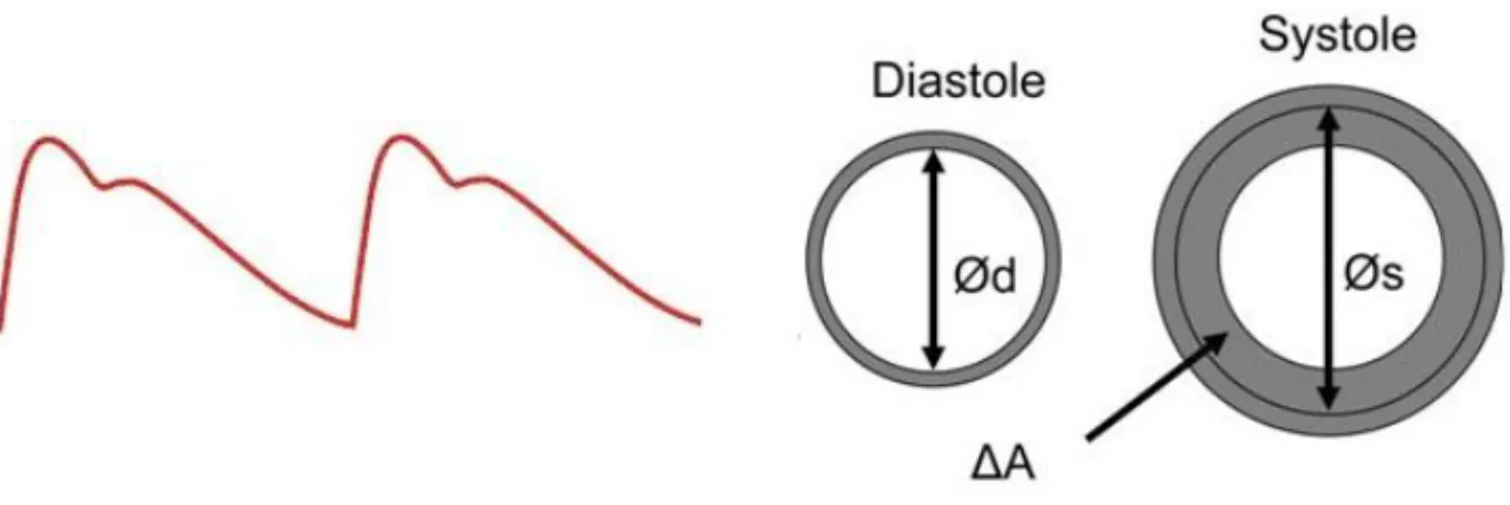
The properties of elastin provide the conditions of the arterial wall to distend during application of force (pressure) and to retract when the force is removed. The force per unit area that causes deformation (distension) is called stress (i.e. blood pressure applies stress on to the arterial wall) and the deformation is called strain and is described as the ratio of the deformation to its original form (Figure 2). Young´s elastic modulus can be used to calculate the ratio between stress and strain in materials that have a linear stress-strain relation.
Figure 2. Aortic distension curve illustrating the diameter change from diastole to systole. ∆A is the increase in area from diastole (Ød) to systole (Øs).
Figure. 3. Pressure-diameter curve of the human AA. Note the transition point indicating a
stiffer wall above this point (Nichols et al. 2011: Data taken from Länne et al.1992b).
In non-linear stress-strain relation materials, as in human arteries, it is favorable to use the slope of the stress/strain curve (i.e. the ratio between change in stress and change in strain) as an incremental elastic modulus. This is due to the properties and the ratio of elastin and collagen in the arterial wall, where elastin, which is elastic, extends and is load bearing at small distensions and collagen, being stiff, extends and is load bearing at larger distensions (Roach and Burton, 1957, Astrand et al., 2011). The arteries are distensible at low pressures and become stiffer with increasing pressure with the transition from compliant to stiff behavior occurring between 80-120 mmHg (Dobrin and Rovick, 1969) (Figure 3).
Elastin has low stiffness and can be lengthened approximately 50-70% while collagen is 100 to 1000 times stiffer than elastin and can only be extended 2-4%. Due to the reduction of the elastin and collagen ratio in the distal arterial tree the arteries in the periphery of the circulation are generally stiffer.
Introduction
the tube, described by the law of Laplace (Pierre S de Laplace, 1749-1827). If the wall has a thickness, the circumferential stress is inversely proportional to the wall thickness, often known as Lamé's equation:
IMT LD/2 Pdiastolic Stress Wall ntial Circumfere (1)
Since wall thickness is included in the formula of incremental elastic modulus and is difficult to measure accurate in vivo, Peterson et al. (1960) established a measure of vascular stiffness that resembles the incremental elastic modulus but neglects wall thickness: the Pressure strain elastic modulus (Ep). Ep is however pressure dependent because it relates pulse pressure to relative diameter change (strain). The relation between the logarithm of relative pressure and strain were linear in vitro however. Based on the above the stiffness index (β) was established by Hayashi et al.(1980) and modified for in vivo use by Kawasaki et al. (1987). The distensibility coefficient (DC) and compliance coefficient (CC) are other equations that describes arterial wall properties (Reneman et al., 1986). The DC is the relative change in arterial diameter during a cardiac cycle for a given increase in pressure and CC of an artery is the absolute increase in cross-sectional area during a cardiac cycle for a given increase in arterial pressure, assuming that the vessel length is constant during the pulse wave. A low CC indicates a reduced buffering capacity and a decrease in DC indicates a reduced elasticity and increased stiffness of the artery.
Effects of aging on arteries
Aging leads to a number of changes in the arterial wall. The optimal proportion of elastin and collagen are at young age. This ratio changes over time and thus also changes vessel wall movement. Aging of the arterial wall involve hyperplasia in the tunica intima but affects primarily the load-bearing tunica media with loss of the orderly arrangement of elastin fibres and laminae. These undergoes thinning, splitting, fraying and fragmentation and degradation of elastin fibres is associated with an increase of collagen resulting in an increase collagen to elastin ratio with age, and deposition of calcium is often seen (Lakatta, 1989, Fonck et al., 2007, McEniery et al., 2009). Collagen is produced throughout life while elastin is not synthesized leading to increased arterial stiffness with age and a decreased arterial distensibility and compliance (Sonesson et al., 1993, Ahlgren et al., 1997). These age-related
changes are mainly seen in the central elastic arteries. Further, as shown in e.g. the human abdominal aorta (AA), the diameter and intima media thickness (IMT) increases (Länne et al., 1994, Astrand et al., 2005). Arterial stiffness leads to an increased systolic blood pressure, which increases the workload of the left ventricle, as well as decreased diastolic blood pressure which reduce coronary perfusion (Laurent et al., 2006).
There is a gender difference regarding arterial stiffness where men are affected by a higher increase in arterial stiffness and cardiovascular morbidity and mortality increases earlier in life. Since arterial disease has different manifestations in the arterial tree in men and women and arterial stiffness is an independent predictor for cardiovascular morbidity and mortality, it is of interest to study differences in the mechanical properties between arteries and gender (Laurent et al., 2001, Boutouyrie et al., 2002).
Aneurysmal disease
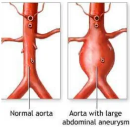
The general definition of an arterial aneurysm is a diameter of 1.5 times that of the normal artery (Figure 4). In the clinical setting, a diameter larger than 30 mm in the AA is considered to be an aneurysm and is a candidate for operation when it exceeds 50-55 mm due to the risk of rupture. Aneurysms are by definition focal but may in some individuals be multiple and associated with generalized arteriomegaly. The pathological process in most aneurysms includes upregulation of proteolytic pathways, inflammation and loss of arterial wall matrix, as reported in cerebral-, thoracic- and abdominal aortic aneurysms (AAA). Aneurysms dilate gradually with an increasing risk of rupture or, as seen in popliteal aneurysms, thrombosis develops with risk of embolization distally (Norman and Powell, 2010).
The most common location for aneurysm is in the AA with a prevalence of approximately 5-6% in men and 1-2% in women, for those older than 65 years
Introduction
thoracic aorta are most commonly seen in the ascending or descending aorta, but are approximately five times less common than in the AA (Gillum, 1995).
Abdominal aortic aneurysm
There are some observations that suggest that the entire vascular tree is abnormal in patients with aneurysmal disease, where AAA has been associated with generalized arteriomegaly, or reduced distensibility at distant arterial sites (Johnsen et al., 2009, Sonesson et al., 1997). Other observations indicate that AAA is associated mainly by abnormalities in the central arteries (Sandgren et al., 2001).
The most common environmental risk factors for aneurysmal diseases are age, male gender and smoking, where the AA appears to be particularly susceptible for smoking (VanderLaan et al., 2004). Previously it was considered that AAA was associated with atherosclerosis, but this relationship is being questioned because aneurysms are rare in locations prone to atherosclerosis, e.g. superficial femoral artery (Norman and Powell, 2010). Furthermore, diabetes mellitus seems to be a negative risk factor for acquiring an AAA, and growth rate of AAA in these patients is lower than in non-diabetics (Sweeting et al., 2012).
There is a strong genetic influence on the development of AAA where different mutated candidate genes have been studied. 1) The mutated Fibrillin-1 (FBNFibrillin-1) gene affects microfibrils, elastogenesis and the transforming growth factor-β (TGF-β) pathway in Marfan syndrome patients with AAA. 2) The mutated collagen type III alpha 1 gene alter the extracellular matrix fibres in Ehlers-Danlos type IV patients with AAA. 3) The mutated elastin gene causes loss of elastin function and alter the extracellular matrix fibres in AAA patients with Cutis Laxa (Norman and Powell, 2010).
Susceptibility genes associated with AAA are studied widely and a
linkage with AAA and a locus in chromosome 12q13.3 which contains the gene for low-density lipoprotein receptor-related protein-1 has been identified (Sakalihasan et al., 2005). Other susceptible genes associated with AAA are 1) a 4G/5G polymorphism in the plasminogen activator inhibitor promoter, 2) a locus on chromosome 19q13 and 3) a locus on chromosome 9p21 (Rossaak et al., 2000, Jones et al., 2002, Boucher et al., 2003, Helgadottir et al., 2008).
Matrix metallopreoteinases (MMP) are enzymes that degrade the extracellular matrix and are involved in normal and diseased tissue remodeling. Studies on MMP genes shows elevated levels of MMPs, especially MMP2 and MMP9, in serum and aortic wall of AAA patients, where mRNA levels of MMP3, known as an activator of MMP9, are significantly increased in AAA tissue (Sakalihasan et al., 1996, Carrell et al., 2002). Increased pressure and strain, activates MMP2 and MMP9 which in turn emphasize the possible importance of wall stress (WS) in the remodeling of the aortic wall (Chesler et al., 1999). Further, there is a relationship between blood pressure, increasing aortic aneurysm diameter and thus WS and the risk of rupture (Cronenwett et al., 1990).
Introduction
Figure 5. 3D structure of Fibrillin-1 (Illustration-3).
Fibrillin-1
Fibrillin-1 is a glycoprotein with forty-seven epidermal growth factor (EGF)-like domains, of which forty-three are calcium-binding (Figure 5). The binding of calcium plays a structural role by protecting the molecule from proteolysis by e.g. trypsin and elastase. Since fibrillin-1 is the main component of microfibrils apart from elastin, the microfibrils are weakened when proteolysis of fibrillin-1 occurs. When the Fibrillin-1 (FBN1) gene is mutated the function of EGF is changed with insufficient calcium binding altering the function of fibrillin-1 and thus the strength of the microfibrils (Jensen et al., 2012). Fibrillin-1 has widespread distribution in both elastic and non-elastic connective tissue throughout the body and occurs in e.g. skin, muscles, kidneys, blood vessels. In arteries, microfibrils are associated with elastic fibres that separate smooth muscle cells and elastic lamellae. Microfibrils also function as a skeleton for the deposition of amorphous elastin (tropoelastin) and provide load-bearing function in the arterial wall (Sherratt et al., 2001). Mutations in the FBN1 gene, located on chromosome 15, cause the connective tissue disorder Marfan syndrome with increased aortic stiffness, elevated pulse pressure and aortic root dilatation (Dietz et al., 1991, Lee et al., 1991, Jeremy et al., 1994).
Previous studies have revealed a potential connection between polymorphisms within the FBN1 gene and aortic impedance. The 2/3 genotype has been shown to be associated with elevated arterial pulse pressure (PP) in middle-aged men and with systolic blood pressure (SBP) in men with AAA (Powell et al., 1997, MacSweeney et al., 1996). A more recent study in mainly
male patients with coronary artery disease (CAD) has shown an association with increased pulse pressure, large artery impedance and the 2/3 genotype (Medley et al., 2002). On the other hand, another study showed that the 2/3 genotype was not associated with increased aortic pulse wave velocity (PWV) in apparently healthy subjects (Yasmin et al., 2006).
Marfan syndrome
The Marfan syndrome (MFS) was first reported by the French pediatric Dr. Antoine Bernard-Jean Marfan in 1896, where he described a 5-year-old girl with skeletal features e.g. long thin limbs and fingers (Marfan, 1896). MFS is a connective tissue disorder that is characterized by abnormalities in the assembly of elastic fibers, involving the skeletal, ocular and cardiovascular system and the major cause of Marfan syndrome is mutations in the FBN1 gene located on chromosome 15q21.1 (Tsipouras et al., 1992).
Due to structural defects and a decrease in the content of aortic elastin, the aorta degenerates prematurely and ascending aorta dilates and finally ruptures. Another mechanism related to abnormalities in synthesis of fibrillin-1 is a defect in production of the latent transforming growth factor-β (TGF-β), caused by fibrillin-1 (Pearson et al., 2008). Increased serum levels of TGF-β have been noted in MFS patients, and in mouse models, antibodies against TGF-β prevent development of mitral valve disease and aortic aneurysm (Keane and Pyeritz, 2008, Matt et al., 2009). Dilatation of the aorta and increased aortic stiffness in MFS patients has been reported and blood pressure reduction has been shown to delay aortic dilatation in Marfan syndrome (Jeremy et al., 1994, Sonesson et al., 1994a, Baumgartner et al., 2006, Yetman, 2007, Williams et al., 2008).
Prominent subjects with Marfan syndrome are Abraham Lincoln (1809-1865) US president and Nicolo Paganini (1782-1840) Italian violinist and composer (McKusick, 1991, Schoenfeld, 1978).
Introduction
Angiotensin-converting enzyme
Angiotensin-converting enzyme (ACE), or kininase II, plays an important role in blood pressure regulation and electrolyte balance by converting angiotensin I (Ang I) to angiotensin II (Ang II), a potent vasoconstrictor. Ang II has significant impact on several important processes in the human body. It stimulates release of aldosterone from the adrenal gland which constricts the renal arterioles leading to an increasing salt and water retention in the kidneys and in the blood vessels. Ang II affects vascular remodeling through induction of smooth muscle cell growth, up-regulation of growth factors and the synthesis of extracellular matrix proteins (Campbell-Boswell and Robertson, 1981, Naftilan et al., 1989, Tokimitsu et al., 1994).
The somatic ACE is expressed in many tissues including cardiovascular system, kidneys, intestine, adrenal glands, liver etc and testicular ACE is only expressed in sperm (Ehlers et al., 1989). In the cardiovascular system ACE is found either bound to the cell membrane of different cell types or in a soluble form in the blood. ACE is cleaved from the endothelial cell membrane and circulates in the blood (Oppong and Hooper, 1993, Beldent et al., 1993). It is still unknown if the circulating level of ACE reflects the level of ACE in tissues.
The ACE gene is located on chromosome 17q23 and there is a variation within the gene, consisting of a 287 base pair insertion/deletion (I/D) polymorphism located in intron 16 which generates three different ACE genotypes: I/I, I/D and D/D (Rigat et al., 1990). The studies on the effect that the ACE I/D genotypes have on CVD are numerous with different conclusions. The first published study that found an association between ACE I/D genotypes and CVD, reported that ACE D/D genotype was more frequent among patients that had suffered myocardial infarction compared to healthy subjects (Cambien et al., 1992). The D/D genotype has also been associated with CAD, hypertension and heart failure, but other studies have failed to confirm these conclusions (Leatham et al., 1994, Lindpaintner et al., 1995, Wang et al., 1996, O'Donnell et al., 1998, Kario et al., 1999, Schut et al., 2004, Agerholm-Larsen et al., 1997). Thus there is no consensus regarding the impact of the ACE genotype on cardiovascular disease.
Aims
AIMS
The aims of this thesis were to investigate:I. The mechanical properties of the popliteal artery in relation to age and gender in healthy subjects.
II. If wall stress of the popliteal artery differs from the adjacent common femoral artery, not being affected by pathological dilatation to the same extent as the popliteal artery.
III. If there are significant associations between the ACE I/D polymorphism, circulating ACE and mechanical properties of the abdominal aorta in elderly subjects.
IV. If variations in Fibrillin-1 genotype are associated with aortic stiffness in male subjects.
V. If the Fibrillin-1 2/3 genotype is associated with presence of carotid plaques and cardiovascular morbidity and mortality in middle-aged subjects.
Materials
MATERIALS
Ethical approval
The studies were approved by the Ethic committee of Lund University (Paper I, II, IV and V) and the Ethic committee of Linköping University (Paper III), Sweden and each participant gave informed consent accordingly to the Helsinki declaration.
Studied subjects
Table I shows a summary of the studied populations in Paper I-V.
Paper I and II
108 subjects were investigated (52 men, range 9-78 years and 56 women, range 9-82 years) in Paper I and of those, 94 subjects were studied in Paper II (45 men, range 10-78 years and 49 women, range 10-83 years). 14 subjects were excluded due to low quality of ultrasound measurements. The subjects were recruited from medical staff, friends and advertising. They were all non-smokers, free from medication, did not have any history of hypertension, cardiopulmonary or renal disease, cerebro-vascular events, diabetes or intermittent claudicatio. The ankle-brachial index was >1 in all subjects. Pregnancy was an exclusion criterion.
Paper III
In 1999, 1130 inhabitants, aged 65-82 years, were invited to participate in an ongoing longitudinal study of elderly people from a rural community in southeast Sweden (Alehagen et al., 2007) were 876 subjects accepted. During 2003 and 2005 a follow-up was performed and 672 subjects were asked to participate in a study regarding mechanical properties of the AA. 452 subjects agreed to participate and ultrasound examinations were successfully performed in 406 subjects (212 men and 194 women) and were included in Paper III. 46 subjects were excluded due to low quality of the ultrasound examination, irregular heart rate, difficulties in obtaining blood samples. Those who choose not to participate in the study indicated as a main reason transportation problem and that they had a long distance to the clinic.
Table I. Study populations used in paper I-V.
Paper IV
79 healthy men (range 28-81 years), first degree relatives of patients with AAA, but not affected to an AAA, were investigated. Subjects with peripheral arterial disease with an ankle brachial index <0.9 and an AA diameter >25 mm were excluded.
A questionnaire was administered to determine the history of previous myocardial infarction, angina, hypertension, diabetes and smoking, and the use of any regular medication was documented.
Paper V
The subjects were recruited from the Malmö Diet and Cancer Study (MDCS) that is a population based prospective cohort study, designed to investigate the association between diet and cancer (Berglund et al., 1993). In short, all inhabitants in the city of Malmö, Sweden, born 1926-1945 (aged 45-69) were invited by mail or by newspaper advertisement to participate. Of an eligible population of 74000, 28449 subjects participated and underwent a health examination and completed a self-administered questionnaire (Manjer et al., 2001). Between November 1991 and February 1994 every second subject that entered the MDCS (n=12445) was invited to participate in a study on the epidemiology of carotid disease, known as the MDCS-Cardiovascular Cohort (MDCS-CC) (Rosvall et al., 2005b). Of the 6103 subjects who participated in the MDCS-CC, 5765 (2424 men, 3341 women) were successfully analyzed regarding their FBN1 genotype. 338 were excluded due to low quality signals during capillary electrophoresis analysis.
N Men/Women Age (range years) Investigated artery Comments
Paper I 108 52/56 9-82 PA Healthy subjects.
Paper II 94 45/49 10-83 PA and CFA Healthy subjects, mainly the
Methods
METHODS
For detailed information about the methods, see Method sections of Paper I-V, respectively.
Ultrasound measurements
Paper I, II and IV
Pulsatile diameter changes during the cardiac cycle in the AA, PA and common femoral artery (CFA) were registered with an ultrasound echo-tracking system (Diamove, Teltec AB, Lund, Sweden) interfaced with a 3.5 MHz and a 5 MHz B-mode real-time linear scanner (EUB 240, Hitatchi, Tokyo, Japan). The ultrasound echo-tracking system was designed with two electronic markers that were aligned with and locked on the echoes from the posterior interface of the anterior wall and the anterior interface of the posterior wall. The markers followed the pulsate movements of the vessel wall. The repetition frequency of the echo-tracking loops was 870 Hz, the time resolution was 1.2 ms and the smallest detectable movement was <10 m (Lindström et al., 1987, Benthin et al., 1991). The echo-tracking system was used in Paper I, II and IV. The pulsatile diameter change during a cardiac cycle together with blood pressure was used to calculate arterial stiffness.
For measuring the IMT and the lumen diameter (LD) of the PA and CFA in Paper I and II, a Philips P700 ultrasound device was used (Philips Ultrasound, Santa Ana, California, US) with a 7,5 MHz linear transducer to visualize the CFA and a 5 MHz linear transducer to visualize the PA. A longitudinal perpendicular image of the vessel was insonated and recorded on a video monitor, three images of good quality were frozen in diastole, according to the prevailing standard of IMT measurements,and the IMT of the far wall as well as the LD were measured manually by tracing a cursor along the echo edges on a section of 10 mm with the aid of the digitizer. This provides approximately 100 boundary points from which the mean value of IMT and LD is automatically calculated (VAP version 2.0, Dept of Appl Electronics, Chalmers University of Technology, Gothenburg, Sweden) (Wendelhag et al., 1991). The accuracy of the technique have showed a good correlation between ultrasound and histology measurement of the arterial wall
Figure 6. Distension curves from the AA determined by the Wall Track System.
(Pignoli et al., 1986). The reproducibility in the PA and CFA measurements were acceptable, coefficients of variation (CV) were 10 and 2 % for IMT and LD respectively (Astrand et al., 2003). The mean value of IMT and LD was calculated based on three images with good recording quality.
Paper III
The Wall Track System (WTS2, Pie Medical, Maastricht, Netherlands) was used on the AA. It was installed in a PC connected to an ultrasound scanner (Esaote AU5, Esaote Biomedica, Florence, Italy), equipped with a 7.5 MHz linear transducer. The Wall Track System (WTS) enables measurements of lumen diameter, pulsatile diameter changes and IMT (Hoeks et al., 1997) (Figure 6).
ECG electrodes were connected to the subject and the artery was visualized in M-mode. The WTS automatically positions two anchors at the posterior and the anterior arterial wall, where manual adjustment could be made, and pulsatile arterial wall movements recorded. Arterial distension waveforms were generated and lumen diameter and pulsatile diameter changes calculated. Using radio frequency signal the WTS can automatically determine end-diastolic IMT of the posterior arterial wall from the interface between the lumen and the tunica intima to the interface between the tunica media and the tunica adventitia.
Methods
Paper V
B-mode ultrasound (Acuson 128 CT system, Mountain View, VA, US) of the right carotid artery (CA) was used to measure atherosclerosis by a trained certified ultrasonographer (Berglund et al., 1994). IMT was measured 1 cm proximal to the bifurcation in the far wall of the right distal common carotid artery (CCA) according to the leading edge principle, using a specially designed computer-assisted analyzing system (Wendelhag et al., 1991). The right CA was scanned within a predefined window compromising 3 cm of the distal CCA, the bulb and 1 cm of the internal and external CA for occurrence of plaques defined as a focal thickening of the IMT>1.2 mm. The intra- and interobserver variation between two measurements of the IMT was 9.0% and 8.7% respectively (Rosvall et al., 2000).
Examination
In Paper I-V the subjects rested in supine position in a darkened and quiet room for 10-15 minutes before blood pressure measurements and ultrasound examination were performed. Differences in blood pressures between the left and right arm were excluded before the investigation.
The right PA was examined in prone position at the site of the popliteal fossa (Paper I and II), the right CFA was examined in supine position at the site of the inguinal fossa (Paper II), with the hip joint as a landmark and the AA was examined in supine position 3-4 cm proximal to the aortic bifurcation (Paper IV). The arteries were visualized in the longitudinal section and care was taken to minimize pressure from the transducer to the skin. A sequence of five representative consecutive diameter cycles was manually chosen and the diameters and the diameter changes were calculated as means of the selected diameter cycles. Each artery was examined three times and the mean value was then calculated. Brachial blood pressure was recorded immediately after ultrasound examination.
In Paper III the AA was examined 3-4 cm proximal to the aortic bifurcation with WTS. Mean values from three consecutive recordings were used.
In Paper V the right distal CCA, the bulb and 1 cm of the internal and external CA was examined.
Measuring mechanical properties of arteries
From the diameter, pulsatile diameter change during a heart cycle and blood pressure the mean diameter, fractional diameter change (strain), pressure strain elastic modulus (Ep), stiffness (β), compliance coefficient (CC) and distensibility coefficient (DC) were calculated based on the following equations (Kawasaki et al., 1987, Reneman et al., 1986, van der Heijden-Spek et al., 2000): Ddiastolic Ddiastolic Dsystolic Strain (2)
Dsystolic Ddiastolic
DdiastolicPdiastolic Psystolic K Ep / (3)
Ddiastolic Ddiastolic Dsystolic Pdiastolic Psystolic / / ln β Siffness (4)
P D D Ddiastolic CC 4 2 2 (5) 2 2 2 Ddiastolic P D D Ddiastolic DC (6)Where Dsystolic and Ddiastolic are the maximum systolic and minimum diastolic diameter (mm). Psystolic and Pdiastolic are the systolic and diastolic
Methods
The circumferential wall stress (WS) was calculated according to the Lamé's equation, an extended equation from the law of Laplace:
IMT LD
Pdiastolic /2
Stress
Wall (7)
Diastolic pressure (Pdiastolic, dyne/cm2) was used since IMT
measurements were performed in diastole. 1 mm Hg equals 1333 dyne/cm². LD, the lumen diameter (cm). IMT, intima-media thickness (cm).
Body surface area (BSA) was estimated according to Du Bois formula (Du Bois and Berlington, 1916):
BSA (m2)= weight 0.425(kg) x height 0.725(cm)x 71.84 (8)
Noninvasive blood pressure measurements
Blood pressure was measured by the auscultatory method with a sphygmomanometer on the upper arm. A cuff with appropriate size was chosen and blood pressure registration was performed with the subjects in supine position immediately after the registration of the pulsatile diameter changes in the AA, PA and CFA (Paper I, II and IV). In Paper III the blood pressure was recorded with an oscillometric method (Dinamap PRO 200 Monitor, Critikon, Tampa, Fl, US). In Paper V, blood pressure was measured auscultatory with a sphygmomanometer on the right upper arm. Blood pressure was measured in both arms to exclude blood pressure differences. Mean arterial pressure (MAP) was defined as the diastolic blood pressure plus one third of the pulse pressure.
When using blood pressure measurements in the calculation of arterial stiffness, it would be favorable to measure it invasively “in situ”, since the blood pressure undergoes transformation in the arterial tree with pressure differences between central and peripheral arteries due to the pulse wave travel and reflections in the arterial tree. Pulse pressure and systolic pressure increase towards the periphery although these differences seem to become less marked later in life due to elastic artery degeneration, decreased distensibility and timing of wave reflection from the lower body (Nichols et al., 2011). Direct invasive measurement of blood pressure in the arteries may be performed, but seems unethical and difficult to use in larger population studies. Instead, we
used the upper arm, and the brachial artery for blood pressure measurements and this could induce an error in the calculations of stiffness/distensibility. However, when comparing invasive pressure in the AA with auscultatory brachial pressure, this showed that auscultatory brachial pulse pressure values were only slightly lower than the invasive aortic pulse pressure leading to a systematic underestimation of Ep and β by 15-20% (Sonesson et al., 1994c). Invasive CFA blood pressure comparison with auscultatory brachial blood pressure shows slightly higher systolic blood pressure and lower diastolic blood pressure in the femoral artery (Ahlgren et al., 2001). There are no available comparisons between invasive pressure in the PA and auscultatory brachial pressure.
Classification of cardiovascular events
(Paper V)
Baseline cardiovascular characteristics were assessed by the self-administered questionnaire and the procedure of follow up in cardiovascular events has been described in detail (Rosvall et al., 2005a, Rosvall et al., 2005b). The National Inpatient Register, the Swedish Hospital Discharge Register, the Stroke Register of Malmö and the National Cause of Death Register were used. The ascertainment of cases and validity of these register has been shown to be high (Socialstyrelsen, 2000, Engstrom et al., 2001). The subjects were followed from baseline examination until first occurring CVD event, emigration from Sweden, or death until December 31st 2008.
CVD events and cause of death were coded in accordance with the 9th
version of the International Classification of Diseases, ICD-9. A CVD event was defined as fatal or nonfatal myocardial infarction (ICD-9: 410), fatal or nonfatal stroke (ICD-9: 430, 431 and 434), or death attributable to CVD (ICD-9: 412 to 414). ICD-9: 390-459 were used to classify CVD death and ICD-9: 140-239 to classify death attributed by tumors. The mean follow up time was 13.2
Methods
Measurement of ACE level (Paper III)
ACE level was determined in plasma using enzyme-linked immunosorbent assay (ELISA) (Quantikine, Human ACE Immunoassay, R&D Systems, Minneapolis, US). The procedure is as follows: monoclonal antibodies specific for ACE are coated on the bottom of a 96-well microplate. Samples are added in to the wells and the available ACE in the samples binds to the antibodies. The unbound substances are removed by washing and biotinylated polyclonal antibodies directed against ACE are added, followed by addition of streptavidine-horseradish peroxidase, which adheres to the polyclonal antibody. A substrate is added, which is converted to a coloured compound by horseradish peroxidase. With a spectrophotometer the intensity of the colour is measured and is proportional to the amount of ACE in the sample. The samples were diluted 1:200. Standards with known concentration of ACE were included in each assay and used to calculate the concentration of ACE in samples. Samples were analyzed in duplicate and re-analyzed if variation from the mean value exceeded 15%. The lower limit of detection was 0.05 ng/ml and intra-assay variation for the analysis of ACE was 6.5%.
Since ACE level was measured in plasma stored at -70° C for 3-5 years degradation of protein might occur, thus the stability of ACE in frozen samples was studied. Plasma from 23 men, recruited from a screening program for AAA at Linköping University Hospital, were stored and frozen at -70° C and ACE level measured after 3 weeks and 12 months. No difference in ACE level was found indicating no degradation of proteins during the first year of freezing.
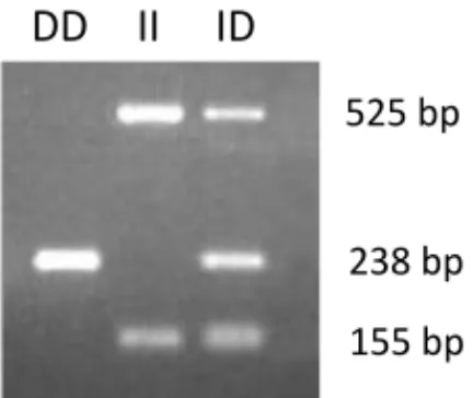
Figure 7. Gel electrophoresis was used to separate the amplified DNA and visualized
by UV-light. One band represents D/D genotype, two bands I/I genotype and three bands I/D genotype.
Genotyping ACE (Paper III)
DNA was isolated from peripheral blood cells using QIAmp DNA Mini Kit (Qiagen, Hilden, Germany) according to the manufacture’s protocol for preparation for polymerase chain reaction (PCR) and according to the standard method of genotyping ACE (Cheon et al., 2000, Ljungberg and Persson, 2008). Three primers were used, for the deletion (D) gene the primers detect a 238 base pair (bp) fragment and for the insertion (I) gene two fragments are detected, 525 bp and 155 bp. The amplified DNA was separated by gel electrophoresis using a 1.5% agarose gel stained with ethidium bromide and visualized by ultra violet light (Figure 7).
Genotyping Fibrillin-1 (Paper IV-V)
DNA from peripheral blood cells was prepared for PCR by typing for the variable tandem nucleotide repeat (VNTR) (TAAAA)n in intron 28 of the FBN1
gene on chromosome 15 using the forward primer 5' 6FAM - CAG AGT ACA TAG AGT GTT TTA GGG AGA -3' and the reverse primer 5'- GTT TCT TCC TGG CTA CCA TTC AAC TCC C-3'. 1 µl portion of the PCR product was diluted with 9 µl highly deionized formamide (GeneScanTM 500ROXTM Size
155 bp 238 bp 525 bp
Methods
The forward primer CCT GGC TAC CAT TCA ACT CCC and reverse primer GAG TAC ATA GAG TGT TTT AGGG were used in Paper IV and the FBN1 genotype was identified as described above.
Statistics
Data are presented as means SD and P<.05 was considered as significant.
Paper I and II
Pearson´s correlation coefficient was used to assess the relationship between age and diameter, strain, stiffness, DC, CC, WS and IMT. Differences between genders were tested using analysis of covariance. A multiple exponential regression model was performed. Paired student t-test was used to calculate differences between studied vessels. A multiple regression model was performed on IMT and LD.
Paper III
Comparisons were performed by 2 tests for categorical variables and one-way
analysis of variance (ANOVA) or Student´s t-test was used to compare continuous data between groups, adjusted for potentially confounding factors. Multiple regression analyses were made to assess the effect of ACE genotype and ACE level on DC, CC and stiffness (β), adjusted for potentially confounding factors. Hardy-Weinberg equilibrium was assessed by 2 tests.
Paper IV
Comparisons were performed by 2 tests for categorical variables. Analysis of
variance was used for the three-group analysis of continuous variables. Associations amongst the three FBN1 genotypes and aortic stiffness were adjusted for age, body mass index, mean blood pressure and heart rate (HR) in a regression model.
Paper V
Comparisons were performed by 2 tests. Analysis of variance was used for
continuous variables with post-hoc tests. The Kaplan-Meier method was used to estimate and plot the first CVD event and survival distribution amongst the three FBN1 genotypes. Differences in incident CVD and survival were assessed using the Log-Rank test.
Results
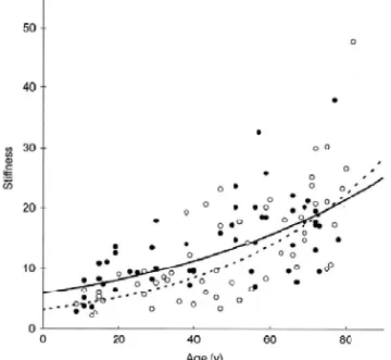
Figure 8. Stiffness (β) of the PA in healthy men (filled circles, solid line) and women (open circles, dashed line) in relation to age. There was a significant correlation between the increase in stiffness (β) and age in men and women, with higher stiffness values in men.
RESULTS
For detailed information about the results, see Result sections of Paper I-V, respectively.
Mechanical properties of the popliteal artery
108 subjects were successfully investigated and the mean diameter of the PA was 7.43 mm (confidence interval (CI); 7.16-7.71 mm) and 6.33 mm (CI; 6.11-6.56 mm) for men and women respectively (P<.001). The diameter increased with age in both men and women, with men having 17% larger diameters than women (P<.001). The diameter was also correlated with age in both men (r=0.66, P<.001) and women (r=0.51, P<.001). The fractional diameter change, strain, of the PA decreased exponentially with age in both men (r=-0.64, P<.001) and women (r=-0.67, P<.001), with women having higher strain values than men (P<.01). The CV regarding arterial diameter was 4% and fractional diameter change 24%. Figure 8 shows that the stiffness (β) of the PA increased exponentially with age in both men and women (P<.001 respectively) and that men had higher stiffness (β) values than women (25%, P<.01). Stiffness (β) was mainly influenced by age (45% males and 56% females, P<.01) while systolic blood pressure (SBP) and MAP had minor importance.
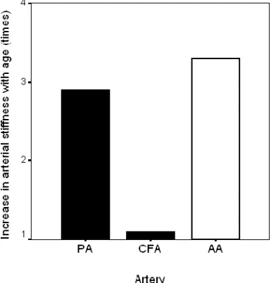
Figure 9. Increase in stiffness (β) between ages 20 and 70 years (times the measured value at 20 years of age) in the PA (muscular artery, present study), and earlier published data regarding the CFA (muscular artery) and the AA (elastic artery).
The DC values decreased with age in both men and women (r=-0.71 and r=-0.80, P<.001 respectively), with men having 24% lower DC values than women (P<.01). DC was mainly influenced by age, 51% and 64% in men and women respectively (P<.01). SBP and diameter were of minor importance, 7% and 4%, (P<.01 and P<.05 respectively). MAP and IMT did not influence DC. The CC decreased with age in men and women (r=-0.40, P<.01 and r=-0.69, P<.001 respectively), without any gender differences.
Figure 9 shows a comparison of the increase in stiffness (β) between ages 20 and 70 years (times the measured value at 20 years of age) in the PA (muscular artery, present study), and earlier published data regarding the CFA (Ahlgren et al., 2001), with subjects from the present investigation included (muscular artery) and the AA (elastic artery) (Sonesson et al., 1993).
Results
Figure 11. Mean WS in the PA (grey bars) and in the CFA (white bars)
in men and women. The PA WS is lower than the CFA in both men and women.
14% higher IMT values than women (P<.001). In adults between the ages 25 to 70 years, the IMT of the PA increased from 0.42 to 0.63 mm (50%) in men and from 0.41 to 0.54 mm (32%) in women. When comparing IMT of the PA and CFA there was no difference seen in women but the IMT of the PA was larger than the IMT of the CFA in men (P<.001).
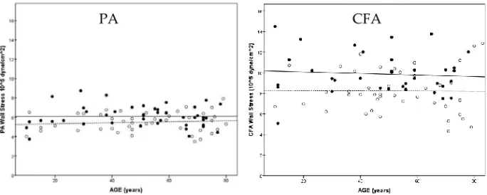
Figure 10 shows the WS at different ages of the PA and CFA. The WS did not increase during age in PA or in CFA. Men had 11 and 20% higher WS values than women in the PA and the CFA (P<.01 and P<.001 respectively). Figure 11 shows the difference in WS in CFA and PA in men and women, where WS was higher in the CFA than in the PA in both men and women (P<.001). 0 1 2 3 4 5 6 7 8 9 10 11 CFA PA CFA PA M e a n W a ll Stre s s (1 0 5d y n e /c m ²) MEN WOMEN *** ***
Figure 10. Wall stress of the PA and CFA in relation to age (men: black circles solid line, and
women: open circles dashed line).
Figure 12. Plasma ACE level according to ACE I/D polymorphism
in men (black circles) and women (grey circles).
Angiotensin-converting enzyme and abdominal
aortic stiffness
406 subjects (212 men and 194 women) were successfully investigated. The use of anti-hypertensive drugs was high (64%) and 23% were using lipid lowering drugs in this elderly study population without gender differences regarding treatment (20% ACE inhibitors (ACEi), 4% angiotensin II receptor blockers, 36% beta blockers and 36% diuretics).
Since ACEi stimulates up-regulation of ACE, subjects with ongoing ACEi treatment (43 men and 40 women) were excluded from the statistical analysis on ACE level.
The frequency of the D allele was 0.50 (0.48 men and 0.53 women) and in accordance with the Hardy-Weinberg equilibrium. Figure 12 shows that the plasma levels of ACE were influenced by ACE I/D polymorphism. The ACE D allele had an impact of 17% (9% in men and 27% in women) of the circulating
Results
Figure 13. Distensibility coefficient and pulsatile diameter change of the AA according to ACE I/D
polymorphism in elderly men.
Figure 13 shows that in elderly men there were significant associations in the subjects carrying the ACE D allele, with lower DC (P=.017) and lower pulsatile diameter change (P=.014) in the AA than in those carrying the ACE II allele.
18 men and two women were identified with an AAA, of these, 10 men were excluded due to low quality of the ultrasound measurements leaving eight men and two women with an AAA in the study population of 406 subjects. When excluding these subjects with an AAA, the DC was still low in men carrying the ACE D allele (P=.036). Further, multiple regression analysis showed in men an association between the ACE D allele and reduced DC (P=.003), CC (P=.045) and increased stiffness (β) (P=.048) in the AA, but no association with circulating ACE levels.
0 2 4 6 8 10 12 II ID/DD D C (10 -3/kPa) 0 0,1 0,2 0,3 0,4 0,5 0,6 0,7 0,8 II ID/DD P u lsa ti le d iam et er c h ange (m m )
*
*
Fibrillin-1 and abdominal aortic stiffness
The distribution of FBN1 genotypes in the 79 men was: 1/2 (n=4), 1/3 (n=1), 1/4 (n=1), 2/2 (n=50), 2/3 (n=10), 2/4 (n=11), 3/3 (n=1) and 3/4 (n=1) and associations between FBN1 genotypes and AA stiffness were calculated in the three common genotypes: 2/2, 2/3 and 2/4. Table II shows the demographics and measured data according to the three genotypes, where subjects of 2/3 genotype had higher SBP (P=.04), PP (P=.04) and MAP (P=.03) than subjects of 2/2 and 2/4 genotypes. Seven subjects were being treated for hypertension (four 2/2, two 2/3 and one 2/4).
FBN1 genotype
2/2 (n=50) 2/3 (n=10) 2/4 (n=11) P
Age (years) 54.7±3.2 56.0±1.7 55.4±2.4 0.91
BMI (body mass index, kg/m2) 25.3±2.1 26.9±2.2 25.3±1.8 0.25
Current smokers (%) 10(20) 2(20) 4(36) 0.42 Systolic pressure (mm Hg) 140±15 158±9 139±16 0.04 Diastolic pressure (mm Hg) 88±10 95±5 87±7 0.08 Mean pressure (mm Hg) 105±11 117±8 105±11 0.03 Pulse pressure (mm Hg) 52±9 61±9 51±11 0.04 Aortic diameter (cm) 1.84±0.22 1.86±0.15 1.87±0.16 0.81
Pressure strain elastic modulus x 10-5 (N/m2) 1.76±0.69 3.04±0.72 1.66±0.55 0.001
The 2/3 genotype subjects also had the highest pressure strain elastic modulus (Ep) (P<.001), highest HR (P<.01) and AA stiffness (β) (P<.001) compared to 2/2 and 2/4 genotype (Figure 14).
After adjusting stiffness (β) for age, body mass index (BMI), MAP and HR the association between AA stiffness and FBN1 2/3 genotype still remained highly
Results
Figure 14. The AA stiffness (β) and HR according to the three common FBN1 genotypes.
Fibrillin-1 and cardiovascular morbidity and
mortality
Four alleles and 10 combinations of FBN1 genotypes were identified in 5765 subjects in the Malmö Diet and Cancer Study (MDCS), i. e. 1/1 (0.02%, n=1), 1/2 (1.1%, n=65), 1/3 (0.1%, n=8), 1/4 (0.3%, n=16), 2/2 (56.4%, n=3254), 2/3 (12.1%, n=698), 2/4 (23.7%, n=1365), 3/3 (0.8%, n=47), 3/4 (2.7%, n=155) and 4/4 (2.7%, n=156), where the most common genotypes were 2/2, 2/3 and 2/4 and accounted for 92.2% (n=5317) of the studied population.
There were no differences between the three genotypes regarding age, blood pressure, smoking habits, lipids, CCA diameter and CCA IMT in men or women at baseline, nor in blood pressure lowering drugs, lipid lowering drugs, anti-diabetes drugs and cardiovascular events in men and women at baseline. 0 5 10 15 20 25 2/2 2/3 2/4 S ti ff nes s ( β) Fibrillin-1 genotype *** 0 20 40 60 80 100 2/2 2/3 2/4 H e a rt ra te ( bea ts /m in) Fibrillin-1 genotype **
Figure 15. The occurrence of plaque in the CA in men according to the three
common FBN1 genotypes (adjusted for age, BMI, SBP and DBP).
Figure 15 shows the occurrence of plaque in the CA where men of 2/3 genotype had higher plaque prevalence than men of 2/2 and 2/4 genotypes; 55% vs. 46% and 50% (P=.03) and after adjusting for factors that affects plaque occurrence such as age, BMI, SBP and DBP the difference remained highly significant (P=.007). There were no differences regarding FBN1 genotype and plaque occurrence observed in women. The odds ratio (OR) for prevalent carotid plaque in men with the 2/3 and 2/2 genotype, respectively, as compared to men with the 2/4 genotype was 1.15 (95% CI: 0.86-1.53) and 0.84 (95% CI: 0.69-1.029), respectively.
Incidence of first CVD events during the follow-up (mean 13.2 years) was 8.6 events per 1000 person-years (12.4 in men and 6.0 in women, P<.001). The corresponding figures for deaths per 1000 person-years were 11.5 (15.1 in men and 9.1 in women, P<.001). Men were affected by a higher number of CVD events than women (Table III).
0 10 20 30 40 50 2/2 2/3 2/4 CA Pl aq u e (% ) FBN1 genotype
Results
Figure 16. Kaplan-Meier plot showing cardiovascular events (MI and stroke) and
mortality according to the main three FBN1 genotypes.
Table III. Incidence (per 1000 person-years) in cardiovascular events (MI and stroke) and
mortality according to the main three FBN1 genotypes.
respectively) between the main three FBN1 genotypes in all subjects, in men or in women (Figure 16).
Men Women
Variables 2/2 genotype 2/3 genotype 2/4 genotype 2/2 genotype 2/3 genotype 2/4 genotype
n Inc n Inc n Inc n Inc n Inc n Inc
Cardiovascular event 206 11.9 47 12.5 91 12.4 143 5.7 32 6.0 67 6.4 MI 117 6.8 25 6.7 59 8.0 63 2.5 15 2.8 32 3.1 Stroke 89 5.1 22 5.9 32 4.4 80 3.2 17 3.2 35 3.4 Mortality 279 16.1 53 14.1 108 14.7 234 9.3 47 8.8 94 9.0 Cardiovascular mortality 77 3.1 12 3.2 34 4.6 46 1.8 9 1.7 23 2.2 n = number of subjects
Discussion
DISCUSSION
The mechanical properties of arteries have a major impact on cardiac work, where the Windkessel effect (buffering function) plays an important role in permitting continuous perfusion of peripheral organs and tissue also during diastole. The composition of the arterial wall influences the mechanical properties of arteries, where the elastin and collagen ratio varies in the arterial system and with age. In central arteries, the dominant component is elastin and in peripheral arteries collagen, with optimal proportions of elastin and collagen at young age, but with an increasing amount of collagen and less elastin with age thus changing the vessel wall movement resulting in increased arterial stiffness, decreased distensibility and compliance (Ahlgren et al., 1997, Sonesson et al., 1993). These age-related changes are mainly seen in the central elastic arteries. Large-artery stiffness is the main determinant of pulse and systolic pressure, and aortic stiffness has independent predicted value for cardiovascular mortality, coronary morbidity and mortality and stroke (Safar et al., 1987, Laurent et al., 2001, Boutouyrie et al., 2002, Laurent et al., 2005, Lacolley et al., 2009).
Popliteal artery, an unusual muscular artery
The most common site of aneurysm development, after the AA, is the PA where almost 30% of patients with popliteal artery aneurysm (PAA) have an AAA (Ravn et al., 2007). Why the PA is more susceptible than other peripheral muscular arteries to aneurysmal disease is at present unknown. One underlying factor of importance may be differences in arterial wall composition compared with other peripheral muscular arteries, which in turn may affect the wall properties. Several studies of mechanical properties on central elastic arteries have shown an age-related increase in stiffness, arterial diameter and intima media thickness (Sonesson et al., 1994c, Hansen et al., 1995, Astrand et al., 2005). However, studies on peripheral muscular arteries are sparse and show no increase in stiffness, indicating less of age-related arterial wall degeneration, e.g. CFA and distal brachial artery (van der Heijden-Spek et al., 2000, Ahlgren et al., 2001, Bjarnegard and Lanne, 2010).
The absence of an age-related increase in arterial stiffness in muscular arteries may seem surprising, because it is well known that the arterial wall structure changes with an increase in collagen and thickening of the arterial wall. There is no reason to believe that age-related histological changes and subsequent remodeling are absent in muscular arteries, inasmuch as age-related dilatation of muscular arteries of similar magnitude to that in elastic arteries has been found (Kawasaki et al., 1987, Sonesson et al., 1993, Hansen et al., 1995, Ahlgren et al., 2001).
The diameter of the muscular PA in healthy subjects increased with age in both men and women with men having larger diameter than women due to larger body size. This gender difference was also observed in the CFA. The age-related arterial dilatation might be influenced by the distending force acting on the arterial wall, the blood pressure. Consequently we found a correlation between blood pressure and arterial dilatation.
The PA strain decreased exponentially with age in both gender, with lower strain values in men. The stiffness of the PA increased exponentially with age, with increased stiffness in men compared to women (Figure 8), as would have been expected from the strain values. Interestingly, when the increase in stiffness (β) between ages 20 and 70 of the PA (muscular artery) was compared to CFA (muscular artery) and AA (elastic artery) the increase in stiffness (β) in the PA was similar to the increase observed in the AA, while the CFA showed no increase at all (Figure 9). Earlier studies in stiffness of the PA in healthy subjects are sparse. Tai et al. (1999) investigated the PA in 11 young and 12 elderly subjects and found an increased stiffness with age. However, the number of subjects included was small and there was no separation between genders. Brodszki et al. (2002) studied the PA in young women but without any attempt to define the relation between stiffness and age or gender. The increased stiffness of the PA differs from the near superficial and common femoral artery, as well as other regions of muscular arteries, such as the radial artery and brachial artery, where the age-related
Discussion
during life, with a fragmentation of elastin without neosynthesization, while collagen is produced throughout life (Lakatta, 1989, Fonck et al., 2007, Nichols et al., 2011).
Incipient atherosclerosis might also lead to an increase in stiffness (Lind et al., 2009). The literature concludes however that the effect of atherosclerosis on arterial stiffness is minor. Another factor that might be of importance is wall thickness, according to the equation of Young´s modulus (Nichols et al., 2011). Thus increased arterial wall thickness may increase stiffness. The IMT of the PA increased with age in both gender, with men having higher IMT than women and in accordance with findings in the femoral artery. Further, men had higher IMT in the PA compared to CFA. IMT was correlated to age but did however not affect the stiffness of the PA wall. Thus, it seems reasonable to assume that the composition of the PA wall may be more similar to central elastic arteries such as the aorta, than to other peripheral muscular arteries. In view of the fact that the stiffness, diameter and IMT increases with age to a similar extent in both PA and AA it may be speculated that this might be the reason for the similar pathology in both regions with a tendency to aneurysm formation.
An increase in blood pressure and diameter leads to increased wall stress (WS) according to the law of Laplace, which in turn activates smooth muscle cells of the arterial wall, leading to an increase in matrix and wall thickness (Ben Driss et al., 1997). Since blood pressure and diameter increases with age, an increased WS in the PA would have been expected. However, no increase was found neither in the PA or nor in the CFA (Figure 10). Further, men had higher WS than women (Figure 11). The absence of WS increase in PA and CFA was probably caused by a compensatory remodeling response with an increase in arterial wall thickness. Interestingly, WS was lower in PA than in the CFA in both genders, despite the fact that aneurysms are more common in the PA than in the CFA (Norman and Powell, 2010). This indicates that other mechanisms than WS are involved in the process of pathological arterial dilatation in the PA. In the AA the WS has been shown to be high, with an age-related increase in WS observed in men but not in women, which may be related to the increased prevalence of AAA in males (Astrand et al., 2005). In diabetic patients however, the WS of the AA is reduced compared to healthy controls, probably due to increased IMT and might be the reason for the reduced risk of aneurysmal disease in diabetic patients (Astrand et al., 2007).