THE ACTIVELY MYELINATING OLIGODENDROCYTE: SIGNALING MECHANISMS AND RESPONSE TO INJURY by JARED TYLER AHRENDSEN B.A., University of Colorado Boulder, 2008 A thesis submitted to the Faculty of the Graduate School of the University of Colorado in partial fulfillment of the requirements for the degree of Doctor of Philosophy Neuroscience Program 2015
This thesis for the Doctor of Philosophy degree by Jared Tyler Ahrendsen has been approved for the Neuroscience Program by Angie Ribera, Chair Wendy Macklin, Advisor Bruce Appel Jeffrey Bennett Paco Herson Mary Reyland Date: 12/18/2015
Ahrendsen, Jared Tyler (Ph.D., Neuroscience) The Actively Myelinating Oligodendrocytes: Signaling Mechanisms and Response to Injury Thesis directed by Professor Wendy B. Macklin ABSTRACT The actively myelinating oligodendrocyte is vital for the normal development and proper function of the central nervous system (CNS). Despite significant progress in recent years, many fundamental questions remain regarding the cellular and molecular properties of oligodendrocytes. In this dissertation, we first sought to identify and characterize putative negative regulators of CNS myelination. We identified Shp2, a protein tyrosine phosphatase, as a potential negative regulator of CNS myelination using a candidate based approach and expression profiling of mRNA and protein during development in wild type and hypermyelinating mutant mice. We then characterized the functional role of oligodendrocyte Shp2 using inducible and conditional transgenic mice. We demonstrated that Shp2 regulated oligodendrocyte differentiation during postnatal development and acted as a transient brake during early myelin formation. Shp2 also regulated the timely remyelination following experimental demyelination. The second objective of this project was to examine the responses of the actively myelinating oligodendrocyte, which are enriched in the brain during late pediatric development, to ischemic injury. Using an established mouse model of experimentally induced reversible
cerebral ischemia, we demonstrated that actively myelinating oligodendrocytes in the late pediatric brain were uniquely resistant to ischemia-induced damage compared to adult mice with the same injury. We showed that this difference could be due to pediatric oligodendrocytes having a more robust anti-oxidant capacity. We also exmined other glial responses unique to pediatric stroke and found that astrogliosis, fibrosis, NG2-cell responses and vascular integrity were all different in pediatric compared to adult animals. Combined, these differences in stroke response led to greater long-term tissue preservation in pediatric mice. Taken together, this thesis provides significant improvements to our understanding of the actively myelinating oligodendrocyte, both during development and following injury. These results could influence our understanding of prominent neurological diseases, such as multiple sclerosis and stroke. The form and content of this abstract are approved. I recommend its publication. Approved: Wendy B. Macklin
ACKNOWLEDGEMENTS First and foremost I would like to thank my mentor, Dr. Wendy Macklin. Her support and guidance throughout my Ph.D. training has proven invaluable. Dr. Macklin allowed me to truly grow as a scientist by giving me the freedom to generate and test my own ideas, while providing valuable feedback and counsel on the feasibility of these ideas. She has instilled in me the skills necessary to pursue an independent career in academic research. I would also like to thank the members of the Macklin lab (both present and past), the Neuroscience and Medical Scientist Training Programs (including administrators, directors, students and faculty), other close professional mentors (Arthur Gutierrez-Hartmann, Angie Ribera and Tim Vollmer, in particular), and my entire thesis committee. Finally, I want to thank my loved ones for their unwavering love and support. In particular, I thank Marissa for her love and commitment to me.
TABLE OF CONTENTS CHAPTER I. INTRODUCTION AND BACKGROUND... 1 Oligodendrocytes: function and development... 1 Akt signaling... 4 Molecular mechanisms regulating myelination: PNS... 6 Molecular mechanisms regulating myelination: CNS... 8 Oligodendrocytes, demyelination and multiple sclerosis... 11 Oligodendrocytes and stroke... 13 Hypotheses... 16 II. IDENTIFICATION OF A PUTATIVE MYELIN BRAKE IN THE CENTRAL NERVOUS SYSTEM... 18 Summary... 18 Introduction... 19 Experimental procedures... 22 Materials... 22 Animals... 22 Western blots... 22 Quantitative real-time polymerase chain reaction (qPCR)... 23 Statistical analysis... 24 Results... 24 Expectations of a putative myelin brake... 24 Developmental expression profiles of PTEN and PHLPP phosphatases... 25
Developmental expression profiles of Ship-1 and Ship-2 phosphatases... 28 Developmental expression profiles of Shp1 and Shp2 phosphatases... 30 Discussion... 31 III. THE PROTEIN TYROSINE PHOSPHATASE SHP2 REGULATES OLIGODENDROCYTE DIFFERENTATION AND EARLY MYELIN FORMATION DURING DEVELOPMENT AND REMEYLINATION... 35 Summary... 35 Introduction... 36 Experimental procedures... 38 Animals... 38 Tamoxifen administration... 38 Lysolecithin induced demyelination... 39 Immunohistochemistry... 39 Cell counting... 41 Western blot... 41 Electron microscopy... 42 G-ratio analysis... 42 Statistical analysis... 43 Results... 44 Shp2 conditional knockout with CNP driven cre-recombination... 44 Delayed oligodendrocyte differentiation in the Shp2 cKO brain... 47 Abnormal myelination in the Shp2 cKO brain... 48
Oligodendrocyte Shp2 is not necessary for long-term maintenance of myelination... 50 Oligodendrocyte Shp2 regulates timely differentiation following lysolecithin-induced demyelination... 54 Discussion... 58 IV. ACTIVELY MYELINATING OLIGODENDROCYTES IN THE PEDIATRIC BRAIN ARE RESISTANT TO ISCHEMIA-INDUCED DAMAGE... 62 Summary... 62 Introduction... 63 Experimental procedures... 65 Animals... 65 Middle cerebral artery occlusion (MCAo)... 66 Infarct Volume Analysis... 66 Immunohistochemistry... 67 Cell counting... 68 Blood vessel analysis... 69 Electron microscopy... 70 G-ratio analysis... 71 Statistical analysis... 72 Results... 72 Cell death, neuronal loss, and oligodendrocyte preservation after pediatric MCAo... 72 Oligodendrocyte precursor cells responses in ischemic tissue after MCAo... 76 Mature oligodendrocytes are resistant to ischemia in the pediatric brain... 80
Myelin damage, axon pathology, and long-term tissue loss are spared in pediatric, but not adult, ischemia... 82 Differential injury progression in pediatric versus adult stroke... 84 Maintained vascular integrity within the ischemic core in pediatric mice... 85 Enhanced protection from oxidative damage in pediatric oligodendrocytes... 87 Discussion... 89 V. DISCUSSION... 96 REFERENCES...108
LIST OF FIGURES FIGURES 1-1. Schematic of Akt signaling... 5 2-1. Regulation of Akt signaling... 20 2-2. Dynamics of myelin formation during development in WT and Akt-DD mice... 23 2-3. Developmental expression profiles of PTEN and PHLPP phosphatases... 26 2-4. Developmental expression profiles of Ship-1 and Ship-2 phosphatases... 28 2-5. Developmental expression profiles of Shp1 and Shp2 phosphatases... 29 3-1. Shp2 conditional knockout using the CNP-cre transgenic line... 43 3-2. Delayed oligodendrocyte differentiation in the Shp2 cKO brain... 45 3-3. Abnormal myelination in the Shp2 cKO brain... 46 3-4. FAK signaling, not Akt/mTOR or ERK signaling, altered in Shp2 cKO corpus callosum... 49 3-5. Oligodendrocyte Shp2 is not necessary for long-term maintenance of myelination... 51 3-6. Efficient knockout of Shp2 from oligodendrocytes in the spinal cord after lysolecithin-induced demyelination... 52 3-7. Delayed oligodendrocyte recruitment and remyelination in icKO spinal cord... 53 3-8. Delayed differentiation of remyelinating oligodendrocytes in Shp2 icKO spinal cord... 55 4-1. Brain regions of interest following MCAo... 68 4-2. No age difference in infarct volume following MCAo... 70
4-3. Cell death after MCAo... 71 4-4. Oligodendrocytes are preserved in pediatric, but not adult, tissue after MCAo... 74 4-5. Myelin damage, axon pathology, and long-term tissue loss after MCAo... 75 4-6. Differential fibrotic and astrogliotic patterns following MCAo in pediatric versus adult mice...77 4-7. Differential gliotic responses following MCAo in pediatric versus adult mice... 78 4-8. Vascular and pericyte responses to MCAo in pediatric versus adult mice... 79 4-9. Decreased oxidative damage and increased anti-oxidant capacity after pediatric MCAo compared to adult... 81 5-1. Model of Shp2 regulation of CNS myelination... 98
LIST OF TABLES TABLES 2-1. Expected developmental profile of a CNS myelin brake... 25
LIST OF ABBREVIATIONS 3-NT 3-nitrotyrosine BACE1 Beta site APP cleavage enzyme 1 (beta secretase) cKO Conditional knockout CNS Central nervous system DAB 3,3’-diaminobenzidine Dlg1 Disks large homolog 1 dpl Days post-lesion EGF Epidermal growth factor EGFP Enhanced green fluorescent protein FDA Food and Drug Administration FOV Field of view GFAP Glial fibrillary acidic protein GSTπ Glutathione S-transferase pi HNLPP Hereditary neuropathy with liability to pressure palsies HO-1 Heme oxygenase-1 IHC Immunohistochemistry icKO Induced conditional knockout i.p. Intraperitoneal MBP Myelin basic protein MCAo Middle cerebral artery occlusion MOG Myelin oligodendrocyte glycoprotein MS Multiple sclerosis MTMR2 Myotubularin related protein 2 mTOR Mammalian target of rapamycin NRG1 Neuregulin-1 OPC Oligodendrocyte precursor cell PDGFRα Platelet derived growth factor receptor alpha PHLPP PH domain and leucine rich repeat protein phosphatase PI3K Phosphatidylinositide-3-kinase PIP3 Phosphatidylinositol-3,4,5-triphosphate PLP Proteolipid protein PNS Peripheral nervous system PTEN Phosphatase and tensin homolog deleted on chromosome 10 qPCR Quantitative real-time polymerase chain reaction Ship-1 SH2 domain containing inositol polyphosphate 5-phosphatase-1 Ship-2 SH2 domain containing inositol polyphosphate 5-phosphatase-2 SHP1 Src homology-2 domain containing protein tyrosine phosphatase-1 SHP2 Src homology-2 domain containing protein tyrosine phosphatase-2 TTC Tetrazolium chloride
CHAPTER I INTRODUCTION AND BACKGROUND1 Oligodendrocytes: function and development Oligodendrocytes are the myelin-producing cells of the central nervous system (CNS). Myelin is the specialized, lipid-rich, multilayered membrane that forms an insulating sheath around axons. Traditionally considered an evolutionary adaptation resulting in rapid and efficient transduction of action potentials via ‘saltatory’ impulse propagation (Geren and Raskind, 1953), myelin is also essential for the proper development and function of the vertebrate nervous system. While optimizing the electrical activity of axons, myelin also clearly provides trophic and metabolic support to ensheathed axons (Nave, 2010). In order for myelin production to occur, either during development or after injury, oligodendrocytes must progress through a well-characterized series of differentiation steps. Oligodendrocyte precursor cells (OPCs) in the developing brain and spinal cord are derived from multiple, distinct cellular foci during late embryonic gestation (Rowitch and Kriegstein, 2010). OPCs are identified histologically by their expression of the platelet-derived growth factor receptor alpha (PDGFRα) and the NG2 proteoglycan. OPCs are highly migratory and 1 Portions of this chapter are reproduced with permission from Ahrendsen JT, Macklin WB (2013) Signaling mechanisms regulating myelination in the central nervous system. Neurosci Bull 29:199–215.
proliferative during late embryonic and early postnatal development in order to populate the entirety of the CNS (de Castro and Bribián, 2005; Moyon et al., 2015). Once located at their final destination, some OPCs persist into adulthood while others differentiate into myelin-producing oligodendrocytes. The differentiation of OPCs into oligodendrocytes is regulated both spatially and temporally. Both intrinsic and extrinsic factors regulate oligodendrocyte differentiation, including protein kinases/phosphatases, transcription factors, soluble growth factors, the extracellular matrix, and neuronal activity/cues (Huang et al., 2013). Furthermore, cells early in the differentiation program, called pre-myelinating oligodendrocytes, will undergo programmed cell death if unable to find an appropriate substrate for myelination and/or do not receive the necessary differentiation signals (Trapp et al., 1997). While the loss of pre-myelinating oligodendrocytes is a normal part of CNS development, differentiation failure is a pathological feature in multiple sclerosis (MS), the most common demyelinating disease of the CNS (Chang et al., 2002). Thus, promoting oligodendrocyte differentiation represents a promising target for promoting recovery after demyelinating injury (Franklin and ffrench-Constant, 2008). Those cells which find axonal substrates for myelination and receive the necessary input will go on to become actively myelinating oligodendrocytes. The process of myelination involves enormous energy expenditure for massive membrane biogenesis to generate concentric, spiral wraps of myelin around axons (Pfeiffer et al., 1993). In mammals, this process occurs in large part during postnatal development (Rorke and Riggs, 1969; Sowell et al., 2003; de Castro
and Bribián, 2005; Bradl and Lassmann, 2010; Moyon et al., 2015) and is modulated by electrical activity (Zalc and Fields, 2000; Wake et al., 2011) as well as axon-derived molecular signals (Taveggia et al., 2010). The maturation of axons and their long-term survival depend on the presence of myelin (Nave, 2010). In turn, the proliferation, migration, survival, and differentiation of myelinating glia require axon-derived signals (Nave and Trapp, 2008), and the long-term maintenance of the myelin sheath also depends on axonal signals (Bremer et al., 2010). Thus, there exists extensive bidirectional signaling between oligodendrocytes and axons. It is well-established that myelin thickness is directly related to axon diameter (Donaldson and Hoke, 1905; Friede, 1972). Optimal conduction by myelinated axons is achieved by adjusting the length and thickness of the myelin sheath to the axon diameter (Waxman, 1997). An axon with either too little or too much myelin will not function optimally. Therefore, precise molecular mechanisms exist that regulate myelin production. After producing the requisite amount of myelin, the actively myelinating oligodendrocyte transitions into a myelin maintenance stage. While still capable of producing myelin, the rate of production and turnover of myelin decreases substantially (Quarles et al., 2006). Recent single cell RNA-seq data suggest that there are as many six different subclasses of oligodendrocyte, including the myelinating oligodendrocyte and terminally differentiated postmyelination oligodendrocyte (Zeisel et al., 2015). Little is known about these subclasses of mature oligodendrocytes and what regulates the transition from a myelinating cell to a postmyelinating cell.
Akt signaling Akt (also known as Protein Kinase B) is a serine/threonine protein kinase is a downstream effector of the lipid kinase phosphatidylinositol 3-kinase (PI3K) and an upstream activator of mammalian target of rapamycin (mTOR) (Figure 1-1) (Burgering and Coffer, 1995; Franke et al., 1995). Akt signaling regulates many aspects of cellular function, including proliferation, cell growth, survival, metabolism, and migration (Yang et al., 2010a). Thus, it is not surprising that disrupted Akt signaling is often a cause of cancer in many tissues. However, Akt signaling is a vital part of normal cellular function so understanding how it is regulated offers unique insight into controlling the action of Akt for therapeutic purposes. The mammalian genome encodes three separate genes for Akt. The rate limiting step in activating the kinase activity of Akt is binding of the lipid second messenger phosphatidylinositol (3,4,5)-triphosphate (PIP3) (generated by activated PI3K in response to growth factors and other extracellular stimuli) to the pleckstrin homology (PH) domain of Akt and the subsequent translocation of Akt to the plasma membrane (Kandel and Hay, 1999). Akt activation is promoted by phosphorylation at threonine 308 by protein dependent kinase-1 and mTOR complex 2 (mTORC2) at serine 473 (Alessi et al., 1996; Sarbassov et al., 2005; Jacinto et al., 2006). Both phosphorylation events are necessary for full activation of Akt. Akt kinase activity has many cellular targets (Kim et al., 2005). In particular, Akt directly phosphorylates and inactivates tuberous sclerosis complex 2 (TSC2)
(Manning et al., 2002). TSC2 is an upstream inhibitor of mTOR, thus Akt activity promotes mTOR activation through inhibition of TSC2 (Inoki et al., 2002). mTOR can then activate proteins such as 4E-BP1 and S6-kinase in order to regulate protein synthesis and cell growth (Hay and Sonenberg, 2004). Regulation of Akt signaling is accomplished at many levels by several proteins. Phosphorylation events have positive effects on Akt activity, both upstream on lipid second messengers and on the Akt protein. Therefore, downregulation of Akt signaling is often accomplished at the molecular level by removal of phosphate groups (dephosphorylation) by phosphatases (Figure 1-1, 2-1). PTEN is the classic negative regulator of Akt signaling, which acts as a lipid phosphatase and decreases levels of PIP3 by catalyzing the formation of PIP2 (Stambolic et al., 1998). Signaling regulators can themselves be regulated by phosphorylation/dephosphorylation events and interactions with scaffolding proteins. For example, PTEN phosphatase activity is upregulated when phosphorylated at three sites on its C-terminal tail domain (S380/T382/T383); however this also renders PTEN less stable (Vazquez et al., 2000). PTEN activity is also upregulated by binding to certain scaffolding proteins such as Nherf (Takahashi et al., 2006). Binding to scaffolding proteins can regulate the stability of other proteins and can also sequester them to certain subcellular locations. The regulation of Akt signaling by other phosphatases is discussed more thoroughly below and in Chapter II. Finally, Akt signaling can be regulated by other post-translational modifications, such as ubiquitination, a process by which proteins are targeted for degradation (Yang et al., 2010a).
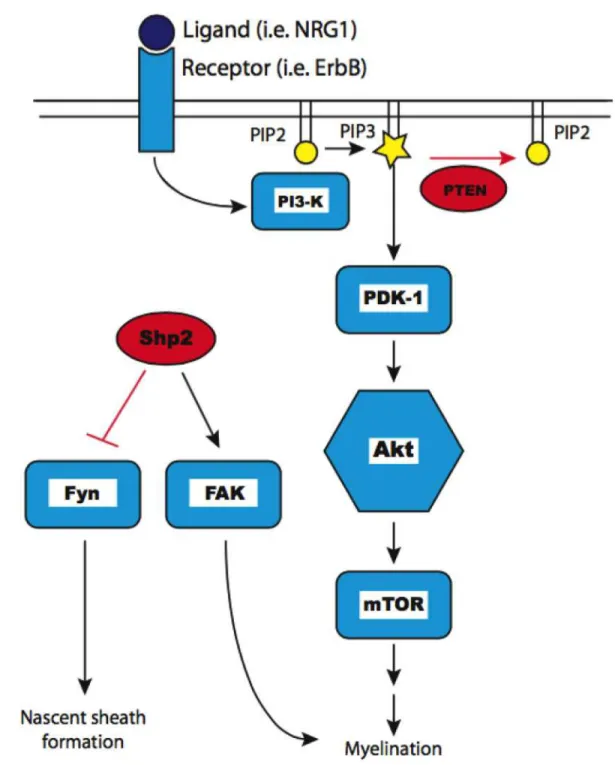
Molecular mechanisms regulating myelination: PNS The molecular mechanisms regulating myelination in the peripheral nervous system (PNS) by myelinating Schwann cells are well studied and provide important insight into potential mechanisms governing myelination in the CNS. In the PNS, Figure 1-1. Schematic of Akt signaling. Extracellular ligand (dark blue) binding a transmembrane receptor and activating the PI3K/Akt/mTOR pathway (light blue icons). Negative regulators indicated in red. Lipid second messengers PIP2 and PIP3 indicated in yellow.
neuregulin 1 (NRG1) type III signaling from the axon membrane can influence whether an axon is myelinated (Taveggia et al., 2005), and the amount of axonal NRG1 type-III regulates peripheral myelin sheath thickness (Michailov et al., 2004). NRG1 signaling acts through ErbB receptors on Schwann cells to regulate PNS myelination (Chen et al., 2006). The peripheral hypermyelination disorder Charcot-Marie-tooth disease type 1A results from mutations leading to overexpression of ErbB2 and ErbB3 receptors (Massa et al., 2006). NRG1 mediated activation of Schwann cell ErbB receptors triggers activation of the PI3K/Akt pathway (Maurel and Salzer, 2000), which then signals though mTOR (Sherman et al., 2012) to promote myelination (Figure 1-1). The genetic knockout of PTEN from myelinating Schwann cells artificially elevates the levels of PIP3 concentrations within Schwann cells, resulting in increased Akt signaling and myelin overproduction (Goebbels et al., 2010; 2012). While there are signals that drive myelination, there must also be mechanisms to counteract, or brake, those signals when the optimal amount of myelin has been formed for any given axon in order to prevent deleterious over production of myelin. The “myelin brake” of the PNS was discovered using a combination of human genetic data along with transgenic mouse technology. Myotubularin-related protein 2 (MTMR2) was identified as the genetic cause of Charcot-Marie-Tooth disease 4B, another hypermyelinating disorder of the PNS (Bolino et al., 2000; Bonneick et al., 2005). This led to the discovery that the scaffolding protein Dlg1 (mammalian discs large homolog 1) interacts with MTMR2 to stabilize PTEN in order to downregulated PI3K/Akt signaling in Schwann cells
after the appropriate amount of myelin had been formed, thereby decreasing myelin production (Cotter et al., 2010). Furthermore, there are multiple levels of regulatory control over myelination as Dlg1 acts as a transient negative regulator of myelination and also serves to increase the expression of DDIT4 (a known negative regulator of mTOR (Ellisen, 2005)) in order to provide sustained downregulation of myelin production (Noseda et al., 2013). Therefore, in the PNS, Dlg1 and DDIT4 downregulate Akt/mTOR signaling and halt the myelination process at the appropriate time, thereby ensuring optimal electrical conductance along myelinated axons and protecting against pathological membrane overproduction. Molecular mechanisms regulating myelination: CNS The role of NRG1/ErbB signaling in the CNS is less clear. Neuregulins mediate survival and differentiation of cultured oligodendrocyte progenitor cells (OPCs) (Canoll et al., 1996; Flores et al., 2000), and spinal cord explant studies demonstrate the requirement for NRG1 signaling in oligodendrocyte development (Vartanian et al., 1999). Other studies suggest that while axonal NRG1 does not direct initial oligodendrocyte differentiation, it promotes myelination in some CNS areas (Taveggia et al., 2008). Similar to the PNS, ErbB2 signaling has also been shown to positively regulate terminal oligodendrocyte differentiation and myelination in vivo (Park et al., 2001; Kim et al., 2003). ErbB4 signaling in oligodendrocytes is quite complex. The complete loss of ErbB4 signaling in neural tube explants increases the number of differentiated oligodendrocytes (Sussman et
al., 2005), whereas the in vivo expression of a dominant negative ErbB4 that binds NRG1, but cannot signal in oligodendrocytes, results in fewer oligodendrocytes and reduced myelin thickness of CNS axons (Roy et al., 2007). While these studies suggest at least regional CNS regulation of oligodendrocyte development or myelination by NRG1, mice with complete NRG1 knockout or elimination of both ErbB3 and ErbB4 receptors in oligodendrocytes have normal CNS myelination, indicating that these factors are not required for normal CNS myelination (Brinkmann et al., 2008). On the other hand, that same study demonstrated that overexpression of either NRG1 type-I or type-III could induce CNS hypermyelination. Thus, NRG1 clearly impacts some elements of CNS myelination. Similar to Schwann cells, PI3K/Akt signaling in oligodendrocytes is activated by neuregulins (Flores et al., 2000; Hu et al., 2006; Luo et al., 2011). The genetic overexpression of a constitutively active form of Akt in oligodendrocytes (Akt-DD) causes CNS hypermyelination, without affecting oligodendrocyte differentiation, proliferation, or survival (Flores et al., 2008). Hypermyelination in these animals is driven by Akt signaling through mTOR, and is reversible upon treatment with the mTOR inhibitor rapamycin (Narayanan et al., 2009). Furthermore, the lack of myelin observed in the BACE1 null mouse, which lacks a neuregulin cleavage enzyme, can be rescued by Akt-DD overexpression (Hu et al., 2006; 2013). In other studies, conditional expression of the mTOR activator Rheb (a downstream effector of Akt) in neural progenitors has been shown to promote myelination in the brain, while conditional Rheb knockout from the same cells inhibits myelination (Zou et al., 2011).
mTOR function in oligodendrocytes, like NRG1/ErbB signaling, is also quite complicated. mTOR is required for the developmentally regulated expression of several myelin proteins and lipid biogenesis enzymes such as those driving cholesterol and fatty acid synthesis (Tyler et al., 2011). The conditional knockout of the entire mTOR complex disrupts oligodendrocyte differentiation and the extent of myelination in a region specific manner (Wahl et al., 2014). Raptor dependent mTOR complex 1 serves as a positive regulator of myelination and the conditional knockout of Raptor also causes region specific myelin abnormalities, even in the presence of functional mTOR complex 2 (Bercury et al., 2014). Together, these results provide strong evidence that Akt signaling through mTOR has significant impact on myelin formation and could represent a master regulator of myelination in the CNS. Again, similar to that observed in the PNS, the conditional knockout of PTEN from oligodendrocytes during development causes dramatic CNS hypermyelination (Harrington et al., 2010) and overproduction of myelin can be induced after active myelination has ceased by deleting PTEN from oligodendrocytes in adulthood (Goebbels et al., 2010). However, unlike in PNS, there are no known human diseases or conditions in which over production of myelin in the CNS is a distinguishing pathological feature. Furthermore, proteins such as MTMR2, Dlg1, and DDIT4 are expressed at very low levels in the CNS and do not show an expression profile that resembles that of a “myelin brake”, as they do in the PNS (Cotter et al., 2010; Noseda et al., 2013; Zhang et al., 2014). Therefore, the identification of the “myelin brake” in the CNS remains as a significant unanswered question.
Oligodendrocytes, demyelination and multiple sclerosis Disruption of oligodendrocytes and the axon-myelin relationship is seen in many congenital and acquired neurological diseases and conditions, including genetic leukodystrophies, perinatal hypoxia/ischemia, stroke, and MS. The most common cause of neurological disability in young adults, MS is characterized by CNS demyelination induced by inflammation and immune responses. Acute demyelinating episodes result in neurological impairment that is generally followed by some functional recovery as the inflammation resolves. Recovery is primarily due to myelin regeneration of demyelinated axons. However, permanent disability is eventually seen in MS, resulting from axonal transection after chronic demyelination or other pathologies (Trapp et al., 1998), although axonal damage can occur at the same time as demyelination (Trapp and Nave, 2008). Clearly, investigating the mechanisms regulating axon-myelin interactions is pivotal in order to improve treatments of demyelinating diseases such as MS. Currently, the only Food and Drug Administration (FDA) approved medications for treating MS are immune modulating agents, which help to prevent immune and inflammatory attacks during the relapsing-remitting stage of MS. However, none of these agents is effective in treating the progressive phase of the disease that results from accumulated axon damage. An additional approach to treating this disease would be to enhance the endogenous repair processes that occur naturally after acute demyelination. Remyelination occurs during early stages of MS and, while rarely complete, allows for some functional recovery (Patrikios et
al., 2006; Franklin et al., 2012). However, after multiple episodes of demyelination, repair fails, resulting in progressive neurodegeneration (Franklin, 2002). The process of remyelination shares many features with developmental myelination (Fancy et al., 2011a). Therefore, a thorough understanding of the mechanisms regulating developmental myelination may help target remyelination therapeutically, promoting functional recovery and preventing MS progression. Currently, drug designers have been unsuccessful in creating pharmaceuticals that promote remyelination in human patients. The signaling mechanisms regulating remyelination are poorly understood. Although conditional knockout of PTEN from oligodendrocytes causes developmental hypermyelination, it has no effect on remyelination (Harrington et al., 2010). However, in wild type mice treated with rapamycin, oligodendrocyte differentiation and remyelination are delayed after cuprizone induced demyelination, suggesting that mTOR signaling is important in the remyelination process (Sachs et al., 2014). While repeated episodes of de/remyelination affect an oligodendrocyte’s ability to repair, there also exist age-associated differences influencing remyelination efficiency (Franklin and ffrench-Constant, 2008). Compared to adult mice, older mice are less efficient and juvenile mice are more efficient at repairing a demyelinated lesion (Shields et al., 1999; Pfeifenbring et al., 2015). Aging affects the intrinsic ability of an OPC to differentiate after a demyelination injury (Shen et al., 2008). There are also circulating factors in the blood that affect remyelination efficiency, such that an old mouse exposed to a youthful circulation improves the
older mouse’s ability to remyelinate (Ruckh et al., 2012). Thus, biological age has both cell intrinsic and extrinsic influences on remyelination. Oligodendrocytes and stroke An ischemic stroke is generally caused by an obstruction of blood flow to an area of the brain, thereby disrupting the delivery of nutrients and oxygen as well as the elimination of waste. Without timely restoration of blood flow, the infarcted tissue becomes necrotic, resulting in permanent neurological disability or death. In addition, even the restoration of blood flow causes production of reactive oxygen species, which contribute additional damage from significant oxidative stress to injured tissue (Allen and Bayraktutan, 2009). The human brain consists of nearly equal proportions of gray matter, which contains neuronal cell bodies, as well as white matter, consisting primarily of oligodendrocytes and myelinated axons (Zhang and Sejnowski, 2000). White matter also has a metabolic rate that is only modestly less than that of gray matter (Nishizaki et al., 1988). Furthermore, injury to myelinated axons in one region of the brain can lead to secondary injury via Wallerian degeneration in distal parts of the brain where the neuronal cell body is located (Thomalla et al., 2005; Kalesnykas et al., 2008). Therefore, white matter injury is a significant component of cerebral ischemia (Matute et al., 2013). Despite the deleterious effects of ischemia on white matter regions of the brain, most stroke research is disproportionately focused on the gray matter aspects of stroke.
Oligodendrocytes are known to be sensitive to stroke, and pathological features of oligodendrocyte injury after experimental cerebral ischemia have been observed as early as 30 minutes after arterial occlusion (Pantoni et al., 1996). In addition to oligodendrocyte dysfunction and cell death, stroke also causes myelin loss and axon injury (Zhou et al., 2013; Hinman et al., 2015). These cellular pathologies result in long term behavioral abnormalities, including complex cognitive impairment and recognition memory deficit (Zhou et al., 2013; Blasi et al., 2014). Therefore, a thorough understanding of stroke’s effects on white matter and oligodendrocytes is pivotal in developing better therapeutic strategies to mitigate long-term damage and promote functional recovery after stroke (Matute et al., 2013). While white matter is sensitive to ischemic injury, the resulting pathology is dependent on the developmental age of the injured brain (Baltan et al., 2008; Baltan, 2009). Ischemic injury in oligodendrocytes and myelinated axons is thought to occur by three interrelated mechanisms: ionic imbalances, AMPA/kainate receptor mediated excitotoxicity, and oxidative stress (Baltan, 2009). The relative contribution of each of these pathological mechanisms varies depending the age at which injury occurs. Following perinatal hypoxia ischemia, late-stage oligodendrocyte precursor cells are selectively vulnerable, presumably to increased sensitivity to oxidative stress (Back et al., 2002a; Haynes et al., 2003). The loss of these late-stage OPCs results in myelination failure due to arrested oligodendrocyte maturation (Segovia et al., 2008). The long-term effects to survivors of perinatal hypoxic/ischemic injury
include motor impairment (i.e. cerebral palsy) and a broad spectrum of cognitive, visual, social, and learning disabilities (Back and Rosenberg, 2014). In the adult, sequential events govern white matter injury following stroke (Baltan, 2009). Ionic imbalances due to reversal of sodium/calcium exchangers must occur before glutamate mediated excitotoxicity takes place (Matute, 2011). Injury can be prevented in experimental models by removal of extracellular calcium, and application of exogenous glutamate on its own is not sufficient to cause white matter injury (Tekkök et al., 2007). Furthermore, excess glutamate interferes with cysteine uptake, thereby reducing cellular glutathione stores and increasing oxidative stress to oligodendrocytes (Oka et al., 1993). The aging brain also responds differently to ischemic injury. Whereas ionic imbalances are a necessary prerequisite for glutamate induced excitotoxicity following stroke in adult animals (Tekkök et al., 2007), in the aged mouse, excitotoxicity occurs independently of ionic imbalances (Baltan et al., 2008). Similarly, advanced age has a profound impact on the outcome of white matter stroke, with increased and prolonged oligodendrocyte death and oxidative damage occurring in aged compared to adult mice (Rosenzweig and Carmichael, 2013). While not as common as stroke occurring later in adulthood, pediatric stroke is thought to affect more than 1,000 children in the United States each year, with likely more cases going undiagnosed and/or misdiagnosed (Tsze and Valente, 2011). The lack of recognition of pediatric stroke could be attributed to the poor understanding that currently exists regarding this condition. The recent development of an experimental model of pediatric stroke is helping address this
issue (Herson et al., 2013). Together, these studies suggest that strategies to protect white matter damage after ischemic injury may be substantially different, depending on the developmental age of the injured tissue. Hypotheses First, this thesis extends the studies on the regulation of CNS myelination, with an added focus on the inhibitory signals that downregulate the myelination process after axons have been appropriately wrapped. A major question in the field of oligodendrocyte biology is how an oligodendrocyte signals to itself to cease myelin production. I hypothesize that negative regulators of Akt signaling in oligodendrocytes downregulate myelination, thereby shifting actively myelinating oligodendrocytes to a myelin maintenance phenotype. I identified the protein tyrosine phosphatase Shp2 as a putative myelin brake in the CNS and using transgenic mouse technology, I examine the role of oligodendrocyte Shp2 as a cell autonomous regulator of CNS myelination. Actively myelinating oligodendrocytes are enriched in the late pediatric brain and the late juvenile period is unique in development as it is the time of maximal myelination (Foran and Peterson, 1992; Paus et al., 1999). During this time, actively myelinating oligodendrocytes generate massive amounts of myelin surface area, have very high metabolic rates, and consume high levels of oxygen and ATP (Harris and Attwell, 2012). Thus, models pediatric stroke allow for detailed analysis of the ischemic effects specifically on actively myelinating oligodendrocytes.
Furthermore, direct comparisons of white matter injury between adult and pediatric animals using the same experimental procedures have not been performed. I hypothesize that actively myelinating oligodendrocytes in the late pediatric brain are particularly vulnerable to ischemic injury. In order to test this hypothesis, I examined the glial responses following experimentally-induced stroke in juvenile mice and compared these responses to adult mice given the same injury.
CHAPTER II IDENTIFICATION OF A PUTATIVE MYELIN BRAKE IN THE CENTRAL NERVOUS SYSTEM2 Summary The coordinated production of myelin is required for proper nervous system development and function. Myelination is tightly regulated and myelin sheath thickness is linked to the caliber of the myelinated axon. Clearly signaling between axons and myelinating glia must establish the amount of myelin needed, i.e., when the ensheathing cell should shift from active myelination to myelin maintenance. The current studies investigate the molecular mechanisms that control CNS myelination. Akt signaling is important for driving CNS myelin production, and transgenic mice that express constitutively active Akt (Akt-DD) in oligodendrocytes are unable to stop active myelination, thereby overriding the natural braking mechanisms of CNS myelination. Thus, this system potentially allows for the identification of the “myelin brake” in the CNS. We hypothesize that negative regulator(s) of Akt signaling may serve as the myelin brake in the CNS. Using a candidate-based approach to examine the developmental expression profiles of putative Akt signaling regulators in both wild type and Akt-DD mice, we identified the protein tyrosine phosphatase Shp2 as a potential myelin brake candidate. 2 Portions of this chapter are reproduced with permission from Ahrendsen JT, Macklin WB (2013) Signaling mechanisms regulating myelination in the central nervous system. Neurosci Bull 29:199–215.
Introduction The production of myelin, the ensheathing membrane that is essential for proper nervous system development and function, is tightly regulated. The thickness of the myelin sheath correlates closely with the caliber of the myelinated axon (Friede, 1972). This correlation suggests that bidirectional interactions between axons and myelinating glia determine the amount of myelin that is generated. Thus, iter- and intracellular signals must direct the increase in myelin gene expression required for proper myelination and also establish when the ensheathing cell should decrease active myelin production and shift to myelin maintenance. Studies in the peripheral nervous system (PNS) have shown that myelin thickness is driven by neuregulin-1 type III (NRG1) expression on axons acting through ErbB receptors on myelinating Schwann cells to increase Akt signaling (Michailov et al., 2004). Akt signaling through mTOR is necessary for myelin sheath production by Schwann cells (Sherman et al., 2012). The Dlg1 scaffolding protein is comparably regulated by axonal NRG1, but serves as a transient brake to this process by stabilizing PTEN, a lipid phosphatase, whose activity then downregulates Akt signaling (Cotter et al., 2010). Sustained downregulation of PNS myelination is accomplished by DDIT4, a negative regulator of mTOR signaling (Noseda et al., 2013).
In the central nervous system (CNS), regulation of myelination appears more complex than in the PNS. The neuregulin/ErbB receptor signaling system is sufficient, but not necessary, to regulate CNS myelination (Brinkmann et al., 2008). Akt signaling through mTOR in oligodendrocytes, the myelinating cells of the CNS, is a prominent signaling axis that drives active myelin production (Flores et al., 2008; Figure 2-1. Regulation of Akt signaling. Extracellular ligand (dark blue) binding a transmembrane receptor and activating the PI3K/Akt pathway (light blue icons). Lipid second messengers PIP2 and PIP3 indicated in yellow. Negative regulators indicated in red. Blunted arrows indicate inhibition. Dashed line indicates indirect regulation.
Narayanan et al., 2009) and could serve as a master regulator of myelination. Transgenic mice that overexpress constitutively active Akt in oligodendrocytes (Akt-DD) remain actively myelinating throughout the animals life, resulting in pathological overproduction of myelin (Flores et al., 2008). In wild type mice, by contrast, the majority of active myelination in the brain occurs during the third and fourth week of postnatal life. Thus, the natural braking mechanisms of CNS myelination have been overridden in these Akt-DD mice and can be used as a powerful tool for identifying putative negative regulators of myelination. We hypothesize that negative regulation of active myelin production is accomplished by inhibiting Akt signaling in oligodendrocytes. The conditional knockout of PTEN from oligodendrocytes also causes over production of myelin in the CNS by upregulating Akt signaling (Goebbels et al., 2010; Harrington et al., 2010). However, unlike the situation in the PNS, there are no known human disorders of myelin overproduction in the CNS. Therefore, the “myelin brake” of the CNS has yet to be identified. While PTEN is a potential candidate, the braking mechanisms that exist in the PNS (Dlg1, DDIT4) are not expressed at levels relevant enough to regulate myelination in the CNS. The current study extends these studies on the regulation of CNS myelination, with an added focus on the inhibitory proteins that downregulate Akt signaling (Figure 2-1). We examined the developmental expression profiles of candidate genes expressed by oligodendrocytes that are implicated in downregulating Akt signaling. Of the candidate genes screened here, the protein
tyrosine phosphatase Shp2 best fit our prediction of a putative “myelin brake” of the CNS. Experimental procedures Materials. Antibodies were obtained from Cell Signaling Technology (GAPDH, PTEN, phospho-PTEN S380/T382/T383, Shp1), Abcam (Shp2), and Bethyl Laboratories (PHLPP). The following Taqman probes were used for qPCR analysis: PTEN (Mm00477208_m1), PHLPP (Mm01295850_m1), Shp1/PTPN6 (Mm00469153_m1), Shp2/PTPN11 (Mm00448434_m1), Ship-1/Inpp5d (Mm00494987_m1), Ship-2/Inppl1 (Mm00802853_m1), and GAPDH (Mm99999915_g1). Animals. All animal experiments were performed in compliance with approved policies of the University of Colorado School of Medicine. Mice expressing constitutively active Akt in PLP expressing cells (Flores et al., 2008) and wild type littermates were anesthetized with isoflurane and the corpus callosum was microdissected, flash frozen in liquid nitrogen, and stored at -80°C. Western blots. Corpus callosum protein lysates were prepared by homogenization in RIPA buffer supplemented with protease inhibitor tablet (Roche) and phosphatase inhibitor cocktail (CalBiochem). 20μg of protein was separated (4-20% SDS-PAGE, BIO-RAD) and transferred to a polyvinylidene fluoride (PVDF)
membrane. Membranes were blocked in 5% BSA in TBS and incubated with the following primary antibodies: PTEN, 1:1000; phospho-PTEN (S380/T382/T383), 1:1000; PHLPP, 1:2500; GAPDH, 1:5000; Shp2, 1:1000; Shp1, 1:1000. Membranes were washed, incubated with the appropriate secondary antibody (LI-COR), and imaged/quantified using an Odyssey Infrared Imaging System (LI-COR). GAPDH was used as a loading control. Three animals per group were used for protein expression analysis. Group means were normalized to P7 WT. Quantitative real time polymerase chain reaction (qPCR). RNA extraction using TRIzol Reagent (Life Technologies) and cDNA synthesis were performed as described previously (Bercury et al., 2014). All qPCR experiments were performed using Taqman probes on a StepOnePlus Real Time PCR System (Applied Figure 2-2. Dynamics of myelin formation during development in WT and Akt-DD mice. Active myelination in the brains of WT animals occurs primarily during the 3rd and 4th weeks of postnatal life (light blue), after which active myelin production slows to maintain steady amounts of myelin (dark blue). In the Akt-DD mouse, active myelination continues throughout the animal’s life (red), never transitioning to a maintenance phase.
Biosciences), according to manufacturer’s instructions. At least three animals per group were used for gene expression analysis and each sample was run in triplicate. The delta(delta(Ct)) method was used to assess expression changes, using GAPDH as an internal control. All samples were run simultaneously and gene expression for each sample was normalized to the average of three P7 WT samples. Statistical analysis. All statistical analyses were performed using unpaired Student’s t-tests. Statistical significance was observed at P < 0.05. Results Expectations of a putative myelin brake. We examined the expression profiles of mRNA and protein levels of putative negative regulators of Akt signaling that are expressed by oligodendrocytes during postnatal development in isolated corpus callosum from wild-type (WT) and hypermyelinating Akt-DD mice. In the PNS, the myelin brakes Dlg1 and DDIT4 are upregulated in a negative feedback mechanism, Table 2-1. Expected developmental profile of a CNS myelin brake.
where protein levels are increased as myelination peaks. Thus, our first expectation was that the expression of a potential ‘myelination brake’, i.e., the signaling system that shifts the oligodendrocyte from active myelination to myelin maintenance, would peak in WT mice as active myelination comes to an end and would decrease to steady-state levels during adulthood, when myelin maintenance is the predominant oligodendrocyte phenotype (Figure 2-2, Table 2-1). In Akt-DD mice, in which active myelination continues throughout the animal’s lifetime (Flores et al., 2008; Narayanan et al., 2009), the myelin brake seems to have been overridden. Therefore, our second expectation was that in Akt-DD mice, we would observe a continual increase in expression of proteins that might act as a myelin brake throughout development, in an effort to curb the continuous myelin production in these mice (Figure 2-2, Table 2-1). Developmental expression profiles of PTEN and PHLPP phosphatases. Because of it’s predominant role in the negative regulation of PNS myelination (Cotter et al., 2010), as well as its effect when genetically deleted from oligodendrocytes (Goebbels et al., 2010; Harrington et al., 2010), we first examined PTEN using this system. However, the expression profile of PTEN did not fit either of the expectations described above (Figure 2-3). PTEN mRNA expression in white matter samples from WT and Akt-DD mice remained relatively unchanged throughout development and into adulthood, and did not differ between WT and Akt-DD mice (Figure 2-3A). At the protein level, PTEN expression was highest at postnatal day 7
Figure 2-3. Developmental expression profiles of PTEN and PHLPP phosphatases. Expression of PTEN and PHLPP mRNA (A and D, respectively) and protein (B and E, respectively), relative to P7 WT is presented. The ratio of phospho-PTEN (T380/S382/S383) to total PTEN relative to the ratio in P7 WT samples is presented (C). Representative western blots shown below the respective quantification. Data for each group represented as the mean ± SEM for three animals. * p-value < 0.05 (unpaired student’s t-test).
(P7) and gradually decreased throughout development, again with no difference between WT and Akt-DD mice (Figure 2-3B). PTEN activity and stability are also regulated by post-translational modifications of its C-terminal tail, where phosphorylation increases PTEN stability but renders the protein less active (Vazquez et al., 2000). When we examined levels of phospho-PTEN (S380/T382/T383) in these samples, we observed a relatively constant ratio of phospho-PTEN to total PTEN from P10 through P120, and that ratio did not differ between wild type and Akt-DD mice (Figure 2-3C). Collectively, there are no differences between wild type and Akt-DD mice in PTEN mRNA or protein expression or in the ratio of phospho-PTEN/PTEN, and the expression profiles observed did not meet our expectations of a bonafide “myelin brake” in the CNS. Although the conditional knockout studies presented above suggest that PTEN may play a role in curbing active myelination (Goebbels et al., 2010; Harrington et al., 2010), the developmental studies presented here suggested that some other molecule(s) may also have an important role in curbing active myelin production in the CNS. We therefore extended our developmental expression profiling studies to other putative regulators of Akt signaling (Figure 2-1). PHLPP (Plekstrin Homology domain Leucine-rich repeat Protein Phosphatase) is a protein phosphatase that dephosphorylates and inactivates Akt directly (Gao et al., 2005), and forms a tumor suppressor network with PTEN and the scaffolding protein Nherf that is disabled in glioblastoma (Molina et al., 2011). The role of PHLPP has not been previously studied in myelinating glia; however, PHLPP has been shown to regulate Akt
signaling in a feedback mechanism mediated by mTOR (Liu et al., 2011a). The expression profile of PHLPP mRNA in WT mice peaked at the end of active myelination (P30), while protein levels peaked earlier, at P14. PHLPP mRNA and protein then decreased to steady-state levels during later developmental time points (Figure 2-3D,E). The expression of PHLPP mRNA increased throughout the time points examined in Akt-DD mice, and it was more than 2-fold increased relative to WT at P90 (Figure 2-3D); however, the protein expression was not different between WT and Akt-DD mice (Figure 2-3E). These results suggested differential regulation of PHLPP mRNA and protein during the development of the corpus callosum, where levels of PHLPP mRNA were increased by constitutively active Akt signaling but protein levels were not. Developmental expression profiles of Ship-1 and Ship-2 phosphatases. Ship-1 and Ship-2 are upstream lipid phosphatases that inhibit the formation of lipid second messengers that activate Akt signaling (Rauh et al., 2004; Dyson et al., 2005). Figure 2-4. Developmental expression profiles of Ship-1 and Ship-2 phosphatases. Expression of Ship-1 and Ship-2 mRNA (A and B, respectively), relative to P7 WT. Data represented as the mean ± SEM for three animals per group. * p-value < 0.05 (unpaired student’s t-test).
In WT mice, Ship-1 mRNA levels remained constant throughout development. mRNA levels in Akt-DD mice matched WT levels at early time points, and then increased dramatically at later time points (P60 and P90) (Figure 2-4A). Ship-1 protein could not be detected via western blot analysis, most likely because of very low expression levels in the tissue. Ship-2 mRNA in WT mice slowly increased during early development and peaked at P30, corresponding to the end of active myelination; mRNA levels then dropped back down to baseline levels at later time points. In Akt-DD mice, Ship-2 mRNA peaked sooner at P21, remained elevated at Figure 2-5. Developmental expression profiles of Shp1 and Shp2 phosphatases. Expression of Shp1 and Shp2 mRNA (A and C, respectively) and protein (B and D, respectively), relative to P7 WT. Representative western blots shown below the respective quantification. Data for each group represented as the mean ± SEM for three animals. * p-value < 0.05 (unpaired student’s t-test).
P30, and then similarly dropped back down to baseline levels at later time points (Figure 2-4B). As with Ship-1, Ship-2 protein expression was too low to be detected. Developmental expression profiles of Shp1 and Shp2 phosphatases. Shp1 (PTPN6) and Shp2 (PTPN11) are nonreceptor type protein tyrosine phosphatases originally described for their ability to regulate MAPK/ERK signaling but have since been shown to regulate multiple intracellular signaling networks (Navis et al., 2010). Shp1 is expressed predominantly in hematopoietic cells (Green and Shultz, 1975); however, mice homozygous for the motheaten loss-of-function mutation in Shp1 display a variety of defects, including hypomyelination, dysmyelination, and decreased numbers of differentiated oligodendrocytes in the brain (Wishcamper et al., 2001; Massa et al., 2004). WT oligodendrocytes have been shown to express functional Shp1, regulating oligodendrocyte differentiation in response to cytokine signaling (Massa et al., 2000). In our analysis, Shp1 mRNA levels peaked at P14 in WT mice, and then slowly decreased during later developmental time points. In Akt-DD mice, a gradual increase in Shp1 mRNA was observed, with very high levels at P90 that differed significantly from WT (Figure 2-5A). In both WT and Akt-DD, Shp1 protein expression increased slowly, peaked at P30, and remained high during adulthood (Figure 2-5B). We examined developmental expression profiles of Shp2 mRNA and protein in order to assess its function during development due to its role in regulating Akt signaling indirectly (Marin et al., 2011; Nagata et al., 2012) and its known role in early oligodendrocyte development (Zhu et al., 2010; Ehrman et al., 2014). In WT
mice, Shp2 mRNA peaked at P30, corresponding to the end of active myelination, and then decreased to steady-state levels during later time points (Figure 2-5C). The same expression pattern was observed with Shp2 protein (Figure 2-5D). In Akt-DD mice, both Shp2 mRNA and protein increased gradually throughout the time points examined, with significant differences between WT and Akt-DD at the mRNA level at P90 and significant differences between WT and Akt-DD at the protein level at P60 and P120 (Figure 4C,D). Like PTEN, Shp2 activity is also regulated by post-translational modifications, where phosphorylation of two carboxy-terminal tyrosine residues relieves basal inhibition of the phosphatase domain (Lu et al., 2001); however, we were unable to detect phospho-Shp2 in our experimental conditions (data not shown). Nevertheless, the expression profile of Shp2 transcript and protein in wild type and Akt-DD mice suggest strongly that this protein may play a role in regulating Akt/mTOR signaling driving myelination as a potential myelin brake in the CNS. Discussion Collectively, the data presented here demonstrate that molecules other than PTEN deserve consideration as potential ‘myelin brake’ candidates in the CNS. PTEN seems to fulfill the role of the ‘myelin brake’ in the PNS, however, many fundamental differences between PNS and CNS myelination exist, only some of which have been highlighted here. In addition to PTEN, other mechanisms may exist to regulate the hypothesized master regulatory function of Akt/mTOR signaling on CNS
myelination. It is likely that cross-talk between Akt/mTOR and other signaling pathways, notably MAPK/ERK (Dai et al., 2014), exist in order to provide intricate regulation over this highly advanced and delicate system. Although PHLPP mRNA met our criteria for a putative myelin brake, PHLPP protein did not. These results suggested a differential regulation of PHLPP at the transcriptional and translational levels and/or differential stability of PHLPP mRNA compared to PHLPP protein. Since PHLPP protein inhibits Akt activity, differences in protein levels are more relevant for our studies than mRNA levels, and we have focused less on PHLPP as a likely myelin brake candidate. Similarly, given the interesting differences in RNA expression levels of Ship-1 and Ship-2, these phosphatases have potential to regulate aspects of Akt signaling in oligodendrocytes. However, because of the difficulty in detecting these proteins, pursuing Ship-1/2 as myelin brake candidates may be difficult. The expression profile for Shp1 protein suggested it could be involved as a myelin brake, as expression peaked at P30 and remained elevated during later developmental stages. Furthermore, Shp1 transcripts were markedly elevated in Akt-DD mice. However, previous studies seem to suggest that Shp1 mutant mice have impaired myelin formation, rather than enhanced (Massa et al., 2000). Finally, Shp2 is a protein tyrosine phosphatase that is traditionally considered a positive regulator ERK signaling (Navis et al., 2010); however, Shp2 is now known to also to negatively regulate Akt/mTOR signaling in some cellular contexts (Marin et al., 2011; Nagata et al., 2012). Furthermore, Shp2 has known functions in oligodendrocyte generation and proliferation in vivo (Zhu et al., 2010;
Ehrman et al., 2014). The expression profile of Shp2 in WT and Akt-DD mice strongly implicate this protein as a potential negative regulator of myelination. Furthermore, its ability to regulate multiple signaling pathways make it an attractive regulator of CNS myelination specifically, due to the inherently complex nature of CNS myelin formation. Myelin overproduction is observed in the PNS in diseases such as Charcot-Marie-Tooth disease (Bolino et al., 2000; Massa et al., 2006) and the hereditary neuropathy with liability to pressure palsies (HNLPP) (Adlkofer et al., 1997). Genetic analyses have helped to pinpoint specific genes responsible for the disruptions and further our understanding of myelination (Cotter et al., 2010; Goebbels et al., 2012). An obstacle impeding progress in understanding myelination in the CNS is the fact that hypermyelination is rarely observed in human disease, and the molecules involved in PNS myelination are not always conserved in CNS myelination (Brinkmann et al., 2008). The hypermyelinating phenotype observed in the CNS as a result of constitutively active Akt signaling in oligodendrocytes (Flores et al., 2008) represents a significant advancement on this front and will serve as a useful tool in elucidating more precise mechanisms of regulatory control over CNS myelination. A key to understanding CNS myelination is to identify cell-autonomous changes within myelinating oligodendrocytes as the cell progresses from initiation of myelination to active myelin accumulation and then to myelin maintenance. Differentiation of oligodendrocytes does not imply myelination per se, and the developmental program of oligodendrocytes appears to be more complex than
traditionally viewed. Achieving a thorough understanding of the molecular mechanisms responsible for the regulation of CNS myelination will undoubtedly improve our knowledge about human myelin disorders and pathology. This heightened understanding will elucidate novel therapeutic approaches that will enable more effective treatments.
CHAPTER III THE PROTEIN TYROSINE PHOSPHATASE SHP2 REGULATES OLIGODENDROCYTE DIFFERENTIATION AND EARLY MYELIN FORMATION DURING DEVELOPMENT AND REMYELINATION Summary Shp2 is a nonreceptor protein tyrosine phosphatase that has been shown to influence neurogenesis, oligodendrogenesis, and oligodendrocyte differentiation. Furthermore, Shp2 is a known regulator of the Akt/mTOR and ERK signaling pathways in multiple cellular contexts, including oligodendrocytes. Its role during later postnatal CNS development or in response to demyelination injury has not been examined. Based on these data, and data presented above, we hypothesize that Shp2 is a negative regulator of CNS myelination. Using transgenic mouse technology, we show that Shp2 promotes oligodendrocyte differentiation and early myelination, but is not necessary for myelin maintenance. Interestingly, although fewer axons were myelinated in conditional knockout mice compared to wild type controls, thicker myelin was observed on myelinated axons. This increase in myelin thickness coincided with increased focal adhesion kinase activation, but not Akt/mTOR or ERK signaling. We also show that Shp2 regulates the timely differentiation of oligodendrocytes following lysolecithin-induced demyelination. These data suggest that Shp2 is a relevant therapeutic target in demyelinating diseases such as multiple sclerosis.
Introduction The coordinated production of myelin is required for proper development and function of the nervous system. However, the molecular mechanisms directing these events in vivo are not well understood. The PI3K/Akt/mTOR signaling pathway is important for driving active myelination in the central nervous system (CNS). The overexpression of constitutively active Akt within oligodendrocytes results in CNS hypermyelination and can be rescued by treatment with the mTOR inhibitor rapamycin (Flores et al., 2008; Narayanan et al., 2009). The signaling mechanisms that downregulate Akt signaling in oligodendrocytes after the appropriate amount of myelin has been formed remain unknown. In order to identify the “myelin brake” of the CNS, we previously screened multiple negative regulators of the Akt signaling for developmental expression profiles suggestive of a role in downregulating the myelination process. The protein tyrosine phosphatase Shp2 was identified as a putative negative regulator of myelination (Ahrendsen and Macklin, 2013). Shp2, also known as PTPN11, is traditionally considered a positive regulator ERK signaling (Navis et al., 2010); however, Shp2 is now known to also to negatively regulate Akt/mTOR signaling in some cellular contexts, including oligodendrocytes (Marin et al., 2011; Liu et al., 2011b; Nagata et al., 2012). Shp2 influences neural and glial specification during early nervous system development (Coskun et al., 2007; Grossmann et al., 2009). In particular, Shp2 expression by oligodendrocytes influences in vitro differentiation of primary
oligodendrocytes via Akt and ERK1/2 signaling (Liu et al., 2011b). Shp2 activity maintains cultured OPCs in a state of proliferation and opposes the pro-differentiation effects of Shp1 (Kuo et al., 2010). Likewise, Shp2 conditional knockout decreases OPC proliferation and generation in vivo, but its effects during later stages of oligodendrocyte differentiation and myelination could not be analyzed due to early postnatal lethality of experimental animals (Zhu et al., 2010; Ehrman et al., 2014). Interestingly, the transgenic expression of a Shp2 gain of function mutation in oligodendrocytes results in elevated numbers of oligodendrocytes precursor cells (OPCs), fewer myelinated axons, and abnormal myelination in the forebrain (Ehrman et al., 2014). Together, these results suggest that oligodendrocyte Shp2 has stage specific effects, influencing OPC proliferation, oligodendrocyte differentiation, and myelination. However, the role of Shp2 during myelination specifically has not been characterized due to early post-natal lethality of transgenic animals. Furthermore, the role of Shp2 during remyelination has not been examined. We hypothesize that Shp2 is a negative regulator of myelination and remyelination by downregulating Akt/mTOR signaling. Using conditional and inducible transgenic mouse technologies, we show here that oligodendrocyte Shp2 influences oligodendrocyte differentiation and early stages of myelination. The effects of Shp2 on early myelination seem to be due to elevated focal adhesion kinase (FAK) signaling, rather than Akt/mTOR. However, oligodendrocyte Shp2 is not necessary for long-term maintenance of CNS myelin. Furthermore, we show that Shp2 is necessary for timely oligodendrocyte differentiation following lysolecithin-induced demyelination. These
studies demonstrate that Shp2 influence the early stages of myelination and could function as part of the braking mechanism of CNS myelination. Furthermore, its role in oligodendrocyte differentiation during remyelination makes Shp2 an attractive therapeutic target for demyelinating disorders, such as multiple sclerosis. Experimental procedures Animals. All experimental protocols were approved by the Institutional Animal Care and Use Committee at the University of Colorado School of Medicine, and conformed to the National Institutes of Health guidelines for the care and use of animals in research. For Cre-mediated recombination of Shp2 in oligodendrocyte lineage cells, mice of either sex carrying the floxed allele of Shp2 (Shp2fl/fl; Mutant Mouse Regional Resource Center (Zhang et al., 2004)) were bred to CNPcre/+ mice (Lappe-Siefke et al., 2003), to generate the following genotypes: CNPcre/+;Shp2fl/fl (experimental animals, cKO), CNP+/+;Shp2fl/fl and CNPcre/+;Shp2fl/+ (wild type controls, WT). For Cre-mediated, tamoxifen inducible recombination of Shp2 in oligodendrocyte lineage cells, Shp2fl/fl mice were bred to PLPcreERT/+ mice (Doerflinger et al., 2003), to generate PLPcreERT/+;Shp2fl/fl and PLP+/+;Shp2fl/fl animals. All animals were maintained on a C57Bl/6 background.
Tamoxifen administration. Tamoxifen (Sigma-Aldrich) was dissolved in sunflower oil/ethanol (10:1) to a final concentration of 10 mg/ml. For
tamoxifen per gram of mouse weight once a day for 10 consecutive days, starting at either post natal day 25 (P25) or P85. As vehicle control, sunflower oil/ethanol (10:1) was injected. For lysolecithin studies, mice received the same dosing for 7 consecutive days starting 3 days after the lysolecithin procedure. Lysolecithin induced demyelination. Lysolecithin surgeries were performed by Danielle Harlow in Wendy Macklin’s lab at the University of Colorado Anschutz Medical Campus. PLPcreERT/+;Shp2fl/fl and PLP+/+;Shp2fl/fl littermates 8-12 weeks old were used for lysolecithin induced demyelination studies. Anesthesia was induced with i.p. injection of Ketamine/Xylazine. Dorsal laminectomy was performed at the T10-11 level, a Hamilton needle was advanced lateral to the central vein, and 1.0 μl of 1% lysolecithin (L-α-lysophosphatidylcholine, Sigma) in saline was injected into the dorsal white matter. Animals were monitored closely following surgery and were sacrificed for analyses at 14, 21, and 35 days post-lesion (dpl). Immunohistochemistry (IHC). Mice were briefly anesthetized with isoflurane and tissue was fixed by transcardial perfusion with PBS followed by 4% paraformaldehyde. Brains and spinal cords were dissected, postfixed overnight, and transferred to cryoprotection solution (20% glycerol in 0.1 M Sorenson’s buffer, pH 7.6). The immunohistochemistry protocol was adapted from previously described studies (Trapp et al., 1997). Tissues were sectioned at 30 μm on a sliding microtome and sections were stored at 4°C in cryostorage solution (30% ethylene glycol, 30% sucrose, and 1% PVP-40 in 0.1 M Sorenson’s buffer). Free-floating sections were
washed in PBS and antigen retrieval was performed with 10 mM sodium citrate (pH 6.0) + 0.05% Tween-20 for 10 minutes at 550 W in a PELCO BioWave Pro tissue processor (Ted Pella). Sections were then washed in PBS, permeabilized with 0.3%-10% Triton X-100 (dependent on specific antibodies), blocked with 5% normal donkey serum, and incubated with primary antibody for 1-3 days at 4°C. The following primary antibodies were used: rabbit anti-Shp2 (Cell Signaling); mouse anti-CC1 (Calbiochem); rabbit anti-MOG (Abcam); mouse anti-SMI94 (MBP, Covance); rabbit anti-Olig2 (a gift from Dr. Charles Stiles, Harvard University); rat anti-PDGFRα (BD Biosciences). For fluorescent IHC, sections were incubated with the appropriate Alexa-fluor conjugated secondary antibodies (Jackson ImmunoResearch) for 2 hours at room temperature and mounted on glass slides in Vectashield Mounting Media (Vector Laboratories). For DAB (3,3’-diaminobenzidine) IHC, sections were incubated with the appropriate biotinylated conjugated secondary antibodies (Jackson ImmunoResearch), followed by incubation with avidin-biotin complex (ABC reagent, Vector Laboratories). Sections were washed and incubated in diaminobenzidine (DAB kit, Vector Laboratories) and the myelin staining was amplified by brief exposure to 0.08% osmium tetroxide. Sections were transferred to 60% glycerol before mounting on glass slides in 100% glycerol. Images for fluorescent IHC were acquired using a Leica TCS SP5 II confocal microscope using identical imaging parameters for ipsilateral and contralateral images. Images for DAB IHC were acquired with a Leica DMR microscope and DFC290HD digital camera using Leica Application Suite (LAS) software (Leica Microsystems).
Cell Counting. For developmental studies, cells were quantified from images acquired in the primary motor cortex. At least 4 images were taken from each hemisphere per stained section, and at least two sections were stained per animal. Four animals per group were analyzed. Oligodendrocytes were quantified by expression of Olig2. Oligodendrocyte precursor cells were identified by co- expression of PDGFRα and Olig2. Mature oligodendrocytes were identified by co-expression of CC1 and Olig2. For demyelination studies, images were taken at the lesion core in the dorsal white matter tracts. At least two sections were analyzed per animal. The Cell Counter plug-in on Fiji software (Schindelin et al., 2012) was used for all cell count analyses. Western blot. Tissue isolated from the oligodendrocyte-enriched corpus callosum was snap frozen in liquid nitrogen and stored at -80°C until lysis. Tissue was lysed in glass homogenizers in radioimmunoprecipitation assay buffer (RIPA, Sigma) supplemented with phosphatase inhibitor cocktail (Calbiochem) and protease inhibitor (Complete-mini; Roche). The lysates were centrifuged at for 10 min at 4°C and the supernatants were collected for protein quantification using a BCA Protein Assay Kit (Thermo Fisher). The lysates were subjected to SDS-PAGE (4-20% gradient gel; BIO-RAD) and the proteins transferred to PVDF membranes. The membranes were blocked with 5% bovine serum albumin in TBS and then incubated with the following antibodies overnight at 4°C (all from Cell Signaling unless otherwise noted): rabbit anti-Shp2; rabbit anti-phospho Akt S473; mouse