DOCT OR AL DISSERT A TION IN ODONT OL OG Y VIVEC A W ALLIN BEN G T SSON MALMÖ UNIVERSIT PERIODONTITIS, C AR O TID C AL CIFIC A TIONS AND FUTURE C ARDIO V ASCUL AR DISEASES IN OLDER INDIVIDU AL S
VIVECA WALLIN BENGTSSON
PERIODONTITIS, CAROTID
CALCIFICATIONS AND
FUTURE CARDIOVASCULAR
DISEASES IN OLDER
© Copyright Viveca Wallin Bengtsson 2019 Coverpage Marie Jönsson
ISBN 978-91-7877-020-5 (Malmö) (print) ISBN 978-91-7877-021-2 (Malmö) (pdf) ISBN 978-91-87973-41-3 (Kristianstad) Holmbergs, Malmö 2019
CONTENTS
ABBREVIATIONS ... 9 LIST OF PAPERS ... 12 ABSTRACT ... 14 Background ...14 Aims ...14 Methods ...15 Results ...15 Conclusion ...16 POPULÄRVETENSKAPLIG SAMMANFATTNING ... 18 INTRODUCTION ... 20Oral health and older individuals ...20
Periodontitis and older individuals ...21
Periodontitis, systemic diseases and older individuals ...22
Periodontitis, cardiovascular diseases (CVDs), mortality and older individuals ...23
Carotid calcifications, atherosclerotic lesions on panoramic radiographs ...25
AIMS ... 27
MATERIAL AND METHODS ... 28
Ethical considerations ...31 Statistical analysis ...32 RESULTS ... 34 Study I ...34 Study II ...36 Study III ...37 Study IV ...42
DISCUSSION ... 45 CONCLUSIONS ... 53 FUTURE RESEARCH ... 55 CLINICAL IMPLICATIONS ... 56 ACKNOWLEDGEMENTS ... 57 REFERENCES ... 60 PAPERS I - IV ... 71
ACS- Acute coronary syndromes AMI- Acute myocardial infarction BMI- Body mass index
BL- Bone loss
BOP- Bleeding on probing CC- Carotid calcification CAL- Clinical attachment level CEJ- Cementoenamel junction CI- Confidence Interval C3- Cervical vertebra 3 C4- Cervical vertebra 4
CVDs- Cardiovascular diseases DS- Doppler Sonography
FDI- World Dental Federation HR- Hazard ratio
ICC- Intraclass correlation
ICD- International classification of diseases JB- Johan Sanmartin Berglund
MI- Myocardial infarction OR- Odds ratio
OO- Old old age cohort 78-96 years
PASW- Predictive Analytics Soft Ware
PC- Personal computer PD- Probing depth
PMX- Panoramic radiograph REP- Rigmor Persson
SD- Standard deviation
SNAC- Swedish National Study of Aging and Care
SPP- Statistical Package for the Social Sciences
SWEAH- The Swedish National Graduate School for Competitive Science on Ageing and Health
UN- United Nations
WHO- World Health Organization WMA- World Medical Association YO- Young old age cohort 60-72 years
This thesis is based on the following four papers, which will be re-ferred to in the text by the Roman numerals I-IV as listed below.
I. Bengtsson VW, Persson GR, Renvert S. Assessment of
ca-rotid calcifications on panoramic radiographs in relation to other used methods and relationship to periodontitis and stroke: a literature review. Acta Odontol. Scand. 2014;72(6):401-12.
II. Bengtsson VW, Persson GR, Berglund J, Renvert S. A
cross-sectional study of the associations between periodon-titis and carotid arterial calcifications in an elderly popula-tion. Acta Odontol. Scand. 2016;74(2):115-20.
III. Bengtsson VW, Persson GR, Berglund J, Renvert S. Carotid
calcifications on panoramic radiographs are associated with future stroke or ischemic heart diseases: a long-term follow-up study. Clin Oral Investig. 2019;23(3):1171-1179.
IV. Periodontitis related to cardiovascular events and
mortali-ty, a long-time longitudinal study. In manuscript.
Reprints of articles I and II were made with kind permission of the publishers. Article III is distributed under the terms of the Creative Commons Attribution 4.0 International License.
An overview of the studies included
Stud y A im D esi gn Sam pl e D at a C olle ct io n M ai n f indi ng s I T o e va lua te t he us e a nd v al ue of pa nor am ic ra di og ra phs in a ss es si ng c ar ot id c al ci fi ca ti ons in r el at ion t o ot he r us ed m et hods ( gol d s ta nd-ard s) . A s eco nd a im w as t o a sse ss t he li te ra-tur e on c ar ot id c al ci fi ca ti ons de fi ne d f rom pa nor am ic r adi og ra phs a nd c onc ur re nt di ag-nos is of s tr oke a nd pe ri odont it is R ev iew 30 a rt i-cl es L it er at ur e se ar ch s cr ee ni ng for publ ic at ions us ing se arc h t erm s su ch a s p an o-ra m ic r adi og ra ph, c ar ot id ca lc if ic at ion, s tr oke a nd pe ri odont it is T he s ens it iv it y f or f indi ng s of c ar ot id c al ci fi ca ti ons on pa nor am ic r adi og ra phs c om pa re d t o D oppl er so nog ra phy v ar ie d be tw ee n 31. 1-100% . T he s pe ci-fic it y f or f indi ng s of c ar ot id c al ci fi ca ti ons on pan o-ra m ic r adi og ra phs c om pa re d t o D oppl er s onog ra-ph y va ri ed be tw ee n 21. 4-87. 5% . I nd iv id ua ls w it h ca ro tid c alc if ic at io n ( CC ) fi ndi ng s f rom pa nor am ic ra di ogr ap h ( PM X ) ha d m or e pe ri odont it is a nd r is k fo r st ro ke II T o e va lua te if pe ri odont it is is a ss oc ia te d w it h th e p res en ce o f ca ro ti d a rt er ia l ca lci fi ca ti on s dia gnos ed on pa nor am ic r adi og ra phs in a n el de rl y popul at ion C ro ss -sect io na l n=499 (313 wom en) C lini ca l a nd r adi og ra phi c ex am in at io n Indi vi dua ls w it h pe ri odont it is ha d a hi ghe r pr ev a-len ce o f ca ro ti d ca lci fic at io ns (χ 2= 4. 05, O R : 1. 5, 95% C I: 1. 0-2. 3, p<0. 05) III T o a ss es s i f ca ro ti d ca lci fi ca ti on s d et ect ed o n pa nor am ic r adi og ra phs a re a ss oc ia te d w it h fu tur e e ve nt s of s tr oke , a nd/ or is che m ic he ar t di se ase s o ve r 1 0– 13 y ea rs in i ndi vi dua ls b e-tw ee n 60 a nd 96 ye ar s Lo ng it ud in al n=726 (350 wom en) C lin ic al, r ad io gr ap hic e x-am ina ti on a nd m edi ca l hi s-to ry A s ig nif ic an t a ss oc ia tio n b et w ee n c ar ot id c alc if ic a-ti ons , a nd f ut ur e e ve nt s of s tr oke a nd/ or is che m ic he ar t di sea ses w er e f ou nd (χ 2=9. 1, O R : 1. 6, 95% C I: 1 .2 -2 .2 , p<0. 002) . I n t he y oung er a ge g roup, t hi s as soc ia ti on w as m or e pr onounc ed ( χ 2=12. 4, O R :2 .4 , 9 5% C I: 1. 5 - 4. 0, p<0. 000) IV T o a ss es s if in div id ua ls ≥ 6 0 ye ar s o f age w it h pe ri odont it is a re m or e l ike ly t o de ve lop s tr oke or i sc he m ic h ea rt d ise ase s o r a re a t hi gh er ri sk of de at h ov er a pe ri od of 17 y ea rs Lo ng it ud in al n=858 (459 wom en) R adi og ra phi c e xa m ina ti on and m edi ca l hi st or y Indi vi dua ls w it h pe ri odont it is ha d a n i nc re as ed r is k fo r i sc he m ic h ea rt d ise ase s i n all in div id ua ls (H R :1 .5 , C I: 1.1 -2 .1 , p=0. 017 ), in w om en ( H R :2 .1 , C I:1 .3 -3. 4, p=0. 002) a nd i n O O ( H R :1. 7, C I: 1. 0-2. 6, p=0. 033) dur ing t he 17 -y ea r f ollo w -up. In di-vi dua ls w it h pe ri odont it is ha d a n i nc re as ed r is k f or all c au se m or ta lit y in a ll in div id ua ls ( H R :1 .4 , C I:1 .2 -1. 8, p=0. 002) , i n m en ( H R :1. 5, C I: 1. 1-1.9 , p=0. 006) , a nd i n Y O ( H R :2. 2, C I: 1. 5-3. 2, p=0. 000) .Periodontitis is a chronic inflammatory disease with a microbiolog-ical etiology affecting the supporting tissues of the tooth. The dis-ease affects approximately 50% of the adult population. The prev-alence of periodontitis increases with age. The complex bacterial infection, as well as an exaggerated host inflammatory reaction, may trigger subclinical atherosclerosis.
The overall aim of the present thesis was to study the associations between periodontitis, cardiovascular diseases and mortality. The specific aims were: I) to evaluate the use and value of panoramic radiographs in assessing carotid calcifications in relation to other used methods (gold standards) and to assess the literature on carot-id calcifications defined from panoramic radiographs and concur-rent diagnosis of stroke and periodontitis, II) to evaluate if perio-dontitis is associated with the presence of carotid arterial calcifica-tions diagnosed on panoramic radiographs in an elderly popula-tion, III) to assess if carotid calcifications detected on panoramic radiographs are associated with future events of stroke, and/or is-chemic heart diseases over 10–13 years in individuals between 60 and 96 years, IV) to assess if individuals ≥ 60 years of age with per-iodontitis are more likely to develop stroke or ischemic heart dis-eases or are at higher risk of death over a period of 17 years.
A literature review based on peer-reviewed studies was performed evaluating the use of panoramic radiographs in assessing carotid calcifications compared to other methods. In study II, III, IV older individuals, 60 years and older participating in the Swedish Na-tional Study of Aging and Care (SNAC) were included in the stud-ies. A dental hygienist performed a dental clinical and radiographic examination. Probing depths (PD) and bleeding on probing (BOP)
was registered. From radiographic panoramic images, the distances
between the alveolar bone level and the cement enamel junction (CEJ) were measured. In study II, a diagnosis of periodontitis was declared, using a composite definition; if a distance between the al-veolar bone level and the CEJ ≥5 mm on panoramic radiographs at >10% of sites and PD ≥5 mm at one or more teeth and with BOP
>20% of teeth. In study IV, an indicator of a history of periodontal
disease was declared if a distance between the alveolar bone level
and the CEJ ≥5 mm on panoramic radiographs at ≥30% of sites.
Evidence of a radiopaque nodular mass in the intervertebral space at or below the vertebrae C3-C4 was identified as carotid calcifica-tion. In addition, a medical research team performed the medical examinations, and a medical doctor (JB) reviewed all medical rec-ords for information about events of stroke and ischemic heart dis-eases. Stroke and ischemic heart diseases were registered according to the ICD 10 codes: ICD 60-69 for stroke and ICD: 20-25 for is-chemic heart diseases. Study I was a review of the literature, in study II, a cross-sectional study design was employed. In studies III and IV, a longitudinal prospective study design was used.
On the use of panoramic radiographs in assessing carotid calcifica-tions in relation to other used methods, the sensitivity and specifici-ty varied between studies published. Furthermore, only a small number of studies were found concerning carotid calcifications and stroke. These studies were primarily retrospective. Four studies were found on the association between periodontitis and carotid calcification.
Study II identified that older individuals with periodontitis had a significantly higher prevalence of carotid calcifications than
indi-viduals who did not have a diagnosis of periodontitis. In study III,
a significant association was found between carotid calcifications on panoramic radiographs and 13- year incidence of stroke using a logistic regression analysis adjusted for confounders (BMI, diabetes
type 2, hypertension) in the 60-72 years. A statistically significant
crude association between radiographic evidence of carotid calcifi-cations and incidence of ischemic heart diseases was found in indi-viduals between 60-72 years. Such an association was, however, not identified among individuals older than 72 years.
In study IV, Cox regression analysis was used, adjusted for con-founders (age group, BMI >30, diabetes type 2, gender, hyperten-sion, history of AMI, history of stroke, periodontitis, smoking) and with a definition of periodontitis as having a distance between the
alveolar bone level and the CEJ ≥5 mm in panoramic radiographs
at ≥ 30% of sites. Periodontitis increased the risk for ischemic heart diseases in all individuals, in women and in the 78-96 years age
group (OO). Associations between periodontitis, and mortality
were found in all individuals, in men and in the 60-72 years age group (YO) in the long term follow-up.
1.
Study I identified that there are studies which have assessed the value of panoramic radiographs in relation to other used methods (gold standards). The sensitivity and the specificity varied, with the specificity being more often higher. Few studies have considered the relationship between radiographic evidence of carotid calcifica-tions and stroke. Four studies identified a relacalcifica-tionship between a diagnosis of periodontitis and carotid calcifications on panoramic radiographs.
2.
Study II identified a significant association between periodontitis and carotid calcification in individuals 60-96 years.
3.
Study III identified that signs of carotid calcifications assessed from panoramic radiographs from the 60-96-year-old individuals were consistent with an incident of stroke and/or ischemic heart diseases over 13 years follow-up.
4.
Study IV identified that periodontitis was associated with future ischemic heart diseases in all individuals, in women and in the 78-96 years age group. Periodontitis was associated with mortality in all indviduals, in men and in the 60-72 years age group.
Munnen är en del av kroppen. Processer och sjukdomar som på-verkar munhålan kan även påverka övriga delar av kroppen. Tand-lossning är en kronisk inflammatorisk sjukdom som startar med bakterier. Bakterieprodukter och inflammatoriska produkter orsa-kar en destruktion av tandens stödjevävnader (parodontit). Om sjukdomen, genom behandling, inte kan stoppas leder det till att tanden lossnar, därför används ibland ordet tandlossning i allmänt tal. Parodontit drabbar en stor del av befolkningen och ökar med stigande ålder. De bakterier, bakterieprodukter och inflammato-riska produkter som är involverade vid parodontit har påträffats i blodkärlen. Det finns många andra sjukdomar som också är in-flammationsdrivna så som hjärtkärlsjukdomar där ateroskleros an-ses vara den huvudsakliga orsaken.
Det övergripande målet med denna avhandling var att studera samband mellan parodontit och hjärtkärlsjukdomar respektive död över tid. Mer specifika mål var att i en övergripande allmän littera-turstudie studera användning och värde av panoramaröntgen för att identifiera karotisförkalkningar. I samma litteraturstudie stude-rades karotisförkalkningar i relation till stroke och parodontit. Med hjälp av röntgenundersökning studerades karotisförkalkning-ar liksom bennivån runt tänderna. Kliniskt undersöktes förekoms-ten av tandköttsfickor och blödning vid ficksondering. Förhållan-det mellan karotisförkalkning och parodontit studerades. Vidare studerades om karotisförkalkningar, som identifierats på röntgen,
kunde sättas i samband med insjuknande i stroke och/eller hjärt-sjukdomar över en uppföljningstid på 13 års. Ytterligare ett mål var att undersöka om individer med parodontit hade större sanno-likhet att utveckla stroke, hjärtsjukdom eller död, under en upp-följningstid på 17 år. Individer 60 år och äldre boende i Karlskrona kommun rekryterades inom projektet "Swedish National Study of Aging and Care" (SNAC). Två tandhygienister utförde kliniska och röntgenologiska undersökningar. En läkare granskade medicinska journaler för att identifiera eventuella stroke eller hjärtsjukdomar. Träffsäkerheten med panoramaröntgen jämfört med andra meto-der varierade kraftig mellan analyserade studier. Ett fåtal studier påträffades avseende samband mellan karotisförkalkning på pano-ramaröntgen och stroke liksom karotisförkalkning och parodontit. I flertalet studier som rörde stroke hade man granskat samband mellan karotisförkalkning och en historia av stroke men inte fram-tida insjuknande i stroke.
Hos individer 60-72 år upptäcktes ett starkt samband mellan karo-tisförkalkning på panoramaröntgen och insjuknande i stroke och/eller hjärtsjukdomar. Individer med parodontit visade sig ha en ökad risk att dö under den 17 åriga uppföljningstiden jämfört med individer utan parodontit. Parodontit visade sig ha ett samband med ökad risk för hjärtsjukdomar.
Slutsatsen av denna avhandling är att;
Äldre individer med parodontit har ökad risk att dö och har oftare karotisförkalkning, jämfört med de som inte har parodontit. Karotisförkalkning på panoramaröntgen har ett samband med in-sjuknande i hjärtkärlsjukdomar.
Oral health is part of the overall health, well-being and quality of life (Glick et al. 2017). For older individuals, oral health is vital to avoid discomfort and pain, tooth loss, and being able to chew food properly to avoid malnutrition (Eke et al. 2016). WHO defines oral health as “a state of being free from chronic mouth and facial pain, oral and throat cancer, oral infection and sores, periodontal (gum) disease, tooth decay, tooth loss, and other diseases and dis-orders that limit an individual’s capacity in biting, chewing, smil-ing, speaksmil-ing, and psychosocial “well-being" (Fisher et al. 2018). A
similar definition by the World Dental Federation (FDI) has been
declared, "oral health is multi-faceted and includes the ability to speak, smile, smell, taste, touch, chew, swallow and convey a range of emotions through facial expressions with confidence and with-out pain, discomfort and disease of the craniofacial complex" (Glick et al. 2017).
Life expectancy worldwide has increased, and most individuals can expect to live beyond 60 years. The proportion of the world’s pop-ulation >60 years is predicted to change from 12% to 22% be-tween 2015 and 2050 (WHO 2018). The demographic shift con-cerns particularly older adults of the developed countries in North America, Western Europe and northeast Asia (Lamster 2016). The definition of the older adult has been debated, but in 2017, the United Nations (UN) agreed to use 60 years as a definition (United Nations Ageing 2017).
In older individuals the presence of complete dentures and partial dentures has steadily decreased over 40 years and the older popula-tion has more remaining teeth at older age (Norderyd et al. 2015, Tonetti et al. 2017, Schwendicke et al. 2018, Wahlin et al. 2018). Wahlin et al. (2018) demonstrated that the number of teeth signifi-cantly increased in a population between 20 to 80 years of age, es-pecially in individuals with severe periodontitis. A decrease in the number of caries affected teeth has also been demonstrated, and this trend is expected to continue (Jordan et al. 2019). Accordingly, the number of filled and decayed teeth has increased in older indi-viduals and will be expected to continue to increase (Norderyd et al. 2015, Jordan et al. 2019).
More remaining teeth and better oral health is considered a result of progress in the prevention and treatment of caries and periodon-titis (Tonetti et al. 2017). The growing aging population and the increasing expectations of good oral health-related quality of life in older individuals will, however, provide a considerable challenge to the healthcare systems (Tonetti et al. 2017).
Periodontitis is a mixed bacterial infection, triggering inflammatory destruction of the teeth supporting tissues (Hajishengallis et al. 2012). The transition from gingivitis, a reversible inflammation in the gingiva, to periodontitis is caused by a disruption in the home-ostasis between genetic (host immune system), environmental and bacterial virulence factors (Harvey et al. 2017). Epidemiologic studies performed in different parts of the world have shown that colonization by certain species of periodontal bacteria, including
Aggregatibacter actinomycetemcomitans (A.a), Porphyromonas gingivalis (P.g), Tannerella forsythia (T.f) and Campylobacter
rec-tus are detected in destructive periodontitis (Papapanou et al.
2014). If untreated, periodontitis may result in tooth loss and ac-counts for one of the biggest reasons to lose teeth in adulthood (Natto et al. 2014).
Until now, periodontitis has been classified as: 1) chronic perio-dontitis, 2) aggressive perioperio-dontitis, and 3) periodontitis as a mani-festation of systemic disease (Armitage, 2004). Recently a new classification has been adopted. Periodontitis is grouped into one single category and based on a multi-dimensional staging based on the severity and complexity and assessment of progression of the disease (Papapanou et al. 2018). Periodontitis is a chronic and cu-mulative disease that affects more often older individuals (Castrejón-Pérez et al. 2014). Data from The NHANES study in US 2009 and 2010, demonstrated that 47% of the population, rep-resenting 64.7 million adults 30 years and older in the US, had per-iodontitis. The prevalence of periodontitis increased with increas-ing age (Eke et al. 2012). The prevalence of severe periodontitis in individuals 80 years and older was in a Swedish population of 50% (Holm-Pedersen et al. 2006). Another Swedish study identi-fied periodontitis in 38% of men ≥ 81 years of age (Renvert et al. 2013). In a Norwegian population 67 years and older, 33% of the subjects had periodontitis, and out of those, 12% had severe peri-odontitis (Norderyd et al. 2012).
During the last 30 years, a new research field, periodontal medicine has been launched. In this field, researchers have studied how peri-odontal disease may influence the individual’s systemic health and vice versa (Williams et al. 2000). Periodontitis has not only local effects on the tooth-supporting tissues. Oral bacteria may also en-ter the bloodstream through the ulcerated periodontal pockets and cause infections in other organs (e.g. endocarditis, lung abscess and pneumonia) (Lockhart et al. 2009, Zhang et al. 2013). Bacteria from the periodontal lesions may cause inflammatory reactions that could influence the progression of systemic diseases, such as diabetes mellitus, atherosclerosis, adverse pregnancy outcomes, rheumatoid arthritis and neurodegenerative diseases (Genco et al. 2010, Glick et al. 2014). Associations between periodontitis and diabetes are well established (Chavarry et al. 2009, Borgnakke et al. 2013, Sanz et al. 2018) and periodontitis is associated with
im-paired glycemic control in individuals with diabetes (Borgnakke et al. 2013).
Development of atherosclerosis is a lifelong process, starting in ear-ly childhood, with clinical manifestations many years after (Niini-koski et al. 2012). Atherosclerosis is the main cause of CVDs (Gasbarrino et al. 2016, Chapman et al. 2017) and CVDs are the
most common cause of death in the US (Xu et al. 2016). CVDs
in-clude all conditions associated with the heart and blood vessels, such as coronary heart disease, stroke and heart failure (Waters et al. 2013).
Periodontitis has been associated with an increased risk for CVDs (Scannapieco et al. 2003, Kinane et al. 2008, Lockhart et al. 2012, Cullinan et al. 2013, Dietrich et al. 2013). The etiopathogenic link between periodontitis and CVDs is considered to be the results of systemic inflammation (Aoyama et al. 2017). Through the ulcer-ated periodontal pockets oral bacteria may enter the bloodstream (Lockhart et al. 2009) resulting in systemic inflammatory and im-munologic responses (Scannapieco et al. 2016) that could trigger the initiation and progression of atherosclerosis (Ross et al. 1999, Libby et al. 2009). Individuals with a diagnosis of acute coronary syndrome (ACS) have demonstrated a significantly higher total oral bacterial load and increased number of species associated with
per-iodontitis, such as Porphyromonas gingivalis, Tanerella forsythia
and Treponema denticola (Renvert et al. 2006). High-sensitivity serum C-reactive protein levels, a mediator for inflammation, have in several studies demonstrated associations to periodontitis (Persson et al. 2005, Kumar et al. 2014). Data exist suggesting that periodontitis is associated with subclinical atherosclerosis (Desvarieux et al. 2003, Söder et al. 2005, Lockhart et al. 2012). Having periodontitis increased the risk for stroke (Reyes et al. 2013, Schenkein et al. 2013, Tonetti et al. 2013). In a review of the literature, periodontitis was reported to be associated with the
oc-currence of stroke (Lafon A et al. 2014) especially in men and in younger subjects (Grau et al. 2004). Periodontitis has also been as-sociated with myocardial heart infarction (Rydén et al. 2016). Few prospective longitudinal studies on the association between periodontitis and CVDs have been reported. In a 3-year follow-up study, periodontitis was predictive of a future ACS event (Renvert et al. 2010). Another 3-year follow-up study demonstrated that in-dividuals with severe periodontitis and a diagnosis of coronary vascular disease attained the combined endpoint (myocardial in-farction, stroke/transient ischemic attack, cardiovascular death and death caused by stroke) more often, although not significant, com-pared to individuals without periodontitis (18.9% versus 14.2%) (Reichert et al. 2016). Patients with periodontitis were reported to have an increased risk of CVDs in a Danish nationwide cohort study with a 15-year follow-up period (Hansen et al. 2016). In the
same study by Hansen et al. (2016) it was reported that
periodonti-tis increased the risk of all-cause mortality within 15 years. Indi-viduals (30-40 years) with periodontitis and missing molars have been reported to have an increased risk for death from diseases of the circulatory system, over 16 years (Söder et al. 2007). Contra-dictory to the results by Hansen et al. (2016) and Söder et al. (2007) survival statistics from a 6-year longitudinal study failed to show that periodontitis predicted mortality in older individuals (Renvert et al. 2015). Missing teeth have been proposed as a surro-gate marker for current or past periodontitis as it reflects an accu-mulation of oral inflammation to which an individual has been ex-perienced throughout life (Holmlund et al. 2012). In a Korean na-tionwide cohort follow-up study of 7.6 years, tooth loss was asso-ciated with an incident of myocardial infarction, heart failure or ischemic stroke, especially in individuals with periodontitis (Lee et al. 2019). A population-based survey over 13 years, demonstrated that ≥ 5 missing teeth were significantly associated with an event of coronary heart disease and acute myocardial infarction whereas no association with stroke was observed (Liljestrand et al. 2015). In another longitudinal study, with a median follow- up time 15.8 years, the number of teeth was significantly related to myocardial
infarction and heart failure but not to an event of stroke (Holmlund et al. 2017). In the study by Liljestrand et al. (2015), missing teeth ≥ 9 was associated with mortality.
Atherosclerotic disease is common in the area where the carotid ar-tery bifurcates into the internal and external carotid arteries (Cheng et al. 2006). Both invasive and non-invasive methods are used to assess the presence and extent of arterial calcifications (Bos et al. 2015). Intravascular ultrasound and intravascular optical co-herence tomography are examples of invasive diagnostic methods, and they may be the most predictable methods in identifying ele-vated risks for stroke or other cardiovascular events (Denzel et al. 2004). Non-invasive diagnostic methods are Doppler sonography (DS), computed tomography (CT) and magnetic resonance imaging (MRI), all able to identify carotid calcifications (Huibers et al. 2015).
Panoramic radiography is a frequently performed radiographic di-agnostic method in dentistry. Carotid calcifications are usually lo-calized in the bifurcation area, posterior, inferior to the mandibular angle, and adjacent to the space between the third or fourth cervi-cal vertebrae (C3 and C4). When the area of the carotid bifurcates is visible, panoramic radiography can also be utilized to identify carotid artery calcifications (Friedlander et al. 1994, Cohen et al. 2002, Persson et al. 2002, Ravon et al. 2003, Tanaka et. al 2006,
Kumagai et al. 2007, Lee et al. 2014, Friedlander et al. 2015). In
3-15% of the adult population, these calcifications can be identi-fied on panoramic radiographs (Friedlander et al. 1994, Carter et al. 1997, Ohba et al. 2003, Johansson et al. 2011). Signs of carotid calcifications are more frequently found in older individuals com-pared to younger individuals (Bengtsson et al. 2016). A high level of agreement in intra-examiner assessments of carotid calcification signs on panoramic radiographs has been reported (Persson et al. 2002). Asymptomatic carotid stenosis of 50% is a risk marker for
future vascular events (Venketasubramanian et al. 2013, Brott et al. 2011). Guidelines recommend secondary vascular prevention and treatment of higher preventive intensity, for individuals with 50% carotid stenosis, even in the absence of vascular events (Ven-ketasubramanian et al. 2013, Brott et al. 2011). Carotid stenosis of 50% or more can be detected in 75% of cases on panoramic radi-ographs (Garoff et al. 2014).
I. To evaluate the use and value of panoramic radiographs in as-sessing carotid calcifications in relation to other used methods (gold standards). A second aim was to assess the literature on ca-rotid calcifications defined from panoramic radiographs and con-current diagnosis of stroke and periodontitis
II. To evaluate if periodontitis is associated with the presence of ca-rotid arterial calcifications diagnosed on panoramic radiographs in an elderly population
III. To assess if carotid calcifications detected on panoramic radio-graphs are associated with future events of stroke, and/or ischemic heart diseases over 10–13 years in individuals between 60 and 96 years
IV. To assess if individuals ≥ 60 years of age with periodontitis are more likely to develop stroke or ischemic heart diseases or are at higher risk of death over a period of 17 years
The first study I, included in this thesis is a review assessing carotid calcifications on panoramic radiographs in relation to other used methods and the relationship to periodontitis and stroke. Studies II, III, IV are based on material from the Swedish National Study on Aging and Care (SNAC) in Blekinge Sweden.
Study 1 was performed as a literature review based on a literature search in PubMed (Medline) up to November 2012. The search was based on the following search terms; (I) (panoramic radiog-raphy and carotid calcification) OR (panoramic radiogradiog-raphy and carotid calcifications) OR (panoramic radiography and carotid ar-tery atheroma); (II) (carotid calcifications and stroke and panoram-ic radiography) OR (carotid calcifpanoram-ication and dental) OR (carotid calcification and stroke and panoramic radiography) OR (carotid calcification and stroke panoramic radiography) and (III) (calcifica-tions and periodontitis and panoramic radiography) OR (carotid calcification and periodontitis and panoramic radiography) OR (carotid calcification and periodontal disease and panoramic radi-ography) OR (periodontitis and carotid calcification) OR (perio-dontitis and carotid calcifications). Hard copy search of additional references was also employed. Only literature published in English was considered. The literature review included studies on the rela-tionship between carotid calcifications identified on panoramic ra-diographs and other methods of assessments and studies on the as-sociation between carotid calcification and stroke and between
ca-rotid calcification and periodontitis. Case reports, review papers and animal research were excluded.
Studies II, III and IV are based on data from the Swedish National Study on Aging and Care (SNAC) in Blekinge. SNAC a national,
multidisciplinary project includes four participating areas: SNAC-Blekinge, SNAC Kungsholmen, SNAC Nordanstig, and SNAC Skåne (GÅS). In all four study areas, research centers conduct a
population study. In an older population, 60 years and over, data
are obtained through repeated medical examinations, tests,
inter-views, and questionnaires regarding health, illness, functional
ca-pability and social situation. In SNAC-Blekinge, an oral health ex-amination is also incorporated. The study has a longitudinal design and uses a common type of study design.
Study individuals were randomly selected from the Swedish popu-lation database representing the ageing popupopu-lation (60–96 years) in Karlskrona municipality (The Swedish Tax Agency electronic da-tabase), Sweden. At baseline (2001), an equal number of randomly selected individuals in age cohorts of 60, 66, 72 and 78 years were included. In the age cohorts of 81, 84, 87, 90, 93 and 96 years, all inhabitants were invited, representing the older population in Karlskrona, Sweden. All individuals were invited by regular mail, and the enrolment occurred between 2001-2003 (baseline). At baseline, the response rate was 62% representing approximately
10% of the population ≥ 60 years. More individuals in the older
age cohorts declined participation.
During the enrolment, at baseline 2001-2003, two dental hygien-ists performed the clinical examinations. The examinations were performed in a dental chair at the research clinic. Probing depth (PD) was measured with a periodontal probe (CP-12, Hu-Friedy Inc. Chicago, IL) at four sites (mesial, buccal, distal and
mid-lingual) per tooth. The deepest PD value of each tooth was used to calculate the proportions of teeth with a probing depth ≥ 5 mm. Re-liability measurements between randomly selected cases for double assessments regarding the inter-observer agreement of PD values between the two clinicians performing the examinations was 0.76
(Cronbach’s α) (95% CI:0.67-0.82; p<0.001). The proportion of
teeth with bleeding on probing (BOP) was registered as bleeding or not per tooth and calculated on an individual level. In study II, per-iodontitis was defined by the extent of bone loss, PD and BOP.
At baseline in 2001-2003, analogue panoramic radiographs (Or-thopantomograph, OP 100 Instrumentarium Tuusula Finland) were taken with a standard exposure of 75kV and 10 mA. An ex-perienced independent examiner (REP) performed the radiographic measurements blind to medical and dental information, age, gender
and survival status. A millimeter graded plastic ruler, a
magnifica-tion viewer (x 2) and a lightbox source, was used to measure the distances between the alveolar bone level and cement enamel junc-tion (CEJ). Alveolar bone loss was measured at the mesial and dis-tal aspect of the existing teeth. The number of interproximal sites with a distance ≥ 5 mm between the alveolar bone level and CEJ was used. Reliability measurements were made for a second read-ing of the panoramic radiographs. The intra-class correlation coef-ficient (ICC) between the intra-observer measurements for the dis-tance between CEJ and the apex was 0.93 (95% CI:0.91-0.96, p<0.01). In study II, a diagnosis of periodontitis was made if a dis-tance between the alveolar bone level and the CEJ ≥ 5 mm on pano-ramic radiographs was detected at >10% of sites and PD ≥ 5 mm at one or more teeth and with BOP >20% of teeth. In study IV, peri-odontitis was defined if a distance between the alveolar bone level and the CEJ ≥ 5 mm on panoramic radiographs was detected at ≥ 30% of sites.
In study II and study III, evidence of a radiopaque nodular mass in the intervertebral space at or below the vertebrae C3-C4 was iden-tified as carotid calcification. Reliability measurements of carotid
calcifications were made through intra-examiner assessments from 100 randomly selected panoramic radiographs resulting in Cronbach´s alpha of 0.91 with an interval between the two sets of readings approximately one year.
Information about events of stroke (cerebrovascular diseases) and ischemic heart diseases were collected from the electronic medical database at the research center of the general hospital in Karlskro-na, Sweden. A physician (JB) annually reviewed the medical rec-ords for all individuals in the study. According to the world health organization (WHO), diseases were classified into "the statistic classification of diseases and related health problems, 10th revi-sion" (ICD 10) (WHO 2019). These classifications are, for ischem-ic heart diseases (including myocardial infarction) classified ICD 10: 20-25 and for cerebrovascular diseases (including stroke) ICD 10: 60-69.
The included studies comply with the ethical regulations for re-search described by the World Medical Association (WMA), The Declaration of Helsinki with amendments (WMA 2013). All the four different ethical considerations were fulfilled: the information, the consent, the confidentiality and the utility requirement. The studies were based on voluntary participation. All study individuals signed an informed consent to the registration of collected infmation. All information was anonymized, coded and stored in or-der to protect the integrity of the study individuals. The principal investigator was the only one having access to the unique code key. Permission for the studies has been requested and granted by the Regional Research Ethics Committee at the Lund University (LU dnr LU-604-00 and 744-00). All the authors had full access to the data set.
Statistical computations were carried out using Statistical Package for the Social Sciences (SPSS) Predictive Analytics Software (PASW) (version 22.0, 23.0 and 25.0 SPSS Inc., Chicago, IL, USA) for Windows.
Descriptive statistics were used to analyze the data. Independent t-tests (equal variance not assumed) and non-parametric t-tests (Mann-Whitney U-test) were performed to assess group
differ-ences. Pearson´s χ2 test and Mantel-Haenzel common odds ratio
were also used. Significance was declared at p<0.05. The SPSS PASW version 22.0 for an Apple computer was used in the analy-sis.
The SPSS PASW version 23.0 for Personal Computer (PC) comput-er was used in the analysis. The data were analyzed using descrip-tive and inferential statistics. The data were assumed not to follow a normal distribution pattern, and therefore, both independent t-test (equal variance not assumed) and nonparametric t-tests (Mann-Whitney U test) was performed to assess group differences. Pearson
χ2 test and Mantel-Haenzel odds ratio were used to analyze di-chotomous data. The data were also studied by binominal logistic regression and adjusted for BMI, diabetes type 2, and hyperten-sion. The Kaplan–Meier estimator (log rank Mantel–Cox) was used to study events of stroke and ischemic heart diseases in study individuals with or without radiographic evidence of carotid calci-fications during the study period. Significance was set with α at p<0.05.
The SPSS PASW version 25.0 for PC was used in the analysis. The data were analyzed using descriptive and inferential statistics. Di-chotomous data were analyzed using Pearson χ2 test, and Mantel–
Haenszel common odds ratio. Cox regression analysis, using the enter method, was used to study adjusted associations (age group, BMI, diabetes type 2, gender, hypertension, history of AMI, history of stroke, periodontitis). Proportional hazards assumption was evaluated graphically with “log-log” plots. Time was defined as months from inclusion (dental examination) to either death, stroke, ischemic heart diseases outcome or censoring due to emigration, death or end of follow-up. Statistical significance was declared at p<0.05.
Most of the studies were case series. Doppler sonography (the gold standard) is often used to identify carotid calcifications and was compared to panoramic radiographs in 12 out of the 16 studies. The sensitivity for findings of carotid calcifications on panoramic radiography compared to a finding by Doppler sonography varied from 31.1-100% and the specificity varied from 21.4-87.5% on a patient level. Two studies compared panoramic radiography to an-terior-posterior projection radiography, and both were in total concordance to panoramic radiographs (Friedlander 1995, Fried-lander et al. 2001). One study compared panoramic radiographs with computer tomography with a sensitivity of 22% and a speci-ficity of 90% (carotid artery level) (Yoon et al. 2008). Digital sub-traction angiography versus panoramic radiographs was studied in one study with a sensitivity of 60% and a specificity of 48% (ca-rotid artery level) (Damaskos et al. 2008).
Associations between findings of carotid calcifications on pano-ramic radiographs and stroke were reported in 10 studies. Most of the information was based on case series. In the majority of
stud-ies, the frequency of carotid calcifications on panoramic radio-graphs differed but was mostly low. Three of the studies investigat-ed individuals with a history of stroke. One of them showinvestigat-ed that all 14 individuals had carotid calcifications on panoramic radio-graphs (Christou et al. 2010). In the other two studies, 7/19 (Fried-lander et al. 1994), and 8/40 (Kumagai et al. 2007), individuals with a history of stroke showed carotid calcifications on panoram-ic radiographs. Four studies focused on individuals with carotid calcifications on panoramic radiographs and if they had a history of stroke. The figures varied considerably, 1/42 (Carter et al. 1997), 29/71 (Cohen et al. 2002), 5/29 (Ravon et al. 2003), 3/33 (Tanaka et al. 2006). In one study, no individuals with carotid cal-cification on panoramic radiograph had a history of stroke (Lewis et al. 1999). One study assessed a history of stroke in individuals with carotid calcifications on panoramic radiographs both at base-line and at 5 years follow-up. At basebase-line 5/46, individuals with signs of carotid calcification had a history of stroke and 5 years later, another five individuals had developed stroke (Friedlander et al. 2007). In individuals 80 years old, one study showed that 2/106 individuals with carotid calcifications died from cardiovascular diseases within 3.5 years (Tamura et al. 2005).
Relationships between carotid calcifications and a concurrent di-agnosis of periodontitis were reported in four studies (Persson et al. 2002, Ravon et al. 2003, Beckström et al. 2007, Tiller at al. 2011). The definition of periodontitis varied between these studies. One of the studies focused on periodontitis risk (Tiller et al. 2011). When periodontitis was defined as alveolar bone loss ≥ 4 mm between the CEJ and the bone level at ≥ 30% of sites, an association was shown with carotid calcification on panoramic radiographs (Persson et al. 2002, Ravon et al. 2003) and a dose-response rela-tionship between the size of the carotid calcification and the severi-ty of periodontitis has been reported (Ravon et al. 2003). If perio-dontitis was defined as >1 mm bone loss, 25.7% of individuals with bilateral carotid calcifications had periodontitis compared to
10.4% in individuals without carotid calcifications on panoramic radiographs (Beckström et al. 2007). Measuring periodontitis risk and radiographic evidence of carotid calcification, no associations were found when multivariate regression analysis was performed (Tiller et al. 2011).
In the study, 714 individuals with ≥ 10 teeth as assessed from the panoramic radiographs were included. The area of interest for identifying carotid calcifications was not readable from 215 pano-ramic radiographs (30.1%), leaving 499 individuals in the study (313 women; 62.7%). When separating the individuals into a young-old (YO) age cohort, 60-72 years, 293 (58.7%) individuals were included and in the old-old (OO) age cohort 78-93 years, 206 (41.3%) were included. Individuals had on average 21.3 remaining teeth (SD±5.1). Teeth with bleeding on probing (BOP) was on av-erage 25.3% (SD±22.6%). A combined definition for periodontitis was used (if a distance between the alveolar bone level and the CEJ
≥ 5 mm could be identified on the panoramic radiographs at >10%
of sites and PD ≥ 5 mm at one tooth or more and with bleeding on probing at >20% of teeth). Using this definition of periodontitis 91 out of the 499 (18.4%) study individuals were diagnosed as perio-dontitis patients. A diagnosis of perioperio-dontitis was more frequent in
men (Pearson χ2=12.9; p< 0.001) with an OR of 2.0 (95% CI:
1.4-2.9, p <0.001). The prevalence of periodontitis was higher in the OO age cohort with an OR of 1.8 (Pearson χ2 = 10.1, 95% CI: 1.3-2.6, p <0.001). No statistical differences were found in BOP between the two age cohorts.
Radiographic signs of carotid calcification identified from pano-ramic radiographs were found in 195/499 individuals (39.1%).
Men had more radiographic signs of carotid calcifications com-pared to women (Pearson χ2 = 4.6, p <0.05), with an OR of 1.5 (95% CI:1.0-2.2, p <0.05). When comparing the age cohorts, the OO had significantly more signs of carotid calcifications with an
OR of 1.7 (95% CI:1.1-2.4, p <0.01). In the younger age cohort,
among men (Pearson χ2 = 5.2, p <0.05) with a likelihood of 1.8 (95% CI:1.1- 2.9, p <0.05).
Statistical analysis demonstrated that individuals with periodontitis
had a higher prevalence of carotid calcification (Pearson χ2 = 4.05
p <0.05) and with an OR of 1.5 (95% CI: 1.0-2.3, p <0.05).
The study included 726 individuals with a readable panoramic ra-diograph. The participants were divided into young-old (YO) 60-72 years, including 350 study participants and old-old (OO) 78-96 years including 376 participants. During the follow-up period (2001-2014) 66/350 (18.9%) in the YO and 285/376 (75.8%) in the OO age group had died.
In all individuals, a significant association between signs of carotid calcifications on panoramic radiographs at baseline and a future event of stroke and/or ischemic heart diseases was identified (χ2 = 9.1, OR:1.6, 95% CI:1.2-2.2, p <0.002). Specifically, in the young-er age group this was more pronounced (χ2 = 12.4, OR:2.4, 95% CI:1.5-4.0, p <0.000). In the same group, a significant association between carotid calcifications and an event of stroke was found (χ2 = 4.5, OR:2.3, 95% CI:0.9-6.2, p= 0.03).
At the baseline examination, a history of stroke and/or ischemic heart diseases was reported in 91/726 (14.3%) individuals. Radio-graphic signs of carotid calcifications were found in 45/91 (49.5%) of these individuals. The gender distribution of such signs was 13/45 (28.9%) in women and 32/45 (71.1%) in men.
Data on the remaining 635 individuals who reported no previous
event of stroke and/or ischemic heart diseases at the baseline
exam-ination is described in a sub-analysis. Carotid calcification was
identified in 238/635 (37.5%) of the study individuals. Carotid calcifications were more frequent in the OO age group 135/319 (42.3%) as compared to the YO age group 103/316 (32.6%) (χ2 = 6.4, OR:1.5, 95% CI:1.1-2.1, p = 0.01). Independent of age,
radi-ographic signs of carotid calcifications were more frequently identi-fied in men (χ2 = 11.1, OR:1.8, 95% CI:1.3-2.5, p = 0.001). Dur-ing 2001-2014, first events of stroke and/or ischemic heart diseases
were identified in 195/635 (30.7%) individuals. Men in the YO age
group had a higher OR for a stroke event than women (OR:3.1, 95% CI:1.0-8.3, p = 0.02). There was no gender difference for is-chemic heart diseases (OR: 1.3, 95% CI:0.7-2.4, p = 0.4) in the same YO group. Data analysis also failed to demonstrate gender differences in the OO age group for the incidence of stroke (OR:1.0, 95% CI:0.6-1.8, p=0.9) or for the incidence of ischemic heart diseases (OR:1.4, 95% CI: 0.9-2.4, p=0.12).
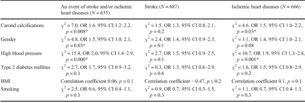
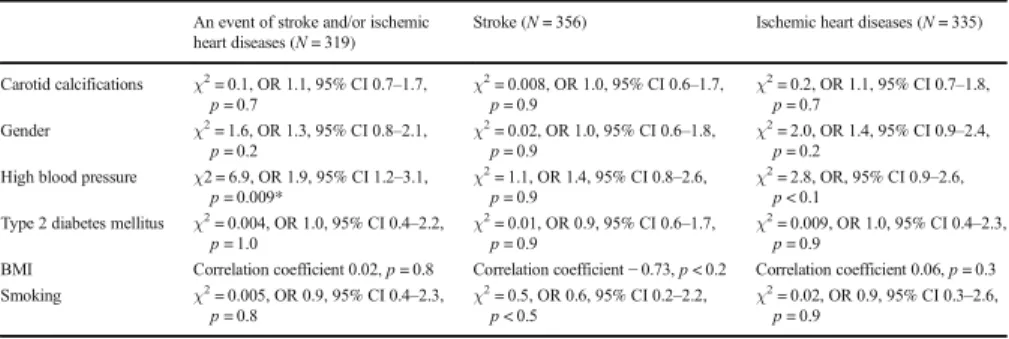
A significant association was identified by logistic regression, crude and adjusted (BMI, diabetes type 2, hypertension), in the YO be-tween carotid calcifications on panoramic radiographs and stroke and/or ischemic heart diseases crude (OR:2.4, 95% CI:1.5-4.0, p= 0.000), and adjusted (OR:1.9, 95% CI:1.1-3.5, p = 0.03) (Table I). In the OO group, only a history of hypertension was significantly associated with future stroke and/or ischemic heart diseases.
Table 1. Associations between 10-13 year incidence of stroke and/or ischemic heart diseases and different independent variables. Logistic regression in 60-72 years without a history of stroke and/or ischemic heart diseases reported at baseline
BMI= Body mass Index * = p<0.0.5
Independent variables at baseline OR 95%CI p-value
BMI 1.0 1.0-1.1 0.14
Carotid calcifications 1.9 1.1-3.5 0.03* Diabetes mellitus type 2 2.1 0.7-5.9 0.16
Data on causes of death was not available. Among those who died during the course of the study, 47.0% had positive signs of radio-graphic carotid calcifications compared to 31.2% in the material that were survivors. For those with signs of carotid calcifications at baseline, the OR of death before the endpoint was 2.0 (95% CI: 1.4-2.7, p <0.001).
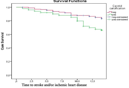
Relationships between positive or negative signs of carotid calcifi-cation and cumulative events of a first event of stroke and/or the first event of ischemic heart diseases were illustrated by Kaplan-Meier survival analysis (Figures 1 and 2). Study participants YO with baseline radiographic carotid calcifications and no previous history of an event had a mean cumulative stroke and/or ischemic heart diseases survival time of 12.1 years compared to those with-out carotid calcifications (mean cumulative survival time 13.0 years) (log rank Mantel–Cox χ2 = 10.7, p = 0.001). YO individuals with baseline radiographic evidence of carotid calcifications and no history of an ischemic event had a mean cumulative ischemic heart diseases survival time 12.5 years compared to those without carot-id calcifications (mean survival time was 13.2 years) (log rank Mantel–Cox χ2 =9.5, p = 0.002).
Time to stroke and/or ischemic heart disease
Figure 1. Kaplan-Meier curves of 60-72 year old individuals with no history of stroke and/or ischemic heart diseases, comparing individuals with and without carotid calcification. 13 year cumulative stroke and/or ischemic heart diseases survival
Time to ischemic heart disease
Figure 2. Kaplan-Meier curves of 60-72 year old individuals with no history of ischemic heart diseases, comparing individuals with and without carotid calci-fication. 13 year cumulative ischemic heart diseases survival.
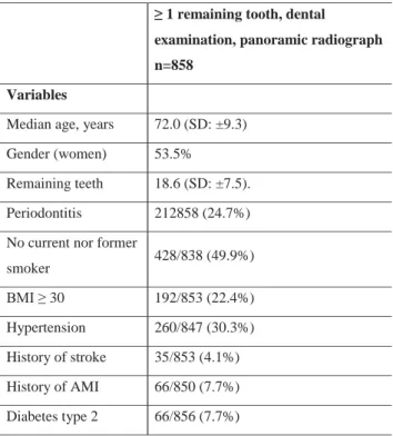
Data were derived from 858 individuals (women 53.5%). The ages at baseline varied between 60-93 years with a median age 72.0 years (SD: ±9.3). Individuals had, on average, 18.6 remaining teeth (SD: ±7.5). Approximately half of the individuals 428/838 (51.1%) reported that they did not or had never smoked. During the 17-year follow-up 492/858 (57.3%) died, and 51/858 (5.9%) of the individuals moved away from the Karlskrona area. Periodontitis was defined as bone loss ≥ 5 mm at ≥ 30% of sites. At the baseline examination, periodontitis was diagnosed in 212/858 (24.7%). Periodontitis was more frequent among men (57.1%) (OR:1.8, 95% CI:1.3-2.4, p= 0.000). If dividing into a young-old (YO) age cohort (60-72 years), 471/858 (54.9%) were included, and in an old-old (OO) age cohort (78-96 years), 387/858 (45.1%) were in-cluded.
The cumulative incidence of at the least one stroke during the 17-year follow-up was 118/858 (13.8%), and for at the least one is-chemic heart disease episode, the cumulative incidence was 203/858 (23.7%). Concerning gender, men and women almost equally developed stroke 60/118 (50.8% men) and ischemic heart diseases 102/203 (50.2% men). The incidence of stroke per year was 24.86, which corresponds to 2898 strokes per 100 000 per-sons and year. The same figures for ischemic heart diseases was 57.2 incidents per year and 6668 per 100 000 and year.
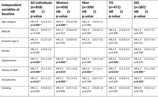
Cox regression analysis was used with periodontitis as an inde-pendent variable. Incidence of death, the first event of a stroke or ischemic heart diseases as the dependent variables were used,
ad-justed for independent variables: age group, BMI >30, diabetes
type 2, gender, hypertension, a history of acute myocardial infarc-tion (AMI), a history of stroke and smoking. Periodontitis in-creased the risk for ischemic heart diseases in all individuals (HR:1.5, CI:1.1-2.1, p=0.017) (Figure 3). If having periodontitis and being a woman (HR:2.1, CI:1.3-3.4, p=0.002) or having peri-odontitis and being in the OO age group (HR:1.7, CI:1.0-2.6, p=0.033) there was also an increased risk for ischemic heart
diseas-es. In men and in the YO age group no association between perio-dontitis and ishemic heart diseases could be demonstrated (HR:1.1, CI:0.7-1.7, p=0.749) respectively (HR:1.3, CI:0.8-2.2, p=0.229). No association between periodontitis and stroke could be identi-fied. Concerning death periodontitis increased the risk for all cause mortality in all individuals (HR:1.4, CI:1.2-1.8, p=0.001) (Figure 4). If having periodontitis and being a man (HR:1.5, CI:1.1-1.9, p=0.006) or in the YO age group (HR:2.2, CI:1.5-3.2, p=0.000), the risk for death was also increased.
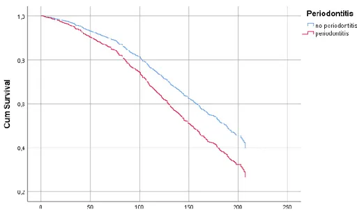
Figure 3. Cox regression curves: 17 year cumulative ischemic heart diseases survival of the total population, comparing individuals with and without peri-odontitis.
Figure 4. Cox regression curves: 17 year cumulative death survival of the total study population, comparing individuals with and without periodontitis.
Periodontitis and cardiovascular diseases are both chronic diseases. It is therefore not surprising that such conditions are more com-mon in older individuals. In part, increasing longevity can be at-tributed the extensive research resulting in better interceptive care. Nevertheless, in most cases medical and dental clinicians can alle-viate symptoms, prolonging the disease processes and consequences of disease over an extended time. Changes in diagnostic criteria over time, changes in therapies, changes in life-style, changes in so-cio-economic factors, and other confounding factors that may af-fect the clinical expression of both periodontitis and cardiovascular diseases are challenging to control for using statistical modelling alone. Until health sciences have identified a majority of the etio-logical factors and effects of confounding factors, it may be diffi-cult to go beyond studies considering associations between differ-ent disease complexes.
Periodontitis driven by inflammation, like many other diseases, in-cluding CVDs (Christodoulidis et al. 2014), has been shown to trigger subclinical atherosclerosis through an inflammatory pro-cess. Atherosclerosis is the main reason for CVDs (Cheng et al. 2006, Desvarieux et al. 2003). One type of atherosclerotic lesion is the carotid calcification (van Gils et al. 2012), which most often is identified in Doppler sonography (Denzel et al. 2004). The carotid calcification can be identified on panoramic radiographs (Fried-lander et al. 1994, Cohen et al. 2002, Persson et al. 2002, Ravon et al. 2003, Tanaka et al. 2006, Kumagai et al. 2007, Lee et al. 2014,
Friedlander et al. 2015, Bengtsson Wallin et al. 2016, Bengtsson Wallin et al. 2019) and more often in older individuals (Bengtsson Wallin et al. 2016, Bengtsson Wallin et al. 2019). In study I, it could be concluded, that there are only a few well-designed studies in older individuals investigating carotid calcifications on pano-ramic radiographs and associations with periodontitis and associa-tions with a history of stroke. The specificity, when identifying
ca-rotid calcifications was in most studies high, meaning that the
ab-sence of radiographic evidence of carotid calcifications on pano-ramic radiographs is consistent with negative findings using Dop-pler sonography. To identify carotid calcifications against DopDop-pler sonography the sensitivity varied between the different studies.
There was a shortage of longitudinal or clinical follow-up studies, specifically beyond assessments with Doppler sonography. It is, therefore, no surprise that one significant meta-analysis 2012 iden-tified that an association between periodontitis and atherosclerotic heart disease exists independent of known confounders. No studies have, however, identified a causative relationship (Lockhart et al. 2012).
In a cross-sectional study (Study II) an association between perio-dontitis and carotid calcifications on panoramic radiographs was shown. Similar results have been shown in other studies (Persson et al. 2002, Ravon et al. 2003, Beckström et al. 2007). In two of the studies (Persson et al. 2002, Ravon et al. 2003), the definition of
periodontitis was; alveolar bone loss > 4 mm between the
ce-mentoenamel junction (CEJ) and bone level at ≥ 30% of sites. In our study a composite definition of periodontitis was used. Alt-hough using different definitions of periodontitis, the results indi-cate a statistical association between periodontitis and carotid cal-cifications.
In study III, it was demonstrated that individuals with carotid cal-cifications on panoramic radiographs at baseline, over 10-13 years developed events of stroke and/or ischemic heart diseases more of-ten than individuals without carotid calcifications and this was es-pecially true in the younger age cohort (YO), 60-72 years. These
associations also remained when adjusted for confounders. Men 60-72 years, without previous events, had a higher OR of 3.1 to develop stroke, compared to women. This association may be re-lated to the finding of more carotid calcifications on panoramic ra-diographs in men. There are several studies demonstrating associa-tions between carotid calcificaassocia-tions on panoramic radiographs and a history of stroke (Friedlander et al. 1994, Carter et al. 1997, Lewis et al. 1999, Ravon et al. 2003, Kumagai et al. 2007, Chris-tou et al. 2010). A few prospective studies have also demonstrated an association between presence of carotid calcifications and stroke (Cohen et al. 2002, Friedlander et al. 2007, Johansson et al. 2015). Two studies with a follow-up of approximately 3 years studied male veterans > 55 years, and found associations between carotid calcifications on panoramic radiographs and an incidence of MI, stroke, TIA, revascularization, and angina (Cohen et al. 2002, Friedlander et al. 2007). Another recent study over 5 years demon-strated that the risk of future vascular events was higher in indi-viduals with carotid calcifications on panoramic radiographs com-pared to individuals without such signs (Johansson et al. 2015). This is in line with the results of our study with a follow-up of 10-13 years. Our study is, as far as we know, the only long-term-study with a follow-up time of 10 or more years in individuals 60 years and older investigating associations between carotid calcifications on panoramic radiographs and new events of medically diagnosed stroke and/or ischemic heart diseases. In the YO age cohort with carotid calcifications, the mean cumulative stroke and/or ischemic heart disease survival time was approximately 1 year lower and should be considered clinically significant. Among individuals with carotid calcifications, more individuals died over time, with an OR of 2.0 for reasons not known but most likely many died due to CVD events.
According to the result in study IV, individuals 60 years and older with periodontitis had a higher risk to die in the 17-year follow-up period compared to individuals without periodontitis. Approxi-mately half of the individuals died during the long-term follow-up period, but we do not have access to the reasons for death. The
main reason for death in the US is coronary heart diseases, and the second reason is stroke (Benjamin et al. 2018). Accordingly, it is likely that some individuals in the study had CVD events that re-sulted in a lethal outcome and therefore no cardiovascular event was registered and likewise deaths due to CVDs could never be clarified. In a registerbased study it was demonstrated that perio-dontitis increased the risk for all cause mortality within 15 years (Hansen et al. 2016). An association between periodontitis and mortality in the 60-72 years (YO) was also shown in our study. Söder et al. (2007) demonstrated that young individuals (30-40 years) with periodontitis and missing molars had an increased risk for early death over 16 years. Some of the studies have used miss-ing teeth as a proxy for periodontitis since if periodontitis is not treated or arrested the consequence will be missing teeth and miss-ing teeth due to periodontitis may in a way reflect an inflammatory burden (Holmlund, & Lind, 2012). Lee et al. (2019) demonstrated that edentulous individuals had the highest cardiovascular risk. Periodontitis has been reported as one of the major causes for
tooth loss in adulthood (Natto et al. 2014).It is, however, difficult
to be sure of the reason for missing teeth unless it is clearly report-ed in the dental record. Bone loss as usreport-ed in the present study to define periodontitis is, however, a very strong indicator of that the patient have had periodontal inflammation and using periodontal bone loss instead of missing teeth as a proxy of inflammatory bur-den over time seems more relevant.
The second main finding in study IV was that periodontitis in-creased the risk for ischemic heart diseases in the total population over the 17-year. In women with periodontitis, the OR for the in-cidence of ischemic heart disease was 2.1. This is in agreement with data from Stramba et al. (2006) reporting that women after the menopause, have a higher incidence of MI compared to age-matched men. There are few other longitudinal studies investigat-ing associations between periodontitis and CVDs. In a 3-year fol-low-up study by Renvert et al. (2010), including individuals that already had been diagnosed with ACS, an association between per-iodontitis and recurrent ACS was shown (Renvert et al. 2010). In
study IV individuals were randomly selected from the population. A register-based study with a 15-year follow-up identified that pa-tients with periodontitis reported an increased risk of CVDs (Han-sen et al. 2016). In this study, individuals >18 years were consecu-tively included. A hospital diagnosis of chronic or acute periodonti-tis, based on ICD codes was registered and also the subsequent in-cidents of CVDs. Compared to our study the included ages differed and the definition of periodontitis was more vague with no infor-mation regarding the severity. In a Korean nationwide cohort fol-low-up study of 7.6 years, a dose-dependent association between missing teeth and incidence of myocardial infarction, heart failure and ischemic stroke was found, especially in individuals with peri-odontitis (Lee et al. 2019). The definition of periperi-odontitis was in the study by Lee et al. (2019) not mentioned and the study individ-uals included were from 20 years with a history of a CVD event. Those differences makes this study not comparable to our study. In study IV the distance between the alveolar bone level and the CEJ ≥ 5 mm at ≥ 30% of sites on panoramic radiographs was used as the definition of periodontitis. This definition reflects the effects of periodontitis. It is reported that progressive periodontitis among older individuals includes bone loss, attachment loss and progres-sive gingival recession rather than deep periodontal pockets (Die-trich et al. 2006).
With the above mentioned definition 24.7% of the individuals were diagnosed as having periodontitis. In other studies the preva-lence of periodontitis varies from 33%-70% in older individuals, all with different criteria for periodontitis and different ages (Holm-Pedersen et al. 2006, Eke et al. 2012, Norderyd et al. 2012, Renvert et al. 2013). The prevalence of periodontitis in this study with an older population was low. Our definition of periodontitis included a more severe stage of periodontitis as a bone loss of ≥ 5 mm from the CEJ to the alveolar bone level indicates a definitive bone loss even accounting for the measurement error. A require-ment in our definition was also that the bone loss should be pre-sent at ≥30% of sites, which corresponds to a general disease
dis-tribution. Other reasons for the low prevalence of periodontitis can be that the very sick and immobile individuals were not included in the study as it was not possible to obtain a panoramic radiograph at the research centre. While periodontitis is a cumulative disease, more individuals develop periodontitis with age (Eke et al. 2016). A prerequisite to having a diagnosis of periodontitis in the present study was the presence of teeth. An inclusion criterion for being in-cluded in the present study was presence of one tooth or more. In-dividuals affected by periodontitis in younger ages are the most sensitive individuals with a hyperactive responsive immune and in-flammatory system (Fine et al. 2018). The most susceptible indi-viduals with the rapid form of periodontitis progression are often younger and have probably fewer teeth or are even edentulous with older ages and therefore were not included in the study. A stronger association between periodontitis and CVDs has been shown in younger individuals compared to older individuals (Lee et al. 2019, Schenkein et al. 2013). This could be one explanation for the lack of association between periodontitis and incidence of stroke in the present study, as it concerns older individuals.
A strength with the comprehensive SNAC study is that it has a long-term follow-up of the individuals. It is valuable that both a medical and a dental examination were performed with the same time intervals. The clinical examinations were performed as a full mouth examination and not as partial examinations of specific teeth or areas. Partial mouth examinations have been shown to underestimate the disease (Kingman et al. 2008). Most of the pub-lished studies so far are cross-sectional studies. Several other stud-ies have identified associations between periodontitis and CVDs (Persson et al. 2003, Scannapieco et al. 2003, Renvert et al. 2006, Kinane et al. 2008, Lockhart et al. 2012, Cullinan et al. 2013, Die-trich et al. 2013). Using a long-term follow-up should increase the possibility to explore true associations between diseases that may be overlooked in cross-sectional studies. Not many studies have this opportunity because they are time-consuming and expensive. There are also limitations in the present studies. When studying as-sociations in older individuals over a long time, many individuals