Milda Dambrauskaite
Supervisor: Jonas Rönnberg, SLU, Southern Swedish Forest Research Centre
Assistant supervisor: Khaled Youssef, SLU, Southern Swedish Forest Research Centre
Incidence of Heterobasidion spp. in
Scots pine on “low risk” sites
in Southern Sweden
Swedish University of Agricultural Sciences
Master Thesis no. 309
Southern Swedish Forest Research Centre
Alnarp 2019
Swedish University of Agricultural Sciences
Master Thesis no. 309
Southern Swedish Forest Research Centre
Alnarp 2019
Milda Dambrauskaite
Supervisor: Jonas Rönnberg, SLU, Southern Swedish Forest Research Centre
Assistant supervisor: Khaled Youssef, SLU, Southern Swedish Forest Research Centre
Examiner: Magnus Löf, SLU, Southern Swedish Forest Research Centre
Master thesis in forest management, EX0928 EUROFORESTER - Master Program SM001
Incidence of Heterobasidion spp. in
Scots pine on “low risk” sites
Incidence of Heterobasidion spp. in Scots pine on “low risk”
sites in Southern Sweden
Milda Dambrauskaite
Supervisor: Jonas Rönnberg, SLU, Southern Swedish Forest Research Centre Assistant supervisor: Khaled Youssef, SLU, Southern Swedish Forest Research Centre Examiner: Magnus Löf, SLU, Southern Swedish Forest Research Centre
Credits: 30 credits
Level: Advanced level, A2E
Course title: Master thesis in Forest Science Course code: EX0928
Programme/education: Euroforester - Master Program SM001 Course coordinating department: Southern Swedish Forest Research Centre Place of publication: Alnarp
Year of publication: 2019
Online publication: https://stud.epsilon.slu.se
Keywords: Heterobasidion annosum, incidence, Scots pine, spore
infection, thinning, Southern Sweden.
Swedish University of Agricultural Sciences Faculty of Forest Sciences
Abstract
Heterobasidion spp. fungi cause pathogenic infections in woody plants throughout the
northern hemisphere, causing the most damage in the Boreal forest zone. In Sweden Scots pine, being the second most economically important tree species, suffers a considerable amount of damage as Heterobasidion spp. fungal infections hinder the annual increment of a tree. This causes significant financial losses.
The aim of this research is to determine Heterobasidion spp. incidence in Scots pine stands growing on sites, considered unlikely to be infected in Southern Sweden.
In this study Scots pine trees, growing on “low risk” sites in Southern Sweden were sampled. 15 trees were sampled - 5 samples discs per tree were taken from the root systems of each. In total 75 trees were sampled. Sample discs were taken at 20-75 cm from root collar. Each tree was measured for volume calculations and visually evaluated for defoliation. Afterwards, samples were incubated at room temperature and checked for conidiophore presence using a stereo microscope.
The results revealed that Heterobasidion spp. infection was present in Scots pine trees on two out of five sampled stands. There was no correlation found between the sampled root diameter and the infection presence. Defoliation proved not to be a viable method for diagnosing the Heterobasidion spp. infection on living Scots pine trees. Current forest management practices in Sweden are not adapted for Heterobasidion spp. infection control, causing significant financial losses for the forest owners. This could be prevented by thinning the stands in winter as well as treating the stumps after each thinning.
Keywords: Heterobasidion annosum, incidence, Scots pine, spore infection, thinning,
Table of Contents
1. Introduction ... 6
1.1. Background information ... 6
1.2. Biology, ecology and characteristics ... 6
1.3. Host trees and disease ... 7
1.4. Economic impact and control ... 10
1.5. Hypothesis ... 11
Theoretical background, method, results: ... 12
2. Materials and methods ... 12
2.1. Site description ... 12
2.2. Sample collection and handling ... 13
2.3. Data collection and analysis ... 15
3. Results ... 16
3.1. Infection frequency ... 16
3.2. Correlation to stand data ... 16
3.3. Root diameter ... 17
3.4. Distance from root collar... 18
3.5. Defoliation ... 19
3.6. Weather correlation ... 20
4. Discussion ... 20
4.1. The infection presence ... 20
4.2. The effect of thinning season ... 22
4.3. Visual evaluation ... 22
5. Practical implications ... 23
6. Acknowledgements ... 24
Table of Figures
Figure 1. Global distribution of Heterobasidion annosum complex (Korhonen, 2004) ... 7
Figure 2. a) Colonization of a fresh stump and wound by H. annosum s.l. spores. b) Spread of the pathogen by grafting (Nemesio-Gorriz, 2015). ... 8
Figure 3. Research site distribution in Southern Sweden ... 12
Figure 4. Measurement procedures on field (from left to right: (A) bark thickness measurement; (B) DBH measurement) ... 14
Figure 5. Sampling procedures (From left to right: (A) Brushing off the soil; (B) spraying 70 % ethanol solution; (C) cutting the disc using a hand saw) ... 15
Figure 6. Examining samples in the laboratory (From left to right: (A) root disc with lines under a microscope; (B) Heterobasidion spp. conidia seen using a stereo microscope) ... 16
Figure 7. Standing volume and Site index for each site... 17
Figure 8. Sampled root disc diameter and infection presence correlation (where 1 - present, 0 - not found). ... 17
Figure 9. Average sample distance from root collar for each site ... 19
Figure 10. Defoliation in infected stands - amount of healthy an infected trees compared .... 19
Table 1. Information of research stands ... 13
Table 2. Defoliation scale (Hanisch & Kilz, 1991) ... 13
Table 3. Infection frequency (%) by root diameter class (1-6) ... 18
1. Introduction
1.1. Background information
Basidiomycetes of the the Heterobasidion genus cause root and butt rot, which decrease timber quality as well as reduces growth in conifers. A host genotype in Pinaceae family completely resistant to the pathogen is yet to be discovered (Stenlid et al., 2006). Due to that finding a way to prevent and control infections is vital for current forest management.
Heterobasidion has been found to cause decreased volume increment in infected pine trees of
approximately 12.6% (Wang, 2012). Thus an infection in the stand can cause significant monetary losses to the forest owner. Despite that treating Scots pine stumps to prevent such losses is not a highly applied procedure amongst forest owners in Sweden. Even though economically Scots pine is the second most important tree species in Sweden, this issue has received little attention thus far.
It is widely known that Heterobasidion spp. infections on coniferous trees are more prominent on sites with sandy soils, however little research has been done to determine its spreading potential and effect on tree growth on other sites with clayey or loamy soils.
1.2. Biology, ecology and characteristics
Heterobasidion annosum sensu lato (s. l.) is a complex of host-specific species
(Garbelotto & Gonthier, 2013). Eurasian groups were described as H. annosum sensu stricto (s.s.), H. abietinum Niemelä and Korhonen and H. parviporum Niemelä and Korhonen (Otrosina & Garbelotto, 2010). Prior to current classification the species were known as intersterility groups (ISGs). Three of them were recognised in Eurasia: P (H. annosum), S (H.
parviporum) and F (H. abietinum). H. annosum is widely distributed across the Northern
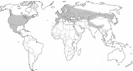
Hemisphere, most commonly found in the coniferous forest zone and in coniferous stands in the deciduous forest zone, however it has been reported in the Southern Hemisphere as well (Asiegbu et al., 2005). In Europe H. annosum is present in most of the coniferous forests, except pine forests in the south and the northernmost ones (Figure 1). The hazardous sites are known to have high sand content and good aeration (Rishbeth, 1951, 1963).
Figure 1. Global distribution of Heterobasidion annosum complex (Korhonen, 2004)
In Sweden Heterobasidion annosum is found throughout the whole territory of the country, while H. parviporum is only found in the southern part, Mälardalen being the north-most point where the infection has been discovered (Karlsson, 1993).
1.3. Host trees and disease
Heterobasidion annosum species complex is classified into different species
according to their host preferences: H. annosum attacks pine, H. parviporum - spruce and H.
abietinum – fir. Even though Scots pine is mostly attacked by H. annosum, young trees have
been found to be attacked by H. parviporum (Asiegbu et al., 2005).
Heterobasidion spp. fungi are most often dispersed by spreading and germination of
haploid reproductive spores called basidiospores. They are produced on a basidium via meiosis (Deacon, 2013). Basidiospore distribution rate is highly dependent on environmental factors such as air currents, water regime and spore physiology. Rain water often enables basidiospores of Heterobasidion spp. to enter lesions of tree roots or stem and to infect the area (Manion, 1991). Basidiospores can also be dispersed by burrowing animals and birds. In addition to that humans often aid the dispersal of pathogens by transferring basidiospores on their tools or clothing or by moving seeds or plants from one area to another (Dapkevičius et
al., 2006).
Heterobasidion spp. occurs in anamorphic (asexual reproductive) phase as
conidiophores with swollen heads that are formed directly on the mycelium. The conidia appear oval and translucent to white in colour. Conidiophores are classified as short term spores - they do not contain a lot of nutrients and cannot survive for a long period of time. Thus they are meant for rapid distribution and infection. Conidiospores are considered to be a
short-distance spread adaptation, as they are not actively released as basidiospores (Carlile & Watkinson, 1994). In the nature they are hardly noticeable, but can be found in bark beetle made holes on young pine trees. It is thought to take little part in natural dispersal. Culture can only produce conidia at temperatures from 0 to 28 °C (optimum 22.5 °C) in light or dark if the relative humidity is 95% or more (Rishbeth, 1951). H. annosum conidia are rarely ever found in natural conditions; however it is relatively easily cultured in a laboratory on agar or other media (Pegler & Waterson, 1968). This can be used in order to identify the fungus species complex. For identification of the specific species further analysis is needed.
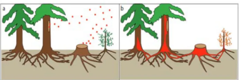
Forest trees are especially susceptible to fungal pathogens after both natural (storms, droughts, etc.) and anthropogenic disturbances (thinnings and other forest operations). Incidence of Heterobasidion spp. inside a tree can last up to several decades as the fungi resides in the heartwood without disturbing any vital processes of the tree (Stenlid et al., 1994). Due to that an infected tree cannot be identified visually.
Freshly cut stumps of conifer trees can be colonized by airborne spores of Heterobasidion
annosum that later grow down into the roots of the stump and infect living trees by root
contact (Piri & Hamberg, 2015) (Figure 2).
Figure 2. a) Colonization of a fresh stump and wound by H. annosum s.l. spores. b) Spread of the pathogen by grafting (Nemesio-Gorriz, 2015)
H. annosum basidiospores infect fresh stumps and wounds when the weather is warm, but not
too dry and hot, the stumps can be infected up to 3-4 weeks after felling (Manion, 1991).
Heterobasidion spp. rhizomorphs are able to penetrate the periderm barrier and infect a tree
(Pegg & Ayres, 1987). The mycelium can infect healthy trees via root contact or grafts by vegetative growth (Oliva et al., 2013). In commercial forest sites, where pine trees are planted at close proximity to each other, roots can often become grafted, creating one large root system with many stems. Such conditions are beneficial for H. annosum to spread through root contact (Lakatos et al., 2014). In order to defend from the infection, the tree produces resin as well as phenolic compounds - if a tree has high vitality the penetration is more likely to be stopped. Butt rot is rarely seen on pine trees, however it can be observed in older trees (Baral et al., 2016).
The infection in colonised stumps may remain for many years, enabling the infection to spread (Asiegbu et al., 2005). However, pine stumps, attacked by H. annosum, tend to rot faster than other species like larch and spruce. Due to that the infection persists for a relatively shorter period of time (Dapkevičius et al., 2006).
In most Pinus species only the roots and the stem base are normally colonised by the fungus, due to their resinous heartwood, thus resulting in sudden death or windthrow. Trees of all ages are susceptible to infections. The roots and stem base can be affected by decay and become resinous, but it doesn’t go up the stem (Vollbrecht et al., 1995). Infected pines have been observed to have reduced growth as the fresh needles tend to be shorter and fall earlier than of uninfected trees. Before dying, infected Pine trees are known to have shorter, sparser needles that eventually tend to fall (Phillips & Burdekin, 1992). Fruiting bodies are known to appear on the base of the infected tree and on infested stumps. Root symptoms are similar to symptoms of the trunk: resin exclusion and characteristic rot forms. On some sites
Heterobasidion annosum was found to kill pine trees, while on others it caused root or butt
rot without killing the tree. It reduces growth and increment on alkaline soils as it kills the roots, Symptoms (needle shortening and crown thinning) on pine trees appear shortly before they die. However, the tree can survive for several decades while being infected and that makes the growth reduction important when it comes to economic value of the stand and potential profit (Phillips & Burdekin, 1992).
Important factors for infection spread and development include soil conditions, tree age, former land use and thinning intensity. It was found that the highest losses in pine stands where soil pH was 6 or more (Low & Gladman, 1960). Even though soil moisture is important, H. annosum can kill trees in very dry areas on acid soil as well. More severe attacks have been recorded on light soils compared to heavy (Jørgensen et al., 1939; Anon, 1963). Plants with poor water supply are more likely to die, thus low soil moisture in dry summer may increase the chance of trees being infected by the fungus. Low levels of organic matter in the soil might be one of the reasons for moisture deficiencies. H. annosum on forest land is found to have the fastest infection rate on sites, previously used to grow conifers since often the fungus is already present in the stumps. On such sites the development of the disease tends to be more aggressive with each conifer rotation. While on sites used to grow hardwood trees the development of the disease seems to be hindered (Otrosina & Cobb, 1989). The effect of thinning is considered to be prominent due to the amount of stumps correlating with the rate of infection (Phillips & Burdekin, 1992). The more stumps there are exposed to H. annosum spores, the higher financial losses are to be expected. However the
most severe infections are seen on land that was previously used for agriculture. In addition to that in dry areas with high pH tree mortality can be high. In regards to the age of trees, young trees are found to die rapidly more often while older trees are more prone to long-lasting effects of the disease (Gibbs et al., 2002).
On living trees H. annosum infection cannot be distinguished from other rot diseases by visual examination (Woodward et al., 2002). H. annosum may kill up to two thirds of the root system of the tree before any crown symptoms, such as crown thinning, shortened needle length or foliage becoming yellowish, begin to show (Phillips & Burdekin, 1992).
Due to forest management practices and other anthropological impact on forests, the natural equilibrium between the fungus and its host has become impaired. Therefore control measures have to be taken in order to maintain productive forests (Schulze & Bahnweg, 2008).
1.4. Economic impact and control
Since H. annosum’s dispersal method is highly favoured by modern forest management practices, it has become the most important conifer root pathogen across Europe. In addition to that vast monocultures of coniferous trees create great dispersion possibilities for this pathogen. Nowadays the majority of forest operations in Sweden are performed using machinery which has to be run all year round in order to be economically feasible. This increases the possibility for H. annosum to spread during the warm seasons (Kärhä et al., 2018).
In order to control H. annosum, a multitude of methods have been applied over time. Stump or wound treatment, winter cutting, stump removal and change of tree species have been the most used in the Nordic countries. However, such methods as prescribed burnings have also been explored (e.g. in Finland). In sites that are considered to be highly infected rotation age is often shortened to minimize economic losses. Stump extraction from the infected site is rarely used nowadays because of high costs as well as high impact on the environment caused by such operation. Winter cuttings are quite often used to decrease the chances of infection as sporulation is not possible when the temperature is below 0 °C (Phillips & Burdekin, 1992). In Spruce stands harvested in summer or spring stump treatment is often applied, however pine stumps are not treated in Sweden. Chemical treatments such as 10 % Sodium nitrate (NaNO2) and creosote have been tested in Denmark (Yde-Andersen, 1982), however these
toxicity. Deacon (2013) suggests that the simplest way to avoid stump infection is treating the surface with such phytotoxic chemicals as urea or boron-containing compounds. However, such control procedures are not environmentally safe particularly when the area is in vicinity to a domestic water supply. Later on Pine stump inoculation with Phlebiopsis gigantea has been found to be effective in restraining H. annosum growth on Pine stumps as it is able to rather quickly occupy a large part of the stump surface and outcompete H. annosum (Rishbeth, 1963). Thus, a biological treatment Rot-Stop® was introduced, consisting of dried
Phlebiopsis gigantea spores that grow on the surface of a treated stump and, functioning as a
competitor for H. annosum, prevents its growth without affecting the living trees (Lakatos et
al., 2014). Nowadays mechanised stump treatment using a special device attached to the
harvester is most commonly used. Change of tree species is often undesirable by forest owners because of strong tradition as well as low market potential for timber.
P. gigantea was found to antagonise H. annosum through hyphal interference. By causing a
loss of normal membrane integrity shortly after first contact P. gigantea deactivates the hyphae of H. annosum and by doing so eliminates the potential competitor for the same substrate. In addition to that P. gigantea is one of the few Basidiomycota able to sporulate in laboratory culture (Deacon, 2013).
Once H. annosum is established in the stand it becomes nearly impossible to eradicate. The only known way to do so is to remove all infected stumps and major root from the stand. This method is highly labour intensive, expensive and inconvenient. Due to that control and preventative measures are normally promoted.
Manion (1981) suggests that there are no good recommendations for severely infected stands, so it is important to recognize the presence and seriousness of this problem at its early stages. Otherwise the productivity of a conifer stand can be destroyed and significant financial losses are inevitable.
1.5. Hypothesis
● Heterobasidion spp. will be found in Pine stands growing on sites, considered unlikely to have infections in Southern Sweden.
● Infected trees will have higher levels of defoliation than healthy trees.
● Heterobasidion spp. incidence throughout the sampled stands will depend on the thinning season.
The aim of this study is to investigate the effect of Heterobasidion spp. on Scots pine stands, growing on”unlikely” sites.
Theoretical background, method, results:
2. Materials and methods
2.1. Site description
In southern Sweden 5 Scots pine sites were randomly selected according to the following criteria:
● The site was not known to have Heterobasidion spp. present;
● The soil on site was moraine with mixed fractions (clayey or loamy texture); ● Scots pine trees constitute to at least 80 % of all trees in the stand;
● The stand has been previously thinned in summer or late spring; ● At the time of sampling either second or third thinning was going to be
executed.
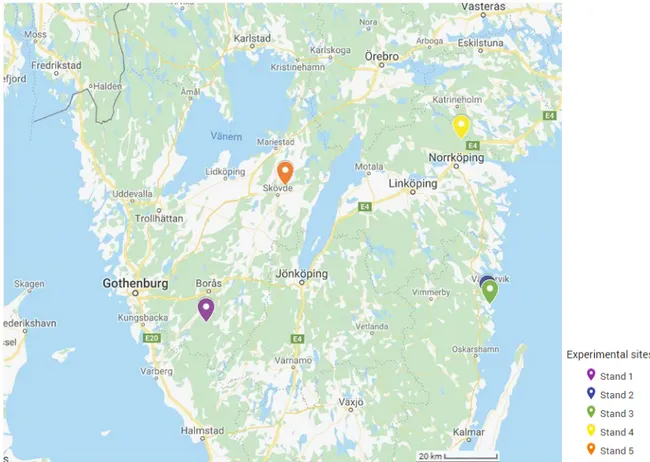
Selected stands were widely distributed across Southern Sweden (Figure 3). This way a better area coverage could be ensured.
Information about each stand was collected and assessed prior to fieldwork (Table 1). During this part of the stand selection process several stands were rejected as unfit for the aim of this research.
Table 1. Information of research stands
Site Location Stand age Site index Area, ha Standing volume, m3/ha
1 15km away from Svenljunga 38 T25 4.2 157
2 Västervik 52 T18 2.6 160
3 Västervik 47 T21 8.5 159.2
4 Norrköping 42 T29 9.2 191
5 Skövde 37 T24 5 191
This information was later used for data analysis and to assist the result interpretation
2.2. Sample collection and handling
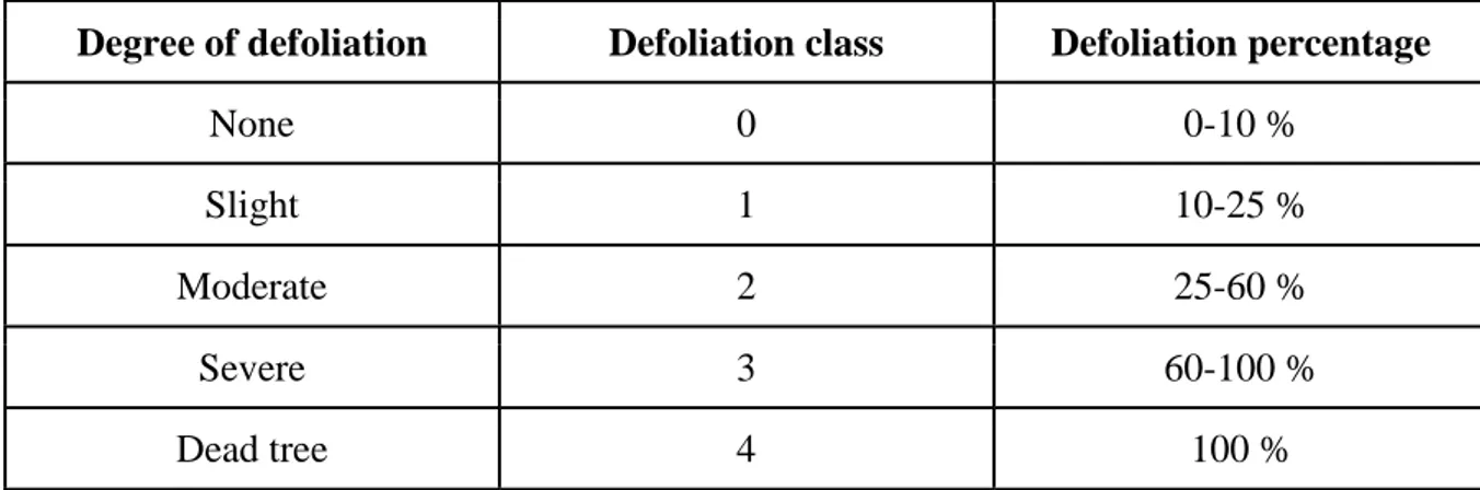
On each site 15 trees were selected by a representative of the forest owner or a forest manager. The trees were selected for thinning with the intention of having the best quality timber at the time of final felling. In addition to that an even distribution throughout the stand was kept in accordance with machine capabilities. Each tree was visually analysed from a viewpoint with good crown visibility to assess the level of defoliation. This was needed to get an overview of crown condition that could indicate the loss of tree vitality (Wulff, 2011). In each stand a local reference tree was selected to be used in comparison with other trees. The reference tree was representative of typical crown morphology in the stand and had 0-10 % defoliation. In order to determine the defoliation a scale of 0 to 4 was used for visual evaluation where 0 represents 0-10 % needle loss and 4 represents 100 % loss and thus is used for dead trees (Table 2):
Table 2. Defoliation scale (Hanisch & Kilz, 1991)
Degree of defoliation Defoliation class Defoliation percentage
None 0 0-10 %
Slight 1 10-25 %
Moderate 2 25-60 %
Severe 3 60-100 %
After defoliation assessment a compass was used to mark the north-most point of the tree, so the general North-East direction could be followed during sample collection.
In order to extract samples from pine roots, selected trees were uprooted using forest machinery (either harvester or a bulldozer). When the trees were down diameter at breast height (DBH), bark thickness, height and stem length to the first living branch were measured. DBH was measured cross-calipering, at 1.30 m above ground level. At the same height bark thickness was measured three times at different parts of the stem circumference using a bark gauge. This was done starting at a random point and moving approximately 120° between each measuring point around the stem. Subsequently a measuring tape was used to measure stem length from ground level to the base of the first living branch as well as the total height of the tree.
Figure 4. Measurement procedures on field ((A) bark thickness measurement; (B) DBH measurement)
Before taking the samples from the root systems of the trees a cleaning process was executed. During this process remaining vegetation, soil and small roots (<0.5 cm in diameter) were removed. After this the root systems were thoroughly cleaned using a garden spade, 5 sampling areas were selected at a 0-75 cm distance from the root collar. Sampling areas were selected close to lesions or other potential entrance points if such were visible. Samples were taken starting from the North-facing root going East-wise. Taproot, if identified, was sampled
last. Before sampling the area was meticulously cleaned using a brush and sprayed with 70 % ethanol solution to avoid cross-contamination from soil.
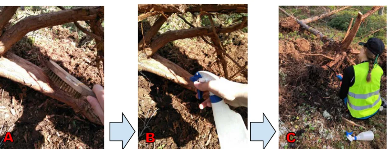
Figure 5. Sampling procedures ((A) Brushing off the soil; (B) spraying 70 % ethanol solution; (C) cutting the disc using a hand saw)
A hand saw blade was also sprayed with ethanol solution before every cut. 2-5 cm discs were cut and stem facing side was marked on the side using an arrow. Each sample was immediately put in a labelled bag where it was stored during the sampling process.
2.3. Data collection and analysis
After the sampling process was complete, samples were stored in the refrigerator (approx. 5 °C) until the incubation process was initiated.
To begin incubation samples were randomly taken out of the refrigeration chamber and stored at room temperature in a dark place for 7-10 days. In this case the majority of samples were incubated fresh from the field. When the incubation process was complete, samples were checked in a random order for Heterobasidion spp. conidiophore presence by using a stereo microscope. Each side of the disc was carefully examined at 20 – 40 (45) times magnification. If conidiophores were found, the presence was noted. If no conidiophores were found, but the infection was suspected incubation was continued for an additional 3-5 days. After the secondary incubation period samples were checked again. In addition to that the diameter of each disc was cross-measured.
Figure 6. Examining samples in the laboratory ((A) root disc with lines under a microscope; (B) Heterobasidion
spp. conidia seen using a stereo microscope)
Stem volume of each tree was calculated using a formula for pine growing in Southern Sweden by Näslund (1947). Data was further analysed using RStudio an MS Excel. Using R studio software ANOVA analysis was performed and using MS Excel “Pivot table” as well as other functions were applied.
3. Results
3.1. Infection frequency
Heterobasidion spp. infections in pine roots were found in 2 out of 5 sites: on site No. 4 (near Norrköping) and site No. 5 (near Skövde). In total 2.9 % of samples were infected: 9 samples were found to be infected on site No. 4 and 2 samples on site No. 5. Overall 9.3 % of trees were infected. The rate of infected trees on site No. 4 was 33.3 % and on site No. 5 it was 13.3 %.
3.2. Correlation to stand data
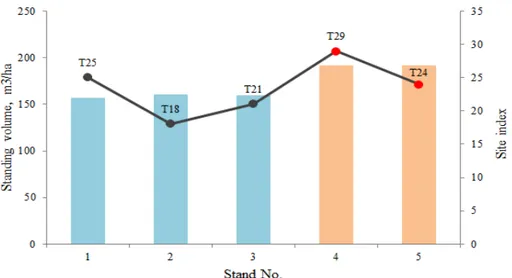
The stand data revealed that the infected stands were growing on sites, that were more fertile than the average of T23 (Fig. 6). In addition to that the infected stands both had 19.4 m3/ha higher volume than the average overall standing volume of all stands.
Figure 7. Standing volume and Site index for each site
The age of the stand, however, did not prove to be of importance during this research. All stand samples were between 37 and 52 years old. The average age of the infected stands was 40 years old, while the overall average age was 43.
3.3. Root diameter
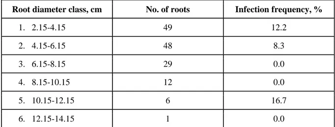
Sampled root diameter ranged from 2.2 to 12.6 cm. ANOVA analysis showed that no significant difference (P=0.915) in infection presence in different diameter roots and was found (Figure 8).
The infection frequency in the lower diameter classes (2.15 - 4.15 and 4.15 - 6.15 cm) was found to be the highest adding up to 90.9 % of all infections found (table 3). However the highest number of root samples (66 % of the total number) do fall into the lower diameter classes, making the average sampled root diameter 5.5 cm.
Table 3. Infection frequency (%) by root diameter class (1-6)
Root diameter class, cm No. of roots Infection frequency, %
1. 2.15-4.15 49 12.2 2. 4.15-6.15 48 8.3 3. 6.15-8.15 29 0.0 4. 8.15-10.15 12 0.0 5. 10.15-12.15 6 16.7 6. 12.15-14.15 1 0.0
Only one sample was found to be infected in higher diameter classes (10,15-12,15 cm).
3.4. Distance from root collar
The average distance from the root collar was found to be 42.2 cm (max. 75 cm, min. 10 cm). Since infections were found in 2 out of 5 stands, a subset of the data of those stands was made and a Pearson's chi-squared test was performed. The results of the test revealed that there is no correlation between the sampling area distance from the root collar and the infection presence (P=0.379).
Figure 9. Average sample distance from root collar for each site
The average sampled root disc diameter ranged between 2.15 and 12.55 cm.
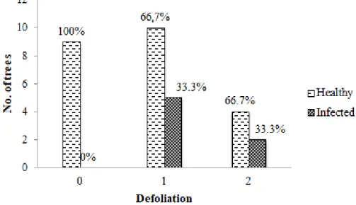
3.5. Defoliation
Since the infection was found in 2 stands, defoliation of healthy and infected trees on those sites was compared. As it can be seen in Figure 10 all infected trees had some level of defoliation as in the group of non-defoliated trees the infection rate was 0 %. However both 10-25 % (1) and 25-60 % (2) defoliation groups had the same ratio of infected trees (33.3 %).
Overall 33 % of all trees in infected stands were assigned to group 0 of defoliation, 47 % - to group 1 and 20 % - to group 2. In stands where no infected trees were found similar tendencies were observed. 64 % of all trees had some level of defoliation. Most of defoliated trees (46 %) had 10-25 % (1) defoliation. Remaining 18 % were found to have a higher level of defoliation (25-60 %) and were assigned to group 2.
3.6. Weather correlation
The stands were visited at the time of either second or third commercial thinning. According to Thor et al. (2003), in Sweden Heterobasidion spp. spore dispersal occurs throughout the growing season during the months of April through September. Due to that obtained stand data was analysed by the season of the last thinning.
Table 4. Thinning season and mean tree volume by site
Site No. of
thinnings Last thinning
Thinning season
Mean tree volume, m3
1 3 2016 April-May Summer 0.21
2 N/A N/A N/A 0.18
3 N/A N/A N/A 0.17
4 2 2007 November Winter 0.31
5 2 2009 June Summer 0.15
Stand 5 was infected even though the thinning was performed during the Winter season, while stand 1 was not found to be infected despite being thinned during the growth season. It is clear that in the infected stands mean tree volume was very varied, therefore the size of the tree was not important for infectivity.
4. Discussion
4.1. The infection presence
This study revealed that Heterobasidion spp. infection was found to be present in stands, growing on sites that are considered to be unlikely to have infection. This is important because currently these stands are not managed in accordance to recommendations for
Heterobasidion spp. prevention thus causing significant financial losses for the landowner.
The stands that were found to be infected both had higher than average site indexes as well as higher than average standing volumes. It is likely that there were more trees per hectare in
such stands compared to others. This could mean that the roots of the trees were closer to each other, increasing the likelihood of root contact and grafting. Due to that the infection had better spreading possibilities.
There was no correlation found between root diameter and infection presence. This means that all roots have similar risk to get infected by Heterobasidion spp. regardless of the diameter. However, roots smaller than 2 cm in diameter were not sampled during this research. Thus the correlation for smaller roots is unknown and further investigation is needed.
Most of the roots sampled are in the lower diameter classes. That is due to the sampling procedure, during which a hand saw was used and that limited the possibility to sample large diameter roots. In addition to that, samples were taken where the roots had lesions, root contact with stump roots or other potential infection entrance points. The infection frequency throughout the diameter classes does not appear to follow any trends.
Even though in this study sample distance from root collar proved not to be important for infection presence, previous studies (Wang, 2012) have found that that Hetrobasidion spp. infections in Scots pine are most likely to be discovered 0 - 75 cm away from the root collar. The samples in this study were collected according to that - no samples were taken more than 75 cm away from the root collar. This way the likelihood of discovering the infection in the roots was increased.
The fruiting bodies of Heterobasidion spp. were found on dead trees in a stands adjacent to the sampled stand No. 3. This proves that the spores must have been present and the infection was very likely to be active in the area. The fact that no infected samples were found on this site implies that either the infection has moved from roots or it was missed by selecting healthy trees to sample. Another possible cause for this could be that the infection was only present in a small part of root system of a tree, making it highly unlikely to be discovered. In addition to that the area of stand 3 was relatively large (8,5 ha), so the 15 trees that were sampled contributed to a very little part of the trees in the stand. Thus, increasing the possibility to miss the infected trees during the sampling process. However it is also possible, that the thinning of the stand was performed during the winter season which prevented the infection from spreading.
Such limiting factors as a rather low number of samples and not knowing the exact soil morphology, chemistry and ground water levels were present. The number of samples had to be reduced due to limited access to the forests because of the drought of the previous
summer. However, even with limited possibilities, the overall sample plot distribution throughout Southern Sweden was appropriate. The sampled stands were growing on sites with a wide range of site indexes from T18 to T29. Soil, being one of the most influential factors in the development of Heterobasidion spp. as well as other root diseases (Kuhlman et
al., 1976), was considered on a wider perspective during this research. Such factors as soil
pH levels, morphology, chemistry and ground water levels are too varied over the stand to be considered, as the results could only be applied for the stand sampled. Therefore, even without knowing such specific information, general conclusions could be made.
4.2. The effect of thinning season
Despite the fact that stand 4 was thinned during the winter season, the infection was found to be present there. Previous studies (Rönnberg et al., 2006) have shown that Scots pine stands, thinned during the winter had a lower Heterobasidion spp. infection incidence rather than stands thinned during the growing season. However, it is known that even though the optimal temperature for Heterobasidion spp. growth 23-26 °C, it grows considerably well at lower temperatures, for example it was found to grow at 21 % of optimum at 8 °C (Cowling & Kelman, 1964). In addition to that Heterobasidion spp. infectivity is also highly dependent on rainfall as higher precipitation can reduce the number of airborne spores present in the area (Farr & Hard, 1987). During rainfall many spores are washed away from the stump surface. Furthermore, high moisture content reduces oxygen availability for the remaining spores evidently lowering the growth rate of the fungus (Shaw III, 1989). The exact spore deposition in the air is virtually impossible to predict, since there are countless factors that can influence it.
4.3. Visual evaluation
It has been found that the infected Scots pine trees did not appear to have higher levels of defoliation than healthy trees. Thus it was confirmed that visual evaluation of Scots pine crown condition cannot be used to determine whether a specific tree is infected by
Heterobasidion spp. or not. Despite the fact that the infected tree crowns were defoliated
(10-60 %) these results cannot be used to diagnose the infection as there are many other possible causes for tree crown defoliation. At late stages of infection in a stand the proportion of dead and dying trees can be used as an indicator of infection presence (Kurkela, 2002). In addition
to that the visual evaluation used to determine defoliation is subjective as it depends on the perspective of the one performing the evaluation of crown condition. Despite that, alongside further tree health information inspection, defoliation data can be used to detect general changes in forest health condition (Wulff, 2011). Therefore, training is important to obtain as precise results as possible. Thus, other methods should be used in order to diagnose
Heterobasidion spp. infections in Scots pine trees.
4.4. Control measures
Even though many control measures for Heterobasidion spp. have been tested worldwide, currently two main methods to prevent the damage are used: winter harvest or thinnings and
Phlebiopsis gigantean Fr. (Rot-Stop®) stump treatment. Currently a stump treatment done at
the time of a thinning cost approximately 600 – 700 SEK/ha (removing 30% of standing volume) (Pettersson, 2013). Considering the financial losses due to reduced growth in Scots pine stands this seems like a feasible option to prevent them. However the question about the effect of mixed stands on Heterobasidion spp. infectivity and spreading is still widely discussed. In Finland it has been found that the damage of H. annosum to Norway spruce has been significantly lower in mixed stands compared to pure stands, however other factors (e.g. management regime) proved to be more influential than the stand composition (Peri et al., 2008). Similar results can be expected in mixed Scots pine stands. In addition to that changing the tree species in the stand is most often not economically feasible as the timber quality of the main tree species is often negatively affected (Lygis et al., 2004). Yet the effect of mixed stands is highly unexplored and more research is needed to determine its effect as there are countless ways to apply it in practice. There are many ways to prevent the
Heterobasidion spp. infection from spreading in Scots pine stands, however currently
basically none of them are used in practice in Sweden. This allows the infection to spread undisturbed and caused major financial losses every year.
5. Practical implications
Heterobasidion spp. infected Scots pine trees were discovered on two out of five sampled
stands located on “low risk” sites in Southern Sweden. This means that it is possible for stands growing on moraine soils with mixed fractions (clayey or loamy texture) to be
infected, leading to annual increment losses in such stands. In order to prevent that, stump treatment after thinning’s should be encouraged amongst forest owners and managers.
More in-depth analysis of Heterobasidion spp. infectivity dependence on soil characteristics is needed. In addition to that more continuous research throughout Sweden could potentially bring some clarity about future trends.
6. Acknowledgements
I would like to hereby thank my supervisor Jonas Rönnberg and my co-supervisor Khaled Youssef for their support and assistance. Special thanks to InterAgro Skog AB for providing financial support. I would also like to extend my thanks to all the all companies and private forest owners for allowing access to their forests and enabling us to take necessary samples.
7. References and citations
Anon, F. (1963). Forest disease research. Southern Forest Experiment Station.
Asiegbu, F. O., Adomas, A. & Stenlid, J. (2005). Conifer root and butt rot caused by Heterobasidion annosum (Fr.) Bref. s.l. Molecular Plant Pathology, 6(4), pp 395–409. Baral, B., Kovalchuk, A. & Asiegbu, F. O. (2016). Genome organisation and expression profiling of ABC protein-encoding genes in Heterobasidion annosum s.l. complex.
Fungal biology, 120, pp 376–378.
Carlile, M. J. & Watkinson, S. C. (1994). The fungi. 2. ed Academic Press.
Cowling, E. B. & Kelman, A. (1964). Influence of temperature on growth of Fomes annosus isolateS. Phytopathology, 54.
Dapkevičius, Z., Vasiliauskas, A. & Žiogas, A. (2006). Miško fitopatologija. Kaunas, Lithuania: Lututė.
Deacon, J. W. (2013). Fungal Biology. 4. ed Edinburgh, UK: John Wiley & Sons.
Garbelotto, M. & Gonthier, P. (2013). Biology, epidemiology, and control of Heterobasidion species worldwide. Annual Review of Phytopathology, 51(1), pp 3–15. Garbelotto, M., Ratcliff, A., Bruns, T. D., Cobb, F. W. & Otrosina, W. J. (1996). Use of Taxon-Specific Competitive-Priming PCR to Study Host Specificity, Hybridization, and Intergroup Gene Flow in Intersterility Groups of Heterobasidion annosum. 86(5), pp 543– 550..
Gibbs, J. N., Greig, B. J. W. & Pratt, J. E. (2002). Fomes root rot in Thetford Forest, East Anglia: past, present and future. Forestry, pp 74–76.
Hanisch, B. & Kilz, E. (1991). Monitoring of Forest Damage: Spruce and Pine.
Jørgensen, C. A., Lund, A. & Treschow, C. (1939). Undersafgelser over rodfordaerveren, Fomes annosus (Fr.) Cke. K. Vet.-og Landbohtfisk. Aarsskr. Applied mycology, 18, pp 772–773.
Karlsson, J.O. (1993). Genetic structure of populations of root rot fungi with special emphasis on Heterobasidion annosum. Diss. Uppsala: Swedish University of Agricultural Science.
Korhonen, K. (2004). Global distribution of Heterobasidion annosum complex .
Kuhlman, E. G., Hodges, C. S. & Froelich, R. C. (1976). Minimizing losses to Fomes
annosus in the southern United States. Asheville, N.C., USA: Southeastern Forest
Experiment Station.
Kurkela, T. (2002). Crown Condition as an Indicator of the Incidence of Root Rot Caused by Heterobasidion annosum in Scots Pine Stands. Silva Fennica, 36((2)).
Kärhä, K., Koivusalo, V., Palander, T. & Ronkanen, M. (2018). Treatment of Picea abies and Pinus sylvestris Stumps with Urea and Phlebiopsis gigantea for Control of Heterobasidion. Forests, 9(139).
Lakatos, F., Mirtchev, S., Mehmeti, A. & Shabanaj, H. (2014a). Handbook of the major
forest pests in southeast Europe. Pristina,: food and Agriculture Organization of the
United Nations.
Lakatos, F., Mirtchev, S., Mehmeti, A. & Shabanaj, H. (2014b). Manual for visual
assessment of forest crown condition. Pristina: Food and agriculture organization of the
United Nations.
Low, J. D. & Gladman, R. J. (1960). Fomes annosus in Great Britain; an assessment of the situation in 1959. Forest Record, 41, pp 20–22.
Lygis, V., Vasiliauskas, A., Stenlid, J. & Vasiliauskas, A. (2004). Silvicultural and pathological evaluation of Scots pine afforestations mixed with deciduous trees to reduce the infections by Heterobasidion annosum s.s. Forest Ecology and Management, vol. 201 (2–3), pp. 275–285.
Manion, P. D. (1991). Tree disease concepts. 2nd. ed Cornell University.
Nemesio-Gorriz, M. (2015). Molecular responses to Heterobasidion annosum s.l. in Picea abies. Diss. Uppsala: Swedish University of Agricultural Sciences.
Näslund, M. (1947). Funktioner och tabeller för kubering av stående träd. Tall, gran och
björk i Södra Sverige samt i hela landet. Stockholm: Statens Skogsforskningsinstitut.
Oliva, J., Boberg, J. B., Hopkins, A. J. M. & Stenlid, J. (2013). Concepts of epidemiology
of forest diseases.(Gonthier, P. & Nicolotti, G., Eds) Uppsala, Sweden: CAB
International.
Otrosina, W. J. & Cobb, F. W. (1989). Biology, Ecology, and Epidemiology of Heterobasidion annosum., California, USA, 1989. pp 26–31. California, USA.
Otrosina, W. J. & Garbelotto, M. (2010). Heterobasidion occidentale sp. nov. and Heterobasidion irregulare nom. nov.: a disposition of North American Heterobasidion biological species. Fungal Biol, 114(1), pp 20–23.
Pegg, G. F. & Ayres, P. G. (1987). Fungal Infection of Plants. Cambridge University press.
Pegler, D. N. & Waterson, J. M. (1968). Heterobasidion annosum. Descriptions of Fungi
and Bacteria. UK: CABI Bioscience.
Peri, T., Korhonen, K. & Sairanen, A. (2008). Occurrence of heterobasidion annosum in pure and mixed spruce stands in Southern Finland. Scandinavian Journal of Forest Research, vol. 5 (1–4), pp. 113–125.
Pettersson, M. (2013). Stump Treatment with the Root Rot Antagonist Phlebiopsis gigantea: - Sensitivity of P. gigantea Spores to High Pressure Stress - Reduced Water
Consumption for Stump Treatment. Diss. Alnarp: Swedish University of Agricultural Sciences.
Phillips, D. H. & Burdekin, D. A. (1992). Diseases of Forest and Ornamental Trees. Palgrave Macmillan UK.
Piri, T. & Hamberg, L. (2015). Persistence and infectivity of Heterobasidion parviporum in Norway spruce root residuals following stump harvesting. Forest Ecology and
Management
Rishbeth, J. (1951). Observations on the Biology of Fomes annosus, with Particular Reference to East Anglian Pine Plantations: III. Natural and Experimental Infection of Pines, and Some Factors affecting Severity of the Disease. Annals of Botany, 15(2), pp 221–246.
Rishbeth, J. (1963). Stump protection against Fomes annosus. Annals of Applied Biology, 52(1), pp 63–77.
Rönnberg, J., Petrylaitė, E., Nilsson, G. & Pratt, J. (2006). Two studies to assess the risk to Pinus sylvestris from Heterobasidion spp. in southern Sweden. Scandinavian Journal
of Forest Research, 21, pp 407–410.
Schulze, S. & Bahnweg, G. (2008). Critical review of Identification Techniques for Armillaria spp. and Heterobasidion annosum Root and Butt Rot Diseases. Journal of
Phytopathyology, 146(2–3), pp 61–72.
Shaw III, C. G. (1989). Is Heterobasidion annosum Poorly Adapted to Incite Disease in
Cool, Wet Environments? Berkeley, CA, USA: U.S. Department of Agriculture.
Stenlid, J., Karlsson, J. O. & Högber, N. (1994). Interspecific genetic variation in Heterobasidion annosum revealed by amplification of minisatellite DNA. Mycologic
Research, 98, pp 57–63.
Stenlid, J., Karlsson, M., Lind, M., Lundén, K., Adomas, A., Asiegbu, F. & Olson, Å. (2006). Pathogenicity in Heterobasidion annosum s.l. Proceedings of Forest pathology
research in the Nordic and Baltic countries 2005, Biri, Norway, 2006. Biri, Norway:
Skogforsk.
Thor, M., Möykkynen, T., Pratt, J. E., Pukkala, T., Rönnberg, J., Shaw III, C. G., Stenlid, J., Ståhl, G. & Woodward, S. (2003). Modeling infection and spread of Heterobasidion annosum in coniferous forests in Europe.
Vollbrecht, G., Johansson, U., Eriksson, H. & Stenlid, J. (1995). Butt rot incidence, yield and growth pattern in a tree species experiment in southwestern Sweden. Forest Ecology
and Management, 76, pp 87–91.
Wang, L. (2012). Impact of Heterobasidion Spp. Root Rot in Conifer Trees and
Assessment of Stump Treatment: with Emphasis on Picea Abies, Pinus Sylvestris and Larix × Eurolepis. Diss. Sweden: Swedish University of Agricultural Sciences.
Woodward, S., Stenlid, J. & Karjalainen, R. (2002). Heterobasidion annosum. Biology, Ecology, Impact and Control.(Hutterman, A., Ed) Plant Pathology, 48(4).
Wulff, S. (2011). Monitoring Forest Damage. Diss. Swedish University of Agricultural Sciences.
Yde-Andersen, A. (1982). Stump protection with urea against Fomes annosus in Norway spruce.