1
MASTER THESIS PROJECT
Microtubule associated
proteins 1B and 1S;
interactions with NR1 and
NR3A
Stefan Björklund 1/15/2008
Supervisors
Maria Eriksson, M. Sc.
Eirikur Benedikz, associate professor Erik Sundstrom, associate professor
Karolinska institutet, Novum, Department Neurobiology, Care science and society. Division of neurodegeneration, Huddinge.
Magnus Neumuller, PhD.
2
Abstract
In previous studies the carboxyl-terminus of microtubule-associated protein 1S was shown to interact with the N-methyl-D-aspartate receptor subunit NR3A (Eriksson et. al.)1. In
this study, interactions between three truncations of the microtubule-associated proteins 1B and one truncation of the microtubule-associated protein 1S carboxyl-terminus and the N-methyl-D-aspartate receptor subunits NR1 and NR3A were examined. The study
showed that an interaction occurred between amino acids 2167 to 2365 of the
microtubule-associated protein 1B and NR3A. That region of microtubule associated protein 1B corresponds to a microtubule-binding region in the light chain. It has been shown in earlier studies (Reviewed in Halpain S. et a12, Riederer, BM. et.al3.) that the light chain is a active part of the protein that have been post translational cleaved. The MAP 1 proteins are present in all tissue but has higher concentrations in the Post Synaptic Density of neurons in the central nervous system. The N-methyl-D-aspartate receptors are
present in glial cells and in the dendritic shafts of the central nervous system neurons (Eriksson et. al.)1 . The diseases were these proteins may play a part is mainly memory destructive diseases such as Alzheimers disease and in muscular dystrophy, but these assumptions are still being speculated.
3
Acknowledgement
1.
I would like to thank Maria, Eirikur, and Erik for help and guidance of my project and for letting me come to their research group to do my examination project.4
Introduction
1.1 The N-methyl-D-aspartate (NMDA) receptor
NMDA receptors belong to the ionotropic glutamate receptor family along with α-amino-3-hydroxy-5-methylisoxazole-4- propionic acid (AMPA) and kainate receptors. The NMDA receptor is composed of four different subunits (NR1, NR2A-D, and NR3A-B) that create a tetrameric assembly4.The NMDA receptor subunits have the same topology. They have a large extracellular N-terminus a membrane region that are composed of three transmembrane segments and one re-entrant domain, which are connected by extracellular and intracellular loops and a intracellular C-terminus as seen in figure 2.
NMDA receptors need both glycine and glutamate to be activated. When the plasma membrane is depolarized a Mg2+ ion that blocks the channel is released and ions can pass through the pore. NMDARs have a high
permeability to Ca2+ ions and the permeability is partially modulated by Zn2+ ions that acts as a partial antagonist on the NR2 subunit5.
The NMDA receptors are associated with the
postsynaptic density (PSD), which is located underneath the plasma membrane of excitatory synaptic connections usually found on dendritic spines6. However recent studies suggest that NMDA receptors also can be found on glial cells, which indicates that NMDARs can have a role in diseases such as multiple sclerosis and cerebral palsy (reviewed by Lipton, SA. Et al7). Such diseases affect oligodendrocytes that create the myelin sheets on neurons7.
The PSD is characterized by a dense network of different protein complexes. The different proteins that are associated with the PSD are cell-adhesion proteins, scaffolding proteins, membrane receptors such as NMDA and AMPA and different signalling molecules (kinases and phosphatases)6. The PSDs are under constant rearrangement and this seems to be one of the necessary functions for long term memory formation, since the
rearrangement leads to synaptic regulation and plasticity5. Actin and actin-binding proteins like the NMDARs are involved in the dynamic rearrangement of the synapses. Overexpression or downregulation of the actin binding proteins alter the shape and number of synaptic contacts5. Post synaptic density protein-95 (PSD95) interacts with NR1 and NR2 and is involved in transportation and stabilization of NMDARs at the synapse6.
The NMDARs are assembled in the endoplasmatic reticulum and transported with motor proteins (kinesines and dyneines) along the microtubule lattice in the dendritic shafts4.
1.1.1 The NR1 subunit
The NR1 subunit can be expressed as eight different variants due to alternative splicing of the transcript and has an ability to bind glycine4. NR1 is expressed at the plasma
membrane and the NR1 subunit is required as at least one of the subunits in forming a functional NMDA receptor, but cannot form a functional receptor of only NR1 subunits4. Expression of different splice variants varies depending on developmental stage, location and the composition of NMDAR signalling complexes, that might be regulated by
different splice variants8.
Figure 2. Displaying the structure of NMDA receptors.
(Picture taken from: Prybylowski, K., and Wenthold, R3)
5 1.1.2 The NR2 subunits
Four forms of the subunit NR2 have been cloned from different genes (NR2A-D) and the NR2 subunits contain a glutamate binding site. NR2A has been found in the forebrain and during brain development the expression level of the protein increases4. NR2B on the other hand, is the dominating NR2 subunit during early development, whereas the NR2C subunit is mainly found in the mature cerebellum and NR2D is mostly expressed in the thalamus4.
1.1.3 The NR3 subunits
NR3 is the most recently discovered NMDA receptor subunit. It has two different forms which are encoded by two different genes, NR3A and NR3B. When a functional NMDA receptor containing the NR3A subunit is activated it shows reduced Ca2+ permeability and lower sensitivity to Mg2+ block, compared to NMDA receptors lacking the NR3A subunit. The NR3A subunit is most prominently expressed during the early postnatal period in mice. Transgenic mice lacking the NR3A subunit show increased dendritic branching, which indicates that NR3A can play a role in synaptic formation and regulation9.
The N-methyl-D-receptor subunit 3A (NR3A) is known to interact with the C-terminus of microtubule associated protein 1S1 (MAP1S) and 1B (MAP1B).
1.2 The Microtubule associated protein (MAP) 1 family
The MAP1 family consists of three members MAP1A, MAP1B and MAP1S.
The main function of the MAP1 family is support and stabilization of the microtubules by binding to the microtubule lattice. MAP1 proteins also interact with filamentous actin (F-actin).When MAP1B and MAP1S are translated they form a precursor protein, which is post-translationally cleaved into one light and one heavy chain by proteolytic enzymes2.
1.2.1 Microtubule-associated protein 1B (MAP1B)
Human MAP1B is composed of 2464 amino acids and has a molecular weight of 320-330 kDa. The heavy chain (HC) comprises aa 1 to 2100 and has an actin binding region (aa 1-517) and an tubuline binding domain (aa 517-848). The light chain (LC) reaches from aa 2100-2464, it also has an actin binding domain (aa 2336-2459) and an tubulin binding domain (aa 2210-2336)3.
MAP1B is mostly expressed in neurons where it is thought to be important in axon guidance and development of dendrites especially during neuronal development when large amounts of MAP1B have been found in the axon. When the MAP1B gene was completely knocked out it resulted in complete absence of the corpus callosum10, which is a region of the brain that mostly contains axons10, connecting the two hemispheres.
After expression of the MAP1B precursor protein, it is proteolytically cleaved into the HC and LC and when the proportions of the different chains where measured it revealed that the LC was found in 6-8x higher concentrations, suggesting that the half-life of the LC is longer3.
1.2.2 Microtubule-associated protein 1S (MAP1S).
MAP1S (101-112 kDa) is considerably smaller than MAP1A and MAP1B (320-330 kDa) , mainly due to its shorter exon 5. It is also the most recently identified member of the MAP1 proteins and has previously been named C19ORF511 and VCYip21. When the amino acid sequence of the MAP1 members were compared it was discovered that they contained three regions of high homology, which are shared among the MAP1 proteins. In newborn mice the levels of MAP1S were high, unlike MAP1B where the levels increase during the first few days after birth and then drop to lower levels12.
6 In this study the main focus lied on determining which regions of the C-termini in
MAP1S and MAP1B that interact with NR3A. It was also investigated if the N-methyl-D -aspartate receptor subunit 1 (NR1) subunit can bind to either MAP1S or MAP1B and if MAP1B interacts with either of these. The C-terminus of MAP1B previously shown to bind NR3A was truncated and divided into three regions; the heavy chain (HC), the microtubule-binding region (MT) and the MAP1 homology domain 3 (MH3) region, which is the third homology region of all MAP1 proteins that bind microtubules (see figure 1 for details). One region of MAP1S was studied which was the HC region.
Figure 1. Schematic picture of the MAP1B and MAP1B proteins
The different regions which were cloned and studied are indicated by black. The regions which are most highly conserved among the MAP1
family members have been termed12 MAP1 homology domains, and are indicated by no 1, 2 and 3.
2. Material and Methods
2.1 MaterialsPrimers Brand Sequence MAP1B
5´ HC Thermo scientific CCCTGATGTGTCCTAGGTGGACCCAGAGGCC 3´ HC Thermo scientific GGCCTCTGGGTCCACCTAGGACACATCAGGG
5´ MT Thermo scientific GCTACATTCCTAACCACAGCAATAGTTAGAATGTTGATGTGG 3´ MT Thermo scientific CCACATCAACATTCTAACTATTGCTGTGGTTAGGAATGTAGC 5´ MH3 Thermo scientific CGGCCAAGACCGCCACTGGAATTCCAGGAACTACCAAGACG 3´ MH3 Thermo scientific CGTACATTCCTAACCACAGCAATAGTTAGAATGTTGATGTGG MAP1S
5´ HC Thermo scientific CCCATCCAGCATCTGCTAGGTGGACCCCGAGATGC 3´ HC Thermo scientific GCATCTCGGGGTCCACCTAGCAGATGCTGGATGGG
pGEX-6P-2
5´ Thermo scientific GGG CTGGCAAGCCACGTTTGGTG 3´ Thermo scientific CCG GGAGCTGCATGTGTCAGAGG
7
Chemical Brand Chemical Brand 10x PCR reactionbuffer Roche Antibodies
10x SureCut buffer Roche α-GST GE Healthcare Agarose SaveenWerner
α-NR3A. Custom made by Zymed invitrogen coop
Ampicillin Roche α-NR1 BD pharmingen bis-acrylamide Hintze anti-goat GE healthcare Fetal calf serum Gibco anti-rabbit HRP GE healthcare Complete protease inhibitor EDTA free Roche anti-mouse HRP GE healthcare DMEM F-12 glutamax medium Gibco Kits
DNA dye Gibco Quickchange Site directed mutagenesis kit Stratagene Eco RI Roche EZNA Plasmid miniprepp DNA kit I Omega Formamide Applied bios. Qiagen miniprepp DNA kit Qiagen GSH sepharose GE healthcare Qiaquick gel extraction kit Qiagen Im media Amp agar Invitrogen BCA protein assay Pierce
IPTG GE healthcare Big dye sequencing kit v1.1 and 3.1 Applied biosystem MagicMark Western protein standard GE healthcare ECL-plus GE healthcare NP-40 Merck
Oligo nucleotides Roche Pfu polymerase Stratagene pGex-6P-2 vector GE healthcare Precision plus protein standard Biorad Sample buffer Sigma SOC medium Invitrogen Sodium acetate GE healthcare SYBR green Invitrogen T4 dilution buffer Roche T4 ligase Roche T4 ligation buffer 10x Roche Taq polymerase Roche TBE 10x Invitrogen Triton X-100 Merck Tween Merck Cells Brand XL-1 blue Supercompetent cells Stratagene XL-10 Ultracompetent cells Stratagene BL-21(DE3) Supercompetent cells Stratagene HEK293 transfected cells
8 2.1.2 Instruments
2.2 Experimental procedure
2.2.1. Creating the cDNA constructs.
The starting constructs for this project were composed of the pGEX-6P-2 vector with fragments encoding the C-terminal parts of MAP1S (bp 2115-2526) or MAP1B (bp 6501-7404) (see appendix 1 for sequences) previously identified to bind to NR3A in a yeast two- hybrid screen.
Site directed mutagenesis was used for inserting mutations in the MAP1B and MAP1S cDNAs thus creating the constructs MAP1B HC (bp 6501-6657), MT (bp 6501-7096), MH3 (bp 7014-7404) and MAP1S HC (2115-2526). Stop codons for the constructs MAP1B HC, MT, and MAP1S HC regions where created and an EcoRI cleavage site for the MAP1B MH3 region was added, see table 1 for details.
The mutagenesis mastermix: 5µl of 10x reaction buffer
40ng of dsDNA (2,3 µl of MAP1B and 1,6 µl for MAP1S HC construct) 1 µM of forward primer*
1 µM of reverse primer* 2 mM of dNTPs
+40,7 µl of dH2O for the MAP1B mastermix and
+41,4 µl of dH2O for the MAP1S mastermix
To each reaction 1µl of PfuTurbo DNA polymerase (2,5U/µl) where added. *See table X for primer sequences..
Table 1. Location and sequence of the primers used for the mutagenesis and sequencing Protein Primer sequence *
MAP1B bp 6741-7648 Heavy chain (HC) bp 6883-6913 5`primer CCCTGATGTGTCCTAGGTGGACCCAGAGGCC bp 6913-6883 3`primer GGCCTCTGGGTCCACCTAGGACACATCAGGG Microtubule region (MT) bp 7323-7364 5`primer GCTACATTCCTAACCACAGCAATAGTTAGAATGTTGATGTGG bp 7364-7323 3`primer CCACATCAACATTCTAACTATTGCTGTGGTTAGGAATGTAGC MH3 region Nanodrop ND-1000 spectrophotometer ABI prism 3200 (ABI)
CCD Camera LAS-3000 (Fujifilm) PCR cycler PTC-200 (MJ Research) Sonicator Soniprep 150 (MSE) Safire Plate reader (Tecan)
9
bp 7233-7273 5`primer CGGCCAAGACCGCCACTGGAATTCCAGGAACTACCAAGACG bp 7273-7233 3`primer CGTCTTGGTAGTTCCTGGAATTCCAGTGGCGGTCTTGGCCG
bp 2120-3187 MAP1S Heavy chain (HC)
bp 2517-2551 5`primer CCCATCCAGCATCTGCTAGGTGGACCCCGAGATGC bp 2552-2517 3`primer GCATCTCGGGGTCCACCTAGCAGATGCTGGATGGG
pGEX-6P-2
bp 869-891 5`primer GGGCTGGCAAGCCACGTTTGGTG bp 1057-1035 3`primer CCGGGAGCTGCATGTGTCAGAGG
The mutagenesis PCR program: 1. 95o 30sek
2. 95o 30sek 3. 62o 1min 4. 68o 11min
Step 2-4 where cycled 16 times.
A total of 5 mutagenesis reactions were started, see table 1 for the location of the mutation on each construct.
When the mutagenesis PCR was finished the product was digested with 1 µl of Dpn I (10 U/µl) restriction enzyme. The reaction was incubated for 3 hours at 37o C.
Pfu Turbo, DNA polymerase is not a proof reading polymerase therefore it does not correct mismatching sequences. Because of this the Pfu DNA polymerase will nick the plasmid DNA due to mismatching primers. Dpn I restriction endonuclease is then used to digest the methylated and hemimethylated non mutated circular parental DNA. 5 µl of every digested product was mixed with 50 µl of XL-10 supercompetent cells in separate tubes. The reaction mixture was incubated on ice for approximately 30 minutes, heat shocked for 45 sec in 42o C and placed on ice. To grow the transformants 0.5 ml of SOC medium was added and the cells were incubated at 37o C for 1 hour with shaking at 160 rpm. 250 µl of the suspended transformants were plated on LB-agar plates with ampicillin (100 µg/ml) and incubated at 37o C for approximately 24 hours. Colony formation could be seen with the constructs MAP1B HC, MT, MH3 and MAP1S HC. 3 colonies from each plate where picked and inoculated in 5 ml of LB medium and grown over night at 37o C and 170 rpm to get more product.
2.2.2. DNA purification and analysis.
For the DNA extraction and purification, the E.Z.N.A plasmid mini preparation kit I was used, see appendix 2 for protocol.
All of the DNA mini preparations where concentration determined by spectrophotometry measuring the absorbance at 260 nm see Table 3 for results.
Table 3. DNA concentrations
cDNA concentration (ng/μl)
MAP1B HC 34.4
MAP1S HC 28.8
MAP1B MT 29.19
10 The MAP1B HC, MT and MAP1S HC and the unmutated vector preparations were
controlled by running a 1% agarose gel at 75 V for 1 hour. 5 µl of DNA dye were mixed with 7 µl of DNA.
To verify that the MAP1B MH3 mutation had been obtained, a control cleaving of the constructs were performed. This was done by picking three colonies from the MAP1B MH3 mutagenesis plate, growing them 5 ml of LB medium and preparing the DNA, as previously described. The concentration of the MAP1B MH3 DNA preparation was determined with the Nanodrop spectrophotometer to 37.4 ng/µl. 2 µg of DNA was mixed with 4 µl of 10 x SureCut buffer H and 10 U of EcoRI restriction endonuclease and incubated for 5 h at 37oC. 40 µl of the MAP1B MH3 along with 10 µl of DNA dye was analysed on a 1,5% agarose gel for 1 h at 75 V and then incubated with 5 µl SYBR green and 50 ml of 1 x TBE. Cleavage with EcoRI removed the HC and MT regions from the plasmid, leaving the pGEX-6P-2 vector with the MH3 region linearized.
The pGEX-6P-2 vector with the MAP1B MH3 fragment on the gel was extracted with a scalpel, the gel slice weighed 150 mg. For DNA extraction the QIAquick Gel extraction kit was used, see appendix 2 for protocol.
2.2.3 DNA sequencing using Big Dye v3.1 sequencing kit
Big Dye v 3.1 is a sequencing kit using Sanger sequencing principles. To be sure that the mutagenesis had incorporated the desired mutation every sample was sequenced using Big Dye v 3.1 .The primers pGEX 5` or pGEX 3` (see table 1) were used depending on
location of the mutation. For the MAP1B HC, MT and MAP1S HC the pGEX 5`primer was used and for the MAP1B MH3 the pGEX 3`primer was used.
For every reaction the following mastermix was prepared:
Ready reaction premix 8 µl Big Dye 5x reaction buffer 4 µl Primer 20 µg
Template 200 ng dH2O to 20µl
.
The PCR program for the sequencing reaction was:
1. 96oC for 1min 2. 96oC for 10 sec 3. 58oC for 15 sec 4. 68oC for 30 sec 5. 68oC for 7 min
Step 2-4 was cycled 25 times
The PCR products were precipitated by adding 1 µl of sodium acetate and 50 µl of 95% ethanol to each reaction and incubating at room temperature for 30 min. The samples were centrifuged for 20 min at 13000 rpm. The supernatant was removed by vacuum suction and the pellet washed with 70 µl of 70% ethanol before centrifuged for 5 min at 13 000 rpm. The supernatant was removed by vacuum suction and the samples were left to dry for 30 min in room temperature. The samples were resuspended in 10 µl of formamide and sequenced on Abi Prism 3100 Genetic analyser. See appendix 1 for sequences.
11 2.2.4. Transformation and expression of MAP1S and MAP1B constructs in E.coli BL-21(DE3) gold cells.
The constructs, that were induced and expressed where MAP1B HC, MH3, MT, MAP1S HC and the constructs encoding the whole C-terminus named MAP1B mini(see figure 1). 200 ng of the DNA preparations was mixed with 50 µl of BL-21(DE3) cells. The reactions were incubated for 30 min on ice then heat shocked for 45 sec at 42o C and placed on ice for 2 min. 500 µl of LB medium was added to each construct and incubated for 1 h at 37o C. 250 µl of each reaction were plated on LB plates with ampicillin and incubated over night at 37o C. One colony from each plate was inoculated with 5 ml of LB medium without ampicillin and incubated over night at 37o C with 225 rpm shaking.
1.5 ml of each over night culture was transferred to 150 ml of fresh LB medium and incubated for approximately 7 h at 37o C with shaking when Isopropyl
β-D-1-thiogalactopyranoside (IPTG) was added to the suspension to a final concentration of 1 mM to induce production of fusion proteins in BL-21(DE3) gold cells. The cultures were incubated another 2 hours before each suspension was centrifuged for 10 min at 4 000 rpm and the supernatant was discarded. The pellets were resuspended in 7.5 ml PBS with 375 µl 50 x complete protease inhibitor. All of the constructs were sonicated 5 sec on and 5 sec x 6 at 14-18 microns. The constructs were centrifuged for 10 min at 4 000 rpm and the supernatant was saved for use in the pulldown experiments.
The proteins that were expressed are fusion proteins that have a GST tag at the N-terminal of the protein. The fusion proteins were separated by western blot on a 10% SDS-PAGE gel for approximately 1 h at 200 V (see Table 3) in electrode buffer* , transferred to a nitrocellulose membrane for 2 h at 200 mA in transfer buffer with methanol*. The membranes were blocked in PBS-B* with a 0.5 % milk solution for 15 min, washed 1 x 15 min in PBS-B with 0.1 % Tween and then 4 x 5 min in the same buffer. The membrane was incubated with GST antibody (1:4000 dilution) over night in room temperature, then washed 5 times in PBS- B with 0.1% Tween, incubated with goat antibody (1:4 000 dilution)in PBS-B* with 5% milk for 1 h in room temperature and then washed 4 times in PBS-B with 0.1% tween*. The membrane was developed with the ECL-plus kit and analysed with a CCD camera.
*See Table 3 for recipes on the different solutions. Table 3. Western blot solution recipes.
Tris glycine SDS methanol water Electrode buffer 9 g 43 g 3 g Transfer buffer 6.05 g 28.82 g 400 ml 1600 ml NaCl Na2HPO4x7 H2O KH2PO4 KCl water PBS-B 10x stock 80 g 11.5 g 2 g 2 g
1000 ml
SDS gel SDS bis-acrylamide Tris pH 6.8 Tris pH 8.8 APS Temed water Stacking gel 20 µl 670 µl 250 µl 20 µl 2 µl 1.35 ml Separation gel 7% 50 µl 1.2 ml 1.25 ml 50 µl 3.5 µl 2.5 ml
2.2.5. Culturing of HEK293 cells stably expressing NR3A and NR1.
The NR3A and NR1 proteins were purified from HEK293 transfected cells, cultured in DMEM Glutamax F-12 medium with fetal calf serum. When the cells were confluent the media was removed and the cells washed three times with PBS. Before being removed from the flasks wall with a cell scraper and suspended in PBS. The cell suspensions were centrifuged for 10 min at 4 700 rpm followed by removal of the supernatant and
12 resuspension of the cell pellet in PBS with 2% Triton X-100, 2% NP-40 and 1 x Complete protease inhibitor. The suspensions were incubated for 1 h on ice with occasional
vortexing and centrifuged at 13 000 rpm for 10 min and the resulting supernatant containing the solubilised NMDA receptor subunits was saved. The total protein concentration of the supernatants was determined by using a BCA protein assay. The supernatants were also analyzed by western blot as previously described. 1 ml of NR1 (1:500) or NR3A (1:2000) polyclonal antibodies was added to the membranes and they were incubated over night in room temperature. For NR1, anti-mouse antibody was used as secondary antibody (1:500) and anti-rabbit for NR3A (1:500) in PBS-B with 5% milk and incubated for 1 h. The membranes were washed in PBS-B with 0.1 % Tween* for 15 min and then for an additional 3 x 5 min in PBS-B with 0.1 % Tween. The membranes were developed in ECL solution and analysed with a CCD camera.
The cell lysates contained NR1 and NR3A proteins and they could thus be used for the GST-Pulldown. The total protein concentration was determined by BCA protein assay. A standard curve was prepared with bovine serum albumin (BSA) and mixed with a working reagent that created a coloured complex. The colour intensity reaches its maximum at 560 nm and is proportional to the total protein concentration.
Three stocks with NR1 and NR3A of the following concentrations for NR1; 6.95, 5.5 and 6.2 µg/µl, and for NR3A; 7, 8.4 and 6.8 µg/µl were prepared.
*See Table 3 for recipes on the different solutions.
2.2.6. The glutathione-S-transferase (GST) Pulldown
This method was used to investigate the protein-protein interaction by expressing a protein with a glutathione-S-transferase (GST) tag attached to the N-terminal end, thus creating a fusion protein. The fusion protein was adsorbed to the glutathione-sepharose via the GST-tag. The sepharose was then incubated with samples of cell lysates, After the incubation, the sepharose was washed and associated proteins were then eluted. The eluate was analysed by SDS-PAGE and western blot..)
2.2.7 The GST pulldown experiment
The GSH sepharose comes in a 80% ethanol slurry and was equilibrated in PBS. The slurry was centrifuged, the supernatant removed and the sepharose pellet was washed with PBS 4 x 2 min with occasional vortexing. A 50% sepharose slurry in PBS was prepared. Equal amounts of fusion protein (200 µl of the pGEX-6P-2 vector protein, 400 µl of MAP1B HC, MT, mini MAP1S HC or 1000 µl of the MAP1B MH3), 75 µl of the 50% sepharose solution, 40 µl of 50 x Complete protease inhibitor and PBS was added to a final volume of 1 ml for each reaction. The mixtures were incubated under rotation at 4o C for approximately 3 h.
The NR3A and NR1 cell lysates were pre-cleared to reduce background noise during analysis by mixing 3500 µg of protein, 350 µl 50% sepharose and 280 µl of 50 x complete protease inhibitor and PBS to 7 ml. NR1 and NR3A were precleared under rotation at 4o C for 30 min.
The fusion protein mixtures were centrifuged at 13 000 rpm for 30 sec and the
supernatants removed. The sepharose pellets containing the fusion protein were washed in PBS with 0.5% Triton X-100 for 2 x 5 min. The pre-cleared NR1 and NR3A proteins were centrifuged for 3 min at 4 700 rpm and the supernatants were saved. 1 ml of the NR1 and NR3A pre-cleared solution was added to each of the fusion protein pellet and
incubated over night at 4o C under rotation. In the morning the samples were centrifuged for 1 min at 13 000 rpm, the supernatants were removed and the pellets washed with 1 ml of Triton X-100 in PBS for 4 x 15 min under rotation.
13
3. Results & Discussion
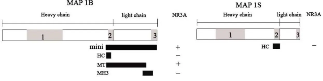
The MAP1B consist of 2464 amino acids and the MAP1S of 1092 amino acids. The MAP1B mini construct comprise amino acid 2167-2549. This region has been identified during earlier studies to interact with the NR3A subunit (unpublished observation). The MAP1B HC comprise amino acid 2167-2304, the MT comprise amino 2167-2503 and the MH3 construct comprise amino acid 2411-2549.
The MAP1S HC construct comprise amino acid 706-760, in earlier studies the region of MAP1S comprising amino acid 706-1062 has been seen to interact with the NR3A subunit. Due to difficulties in mutating and cloning the whole region in which NR3A interacted with MAP1S no results could be obtained
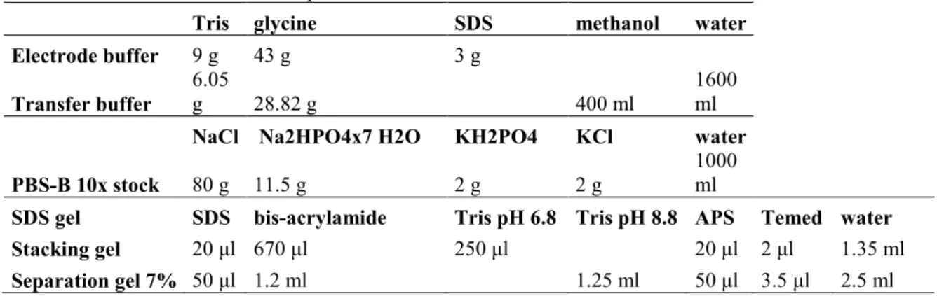
When the partial MAP1B and MAP1S gene fragments had been cloned and the DNA preparations had been purified, the constructs were analysed on a 1% agarose gel to confirm that their sizes were correct ( figure 3).
Figure 3.Gel electrophoresis confirming that the MAP1B HC, MT and MAP1S HC mutated constructs were the right size after the cloning reaction.
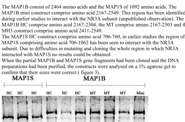
Three clonesof the MAP1B MH3 cDNA construct were control cleaved with EcoRI to see that the cloning been successful and the desired construct could be obtained as seen in figure 4, one of the replicates that underwent the cloning reaction had the Eco RI site incorporated.
14
Figure 4. Gel electrophoretic analysis of three MAP1B MH3 samples that underwent cloning reaction and then were
cleaved with EcoRI. As seen on the gel only the middle reaction was successful were the linearized pGex-6P-2 vector is
the upper band and the construct with the incorporated EcoRI site is the lower band
When the correct sequence of the DNA constructs had been confirmed by sequencing (see results in Appendix 1) they were expressed as fusion proteins in the BL-21(DE3) gold E.coli. The western blot analysis showed that the sizes of the expressed fusion proteins were correct (figure 5).
Figure 5. Western blot analysis of the expressed fusion proteins containing the amino acids which were used in the GST pulldown to see interactions with the NR3A C-terminus. They are probed with a GST antibody which interacts with the GST tag that was added to the peptides when their coding sequence were inserted in the pGEX-6P-2 vector.
The GST pulldown experiments with NR3A showed that MAP1B MT (amino acid 2167-2505) interacted with this receptor domain as seen in figure 6 and 7. The regions lacking the microtubule binding region (MAP1B or MAP1S) were not able to bind to NR3A. This indicates that the MT region is essential for the interaction between NR3A, MAP1B and MAP1S.
When GST pulldown experiments were done between NR1 and MAP1B, MAP1S all fusions proteins showed interaction with NR1. This result is highly unlikely. A more trustworthy explanation would be that unspecific binding occurs and the method used to test for the interaction has to be questioned.
15
Figure 6. Western blot analysis showing which fusion proteins that interact with NR3A.The MAP1B MT protein (aa 2167-2365) was able to bind NR3A but the MAP1B HC, MH3 and MAP1S HC were not.
Figure 7. Schematic picture showing the MAP1B and MAP1S proteins and what regions the fusion proteins comprises and were interaction with NR3A occurs. (Not drawn to scale).
The cloning of truncated parts of the MAP1B and MAP1S genes was successful. The encoded peptides were also successfully expressed the peptides seems to be useful in interaction studies.
The pulldown experiment showed an apparently specific interaction between the MT region of MAP1B and NR1. This is consistent with earlier observations that the light chain of MAP proteins NMDA receptors1 and the present study has narrowed down the interactive region to the MT sequence.
More extensive studies on the interactions between MAP1S and NR3A but also between MAP1B and NR1 should be made. Due to lack of time no conclusions on the MAP1B C-terminus interaction with could be drawn. The MAP1B MT sequence should be truncated further to determine with higher accuracy what sequence of MAP1B that interact with NR3A. Two more fusion proteins for MAP1S where planned to be analysed for
interaction with NR3A and NR1 but problems with mutagenesis and cloning occurred, so it would be satisfactory in the future to analyse interactions between different fusion proteins of the C-terminus of MAP1S and NR3A and NR1. It would also be interesting to see if any interactions occur in the MAP1 homology region 1.
The function of this interaction in vivo may be that MAP1B can transport NR3A along the microtubules along the dendritic shafts. Eriksson, M. Et. al. have shown that the
intracellular C-terminus of NR3A interact with MAP1S, suggesting that this protein have a central role in regulation of the NR3A subunits sites1.
16
4. Reference
1. Eriksson, M. Samuelsson, H. Samuelsson EB. Liu, L. McKeehan, WL. Benedikz, E. Sundström, E. The NMDAR subunit NR3A interacts with microtubule-associated protein 1S in the brain. Biochemical and Biophysical Research communications 361 (2007) 127-132.
2. Halpain, S. Dehmelt, L. The MAP1 family of microtubule-associated proteins. Genome biology (2006) 7:224.
3. Riederer, BM. Microtubule associated protein 1B, a growth-associated and phosphorylated scaffold protein. Brain research bull. 71 (2007) 541-558.
4. Prybylowski, K. Wenthold, R J. N-methyl-D-aspartate receptors: Subunit assembly and trafficking to the synapse. Journal of biological chemistry 279 (2004) 9673-9676. 5. Paoletti, P. Neyton, J. NMDA receptor subunits: function and pharmacology.
Pharmacology (2007) 7:39-47.
6. Boeckers, T,M. The Postsynaptic density. Cell tissue Res (2006) 326:409-422. 7. Lipton, SA. NMDA receptors, Glial cells, and Clinical medicine. Neuron. (2006)
6:9-11.
8. Bradley, J. Carter, SR. Vikram, RR. Wang, J. Finkbeiner, S. Splice variants of the NR1 subunit differentially induce NMDA receptor-dependent gene expression. Journal of Neuroscience (2006) 26(4):1065-1076.
9. Das, S. Sasaki, YF. Rothe, T. Premkumar, LS. Takasu, M., Crandall, JE. Increased NMDA current and spine density in mice lacking the NMDAR subunit NR3A. Nature (1998) 393:377-81.
10. Meixner, A. Haverkamp, S. Wässle, H. Führer, S. Thalhammer, J. Kropf, N. Bittner, RE. Lassmann, H. Wiche, G. Propst, F. MAP1B is required for axon guidance and is involved in the development of the central and peripheral nervous system. Journal of cell biology (2000) 6:1169-1178.
11. Liu, L. Vo, A. McKeehan, WL. Distinct structural domains within C19ORF5 support association with stabilized microtubules and mitochondrial aggregation and genome destruction. Cancer res.(2005b) 65, 1830-1838.
12. Orban-Nemeth, Z. Simader, H. Badurek, S. Trancikova, A. Propst, F. Microtubule-associated protein 1S, a short and ubiquitously expressed member of the microtubule-associated protein 1 family. Journal of biological chemistry (2005) vol.280, No. 3, pp. 2257-2265.
1 Appendix 1. Sequences of cDNAs and pGex-6P-2.
pGEX-6P-2 ACGTTATCGACTGCACGGTGCACCAATGCTTCTGGCGTCAGGCAGCCATCGGAAGCTGTGGTATGGCTGT GCAGGTCGTAAATCACTGCATAATTCGTGTCGCTCAAGGCGCACTCCCGTTCTGGATAATGTTTTTTGCG CCGACATCATAACGGTTCTGGCAAATATTCTGAAATGAGCTGTTGACAATTAATCATCGGCTCGTATAAT GTGTGGAATTGTGAGCGGATAACAATTTCACACAGGAAACAGTATTCATGTCCCCTATACTAGGTTATTG GAAAATTAAGGGCCTTGTGCAACCCACTCGACTTCTTTTGGAATATCTTGAAGAAAAATATGAAGAGCAT TTGTATGAGCGCGATGAAGGTGATAAATGGCGAAACAAAAAGTTTGAATTGGGTTTGGAGTTTCCCAATC TTCCTTATTATATTGATGGTGATGTTAAATTAACACAGTCTATGGCCATCATACGTTATATAGCTGACAA GCACAACATGTTGGGTGGTTGTCCAAAAGAGCGTGCAGAGATTTCAATGCTTGAAGGAGCGGTTTTGGAT ATTAGATACGGTGTTTCGAGAATTGCATATAGTAAAGACTTTGAAACTCTCAAAGTTGATTTTCTTAGCA AGCTACCTGAAATGCTGAAAATGTTCGAAGATCGTTTATGTCATAAAACATATTTAAATGGTGATCATGT AACCCATCCTGACTTCATGTTGTATGACGCTCTTGATGTTGTTTTATACATGGACCCAATGTGCCTGGAT GCGTTCCCAAAATTAGTTTGTTTTAAAAAACGTATTGAAGCTATCCCACAAATTGATAAGTACTTGAAAT CCAGCAAGTATATAGCATGGCCTTTGCAGGGCTGGCAAGCCACGTTTGGTGGTGGCGACCATCCTCCAAA ATCGGATCTGGAAGTTCTGTTCCAGGGGCCCCTGGGATCCCCAGGAATTCCCGGGTCGACTCGAGCGGCC GCATCGTGACTGACTGACGATCTGCCTCGCGCGTTTCGGTGATGACGGTGAAAACCTCTGACACATGCAG CTCCCGGAGACGGTCACAGCTTGTCTGTAAGCGGATGCCGGGAGCAGACAAGCCCGTCAGGGCGCGTCAG CGGGTGTTGGCGGGTGTCGGGGCGCAGCCATGACCCAGTCACGTAGCGATAGCGGAGTGTATAATTCTTG AAGACGAAAGGGCCTCGTGATACGCCTATTTTTATAGGTTAATGTCATGATAATAATGGTTTCTTAGACG TCAGGTGGCACTTTTCGGGGAAATGTGCGCGGAACCCCTATTTGTTTATTTTTCTAAATACATTCAAATA TGTATCCGCTCATGAGACAATAACCCTGATAAATGCTTCAATAATATTGAAAAAGGAAGAGTATGAGTAT TCAACATTTCCGTGTCGCCCTTATTCCCTTTTTTGCGGCATTTTGCCTTCCTGTTTTTGCTCACCCAGAA ACGCTGGTGAAAGTAAAAGATGCTGAAGATCAGTTGGGTGCACGAGTGGGTTACATCGAACTGGATCTCA ACAGCGGTAAGATCCTTGAGAGTTTTCGCCCCGAAGAACGTTTTCCAATGATGAGCACTTTTAAAGTTCT GCTATGTGGCGCGGTATTATCCCGTGTTGACGCCGGGCAAGAGCAACTCGGTCGCCGCATACACTATTCT CAGAATGACTTGGTTGAGTACTCACCAGTCACAGAAAAGCATCTTACGGATGGCATGACAGTAAGAGAAT TATGCAGTGCTGCCATAACCATGAGTGATAACACTGCGGCCAACTTACTTCTGACAACGATCGGAGGACC GAAGGAGCTAACCGCTTTTTTGCACAACATGGGGGATCATGTAACTCGCCTTGATCGTTGGGAACCGGAG CTGAATGAAGCCATACCAAACGACGAGCGTGACACCACGATGCCTGCAGCAATGGCAACAACGTTGCGCA AACTATTAACTGGCGAACTACTTACTCTAGCTTCCCGGCAACAATTAATAGACTGGATGGAGGCGGATAA AGTTGCAGGACCACTTCTGCGCTCGGCCCTTCCGGCTGGCTGGTTTATTGCTGATAAATCTGGAGCCGGT GAGCGTGGGTCTCGCGGTATCATTGCAGCACTGGGGCCAGATGGTAAGCCCTCCCGTATCGTAGTTATCT ACACGACGGGGAGTCAGGCAACTATGGATGAACGAAATAGACAGATCGCTGAGATAGGTGCCTCACTGAT TAAGCATTGGTAACTGTCAGACCAAGTTTACTCATATATACTTTAGATTGATTTAAAACTTCATTTTTAA TTTAAAAGGATCTAGGTGAAGATCCTTTTTGATAATCTCATGACCAAAATCCCTTAACGTGAGTTTTCGT TCCACTGAGCGTCAGACCCCGTAGAAAAGATCAAAGGATCTTCTTGAGATCCTTTTTTTCTGCGCGTAAT CTGCTGCTTGCAAACAAAAAAACCACCGCTACCAGCGGTGGTTTGTTTGCCGGATCAAGAGCTACCAACT CTTTTTCCGAAGGTAACTGGCTTCAGCAGAGCGCAGATACCAAATACTGTCCTTCTAGTGTAGCCGTAGT TAGGCCACCACTTCAAGAACTCTGTAGCACCGCCTACATACCTCGCTCTGCTAATCCTGTTACCAGTGGC TGCTGCCAGTGGCGATAAGTCGTGTCTTACCGGGTTGGACTCAAGACGATAGTTACCGGATAAGGCGCAG CGGTCGGGCTGAACGGGGGGTTCGTGCACACAGCCCAGCTTGGAGCGAACGACCTACACCGAACTGAGAT ACCTACAGCGTGAGCTATGAGAAAGCGCCACGCTTCCCGAAGGGAGAAAGGCGGACAGGTATCCGGTAAG CGGCAGGGTCGGAACAGGAGAGCGCACGAGGGAGCTTCCAGGGGGAAACGCCTGGTATCTTTATAGTCCT GTCGGGTTTCGCCACCTCTGACTTGAGCGTCGATTTTTGTGATGCTCGTCAGGGGGGCGGAGCCTATGGA AAAACGCCAGCAACGCGGCCTTTTTACGGTTCCTGGCCTTTTGCTGGCCTTTTGCTCACATGTTCTTTCC TGCGTTATCCCCTGATTCTGTGGATAACCGTATTACCGCCTTTGAGTGAGCTGATACCGCTCGCCGCAGC CGAACGACCGAGCGCAGCGAGTCAGTGAGCGAGGAAGCGGAAGAGCGCCTGATGCGGTATTTTCTCCTTA CGCATCTGTGCGGTATTTCACACCGCATAAATTCCGACACCATCGAATGGTGCAAAACCTTTCGCGGTAT GGCATGATAGCGCCCGGAAGAGAGTCAATTCAGGGTGGTGAATGTGAAACCAGTAACGTTATACGATGTC GCAGAGTATGCCGGTGTCTCTTATCAGACCGTTTCCCGCGTGGTGAACCAGGCCAGCCACGTTTCTGCGA AAACGCGGGAAAAAGTGGAAGCGGCGATGGCGGAGCTGAATTACATTCCCAACCGCGTGGCACAACAACT GGCGGGCAAACAGTCGTTGCTGATTGGCGTTGCCACCTCCAGTCTGGCCCTGCACGCGCCGTCGCAAATT GTCGCGGCGATTAAATCTCGCGCCGATCAACTGGGTGCCAGCGTGGTGGTGTCGATGGTAGAACGAAGCG GCGTCGAAGCCTGTAAAGCGGCGGTGCACAATCTTCTCGCGCAACGCGTCAGTGGGCTGATCATTAACTA TCCGCTGGATGACCAGGATGCCATTGCTGTGGAAGCTGCCTGCACTAATGTTCCGGCGTTATTTCTTGAT GTCTCTGACCAGACACCCATCAACAGTATTATTTTCTCCCATGAAGACGGTACGCGACTGGGCGTGGAGC ATCTGGTCGCATTGGGTCACCAGCAAATCGCGCTGTTAGCGGGCCCATTAAGTTCTGTCTCGGCGCGTCT GCGTCTGGCTGGCTGGCATAAATATCTCACTCGCAATCAAATTCAGCCGATAGCGGAACGGGAAGGCGAC TGGAGTGCCATGTCCGGTTTTCAACAAACCATGCAAATGCTGAATGAGGGCATCGTTCCCACTGCGATGC TGGTTGCCAACGATCAGATGGCGCTGGGCGCAATGCGCGCCATTACCGAGTCCGGGCTGCGCGTTGGTGC GGATATCTCGGTAGTGGGATACGACGATACCGAAGACAGCTCATGTTATATCCCGCCGTCAACCACCATC AAACAGGATTTTCGCCTGCTGGGGCAAACCAGCGTGGACCGCTTGCTGCAACTCTCTCAGGGCCAGGCGG TGAAGGGCAATCAGCTGTTGCCCGTCTCACTGGTGAAAAGAAAAACCACCCTGGCGCCCAATACGCAAAC CGCCTCTCCCCGCGCGTTGGCCGATTCATTAATGCAGCTGGCACGACAGGTTTCCCGACTGGAAAGCGGG
2 CAGTGAGCGCAACGCAATTAATGTGAGTTAGCTCACTCATTAGGCACCCCAGGCTTTACACTTTATGCTT CCGGCTCGTATGTTGTGTGGAATTGTGAGCGGATAACAATTTCACACAGGAAACAGCTATGACCATGATT ACGGATTCACTGGCCGTCGTTTTACAACGTCGTGACTGGGAAAACCCTGGCGTTACCCAACTTAATCGCC TTGCAGCACATCCCCCTTTCGCCAGCTGGCGTAATAGCGAAGAGGCCCGCACCGATCGCCCTTCCCAACA GTTGCGCAGCCTGAATGGCGAATGGCGCTTTGCCTGGTTTCCGGCACCAGAAGCGGTGCCGGAAAGCTGG CTGGAGTGCGATCTTCCTGAGGCCGATACTGTCGTCGTCCCCTCAAACTGGCAGATGCACGGTTACGATG CGCCCATCTACACCAACGTAACCTATCCCATTACGGTCAATCCGCCGTTTGTTCCCACGGAGAATCCGAC GGGTTGTTACTCGCTCACATTTAATGTTGATGAAAGCTGGCTACAGGAAGGCCAGACGCGAATTATTTTT GATGGCGTTGGAATT MAP 1B MH3 GCTCTCTCTCTCTCTAAGACAAATGCCTCTACCTAGCAGAAGTCAGTTGGGTTGGCTAGTGCATGGTTTC CAAATGTTCAGCARAGATGAACTGGGGTAGGTAATTTTGCAGGAATTTGAACCCTATTGTGGAAATACTG TTATTTCCCTTAGCAATCAGCAAAAACTGAGAAATTTAGKGACTTGCATCACCATGACACAGTATAGCAT TTGGCAGGTAATTGTTCAGCGACTTAACTAGATGAATTGACTTTTTAGAAAAGGTGATTTCAAATTGAAG AATTTCTGGAAACAAAGTTCAGATCCTGTGGTGTGG MAP 1B HC GCCCCTCCATCACGGCCGATGCCAATATCGACTCTGAAGACGAGTCGGAAACCATCCCCACAGACAAAAC TGTCACGTACAAACACATGGACCCACCTCCAGCTCCCGTGCAAGACCGCAGCCCTTCGCCACGCCACCCT GATGTGTCCTAGGTGGACCCAGAGGCCTTGGCCATTGAGCAGAACCTGGGCAAAGCTCTAAAGAAAGATC TGAAAGAGAAGACCAAAACCAAAAAGCCAGGTACAAAGACCAAGTCATCTTCACCTGTCAAAAAGAGTGA TGGGAAGTCTAAGCCCTTGGCAGCTTCACCAAAACCAGCGGGCTTGAAAGAATCCTCGGATAAAGTGTCC AGGGTGGCTTCTCCTAAGAAGAAAGAATCTGTGGAAAAGGCAGCAAAACCCACCACCACTCCTGAGGTCA AAGCTGCACGTGGGGAAGAGAAAGACAAGGAGACCAAGAATGCTGCCAATGCCTCTG MAP1B MT CCTAGCARAAGTCAGTTGGGTTGGCTAGTGCATGGTTTCCAAATGTTCAGCARARATGAACTGGGGTAGG TAATTTTGCAGGAATTTGAACCCTATTGTGGAAATACTGTTATTTCCCTTAGCAATCAGCAAAAACTGAG AAATTTAGTGACTTGCATCACCATGACACAGTATAGCATTTGGCAGGTAATTGTTCAGCGACTTAACTAG ATGAATTGACTTTTTAGAAAAGGTGATTTCAAATTGAAGAATTTCTGGAAACAAAGTTCAGATCCTGTGG TGTGG MAP1S HC TTCTGTTCCAGGGGCCCCTGGGATCCCCAGGAATYYGCGGCCGCGWCGACCTGGCCCCCGGTGCGGCAGA CTCAGACSAMSMCACAGAGGGCTTTGGAGTCCCTCGCCACGACCCTTTGCCTGACCCCCTCAAGGTCCCC CCACCACTGCCTGACCCATCCAGCATCTGCTAGGTGGACCCCGAGATGCTGCCCCCCAAGACAGCACGGC AAACGGAGAACGTCAGCCGCACCCGGAAGCCCCTGGCCCGCCCCAACTCACGCGCTGCCGCCCCCAAAGC CACTCCAGTGGCTGCTGCCAAAACCAAGGGGCTTGCTGGTGGGGACCGTGCCAGCCGACCACTCAGTGCC CGGAGTGAGCCCAGTGAGAAGGGAGGCCGGGCACCCCTGTCCAGAAAGTCCTCAACCCCCAAGACTGCCA CTCGAGCGGCCGCATCGTGACTGACTGACGATCTGCCTCGCGCGTTTCGGTGATGA
3 Appendix 2. Protocols .
E.Z.N.A plasmid miniprep protocol
The overnight cultures were centrifuged at 4 000 rpm for 10 minutes and the resulting pellet was saved and the supernatant discarded. The pellet was resuspended in 250 µl of solution 1 with RNase A added. 200 µl of solution 2 was added and the samples were lightly vortexed and incubated for 2 min. To precipitate proteins, 350 µl of solution 3 were added to each sample and centrifuged at 13000 x g for 10 min. The supernatants were transferred to a Hibind miniprepp column and centrifuged for 1 minute at 13000 x g. The flow through was discarded and 500 µl of HB buffer where added to each column and centrifuged at 13 000 x g for 1 minute. The HB buffer was discarded and 700 µl of DNA wash buffer was added to each sample and centrifuged at 13 000 x g for 1 min. The DNA wash buffer flow through was discarded and the columns were centrifuged again for 1 min at 13 000 x g to remove any remaining liquid. 40 µl of elution buffer where added to each column and incubated for approximately 2 min before centrifuged at 13 000 x g for 1 min.
Qiagen qiaquick gel extraction kit
450 µl of QG buffer was added and the reaction was incubated at 50o C for 10 min under regular interval vortexing. 150 µl of isopropanol was added and the reaction mixture was transferred to a QIAquick column and centrifuged for 1 min at 13 000 rpm. 750 µl of PE buffer was added and centrifuged for 1 min at 13 000 rpm. The flow through was discarded and the column was centrifuged for 1 min at 13 000 rpm. The column was transferred to a steril eppendorff tube and 40 µl of EB buffer was added and the DNA was eluated. The concentration was determined to 8 ng/µl. The purified plasmid was ligated back together by adding 10 µl of 5 x T4 DNA dilution buffer, 50 µl T4 ligation buffer and 10 µl T4 DNA ligase and incubation for 30 min at room temperature.