This article was downloaded by: [Stockholm University Library] On: 07 December 2014, At: 07:43
Publisher: Taylor & Francis
Informa Ltd Registered in England and Wales Registered Number: 1072954 Registered office: Mortimer House, 37-41 Mortimer Street, London W1T 3JH, UK
Geomicrobiology Journal
Publication details, including instructions for authors and subscription information:
http://www.tandfonline.com/loi/ugmb20
Siderophore production by microorganisms isolated
from a podzol soil profile
Engy Ahmeda & Sara J. M. Holmstromb a
Postal address: Department of Geological Sciences, Stockholm University, SE-10691 Stockholm, Sweden
b
Postal address: Department of Geological Sciences, Stockholm University, SE-10691 Stockholm, Sweden, Phone: +46 (0)8 16 4751, Fax: +46 (0)8 674 7897, E-mail: Accepted author version posted online: 10 Sep 2014.
To cite this article: Engy Ahmed & Sara J. M. Holmstrom (2014): Siderophore production by microorganisms isolated from a
podzol soil profile, Geomicrobiology Journal
To link to this article: http://dx.doi.org/10.1080/01490451.2014.925011
Disclaimer: This is a version of an unedited manuscript that has been accepted for publication. As a service
to authors and researchers we are providing this version of the accepted manuscript (AM). Copyediting, typesetting, and review of the resulting proof will be undertaken on this manuscript before final publication of the Version of Record (VoR). During production and pre-press, errors may be discovered which could affect the content, and all legal disclaimers that apply to the journal relate to this version also.
PLEASE SCROLL DOWN FOR ARTICLE
Taylor & Francis makes every effort to ensure the accuracy of all the information (the “Content”) contained in the publications on our platform. However, Taylor & Francis, our agents, and our licensors make no
representations or warranties whatsoever as to the accuracy, completeness, or suitability for any purpose of the Content. Any opinions and views expressed in this publication are the opinions and views of the authors, and are not the views of or endorsed by Taylor & Francis. The accuracy of the Content should not be relied upon and should be independently verified with primary sources of information. Taylor and Francis shall not be liable for any losses, actions, claims, proceedings, demands, costs, expenses, damages, and other liabilities whatsoever or howsoever caused arising directly or indirectly in connection with, in relation to or arising out of the use of the Content.
This article may be used for research, teaching, and private study purposes. Any substantial or systematic reproduction, redistribution, reselling, loan, sub-licensing, systematic supply, or distribution in any
form to anyone is expressly forbidden. Terms & Conditions of access and use can be found at http:// www.tandfonline.com/page/terms-and-conditions
ACCEPTED MANUSCRIPT
Title: Siderophore production by microorganisms isolated from a podzol soil profile Author names and affiliations:
Engy Ahmed (Corresponding author)
Postal address: Department of Geological Sciences, Stockholm University, SE-10691 Stockholm, Sweden
E-mail: engy.ahmed@geo.su.se
Phone: +46 (0)8 674 7725
Fax: +46 (0)8 674 7897
Sara J. M. Holmstrom
Postal address: Department of Geological Sciences, Stockholm University, SE-10691 Stockholm, Sweden
E-mail: sara.holmstrom@geo.su.se
Phone: +46 (0)8 16 4751
Fax: +46 (0)8 674 7897
ACCEPTED MANUSCRIPT
Abstract
Siderophore producing bacteria/actinobacteria and fungi were isolated from O- (organic), E- (eluvial), B- (upper illuvial), and C- (parent material) horizons of podzol soil. Siderophores were isolated and hydroxamate type siderophores were detected and quantitated by high-performance liquid chromatography coupled to electrospray ionization mass spectrometry. The molecular identification of siderophore producing isolates showed that there was a high diversity of fungal
and bacterial/actinobacterial species throughout the soil profile. The isolated
bacteria/actinobacteria showed different abilities in the production of ferrioxamines (E, B, G and D). Moreover, the isolated fungal species showed great variety in the production of ferrichromes, coprogens and fusarinines.
Keywords: Bacteria, Fungi, Hydroxamates
ACCEPTED MANUSCRIPT
1. Introduction
Iron (Fe) is an essential element for the growth of almost all living microorganisms since it acts as a catalyst in enzymatic processes, oxygen metabolism, electron transfer, and DNA and RNA synthesis (Hayat et al., 2010; Aguado-Santacruz et al., 2012). Due to the low bioavailability of Fe in the environment, microorganisms have developed specific uptake strategies like production of siderophores (Ahmed and Holmström, 2014a). Siderophores are metal chelating agents with low molecular masses (200 to 2000 Daltons), which provide the microorganisms with an efficient Fe-acquisition system due to their high affinity for Fe(III) complexation (Schwyn and Neilands, 1987; Kraemer, 2004). Although the role of siderophores is primarily to scavenge Fe(III) and make it available to the microbial cell, siderophores could also have other functions. The production of the siderophores could help the microorganisms in their competition for mineral nutrients, in addition to function as a virulence factor to protect the microorganisms against other harmful microorganisms inhabiting in their environment (Lamont et al., 2002; Hibbing et al., 2010; Ahmed and Holmström, 2014a).
Soil microorganisms produce a wide range of siderophores. Bacteria mainly produce three groups of siderophores (catecholates, hydroxamates, and carboxylates) (Matzanke, 1991). Catecholate or phenolate siderophores are cyclic tri-ester of 2,3-dihydroxybenzoylserine, which is characterized by extremely high stability constants (Pollack et al., 1970; Patel et al., 2009). The main catecholate type siderophores, enterobactin, is produced by Escherichia coli and Enterobacter, which are commonly associated with plants (Crowley, 2006). There are also various catecholate siderophores produced by soil bacteria like agrobactin, which is produced by
ACCEPTED MANUSCRIPT
Agrobacterium tumefaciens; dihydroxybenzoic acid by Erwinia sp. and Bacillus subtilis; mycobactins by Mycobacterium, Nocardia, and Rhodococcus, and pyochelins by Pseudomonas spp. (Ratledge, 1987). The second major group of bacterial siderophores is hydroxamates, which have both linear and cyclic compounds containing 1-amino-5-hydroxyaminopentane (Dhungana et al., 2001). Hydroxamates are resistant to hydrolysis and enzymatic degradation in the natural environment due to their hexadentate structure (Winkelmann, 2007). Bacterial hydroxamates such as schizokinen, aerobactin, and ferrioxamines are produced for example by Streptomyces spp., Enterobacteriaceae, and Arthrobacter spp. (Lee et al., 2012). The third group of bacterial siderophores is carboxylates (e.g. rhizobactin) which consist of citrate linked by ornithine. Rhizobactin is mainly produced by Rhizobium sp. and is considered an effective Fe source for plants (Hider and Kong, 2010). There are also certain types of bacterial siderophores containing a mix of hydroxamate and carboxylate groups like pyoverdines, which are commonly produced by Pseudomonas spp. and Azotobacter (Cornelis, 2010). One bacterial strain can produce more than one type of siderophore (Budzikiewicz, 2010). For instance, Pseudomonas spp. produce over 50 types of different pyoverdines besides a variety of other siderophore types, such as pyochelin, salicylic acid, cepabactin, corrugatin, ferribactin, ferricrocin, ornibactin, pyridine-2-6-di-monothyocarboxylic acid and quinolobactin (Cornelis, 2010).
Soil fungi mainly produce four different groups of siderophores included in the hydroxamate family, i.e. ferrichromes, coprogens, fusarinines, and rhodotorulic acids (Winkelmann, 2007). Ferrichromes, the most common type of siderophores produced by soil fungi, are based on a cyclic hexapeptide structure (Leong and Nielands, 1982; Deml et al., 1984). Ferrichromes are further divided into five groups depending on the side chain of the hydroxamate functional
ACCEPTED MANUSCRIPT
group: acetyl (ferrichrome, ferrichrome C, ferricrocin, and ferrichrysin), malonyl (malonichrome), trans-b-methylglutaconyl (ferrichrome A), trans-anhydromevalonyl (ferrirubin), and cis-anhydromevalonyl (ferrirhodin) (Renshaw et al., 2002; Winkelmann, 2007). Common soil fungi that produce ferrichrome type siderophores are U. sphaerogena for ferrichrome (Emery, 1971), Aspergillus fumigatus for ferricrocin (Wallner et al., 2009), Neurospora crassa for tetraglycylferrichrome (Winkelmann, 2007) and Cenococcum geophilum and Hebeloma crustuliniforme for ferrichrysin (Martino and Perotto, 2010). Coprogens consist of a diketopiperazine ring formed by two N5-acyl-N5-hydroxy-L-Orn units (Budzikiewicz, 2010). Coprogens are mainly produced by Trichoderma spp. and N. crassa (Zähner et al., 1963). Fusarinines consist of acyl unit (5- hydroxy-3-methyl-pent-2-enoic acid) that bounds to N5-hydroxy L-ornithine and they are commonly produced by Fusarium spp. (Neilands, 1973; Barry and Challis, 2009). Rhodotorulic acid contains two acetyl groups and has previously been isolated from Rhodotorula pilimanae (Atkin et al., 1970). Some fungi have been observed to produce carboxylate type siderophores such as rhizoferrin (Drechsel et al., 1991, 1992). Rhizoferrin seems to be a characteristic siderophore of a variety of fungal species like Rhizopus, Mucor, Phycomyces, Chaetostylum, Absidia, Cokeromyces, Cunninghamella, Mycotypha, and Mortierella (Winkelmann, 1992; Thieken and Winkelmann, 1992).
Given the great impact of siderophore producing soil microorganisms in mineral weathering, soil formation and plant growth promotion, the present study aims to: a) study the diversity of siderophore producing microorganisms within the horizons of podzol soil, and b) determine the types and concentrations of hydroxamates produced by the isolated microorganisms.
ACCEPTED MANUSCRIPT
2. Materials and Methods 2.1 Sampling site
Soils were sampled in September 2011 near the village Bispgården, central Sweden (63°07′N, 16°70′E). The site is located on a slope (angle 2°) at an altitude of 258 m above sea level and is forested with 80-yr-old Norway spruce (Picea abies) and Scots pine (Pinus sylvestris). The annual average precipitation is 700 mm. The annual average temperature is +2 °C. The bedrock in the area is granite and gneiss. The soil is a typical haplic podzol (FAO, 1990). The soil horizons in the studied soil profile have the following depths: 12 cm for O (organic horizon), 10 cm for E (elluvial horizon), 9 cm for B (upper illuvial horizon) and finally C (parent material). The soil samples for this study were taken from the bulk soil of each specific soil horizon for the whole profile and kept cold (+4 °C) until further analysis. The characteristics of the soil samples such as exchangeable cations, carbon, nitrogen, and moisture content were determined (Table 1).
2.2 Isolation and enumeration of siderophore producing microorganisms from soil
The aerobic and facultative anaerobic siderophore producing microorganisms were isolated from soil solution. Soil solutions were obtained by shaking 5 g of soil in 15 ml of Milli-Q water with glass pearls for 4 h and then centrifuged for 15 min at 5000 rpm. Then the supernatant were serially diluted to minimize the transfer of Fe from the soil solution to the growth plates. 0.1 ml of the 1/1000 and 1/10000 dilutions of the soil solution were plated using the spread plate technique onto two replicates of the appropriate media containing diluted chrome azurol S (CAS) indicator in order to be selective for the isolation of siderophore producing microorganisms. The
ACCEPTED MANUSCRIPT
CAS was diluted in order to avoid a possible growth inhibition caused by its toxicity for some microbial species (Milagres et al., 1999). Fungi were isolated using modified ½MMN (Melin
Norkans agar): Glucose 5 g/l, Malt extract 3 g/l, (NH4)2HPO4 0.25 g/l, KH2PO4 0.5 g/l, NaCl
0.025 g/l, Agar 15 g/l; the inoculated plates were incubated at 28°C for 4-6 days. Bacteria and actinobacteria were isolated using soybroth agar (Bacto Tryptone 17 g/l, Bacto soytone 3 g/l, Glucose 2.5 g/l, NaCl 5g/l, Agar 15g/l) and the inoculated plates were incubated at 28-37°C for 3-7 days. The color change from blue to orange was an indicator of the production of siderophores. When the microorganisms secrete siderophores in the blue colored CAS media, the Fe-siderophore complex is formed. The formation of this complex leads to the release of the free CAS, which is accompanied by a color change from blue to orange (Schwyn and Neilands, 1987). The most effective siderophore producing isolates were selected by choosing those that formed hallows with the greatest width of the orange colored zone around the cultures on the plates.
2.3 Molecular identification of the isolated siderophore producing microorganisms
The isolated microorganisms were individually inoculated into 25 ml of different broth medium according to the microbial type and were incubated on a rotary shaker (200 rpm) for 2-4 days. The biomass was harvested by centrifugation at 3000 rpm for 10 min, then washed twice with sterilized MilliQ water and again centrifuged at 3000 rpm for 10 min to collect the microbial cells. The microbial genomic DNA was extracted by using Genomic DNA Miniprep Kit (Axygen Biosciences, USA) according to the manufacturer’s instructions. The quantity of DNA
ACCEPTED MANUSCRIPT
was measured using a NanoDrop spectrophotometer (ND-1000, Thermo Fisher Scientific, USA) at 260 nm. The molecular identification was carried out by amplification of a set of universal primers. For bacteria and actinomycetes, 518F (5’- CCA GCA GCC GCG GTA ATA CG -3’) and 800R (5’- TAC CAG GGT ATC TAA TCC -3’) primers were used. For fungi, ITS1f (5’- CTT GGT CAT TTA GAG GAA GTA A -3’) and ITS4 (5’- TCC TCC GCT TAT TGA TAT GC -3’) primers were used. The PCR reaction was performed by adding 1µl of template DNA in 50µl of PCR reaction solution (10X MGTM Taq-HF buffer 10µl; 2mM MGTM dNTP mixture 10µl; 10pmol Primer 5µl; MGTM Taq-HF polymerase 1µl; Distilled water). The thermal cycling conditions were as follows: initial denaturation at 95℃ for 5 min, followed by 30 cycles at 94℃ for 1min, 55°C for 1min and 72°C for 1 min and then a final elongation step at 72°C for 10 min. The PCR products were verified by agarose gel electrophoresis. The DNA sequencing was done by the sequencing service Macrogen inc. (http://www.macrogen.com). The obtained sequences were compared to sequences in the NCBI GenBank database using the BlastN (Altschul et al., 1990). The recovered sequences as well as the closest identified relatives were aligned in Molecular Evolutionary Genetics Analysis (MEGA) Software ver. 5.0 using MUSCLE. The phylogenetic trees were generated from the distance matrixes using a neighbor-joining tree-building algorithm (Saitou and Nei, 1987). The robustness of inferred tree topologies was evaluated by using the bootstrap test (1000 bootstrap replication) (Felfenstein, 1985). All the sequences were submitted to the GeneBank and were got accession numbers.
ACCEPTED MANUSCRIPT
2.4 Screening for Fe accumulation by the isolated siderophore producing microorganisms
In order to screen the different abilities of the isolated microorganisms for Fe accumulation as a microbial activity, the isolated microorganisms were inoculated in TSA media plates (Tryptone
15 g/l, Soya peptone 5 g/l, NaCl 5 g/l, agar 15 g/l) amended with a wide range of Fe (FeCl3)
concentrations (40, 60, 80, 100, 140, 160, 180, 200 mg/l) and were incubated for 3-7 days at 28°C. The accumulation of Fe was indicated by the formation of dark haloes around the cultures as a result of the diffusion of dissolved Fe towards the microorganisms (Pümpel et al., 1995). Thus, the more Fe diffusion towards the microorganism the darker and wider halos form. In the evaluation of the result, the detection of the different Fe accumulation capabilities were represented by a scale of one to three cross signs depending on the width and darkness of the zone formed around the cultures.
2.5 Isolation of the siderophores from the isolated microorganisms
In order to isolate the siderophores produced by the isolated microorganisms, all the cultures were inoculated into 10 ml Fe limited glucose broth medium and were incubated on a rotary shaker (200 rpm) for 7 days. Centrifugation at 3000 rpm for 10 min was used to collect the
biomass. A low concentration (1%) of FeCl3 was added to the microbial filtrates during stirring.
Then the brown microbial filtrates were filtrated through 0.45 μm filters (Filtropur S, Sarstedt, Germany). The filtrates were pre-concentrated by freeze-drying (Scanvac cool Safe, 100-9 Pro). The remaining yellow-brown solid dust was dissolved in 1 ml of MilliQ-water after the
ACCEPTED MANUSCRIPT
drying. To remove the high molecular mass compounds (>3000 Da), centrifugal ultrafiltration (3000 Da cutoff) filters (Nanosep 3K Omega, Pall, Mexico) were used. Thereafter extracts were stored at -20°C until further analysis. The pre-concentration and purification method was developed by Holmström et al. (2004).
2.6 Quantification and structure identification of the siderophores using HPLC-ESI-MS
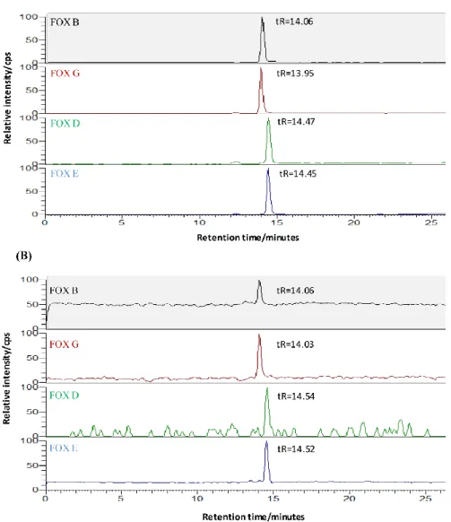
The extracted siderophores were analyzed using a method of Ahmed and Holmström (2014b). Components of the HPLC system (Ultimate 3000 RS, Thermo Scientific, USA) included two pumps with flow rates of 0.030 ml/min for the low pressure gradient pump and of 0.15 ml/min for the high pressure gradient pump. The column compartment (Dionex Ultimate 3000, Thermo Scientific, USA) was set at 10°C. The injection volume of standards and samples was 100 µl. The pre-column was a Syncronis C18 (50 mm x 2.1mm, particle size 1.7 µm, Thermo Scientific, USA) and the separation column was a Hypersil GOLD (100 mm x 2.1 mm, particle size 1.9 µm, Thermo Scientific, USA). The pre-column was eluted to the waste with mobile phase A (11 mM ammonium formate buffer, pH 4.0 and 1% v/v methanol) in order to concentrate and purify the hydroxamate siderophores. After 20 min, the pre-column was followed by back flushing towards the analytical column with a gradient of mobile phase B (11 mM ammonium formate buffer, pH 4.0 and 15% v/v acetonitrile) and mobile phase C (11 mM ammonium formate buffer, pH 4.0 and 5% v/v acetonitrile). The total analysis time was 60 min. The ferric complexes of the tri-hydroxamate siderophores, including ferrichromes, ferrioxamines, coprogens and fusigen (Figure
1) were detected by selected ion monitoring (SIM) of the proton adducts [M + H]+: i.e. m/z 797
for Tetraglycyl Ferrichrome, 771 for Ferricrocin, 741 for Ferrichrome, 801 for Ferrichrysin, 1011 for Ferrirubin, 1011 for Ferrirhodin, 1052 for Ferrichrome A, 614 for Ferrioxamine B, 672 for
ACCEPTED MANUSCRIPT
Ferrioxamine G, 656 for Ferrioxamine D, 654 for Ferrioxamine E, 682 for Neocoprogen II, 793 for Fusigen (lin.), 752 for Neocoprogen I and 821 for Coprogen on a triple quadropole mass spectrometer (TSQ Quantum Access Max, Thermo Scientific, US). The peaks identity were determined by comparing the retention time with the standard of each specific hydroxamate type in the chromatography step, in combination with using the selected ion monitoring (SIM) of the proton adducts that detected the known mass of each specific hydroxamate siderophore. Representative examples of selected ion monitoring chromatograms of a standard and a sample from a bacterial culture showing the four types of ferrioxamine siderophores (B, G, D and E) are shown in Figure 2.
2.7 Data analysis and statistics
The data were normalized and one/two-way ANOVA were analyzed by using XLSTAT (http://www.xlstat.com/en/).
3. Results
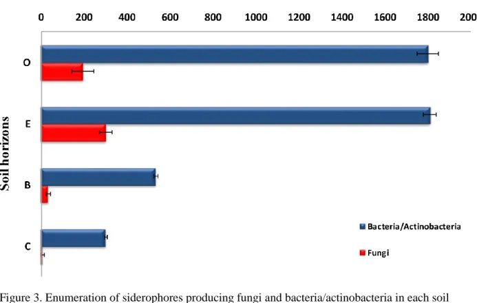
3.1 The enumeration of siderophores producing isolates within the soil horizons
The population of the most efficient siderophore producing microorganisms was isolated and counted from the soil samples collected from the four horizons of podzol (O, E, B, and C) (Figure 3). Viable fungi and bacteria/actinobacteria counts ranged from 5 to 300 and from 300 up to 1810 cfu/g soil, respectively. The highest counts (300 cfu/g soil for fungi and 1810 cfu/g soil for bacteria/actinobacteria) were found in the E-horizon. The lowest counts (5 cfu/g soil for fungi and 300 cfu/g soil for bacteria/actinobacteria) were found in the C-horizon. It was also shown
ACCEPTED MANUSCRIPT
that there was no significant difference (p= 0.3) between the count of bacteria and actinobacteria in O- and E-horizons, while there was a significant difference (p= 0.02) for the fungi counts within the whole profile.
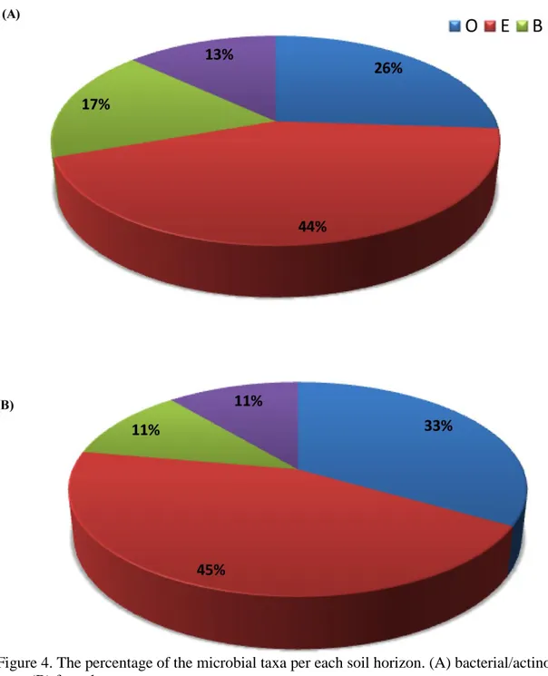
3.2 The diversity and distribution of siderophore producing microorganisms within the soil profile
All of the thirty-one isolated siderophore producing fungi and bacteria/actinobacteria were genetically identified. The molecular identification showed that there was a high diversity of various fungal and bacterial/actinobacterial species throughout the soil profile (Figure 4). The E-horizon had the maximum percentage of the fungal and bacterial/actinobacterial taxa, 45% and 44%, respectively. Then, it was followed by the O-horizon, which had 33% and 26% of the fungal and bacterial/actinobacterial taxa, respectively. On the other hand, B- and C-horizons had the minimum percentages for both the fungal and bacterial/actinobacterial taxa (11% and 17%) and (11% and 13%), respectively.
The isolated soil bacteria/actinobacteria were affiliated with three phylum-level groups, Proteobacteria, Actinobacteria, and Firmicutes (Figure 5). The dominant phyla were Proteobacteria and Actinobacteria. Those phyla contained different members of bacterial families and genus. Families such as Streptomycetaceae (Streptomyces), Nocardiaceae (Nocardia and Rhodococcus), Actinosynnemataceae (Actinosynnema) and Micrococcaceae (Micrococcus) were identified in the Actinobacteria phyla. The Proteobacteria phyla included Pseudomonadaceae (Pseudomonas), Enterobacteriaceae (Klebsiella and Erwinia), and
ACCEPTED MANUSCRIPT
Pseudoalteromonadaceae (Pseudoalteromonas). However, the only genus involved in the Firmicutes phylum was Bacillus, which belongs to the Bacillaceae family. For the bacterial/actinobacterial distribution, Streptomyces spp. were the most common taxa throughout the profile except in the C-horizon. However, Pseudomonas spp. were sparsely detected within the profile and M. luteus was restricted to B- and C-horizons.
The isolated soil fungi were affiliated with two phylum-level groups, Ascomycota and Basidiomycota (Figure 5). The most common phylum, Ascomycota, contained a wide range of fungal families and genera. The isolates of the Ascomycota phylum consisted of families such as Trichocomaceae (Penicillium and Aspergillus), Chaetomiaceae (Thielavia), Incertae sedis (Trichothecium and Epicoccum), Sordariaceae (Neurospora) and Nectriaceae (Fusarium). The Basidiomycota phylum included only Ustilago, which belongs to Ustilaginaceae. For the fungal distribution, Penicillium spp. were found only in O- and E-horizons. All the other fungal taxa were distributed within the profile. For instance, T. roseum and T. terrestris were found in the O-horizon; F. sporotrichioides, N. crassa, and U. maydis were found in E-O-horizon; A. fumigatus was found in B-horizon and E. nigrum was present in C-horizon.
3.3 Fe accumulation by the isolated siderophore producing microorganisms
All the isolated microbial species were screened for Fe accumulation at concentrations between 40 and 200 mg/l as a microbial activity. The fungal and bacterial/actinobacterial species isolated from the different soil horizons showed different degrees of Fe accumulation (Table 2). Most of the microbial species isolated from E- and B-horizons showed a high Fe accumulation ability
ACCEPTED MANUSCRIPT
(i.e. S. pilosus, N. farcinica, A. fumigatus, and F. sporotrichioides). P. stutzeri and K. pneumonia had the highest Fe accumulation ability among the microbial species isolated from O-horizon. The microbial species isolated from C-horizon had the minimum Fe accumulation ability, except for M. luteus.
3.4 The concentration and diversity of siderophores produced by the microbial isolates
Siderophores were isolated from all the fungal and bacterial/actinobacterial isolates. Hydroxamate types (i.e. ferrioxamines, ferrichromes, fusigen, and coprogens) were detected and measured by HPLC-ESI-MS. The isolated bacteria/actinobacteria showed different abilities in the production of the four types of ferrioxamines (E, B, G, and D) (Table 3). All the bacterial/actinobacterial isolates could produce ferrioxamine D, except for M. luteus from (C-horizon). The maximum concentration (19 nM) of ferrioxamine D was produced by S. olivaceus (O-horizon) and the minimum concentration (0.8 nM) was produced by Enterobacteriaceae bacterium (E-horizon). Ferrioxamine G was the second most common hydroxamate type produced by all the isolated bacteria/actinobacteria except for A. mirum (E-horizon) and M. luteus and an Uncultured gamma proteobacterium (C-horizon). The maximum concentration (24 nM) of ferrioxamine G was produced by S. nitrosporeus (B-horizon) and the minimum concentration (2 nM) was produced by P. stutzeri (O-horizon). Ferrioxamine B was produced by most of the bacterial/actinobacterial species isolated from all soil horizons. However, it was completely absent from all the isolates from C-horizon. The maximum concentration (19 nM) of ferrioxamine B was produced by Streptomyces sp. (E-horizon) and the minimum concentration
ACCEPTED MANUSCRIPT
(5 nM) was produced by M. luteus (B-horizon). Few bacteria/actinobacteria isolates produced ferrioxamine E. P. mendocina isolated from B-horizon produced the maximum concentration (11 nM) of ferrioxamine E. Enterobacteriaceae bacterium isolated from E-horizon produced the minimum concentration (0.7 nM).
The isolated fungal species showed different abilities of hydroxamates production (i.e. ferrichromes, coprogens and fusarinines) (Table 3). Most of the fungal isolates produced coprogen with high concentrations that ranged between 101 nM by T. roseum (O-horizon) and 1649 nM by (U. maydis, E-horizon). Coprogen was absent in the extracts in only two of the fungal species T. terrestris (O-horizon and A. fumigatus ((B-horizon). Neocoprogen II was also produced by many of the isolated fungi, especially those that were isolated from the E-horizon. The maximum concentration (6 nM) of neocoprogen II was produced by U. maydis (E-horizon) and the minimum concentration (3 nM) was produced by F. sporotrichioides isolated from the same horizon. Fe-dimerum acid was sparsely detected in the fungal extracts. However, neocoprogen I and fusigen (lin.) were produced only by F. sporotrichioides (E-horizon) with concentrations of 81 nM and 15 nM, respectively. Ferrichromes had much lower concentration compared to coprogens. It was found that the fungal species isolated from O- and B-horizons had the ability to produce most ferrichrome types, except ferrichrome A. Ferrichrome A production was restricted to E. nigrum (C-horizon) with a concentration of 11 nM. However, ferricrocin production was restricted to T. terrestris (O-horizon) with a concentration of 0.2 nM. The maximum concentration among the ferrichrome types was 19 nM of tetraglycyl ferrichrome produced by A. fumigatus (B-horizon).
ACCEPTED MANUSCRIPT
It was observed that the coprogen concentration produced by most of the fungal species represented between 97% and 99% of the total hydroxamates produced, except for T. terrestris and A. fumigatus in which the tetraglycyl ferrichrome represented 99% and 86% of the total hydroxamates produced, respectively. Ferrioxamine D was the most commonly hydroxamate type siderophore produced by the bacterial species. However, the ferrioxamine G concentration produced by most of the bacterial species represented a higher percentage, between 35% and 64% of the total hydroxamates produced, compared to ferrioxamine D even if it was the most commonly produced. The results also showed that most of the fungal species isolated from E-horizon could produce coprogens, whereas most of the fungal species isolated from O- and B-horizon produced ferrichromes. In addition, the bacterial taxa isolated from C-B-horizon produced only a few types of ferrioxamines. It was found that some microbial isolates from the same family but originating from different horizons have various abilities in siderophore production like in Penicillium and Micrococcus species. P. glabrum isolated from O-horizon had the ability to produce a wide range of ferrichromes (i.e. tetraglycyl ferrichrome and ferrrichrysin) and coprogens (i.e. Fe-dimerum acid and coprogen), while P. melinii isolated from the E-horizon produced only ferrichrome and coprogen. In addition, the same species of Micrococcus (M. luteus) isolated from B- and C-horizons showed different abilities of hydroxamates production. M. luteus isolated from the B-horizon produced all four types of investigated ferrioxamines, while M. luteus isolated from the C-horizon produced only ferrioxamine E.
ACCEPTED MANUSCRIPT
4. Discussion
4.1 Production of hydroxamates by soil microorganisms
Among the different siderophore types, the hydroxamates have received much attention since they are highly resistant to environmental degradation due to their cyclic hexapeptide structure (Winkelmann, 2007). It is known that a wide range of soil fungal isolates have the ability to produce one or several types of hydroxamates (Haselwandter and Winkelmann, 2007; Haselwandter et al., 2013). In the present study, both P. glabrum and P. melinii produced coprogen with concentrations of 305 nM and 540 nM, respectively. Besides producing coprogen, P. glabrum secreted tetraglycyl ferrichrome (2 nM), ferrrichrysin (0.01 nM), and Fe-dimerum acid (1 nM), whereas P. melinii produced only ferrichrome (0.6 nM). This is in agreement with earlier studies that have detected ferrichromes and coprogens production by a variety of soil Penicillium isolates (Zähner et al., 1963; Ong and Neilands 1979; Charlang et al., 1981). In addition, Konetschny-Rapp et al. (1988) detected the production of 33 mg/l (corresponding to 43 µM) ferricrocin in the culture filtrate of P. resticulosum. However, other studies showed that a mixture of different hydroxamates (e.g. coprogen, ferricrocin, dimerum acid, and fusarinines) could be produced by P. chrysogenum isolated from soil (Hördt et al., 2000; Winkelmann, 2007). Our findings indicated also that N. crassa could produce different types of coprogens like coprogen (893 nM), Neocoprogen II (6 nM), and Fe-dimerum acid (0.8 nM). An earlier study showed that N. crassa had the ability to produce only coprogen in comparison to Aspergillus and Penicillium, which secreted several other siderophores (Charlang et al., 1981). It was also found that N. crassa produced two main hydroxamates, coprogen and ferricrocin with concentrations of
ACCEPTED MANUSCRIPT
28 mg/l (34 µM) and 3 mg/l (4 µM), respectively (Konetschny-Rapp et al., 1988; Jalal and Van Der Helm, 1991). We isolated F. sporotrichioides, which had the ability to produce variety of hydroxamate types such as tetraglycyl ferrichrome (1 nM), neocoprogen II (3 nM), neocoprogen I (81 nM), coprogen (462 nM), and fusigen (lin.) (15 nM). It has been also observed previously that F. cubense and F. roseum has the ability to produce fusarinines (Diekmann, 1967; Diekmann and Zähner, 1967; Sayer and Emery, 1968). Van der Helm and Winkelmann (1994) detected the production of a low concentration of coprogen (4 mg/l corresponding to 5 µM) by F. dimerum.
A. fumigatus isolated in our study produced tetraglycyl ferrichrome (19 nM), ferrrichrysin (0.04 nM), ferrirubin (4 nM), and Fe-dimerum acid (9 nM). It was observed previously that Asprigillus sp. could produce 0.1 µg/ml (0.13 µM) of ferrichrome, ferricrocin, ferrrichrysin and coprogen (Charlang et al., 1981). However, Konetschny-Rapp et al. (1988) measured 14 mg/l (18 µM) ferricrocin and 27 mg/l (34 µM) tri-acetylfusarinin C in the culture filtrate of Asprigillus. Tri-acetylfusarinin C was produced also by A. fumigatus (Kragl et al., 2007). We did not detect any ferrichromes in the culture filtrate of U. maydis, but we measured a high concentration (1649 nM) of coprogen and (6 nM) of neocoprogen II. Several studies investigated the production of ferrichrome by U. sphaerogena (Emery and Neilands, 1959; Neilands, 1984; Winkelmann 1993; Hider and Kong, 2010).
We scarcely detected both ferrichrome A and ferricrocin in the isolates filtrate. Ferrichrome A was only produced by E. nigrum with concentration 11 nM. This finding might be explained by the presence of three negative charges on the ferrichrome A, which enhances their adsorption to
ACCEPTED MANUSCRIPT
the positive charges on the fungal cell wall; therefore, ferrichrome A was rarely found free in the culture filtrate (Winkelmann, 2007). In addition, ferricrocin was produced only by T. terrestris in a concentration of 0.2 nM. Ferricrocin had a low productivity because it was found commonly to be synthesized as an intracellular siderophore functioning as Fe storage compound and sporulation factor (Matzanke et al., 1987; Winkelmann and Drechsel, 1997; Haas, 2003).
Several soil bacteria could produce more than one type of hydroxamate (Scavino and Pedraza, 2013). Streptomyces species were known to produce different types of ferrioxamines (Muller and Raymond, 1984). In our study, we isolated five different species of Streptomyces which displayed various abilities in the production of ferrioxamines B, G, and D. S. olivaceus produced the maximum concentration (19 nM) of ferrioxamine D, Streptomyces sp. produced the maximum concentration (19 nM) of ferrioxamine B, while S. nitrosporeus produced the maximum concentration (24 nM) of ferrioxamine G. However, S. pilosus and uncultured Streptomyces sp. showed lower ferrioxamines productivity when compared to the other isolates. Previous studies identified ferrioxamine B as the major siderophore produced by S. viridosporus and S. pilosus, and ferrioxamine G as the main siderophore produced by S. coelicolor and S. lividans (Bickel et al., 1960). Other studies found that ferrioxamine E was the most widespread siderophore in several Streptomyces species such as S. glaucescens (Schafft and Diekmann, 1978), S. olivaceus (Meiwes et al., 1990), and S. viridosporus, and S. ambofaciens (Imbert et al., 1995). Recently, Kobayakawa and Kodani (2012) detected the production of ferrioxamines in 78% of 41 Streptomyces strains used in screening.
ACCEPTED MANUSCRIPT
Earlier investigations found that ferrioxamines were not only restricted to the genus Streptomyces but were also characteristic siderophores of several gram negative (e.g. Pseudomonas), gram positive (e.g. Nocardia), and enterobacterial genera (e.g. Erwinia) (Meyer and Abdullah, 1980; Berner et al., 1988; Berner and Winkelmann, 1990; Reissbrodt et al., 1990). Our findings showed that P. stutzeri and Pseudomonas sp. could produce high concentrations (9 nM) of ferrioxamine B and (13 nM) of ferrioxamine G, respectively. However, Essén et al. (2007) showed that P. stutzeri could produce ferrioxamine E as the main siderophore together with ferrioxamine G. In the present study, the isolated N. tenerifensis and N. farcinica produced all the ferrioxamine types except ferrioxamines E. Moreover, both isolates were characterized by high concentrations of ferrioxamine D (17 nM) for N. tenerifensis and (15 nM) for N. farcinica. Ferrioxamine E has been detected previously in N. brasiliensis (Gaeumann et al., 1964). We isolated also E. amylovora, which produced the four types of ferrioxamines in different concentrations, (8 nM) for ferrioxamine B, (12 nM) for ferrioxamine G, (7 nM) for ferrioxamine D, and (2 nM) for ferrioxamine E. Recently, Smits and Duffy (2011) reported the production ability and biosynthetic pathway of the ferrioxamine E in E. amylovora.
We found that some microbial isolates within the same genus and same species, but originating from different soil horizons had various siderophore production capabilities, like for example M. luteus that was isolated from both the B- and the C-horizon. This finding could be explained by that the variety in the siderophore biosynthesis may also be affected by the isolates origin from different soil horizons, which holds specific chemical properties. These findings agrees with earlier results by Fierer et al. (2003), who have suggested that the microbial communities and their metabolic properties in the deep soil horizons function differently from
ACCEPTED MANUSCRIPT
those at the upper soil horizons. Thus, the different habitats within the soil profile may works as exogenous signals that regulate the gene expression of siderophore biosynthesis (Duffy and Défago, 1999).
4.2 Why do the soil microorganisms produce a variety of siderophores?
Most of the isolated bacteria/actinobacteria and fungi produced different types of hydroxamates, and each individual microorganism could produce a set of siderophore types covering a wide range of physicochemical properties. This finding could be explained by under Fe deficiency conditions the Fe operon is depressed and several genes involved in the biosynthesis of the siderophores and their receptors are expressed (Barona-Gòmez et al., 2006). In addition, there is a sequence homology for the different siderophore receptors, so these receptors can recognize the siderophores which have common properties. Therefore several siderophores can be produced by individual microorganisms to obtain Fe from different sources (Pattus and Abdallah, 2000). The diversity in hydroxamate productivity may allow the soil microorganisms to overcome the deficiency of Fe solubility and the competition with other microorganisms for increasing nutrient concentrations (Winkelmann, 2007). Thus, microorganisms that produce various types of siderophores seem to have advantages since the competition for Fe is based on the following (Hider and Kong, 2010): a) the concentration of siderophores produced by the microorganisms, b) the chemical properties of the siderophore with regards to its stability constant in binding with Fe, and c) the resistance to environmental degradation. In addition, the different affinities of hydroxamates for Fe not only enable the microorganisms to chelate it from the soil, but also
ACCEPTED MANUSCRIPT
allow the microorganisms to mobilize the Fe from weaker Fe-siderophore complexes produced by other microorganisms (Desai and Archana, 2011). Therefore, the microorganism producing siderophores that have a higher affinity for Fe can be more successful in growth and survival than the other producers with lower affinity.
4.3 Factors influencing the siderophore production by the isolated microorganisms
The siderophore production by microorganisms in laboratory conditions may vary depending on the Fe availability, carbon source, aeration, and time of cultivation (Hördt et al., 2000). The choice of method for qualitative and quantitative analysis of the siderophores play also an important role in to which siderophore types that could be identified and to which extent they could be quantitated (Hördt et al., 2000). The Fe concentration in the medium is one of the main factors affecting the siderophore production by microorganisms. In the present study, we used Fe limited broth medium that could trigger high production of hydroxamate siderophores by the isolated microorganisms. Our findings agreed with previous studies that suggest that the microbial cultures need to be Fe starved to enhance the expression of the genes responsible for siderophore biosynthesis (Neilands, 1984; Díaz de Villegas et al., 2002). That means that the lower concentration of Fe in the medium leads to higher expression of siderophore biosynthesis genes and thereby a higher concentration of siderophores is produced (Lee et al., 2012). The trace level of Fe needed for obtaining maximum siderophore production is highly species dependent. Neilands (1995) has reported that the siderophore production was induced at Fe concentrations between 0 and 1 μM. In contrast, Díaz de Villegas et al. (2002) found that Fe
ACCEPTED MANUSCRIPT
concentrations >10 M had a negative effect on siderophore production by Pseudomonas aeruginosa. In addition, it was found that the maximum concentration of hydroxamate siderophores production by Micrococcus luteus and Bacillus silvestris was found at a concentration of Fe 0.04 μM (Cabaj and Kosakowska, 2009).
The choice of carbon source in the culture medium has also a great effect on both the concentration and types of siderophores that could be produced (Warner et al., 1981; Bendale et al., 2009). We used glucose broth medium for cultivation the microorganisms in order to estimate the siderophores production since it has recently been shown that using glucose as carbon source in the culture medium could enhance the siderophore production compared to other carbon sources such as lactose and maltose (Santos et al., 2014). On the other hand, it was found that the effect of the carbon source on the siderophore production differ according to the type of the microorganisms (Chincholkar et al., 2007). For instance, the addition of amino acids (glycine, aspartic acid and glutamic acid) to the culture medium increased the siderophore production by fungi, while the medium containing glucose could enhance the siderophore production by bacteria (Chincholkar et al., 2007). However, other earlier studies found that the use of succinate as a carbon source increased the siderophore yields compared to glycerol (Chodat and Gouda, 1961; Meyer and Abdallah, 1978; Rachid and Ahmed, 2005).
The concentration of siderophores produced by microorganisms could depend on other cultivation conditions such as aeration and cultivation period. The aerobic conditions as we used in our study commonly results in a high production of siderophores. The involvement of aerobic metabolism in the production of siderophores is necessary for a high yield of siderophores, since
ACCEPTED MANUSCRIPT
the absence of aeration does not prevent siderophore production but it strongly decreases the concentration (Santos et al., 2014). Siderophores production could also vary according to the time of cultivation. The duration of cultivation in our study was 7 days. It has been previously established for fungi that prolonged cultivation time results in a higher production of ferrichrome siderophores, although it leads to the degradation of some other siderophores like coprogens that degrades to fusigen and dimerum acid (Hördt et al., 2000).
On the other hand, when comparing the qualitative and quantitative siderophore results from different studies conducted at various culturing conditions, you also have to keep in mind that a wide range of analytical methods for the determination of siderophores have been used, which also can affect the recovery of the siderophores among the different studies. Earlier studies that determined the siderophores by microbial assays were very specific but had low a sensitivity (e.g Buyer et al. 1993), while recently HPLC-ESI-MS methods has been used intensively since it gives more accurate and highly sensitive results (Moberg et al., 2003; Haselwandter et al., 2013).
5. Conclusion
Our findings indicate that there is a significant variation in the composition of siderophore producing microorganisms within the horizons of a podzol soil profile. A wide range of fungal hydroxamate siderophores (i.e. ferrichromes, coprogens, and fusigen) and bacterial hydroxamate siderophores (i.e. ferrioxamine B, D, E and G) were found to be produced by the isolated microorganisms and these results may demonstrate the important contributions of these microorganisms in the mineral weathering processes in podzol soil. The high diversity and
ACCEPTED MANUSCRIPT
concentration of hydroxamate siderophore production by the microbial isolates from the different soil horizons strongly suggests that the factors (e.g. Fe availability, carbon source, aeration and time of cultivation) have a great effect on the siderophore biosynthesis by microorganisms. At present, there is still little available information about the environmental factors inducing the microbial siderophore production. Further studies are needed to investigate how the diversity and function of siderophore producing microorganisms change by depth of soil and how it could be linked to the specific properties of the soil horizons.
Acknowledgement
We would like to thank Madelen Olofsson and Dan Bylund (Mittuniversitetet, Sundsvall, Sweden) for taking part in the sampling. We thank Hildred Crill (Stockholm University, Stockholm, Sweden) for English editing. The present study was supported by grants from the Swedish Research Council for Environment, Agricultural Sciences and Spatial Planning (FORMAS) and the Faculty of Science, Stockholm University, Sweden.
ACCEPTED MANUSCRIPT
References
Aguado–Santacruz GA, Moreno–Gómez B, Jiménez–Francisco B, García–Moya E, Preciado– Ortiz RE. 2012. Impact of the microbial siderophores and phytosiderophores on the iron assimilation by plants: a synthesis. Revista fitotecnia mexicana 35: 9-21.
Ahmed E, Holmström SJM. 2014a. Siderophores in Environmental Research: Roles and Applications. Microbial Biotechnology 7: 196-208.
Ahmed E, Holmström SJM. 2014b. The effect of soil horizon and mineral type on the distribution of siderophores in soil. Geochim et Cosmochim Acta 131: 184-195.
Altschul SF, Gish W, Miller W, Myers EW, Lipman DJ. 1990. Basic local alignment search tool. Journal of Molecular Biology 215: 403-410.
Atkin CL, Neilands JB, Phaff HJ. 1970. Rhodotorulic Acid from Species of Leucosporidium, Rhodosporidium, Rhodotorula, Sporidiobolus, and Sporobolomyces, and a New Alanine-Containing Ferrichrome from Cryptococcus melibiosum. Journal of Bacteriology 103: 722-733.
Barona-Gómez F, Lautru S, Francou FX, Leblond P, Pernodet JL, Challis GL. 2006. Multiple biosynthetic and uptake systems mediate siderophore-dependent iron acquisition in Streptomyces coelicolor A3(2) and Streptomyces ambofaciens ATCC 23877. Microbiology 152: 3355-3366.
Barry SM, Challis GL. 2009. Recent advances in siderophore biosynthesis. Current Opinion in Chemical Biology 13: 205-215.
ACCEPTED MANUSCRIPT
Bendale MS, Chaudhari BL, Chincholkar SB. 2009. Influence of Environmental Factors on siderophore production by Streptomyces fulvissimus ATCC 27431. Current Trends in Biotechnology and Pharmacy 3: 362-371.
Berner I, Konetschny-Rapp S, Jung G, Winkelmann G. 1988. Characterization of ferrioxamine E as the principal siderophore of Erwinia herbicola (Enterobacter agglomerans). Biology of Metals 1: 51-56.
Berner I, Winkelmann G. 1990. Ferrioxamine transport mutants and the identification of the ferrioxamine receptor protein (FoxA) in Erwinia herbicola (Enterobacter agglomerans). Biology of Metals 2: 197-202.
Bickel H, Bosshartdt R, Gäumann E, Reusser P, Vischer E, Voser W, Wettstein A, Zähner H. 1960. Über die isolierung und charakterisierung der Ferrioxamine A—F, neuer wuchstoffe der sideramin-gruppe. Helvetica Chimica Acta 43: 2118-2128.
Budzikiewicz H. 2010. Siderophores from bacteria and from fungi. In: Cornelis P, Andrews SC, editors. Iron uptake and homeostasis in microorganisms. Norfolk: Caister Academic press. P 1-16.
Buyer JS, Kratzke MG, Sikora LJ. 1993. A method for detection of pseudobactin, the siderophore produced by a plant-growth-promoting Pseudomonas strain, in the barley rhizosphere. Applied and Environmental Microbiology 59: 677-681.
ACCEPTED MANUSCRIPT
Cabaj A, Kosakowska, A. 2009. Iron-dependent growth of and siderophore production by two heterotrophic bacteria isolated from brackish water of the southern Baltic Sea. Microbiological Research 164: 570-577.
Charlang G, Ng B, Horowitz NH, Horowitz RM. 1981. Cellular and extracellular siderophores of Aspergillus nidulans and Penicillium chrysogenum. . Molecular and Cellular Biology 1: 94-100.
Chincholkar SB, Chaudhari BL, Rane MR. 2007. Microbial Siderophores: State of art. In: Chincholkar SB, Varma A, editors. Microbial Siderophores. Germany: Springer Verlag. P 233-242.
Chodat F, Gouda S. 1961. Contribution a l étude du pigment de Pseudomonas fluorescens Migula. Pathologia et microbilogia 24: 840-847.
Cornelis P. 2010. Iron uptake and metabolism in pseudomonads. Applied Microbiology and Biotechnology 86: 1637-1645.
Crowley DE. 2006. Microbial siderophores in the plant rhizosphere. In: Barton LL, Abadía J, editors. Iron nutrition in plants and rhizospheric microorganisms. Netherlands: Springer. P 169-198.
Deml G, Voges K, Jung G, Winkelmann G. 1984. Tetraglycylferrichrome - the first heptapeptide ferrichrome. FEBS Letters 173:53-57.
Desai A, Archana G. 2011. Role of siderophores in crop improvement. In: Maheshwari DK, editor. Bacteria in Agrobiology: plant nutrient management. Berlin: Springer. P 109-139.
ACCEPTED MANUSCRIPT
Dhungana S, White PS, Crumbliss AL. 2001. Crystal structure of ferrioxamine B: a comparative analysis and implications for molecular recognition. Journal of Biological Inorganic Chemistry 6: 810-818.
Díaz de Villegas ME, Villa P, Frías A. 2002. Evaluation of the siderophores production by Pseudomonas aeruginosa PSS. Revista Latinoamericana de Microbiología 44: 112-117.
Diekmann H, Zähner H. 1967. Konstitution von Fusigen and dessen Abbau zu Δ2 Anhydromevalonsaurelacton. European Journal of Biochemistry 3: 213-218.
Diekmann H. 1967. Stoffwechselprodukte von Mikroorganismen. 56 Mitteilung. Fusigen—ein neues Sideramin aus Pilzen. Archives of Microbiology 58: 1-5.
Drechsel H, Jung G, Winkelmann G. 1992. Stereochemical characterization of rhizoferrin and identification of its dehydration products. Biometals 5: 141-148.
Drechsel H, Metzger J, Freund S, Jung G, Boelaert JR, Winkelmann G. 1991. Rhizoferrin - a novel siderophore from the fungus Rhizopus microsporus var. rhizopodiformis. Biology of Metals 4: 238-243.
Duffy BK, and Défago G. 1999. Macro- and microelement fertilizers influence the severity of Fusarium crown and root rot of tomato in a soilless production system. Hortscience 34: 287-291.
Emery T, Neilands JB. 1959. The Iron-Binding Centre of Ferrichrome Compounds. Nature 184: 1632-1633.
ACCEPTED MANUSCRIPT
Emery T. 1971. Role of ferrichrome as a ferric ionophore in Ustilago sphaerogena. Biochemistry 10: 1483-1488.
Essén SA, Johnsson A, Bylund D, Pedersen K, Lundström US. 2007. Siderophore Production by Pseudomonas stutzeri under Aerobic and Anaerobic Conditions. Applied and Environmental Microbiology 73: 5857-5864.
FAO. 1990. FAO-Unesco Soil Map of the World-Revised Legend. World Soil Resources Reports 60. Food and Agriculture Organization of the United Nations, Rome. 119 p.
Felfenstein J. 1985. Confidence limits on phylogenies: an approach using the bootstrap. Evolution 39: 783-791.
Fierer N, Schimela JP, Holden PA. 2003. Variations in microbial community composition through two soil depth profiles. Soil Biology & Biochemistry 35: 167-176.
Gaeumann E, Prelog V, Vischer E, Bickel H. 1964. Method for producing ferrioxamine. Unites States Patent office 3: 118-823.
Haas H. 2003. Molecular genetics of fungal siderophore biosynthesis and uptake: the role of siderophores in iron uptake and storage. Applied Microbiology and Biotechnology 62: 316-330.
Haselwandter K, Hãninger G, Ganzera M, Haas H, Nicholson G, Winkelmann G. 2013. Linear fusigen as the major hydroxamate siderophore of the ectomycorrhizal Basidiomycota Laccaria laccata and Laccaria bicolor. Biometals 26: 969-979.
ACCEPTED MANUSCRIPT
Haselwandter K, Winkelmann G. 2007. Siderophores of symbiotic fungi. In: Varma A, Chincholkar SB, editors. Microbial Siderophores. Soil Biology Series.vol 12. Berlin: Springer. P 91-103.
Hayat R, Ali S, Amara U, Khalid R, Ahmed I. 2010. Soil beneficial bacteria and their role in plant growth promotion: a review. Annals of Microbiology 60: 579-598.
Hibbing ME, Fuqua C, Parsek MR, Peterson SB. 2010. Bacterial competition: surviving and thriving in the microbial jungle. Nat Rev Microbiol 8: 15–25.
Hider RC, Kong X. 2010. Chemistry and biology of siderophores. Natural Product Reports 27: 637-657.
Holmström SJM, Lundström US, Finlay RD, van Hees PAW. 2004. Siderophores in forest soil solution. Biogeochemistry 71: 247-258.
Hördt W, Römheld V, Winkelmann G. 2000. Fusarinines and dimerum acid, mono- and dihydroxamate siderophores from Penicillium chrysogenum, improve iron utilization by strategy I and strategy II plants. Biometals 13: 37-46.
Imbert M, Béchet M, Blondeau R. 1995. Comparison of the main siderophores produced by some species of Streptomyces. Current Microbiology 31: 129-133.
Jalal MAF, van der Helm D. 1991. Isolation and spectroscopic identification of fungal siderophores. In: Winkelmann G, editor. Handbook of microbial iron chelates. Boca Raton, FL: CRC Press Inc. P 235-269.
ACCEPTED MANUSCRIPT
Kobayakawa F, Kodani S. 2012. Screening of Streptomycetes for Production of Desferrioxamines. Journal of Pure and Applied Microbiology 6: 1553-1558.
Konetschny-Rapp S, Huschka HG, Winkelmann G, Jung G. 1988. High-performance liquid chromatography of siderophores from fungi. Biology of Metals 1: 9-17.
Kraemer SM. 2004. Iron oxide dissolution and solubility in the presence of siderophores. Aquatic Sciences 66: 3-18.
Kragl C, Schrettl M, Abt B, Sarg B, Lindner HH, Haas H. 2007. EstB-mediated hydrolysis of the siderophore triacetylfusarinine C optimizes iron uptake of Aspergillus fumigatus. Eukaryotic Cell 6: 1278-1285.
Lamont IL, Beare PA, Ochsner U, Vasil AI, Vasil ML. 2002. Siderophore-mediated signaling regulates virulence factor production in Pseudomonas aeruginosa. Proceedings of the national academy of sciences of the United States of America 99: 7072–7077.
Lee J, Postmaster A, Soon HP, Keast D, Carson KC. 2012. Siderophore production by actinomycetes isolates from two soil sites in Western Australia. Biometals 25: 285-296.
Leong SA, Neilands JB. 1982. Siderophore Production by Phytopathogenic Microbial Species. Archives of Biochemistry and Biophysics 218: 351-359.
Martino E, Perotto S. 2010. Mineral Transformations by Mycorrhizal Fungi. Geomicrobiology Journal 27: 609-623.
ACCEPTED MANUSCRIPT
Matzanke BF, Bill E, Trautwein AX, Winkelmann G. 1987. Role of siderophores in iron storage in spores of Neurospora crassa and Aspergillus ochraceus. Journal of Bacteriology 169: 5873-5876.
Matzanke BF. 1991. Structures, coordination chemistry and functions of microbial iron chelates. In: Winkelmann G, editor. CRC Handbook of Microbial Iron Chelates. Boca Raton, FL: CRC Press. P 15-64.
Meiwes J, Fiedler HP, Zäihner H, Konetschny-Rapp S, Jung G. 1990. Production of desferrioxamine E and new analogues by directed fermentation and feeding fermentation. Applied Microbiology and Biotechnology 32: 505-510.
Meyer JM, Abdallah MA. 1978. The florescent pigment of Pseudomonas fluorescens Biosynthesis, purification and physical-chemical properties. Journal of General Microbiology 107: 319-328.
Meyer J-M, Abdallah MA. 1980. The siderochromes of nonfluorescent pseudomonads: production of nocardamine by Pseudomonas stutzeri. Journal of General Microbiology 118: 125-129.
Milagres AMF, Machuca A, Napoleão D. 1999. Detection of siderophore production from several fungi and bacteria by a modification of chrome azurol S (CAS) agar plate assay. Journal of Microbiological Methods 37: 1-6.
ACCEPTED MANUSCRIPT
Moberg M, Holmström SJ, Lundström US, Markides KE. 2003. Novel approach to the determination of structurally similar hydroxamate siderophores by column-switching capillary liquid chromatography coupled to mass spectrometry. Journal of Chromatography A. 5: 1-7.
Müller G, Raymond KN. 1984. Specificity and mechanism of ferrioxamine-mediated iron transport in Streptomyces pilosus. Journal of Bacteriology 160: 304-312.
Neilands JB. 1995. Siderophores: Structure and function of microbial iron transport compounds (Short Survey). Journal of Biological Chemistry 270: 26723-26726.
Neilands JB. 1973. Microbial iron transport compounds (siderochromes). In: Eichhorn GL, editor. Inorganic biochemistry, vol. 1. New York: Elsevier Scienc'e Publishing, Inc. P 167-202.
Neilands JB. 1984. Siderophores of bacteria and fungi. Microbiological sciences 1: 9-14.
Ong SA, Neilands JB. 1979. Siderophores in microbially processed cheese. Journal of Agriculture and Food Chemistry 27: 990-995.
Patel AK, Deshattiwar MK, Chaudhari BL, Chincholkar SB. 2009. Production, purification and chemical characterization of the catecholate siderophore from potent probiotic strains of Bacillus spp. Bioresource Technology 100: 368-373.
Pattus F, Abdallah MA. 2000. Siderophores and iron-transport in microorganisms. Journal of Chinese Chemical Society 47: 1-20.
Pollack JR, Ames BN, Neilands JB. 1970. Iron Transport in Salmonella typhimurium: Mutants Blocked in the Biosynthesis of Enterobactin. Journal of Bacteriology 104: 635-639.
ACCEPTED MANUSCRIPT
Pümpel T, Pernfuß B, Pigher B, Diels L, Schinner F. 1995. A rapid screening method for the isolation of metal-accumulating microorganisms. Journal of Industrial Microbiology 14: 213-217.
Rachid D, Ahmed, B. 2005. Effect of iron and growth inhibitors on siderophores production by Pseudomonas fluorescens. African Journal of Biotechnology 4: 697-702.
Ratledge C. 1987. Iron metabolism in mycobacteria. In: Winkelmann G, Van der Helm D, Neilands JB, editors. Iron Transport in Microbes, Plants and Animals. New York: VCH Publishers. P 207-222.
Reissbrodt R, Rabsch W, Chapeaurouge A, Jung G, Winkelmann G. 1990. Isolation and identification of ferrioxamine G and E in Hafnia alvei. Biology of Metals 3: 54-60.
Renshaw JC, Robson GD, Trinci APJ, Wiebe MG, Livens FR, Collison D, Taylor RJ. 2002. Fungal siderophores: structures, functions and applications. Mycological Research 106: 1123-1142.
Saitou N, Nei M. 1987. The neighbor-joining method: a new method for reconstructing phylogenetic trees. Molecular Biology and Evolution 4: 406-425.
Santos S, Neto IF, Machado, MD, Soares HM, Soares, EV. 2014. Siderophore Production by Bacillus megaterium: Effect of Growth Phase and Cultural Conditions. Applied biochemistry and biotechnology 172: 549–560.
Sayer JM, Emery TF. 1968. Structures of the naturally occurring hydroxamic acids, fusarinines A and B. Biochemistry 7:184-190.
ACCEPTED MANUSCRIPT
Scavino AF, Pedraza RO. 2013. The Role of Siderophores in Plant Growth-Promoting Bacteria. In: Maheshwari DK, Saraf M, Aeron A, editors. Bacteria in Agrobiology: Crop Productivity. Berlin: Springer. P 265-285.
Schafft M, Diekmann H. 1978. Cadaverin ist ein Zwischenprodukt der Biosynthese von Arthrobactin und Ferrioxamin E. Archives of Microbiology 117: 203-207.
Schwyn B, Neilands JB. 1987. Universal chemical assay for the detection and determination of siderophores. Analytical Biochemistry 160: 47-56.
Smits THM, Duffy B. 2011. Genomics of iron acquisition in the plant pathogen Erwinia amylovora: Insights in the biosynthetic pathway of the siderophore desferrioxamine E. Archives of Microbiology 193: 693-699.
Thieken A, Winkelmann G. 1992. Rhizoferrin: a complexone type siderophore of the Mocorales and entomophthorales (Zygomycetes). FEMS Microbiology Letters 94: 37-41.
Van der Helm D, Winkelmann G. 1994. Hydroxamates and polycarboxylates as iron transport agents (siderophores) in fungi. In: Winkelmann G, Winge D, editors. Metal Ions in Fungi. New York: Marcel Dekker, Inc. P 39-98.
Wallner A, Blatzer M, Schrettl M, Sarg B, Lindner H, Haas H. 2009. Ferricrocin, a siderophore involved in intra- and transcellular iron distribution in Aspergillus fumigatus. Applied and Environmental Microbiology 75: 4194-4196.
Warner PJ, Williams PH, Bindereif A, Neilands JB. 1981. ColV plasmid-specific aerobactin synthesis by invasive strains of Escherichia coli. Infection and Immunity 33: 540-545.
ACCEPTED MANUSCRIPT
Winkelmann G, Drechsel H. 1997. Microbial siderophores. In: Rehm HJ, Reed G, editors. Biotechnology, 2nd ed., Vol. 7. Weinheim: VCH-Wiley. P 199-246.
Winkelmann G. 1992. Structures and functions of fungal siderophores containing hydroxamate and complexone type iron binding ligands. Mycological Research 96: 529-534.
Winkelmann G. 1993. Kinetics, energetics, and mechanisms of siderophore iron transport in fungi. In: Barton LL, Hemming BC, editors. Iron chelation in plants and soil microorganisms. New York: Academic Press. P 219-239.
Winkelmann G. 2007 Ecology of siderophores with special reference to the fungi. Biometals 20: 379-392.
Zähner H, Keller-Schierlein W, Hütter R, Hess-Leisinger K, Deér A. 1963.
Stoffwechselprodukte von Mikroorganismen 40. Mitteilung. Sideramine aus Aspergillaceen. Archives of Microbiology 45: 119-135.
Table 1. The chemical characterization of the whole podzol soil profile; (O) organic horizon, (E) elluvial horizon, (B) illuvial horizon and C parent material. The average content of exchangeable cations (μmol/g) was determined. Carbon (C %), nitrogen (N %) and moisture (%) content were also estimated. a) Vestin et al., 2008, b) Ahmed and Holmström, 2014b.
Podzol Horizons Ca % Na % Caa μmol/g Ka μmol/g Mga μmol/g Naa μmol/g Feb μmol/g pHb Moistureb content % O 47 1.2 1.7 0.5 0.5 0.2 0.3 4.4 89 E 0.93 0.03 1.7 0.5 0.5 0.2 0.3 4.6 86 B 1.9 0.06 1.5 0.3 0.3 0.2 0.2 5.1 78
ACCEPTED MANUSCRIPT
Table 2. Fe screening of the isolated microorganisms from soil horizons. The different Fe accumulation capabilities were represented by cross sign (+) and were depended on the width and darkness of the zone formed around the cultures. The highest Fe accumulation was represented by (+++), while the lowest Fe accumulation was represented by (+).
C 0.3 0.01 1.3 0.2 0.2 0.2 0.09 5.4 26 Fe Concentration (mg/l) Horizon/Microorganisms 40 60 80 100 140 160 180 200 HORIZON ^O^ Fungi Penicillium glabrum ++ ++ ++ ++ ++ ++ ++ ++ Thielavia terrestris + + + + + + + + Trichothecium roseum ++ ++ + + + + + + Bacteria/Actinobacteria Streptomyces olivaceus ++ ++ ++ + + + + + Pseudomonas stutzeri +++ +++ +++ +++ +++ +++ +++ +++ Nocardia tenerifensis + + + + + + + + Klebsiella pneumoniae +++ +++ +++ +++ +++ +++ +++ +++ Rhodococcus sp. + + + + + + + + HORIZON ^E^ Fungi Penicillium melinii +++ +++ +++ +++ +++ +++ +++ +++ Ustilago maydis ++ ++ + + + + + + Neurospora crassa + + + + + + + + Fusarium sporotrichioides +++ +++ +++ +++ +++ +++ +++ +++ Bacteria/Actinobacteria Enterobacteriaceae bacterium ++ ++ ++ ++ ++ ++ ++ ++ Bacillus thuringiensis + + + + + + + + Pseudomonas sp +++ +++ +++ +++ +++ +++ +++ +++ Streptomyces sp. + + + + + + + + Pseudoalteromonas sp. +++ +++ +++ +++ +++ +++ +++ +++ Actinosynnema mirum ++ ++ + + + + + + Streptomyces pilosus +++ +++ +++ +++ +++ +++ +++ +++ +++ ++ +
ACCEPTED MANUSCRIPT
Nocardia farcinica +++ +++ +++ +++ ++ ++ ++ ++ Erwinia amylovora +++ +++ +++ +++ +++ +++ +++ +++ gamma proteobacterium + + + + + + + + HORIZON ^B^ Fungi Aspergillus fumigatus +++ +++ +++ +++ +++ +++ +++ +++ Bacteria/Actinobacteria Micrococcus luteus +++ +++ +++ +++ +++ +++ ++ ++ Pseudomonas mendocina ++ ++ + + + + + + Streptomyces nitrosporeus +++ +++ +++ +++ +++ +++ +++ +++ Uncultured Streptomyces sp. +++ +++ +++ +++ ++ ++ + + HORIZON ^C^ Fungi Epicoccum nigrum ++ ++ ++ + + + + + Bacteria/ActinobacteriaUncultured gamma proteobacterium + + + + + + + +
Micrococcus luteus +++ +++ +++ +++ +++ ++ ++ ++
Uncultured bacterium + + + + + + + +