1
Using the counterselectable marker pheS* to study
the excision rate and excision patterns of the
pathogenicity island of Enterococcus faecalis V583
Maria Bergdahl
Biomedicine 160 points
University of Kalmar, School of Pure and Natural Sciences Examination project work 20 points
Supervisors:
Michael S. Gilmore, Prof. & Schepens Eye Research Institute,
Janet Manson, Ph.D. Department of optamology, Harvard Medical School.
Boston, MA 02114 USA
Internal supervisor:
Britt-Inger Marklund, Ph.D. School of Pure and Applied Natural Sciences, University of Kalmar
SE-391 83 Kalmar SWEDEN
Examiner:
Kristina Nilsson-Ekdahl, Prof. School of Pure and Applied Natural Sciences, University of Kalmar
SE-391 83 Kalmar SWEDEN
Abstract
The Enterococcus genus consists of natural members of the gastrointestinal tract but they are also opportunistic pathogens. They are a common cause of urinary tract infections but can also cause sepsis and other infections. Enterococcus faecalis and Enterococcus faecium are the most abundant in clinical specimens. Enterococci are a leading cause of nosocomial infections and they have developed resistance against a number of antibiotics e.g. vancomycin. E. faecalis V583 was the first vancomycin resistant isolate reported in the U.S. Movable genetic elements such as pathogenicity islands, PAI, are important for bacterial evolution. PAI:s are large chromosomal fragments mostly seen in pathogenic strains and carry regions such as transposons and insertion elements along with virulence factors and transfer genes. A PAI has been detected in the chromosome of E. faecalis. Excision of PAI:s has been studied for uropathogenic E. coli and frequencies of 10-5 and 10-6 have been reported. In this study the excision rate and excision patterns of E. faecalis V583 was studied using the counterselectable marker pheS*, causing p-Cl-phe sensitivity, and a chloramphenicol resistance gene, cat, inserted at two different positions of the PAI and selecting for excisions by growth on p-Cl-phe. Excision rates of 10-6 and 10-8 were seen based on the p-Cl-phe resistance and chloramphenicol sensitivity. Mutation rate in the pheS* gene was high compared to excision rate which made the method difficult to work with. No obvious excision patterns were detected but the excisions seemed to be limited to the close surroundings of the pheS*/cat insertion.
2
POPULÄRVETENSKAPLIG SAMMANFATTNING
Bakterier finns överallt i vår omgivning och hos oss människor, exempelvis på huden och i vår mag-tarmkanal. Flertalet av dessa är apatogena, d.v.s. orsakar inte sjukdom. Vissa normalt goda bakterier kan dock orsaka sjukdom när de hamnar på fel plats och får tillfälle att orsaka sjukdom s.k. opportunistiska patogener. Ett exempel på detta är bakterien Enterococcus faecalis som finns i mag-tarmkanalen hos friska människor men som när den hamnar på fel plats kan orsaka bl.a. urinvägsinfektion, sårinfektioner och i svåra fall blodförgiftning, s.k. sepsis.
Genetiska egenskaper hos såväl människor som bakterier styrs av arvsmassan, DNA. Hos människor är arvsmassan samlad i 46 kromosomer medan bakterier har en. På senare år har vi lärt oss hur man kan klippa och klistra i exempelvis bakteriers DNA för att introducera egenskaper eller ta bort. Detta används inom forskning för att studera t.ex. bakteriers förmåga att orsaka sjukdom eller anpassning till sin omgivning. Bakterier är mycket duktiga på just anpassning vilket beror på deras förmåga att snabbt förändra sitt DNA ofta genom utbyte med andra bakterier, detta kan bl.a. leda till utveckling av antibiotikaresistens eller nyvunnen förmåga att orsaka sjukdom. Största delen av en bakteries arvsmassa består av konserverade regioner medan andra är mycket föränderliga exempelvis s.k. isertions element, tansposoner och patogenicitetsöar, som har visat sig kunna lämna kromosomen via excision. En patogenicitetsö har hittas hos E.
faecalis. Patogenicitetsöar är delar av kromosomen som ofta innehåller virulensfaktorer,
som gör bakterien patogen och hos vissa bakterier har man påvisat gener för antibiotikaresistens på sådana öar.
I den här studien klistras en gen in i patogenicitetsön hos E. faecalis som gör att bakterien inte kan växa på ett speciellt selektivt media. Detta gör det möjligt att välja bakterier där ön lämnat kromosomen för vidare studier och en excisionsfrekvens bestämmas.
Bakteriekloner där excision förekommit erhölls och excisionsfrekvensen bestämdes till 10-6 till 10-8. Detta är låga frekvenser jämfört med vad man kommit fram till hos andra
3
bakterier. Inga kloner där hela patogenicitetsön lämnat kromosomen kunde detekteras, dock visade det sig att områden precis intill området där genen klistrats in hade försvunnit. Inga tydliga excisionsmönster kunde bestämmas. En hög frekvens av mutationer i den insatta genen gjorde metoden svår att arbeta med.
4
CONTENTS
LIST OF ABBREVIATIONS 6
INTRODUCTION 8
Properties of Enterococcus faecalis 8
Recombination 9
Pathogenicity islands 9
Mobilization of genetic elements and PAI:s 12
Mitomycin C 13
In vitro recombination 13
Phenylalanyl-tRNA synthetase 13
Aim 14
MATERIALS AND METHODS 15 Bacterial strains, media and plasmids 15
PCR program 19
Electrocompetent cells 19
E. coli DH5α 19
E. faecalis V583 19
DNA preparation for Enterococci 20
Labeling of E. faecalis V583 PAI with the 20 selection marker pheS*
Determination of excision rate of pheS*-labeled 23 PAI in E. faecalis
Screening of putative E. faecalis PAI excisionclones 24 and characterization of the deleted region
5
RESULTS 27
Construction of plasmids for site directed mutagenesis 27
Cloning 27
Labeling of E. faecalis V583 PAI with the selection 28 marker pheS* pLR7 29 pLR9 30 Excision experiments 30 V583PAI::pheS*cat 31 V583cylM::pheS*cat 33
Characterization of chloramphenicol sensitive isolates 34
V583PAI:pheS*cat 34
V583cylM::pheS*cat 37
Characterization of chloramphenicol resistant isolates 41
DISCUSSION 43
Conclusions 46
ACKNOWLEDGEMENT 47
6
LIST OF ABBREVIATIONS
aggS Aggregation substance gene
AggS Aggregation substance protein
BHI Brain heart infusion
BLAST Basic local alignment search tool
cat Chloramphenicol resistance gene
cbh Bile acid hydrolase gene
CFU Clonoy forming units
cylM gene encoding post-translation modification of cytolysin
DNA Deoxyribonucleic acid
E. coli Escherichia coli
EDTA Ethylenediaminetetraacetic acid
E. faecalis Enterococcus faecalis E. faecium Enterococcus faecium
esp Enterococcal surface protein encoding gene
exc Excisionase gene
HCl Hydrochloric acid
Int Integrase gene
KCl Potassium chloride
LB broth Lauria Bertani broth
MgSO4 Magnesium Sulfate
Mit. C Mitomycin C
NaCl Sodium chloride
ORF Open reading frame
PAI Pathogenicity island
p-Cl-phe para-chloro-phenylalanine
p-Cl-pheR para-chloro-phenylalanine resistance PCR Polymerase chain reaction
7
PheS phenylalanyl tRNA synthetase a-subunit protein
pheS* mutated phenylalanyl tRNA synthetase a-subunit gene PheS* mutated phenylalanyl tRNA synthetase a-subunit protein
RT Room temperature
tetM Tetracycline resistance gene
8
INTRODUCTION
Properties of Enterococcus faecalis
The genus Enterococcus was at first a member of the Streptococcus genus called group D Streptococcus. Like Streptococcus, Enterococcus are nonmotile, Gram-positive cocci and facultative anaerobes, they mostly exist as single cells or as diplococci compared to
Streptococcus which forms ribbons. Special for Enterococcus are their tolerance to heat,
drying and high salt concentrations. [1, 2] Enterococcus species are natural inhabitants of the human gastrointestinal tract but they are also opportunistic pathogens formerly considered of little clinical importance. [1, 3, 4] Enterococcus and pathogenicity was associated for the first time by MacCallum and Hastings in the late 19th century when they were able to isolate a bacterium they referred to as Micrococcus zymogenes from a case of acute endocarditis. [3-5] Enterococcus mostly causes urinary tract infections but can also cause infections in wounds, the biliary tract, the bloodstream (sepsis), the endocardium and it is associated with infections at indwelling foreign devices (catheter associated). [3] The Enterococcus genus consists of several species but Enterococcus
faecalis and Enterococcus faecium are the most abundant in clinical specimens. [3, 6]
Enterococci are one of the leading causes of nosocomial infections in the United States. and Enterococcus species have developed resistance against a number of antibiotics such as tetracycline, ampicillin and vancomycin. E. faecium accounts for the majority of the antibiotic resistant isolates while E. faecalis is the most frequently isolated species from clinical samples. [3, 4]
The E. faecalis strain MMH594 used in this project is a clinical isolate from a nosocomial outbreak in the mid 1980’s [7] and the strain V583 is the first vancomycin resistant isolate of E. faecalis reported in the United States. [8]
9 Recombination
Recombination is an important event in bacterial evolution. It is a process were genetic elements from one genome are rearranged or incorporated into another genome. Homologous recombination takes place when identical or nearly identical genetic sequences enable base pairing resulting in new combinations of genetic material. The process is complex and considered to be dependent on the RecA protein, which facilitates the donor DNA strand to interact with the recipient DNA. It is a naturally occurring event to incorporate foreign DNA which has been transferred into the recipient cell. Transfer of bacterial DNA takes place through transformation, transduction or conjugation. Conjugation is DNA transfer by cell-to-cell contact and the process requires a conjugation plasmid or transposon. The conjugational F-plasmid in E.
coli has been well studied but conjugative plasmids have been detected for Enterococcus
species as well. Other movable genetic elements important for bacterial evolution are so called conjugative transposons mostly found in gram positive bacteria. Conjugative transposons are chromosomal fragments which have the ability to transfer themselves to the chromosome of a recipient cell. [9]
Pathogenicity islands
E. faecalis has the ability to exchange genetic material with other bacteria and the
genome consists of a considerably high proportion of mobile elements, such as insertion elements, integrated plasmid genes, transposons and integrated phage regions. [2, 3] Of special interest is the approximately 150 kb pathogenicity island (PAI) known to be carried in the chromosome by several pathogenic strains of E. faecalis e.g. MMH594, V586 and V583. [7] PAI’s are large chromosomal fragments that along with transposons and insertion elements contain genes encoding virulence factors and are consequently mostly to be found in pathogenic strains. They are thought to be important in evolution of bacterial pathogens and for adaptation to the environment.[10-12] PAI:s have been detected in many gram negative bacteria such as Escherichia coli and Salmonella species but also in gram positives such as Staphylococcus species. [10, 11, 13] The
10
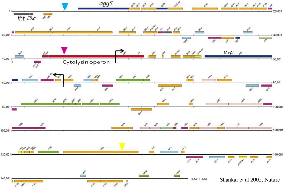
PAI:s usually differ in G+C content from the rest of the chromosome, they are flanked by direct repeats and in addition to the virulence genes often carry a phage lambda-related integrase gene or plasmid-type transfer genes that enables excision and chromosomal integration by homologous recombination. [10, 11] There is evidence of PAI:s carrying antibiotic resistance for example the SRL in Shigella flexneri.[14] The PAI found in pathogenic E. faecalis strains MMH594 and V583, Fig. 1, contains virulence genes such as the cytolysin operon that encodes a hemolysin and aggregation substance, aggS, probably acquired from virulence plasmids, [3, 7] as well as regions which resemble transposons and insertion elements from other gram positive cocci. This raises the idea that these elements may be conquered from other species. [4] A deletion in the PAI of V583 compared to MMH594 has been detected. The deletion is about 17,000 bp in size and includes parts of the cytolysin operon and the esp gene coding for a surface protein. [7] The presence of at least one transfer related gene suggests the possibility of mobilization. [7, 11, 15]
11
Fig 1. Schematic picture of the PAI of E. faecalis MMH594. The region between the black arrows is the about 17,000 bp deletion resulting in the PAI of V583. The ORF:s marked with red represents the cytolsin operon and turquoise ORF:s are movable genetic elements. The blue, purple and yellow arrows indicates the three positions where the pheS*/cat cassette was incorporated in the V583 PAI.
12 Mobilization of genetic elements and PAI:s
It has been shown that some PAI:s are capable of gene transfer using different mechanisms. PAI:s in Pseudomonas species have been known to transfer through conjugation using an integrase. [11] Integrases and excisionases are genes responsible for the ability of fragments to be inserted into or deleted from the chromosome. [11, 14] They belong to the family tyrosine
recombinases which are important for recombination between DNA molecules. [11] Integrases of phage-type have been studied for PAI’s in Escherichia coli 536 and shown to be necessary for excision of these PAI’s. [16] By the loss of a functional integrase gene it is suggested that PAI:s become permanently incorporated into the chromosome. [11] Other genes enabling mobilization of genetic material are plasmid related transfer genes such as the tra operon found in the conjugational F-plasmid of E. coli. [9, 11, 17] A TraG like gene has been identified on the E. faecalis PAI. [7, 18]
Excision of PAI’s has been studied in uropathogenic E. coli, for which deletion of complete PAI’s have been confirmed at frequencies of 10-5
and 10-6 using the counter selectable marker sacB inserted in the PAI’s studied. One of the PAI:s studied was considered to be stably inserted into the chromosome as mutations in the counter selectable marker occurred at higher frequencies than excisions. [10] Excision frequencies of 10-5 and 10-6 have been established for the she PAI and SRL PAI of
Shigella flexneri as well. [12, 19] Parts of the E. faecalis V586 PAI have been shown to
undergo precise excision resulting in the V583 PAI, mentioned earlier, however the PAI itself has not been shown to undergo complete deletions but it is suggested that the complete PAI could transfer. [7, 11]
13 Mitomycin C
Mitomycin C is an anti tumor antibiotic that has been shown to increase the excision frequency of integrative elements in the gram positive Streptococcus thermophilus. It is believed to work through DNA damage which activates the RecA protein that induces proteolysis of a repressor of the excision process. [20]
In vitro recombination
Recombination can also be performed in vitro for example to introduce selectable markers such as antibiotic resistance genes. The antibiotic resistance gene is incorporated into a vector using restriction enzyme digestion and DNA ligation. To enable recombination with the chromosome of the recipient cell, sequences identical to the host’s genome are introduced on both sides of the gene. The bacterial host is transformed with the constructed vector and homologous recombination will conduct introduction of the wanted gene into the recipient chromosome. Selection for the desired phenotype is then carried out. [21]
Phenylalanyl-tRNA synthetase
A counterselectable marker, pheS*, has been developed for studies of E. faecalis. The
pheS gene in E. faecalis encodes the α-subunit of phenylalanyl-tRNA synthetase.
Phenylalanyl-tRNA is responsible for incorporating the amino acid phenylalanine into the growing protein during translation. Kristich and Dunny have introduced a mutation, A312G, in the pheS gene that causes relaxed substrate specifity and allows incorporation of phenylalanine analogues, i.e. p-chloro-phenylalanine, p-Cl-phe, which causes non-functional proteins to be synthesized. Bacteria with the mutated pheS* gene are therefore unable to grow on p-Cl-phe media, while bacteria with the wildtype pheS will grow. [22]
14 Aim
The aim of this project was to study the pathogenicity island of E. faecalis V583 and evaluate a method to determine the rate at which the PAI excises and whether the PAI is a random mix of different genes or consists of several functional pieces. This was carried out by introducing the counterselectable marker pheS* into the pathogenicity island of the chromosome of V583, selecting for p-Cl-phe resistant clones and looking at the excision patterns.
15
MATERIALS AND METHODS
Bacterial strains, media and plasmids
Bacterial strains used in this project were E. coli DH5α, E. faecalis V583, MMH594 and OG1X, see Table 1. E. coli DH5α was grown in Luria-Bertani (LB) broth. E. faecalis V583 was grown in Brain heart infusion (BHI) media. For solid media 15 g/l of agar was added. For selective media, tetracycline was added to a final concentration of 10 μg/ml and/or chloramphenicol to a final concentration of 12 μg/ml for E. coli and 20 μg/ml for E.
faecalis. All antibiotics were from Sigma-Aldrich and all medias were from Difco Laboratories.
The pLR2 vector, fig. 2 and table I, contains a Tetracycline resistance gene, tetM, a chloramphenicol resistance gene (cat) and the pheS* gene, encoding a phenylalanyl-tRNA synthetase with relaxed substrate specificity. cat and pheS* are located as one unit between the XbaI and SalI sites, see figure 2. pLR2 is able to replicate in E. coli but is non-replicative in E. faecalis. Derivatives of the pLR2 vector were used to integrate the
pheS*/cat cassette into the PAI of E. faecalis V583 at three positions, see fig 1. pLR6,
pLR8 and pLR10, fig 2 and table I, are derivatives of pLR2 where the pheS*/cat cassette was flanked with a fragment of the E. faecalis MMH594 PAI next to the selected insertion site, using the EcoRI/BamHI restriction enzyme sites. pLR7, pLR9 and pLR11, fig 2 and table I, are derivatives of pLR2 flanked with PAI fragments next to insert positions 1, 2 and 3 introduced at restriction enzyme sites EcoRI/BamHI and SalI/PstI. Primers used in this study are listed in table II.
16
Fig 2. Schematic picture of the plasmids used to incorporate phe*S/cat cassette into the PAI of E.
faecalis V583. Plasmids pLR6, pLR8 and pLR10 are intermediate plasmids flanked with part of the E. faecalis V583 PAI on one side. pLR7, pLR9 and pLR11 are the final plasmids flanked with parts of the E. faecalis V583 PAI on both sides of the pheS*/cat cassete to enable homologous recombination with
17
Table I. Bacterial strains and plasmids used in this study
Strain or plasmid Description/Relevant characteristic(s) Reference
E. coli DH5α Cloning host * E. faecalis V583 MMH594 OG1
Genome sequenced E. faecalis strain containing PAI.
Genome sequenced E. faecalis strain containing PAI.
Apahtoghenic strain of E. faecalis missing the PAI. * * *
V583PAI::pheS*cat pheS */cat cassette integrated into intergenic region
between exc and aggS In V583
This study
V583cylM::pheS*cat PheS* and cat integrated in cylM of V583 PAI. This study pLR2 Cloning vector containing pheS*, cat and tetM *
pLR6 pLR7
pLR2 derivatives for construction of V583PAI::pheS*cat
This study This study pLR8
pLR9
pLR2 derivatives for construction of V583cylM::pheS*
This study This study pLR10
pLR11
pLR2 derivative for construction of V583ORFEF0592::pheS*cat
This study This study * Michael S. Gilmore, Schepens Eye Research Institute.
18
Table II. Primers used in this study. F = forward primer, R = reverse primer. Primer name Sequence
LR1F CCCAGCTCTAGATTTGAGGTGATAGGTAAGAT LR1R ATTTTTGTCGACAGGAGGAATAATCCTCTGAT LR2F ATTGATGTCGACCAACGTGAATTTAGGTTTTGG LR2R ATAAATGTCGACATCACTTACGTGTATAAAATTA LR4F AAATTAGAATTCGTAACGAATGCGATTTATCC LR4R GTAACTGGATCCTGCTCCTTGAAGTGTTAACG LR5F ACGCCAGTCGACCTTATCGTAAAGAAGGAGCG LR5R CTCTTTCTGCAGACCTAATGTAGGTAGCTTTT
cylM1F No sequence available
cylM1R No sequence available
cylM2F No sequence available
cylM2R No sequence available
EF0592AF CTTTCGTTGCGGAATTCGATGGAGCGAAAAGT EF0592AR CTCTTTAGGATCCACTATAG EF0592BF ACGTAGGTCGACTTCTAAAATGTGGCTTTGGG EF0592BR cylMleft cylMright pheSR pLT06F1 p3TET1 p3TET2 AggSF AggSR ExcF1 ExcR1 IntR1 IntF2 CbhkF CbhkR 5’LR4F 3’LR5R EF0503F EF0503R EF0516F EF0516R EF0527F EF0528R EF0530F EF0530R EF0541F EF0541R TACTTGCTGCAGTGGTTGACGGTCAAAGTCAG GCTGTTGCGGCGACAGCTG GCTGACAATATATCTTCAGATGG TATTCAAGGCAATCTGCCTC AACCGATACCTGAAAACACC CGACGGCCAGTGAATTGTAA TGTTGTGTGGAATTGTGAGC GCGTTCTAATACCGTGGTGA TGCTAAGCCAAGCATTGCAG TGTAGAGAATTCTATGCTAAGACAGCTAGAAA GGCTTTATCTTGGAGCAATATCGAGGGATAACTTTC CTCCAAGATAAAGCCTTGTAATGTTCCTCTTTTCTC GGCTTTATCTTGGAGGVAGCAGAAGCATTAGAAAC No sequence available No sequence available GATAGCGGAACTAATGGATA TCAGCTGGTGCAACTTCTGT CCCAGTAGAGCATGGAAATG GGTTCATCAAAAATCACAAGAC ATCATCGTAGGGTTGATTCC AATGAAGCCAACGACCTAC TGTACGATGGATTGCCTGGT CCACACCACTGATTCCTACA TTCCTGTGGTTAGTGGGGAT TAGGAGTCGTCCCTGTTATC GGCGGAACTGTGCATTTCT TACCGCCAAAAACTGCCTGA
19 PCR program
PCR-reactions were carried out using Hybaid PCR sprint Thermal Cycler, Thermo Scientific. An initial step of denaturation at 95°C for 2 min followed by 35 cycles of denaturation 95°C for 1 min, primer annealing at 55°C for 30 s and DNA extension at 72°C for 1 min. After the last amplification cycle, samples were kept for final extension at 72°C for 5 min and cooled to 4°C.
Electrocompetent cells
E. coli DH5α
125 ml SOB media (Tryptone 20g/l, Yeast extract 5 g/l, 8.5 mM NaCl, 2,5 mM KCl) was inoculated with 0.5 ml fresh over night culture of E. coli DH5α and grown in a 37C shaking incubator at 200 rpm until OD550 0.7. Cells were pelleted at 4C for 10
min at 5000xg, resuspended and washed twice in 125 ml of cold 10 % glycerol. The cells were then pelleted as above and washed with 60 ml of cold 10% glycerol. The cells were once again pelleted and washed in 30 ml of cold 10% glycerol and finally resuspended in 250 L of cold 10% glycerol. 40L aliquots of cells were snap frozen and stored at -70C.
E. faecalis V583
E. faecalis V583 were grown in M17 media (peptone 5 g/l, phytone peptone 5 g/l, lab
lemco 5 g/l, yeast extract 5 g/l, lactose 5 g/l, b-disodium glycerophosphate 19 g/l, ascorbic acid 0.5 g/l, 1mM MgSO4) over night at 37C. 100 ml of SGM17 media
(M17 37.25 g/l, glycine 65 g/l, 0.5 M sucrose) was inoculated with 1 ml E. faecalis V583 over night culture and incubated for 20 hours at 37C without aeration. Cells were pelleted at 4C for 10 min at 1000xg and washed at least three times with 100 ml of chilled electroporation solution (glycerol 100 ml/l, 0.5 M sucrose). Cells were
20
resuspended in 1 ml electroporation solution and 80 µl aliquots were snap frozen and stored at -70C.
DNA preparation for Enterococci
Cells were grown in 10 ml BHI broth over night. The cells were pelleted for 10 min at 1000xg and washed in 1 ml TE buffer (10 mM Tris HCl, pH 7.5, 1mM EDTA) and transferred to an eppendorf tube. The pellet was resuspended in 200 µL of filter sterilized IHB-1 solution (50 mM glucose, 25 mM Tris HCl, pH 8.0, 10mM EDTA) and 50 µL of lysozyme (50 mg/ml) and 10 µL mutanolysin (2500 U/ml) was added and the mixture was incubated for 2 hours at 37 ºC. 100 µL of 20 % sarkosyl and 15 µL of RNAse (10mg/ml) was added and the mixture was incubate for 30 min at 37 ºC. 15 µL pronase (10mg/ml) was added and the sample incubated for additional 30 min at 37 ºC. The volume was brought up to 600 µL with TE buffer and DNA was extract twice with buffer saturated phenol by adding 600 µL buffer saturated phenol. After centrifugation in a table centrifuge at maximum speed for 4 min the top phase was transferred to a new tube. DNA extraction was then carried out three times using phenol:chloroform 1:1 and once with chloroform. The extracted DNA was precipitated using 500 µL of isopropanol and 50 µL 3M sodium acetate, pH 5.2 and the DNA was pelleted for 10 min at maximum speed using a table centrifuge and washed twice with 700 µL 70 % ethanol. The DNA was resuspended in 100 µL TE buffer.
Labeling of E. faecalis V583 PAI with the selection marker pheS*
The PAI of E. faecalis was to be labeled with pheS* at three different positions See fig. 1. For labeling at position 1, plasmid pLR7, see fig 2, was constructed.
To construct pLR6 fragments of the PAI were amplified using primer pair LR4F/LR4R and Taq DNA polymerase (New England Biolab). The 0.8 kb PCR product LR4 of primer pair LR4F/LR4R was purified according to protocol using the Qiagen PCR Purification Kit. The purified PCR product and pLR2 were digested with
21
restriction enzymes EcoRI and BamHI at 37ºC for 3 h. The large fragment of the digested pLR2 vector was purified by electrophoresis on a 1 % agarose gel and extracted according to protocol using the Qiagen Gel Extraction Kit. The digested and purified PCR product was ligated into the EcoRI/BamHI digested pLR2 vector to construct plasmid pLR6. The ligation was carried out using T4 DNA ligase and the mixture was incubated at 16C over night. Electrocompetent E. coli DH5α were transformed with the ligation mixture by electroporation at 2.5 kV. The cells were allowed to recover in 2xYT media (tryptone 16 g/l, yeast extract 10 g/l, 85 mM NaCl) for 50 min at 37ºC. To select for transformed clones of E. coli DH5α 100 μL were plated onto LB agar supplemented with chloramphenicol to a final concentration of 12 μg/ml and tetracycline to a final concentration of 10 μg/ml. Digested vector, ligase added to digested vector and H2O were used as controls. Putative pLR6 plasmids from
colonies growing on the selective plate were purified according to protocol using the Qiagen Miniprep Kit. To check the plasmids for the correct insert they were digested with EcoRI and BamHI at 37ºC for 3 h. The digestion product was run on a 1 % agarose gel. Plasmids with digestion products of the right size were sent for sequencing using primers p3TET1 and p3TET2, at the Massachusetts General Hospital (MGH) core facility and the sequences were analyzed using Cromas Pro and compared to the E. faecalis PAI sequence by a BLAST search.
To construct the plasmid pLR7 the 0.7 kb PCR product of LR5F/LR5R was cloned into the SalI/PstI digested pLR6 in the same way and using the same conditions as described above. All restriction enzymes and DNA ligase were from New England Biolab.
For labeling of the PAI at position 2, see fig. 1, plasmid pLR9 was constructed as described but using primer pairs cylM1F/cylM1R and cylM2F/cylM2R. The 0.6 kb and 1 kb fragments were cloned into the EcoRI/BamHI and SalI/PstI sites of pLR2.
22
To introduce the pheS* at position 3 of the V583 PAI, see fig. 1, attempts were made to construct the plasmid pLR11 using primer pairs EF0592AF/EF0592AR which gave a 1.2 kb PCR product and EF0592BF/EF0592BR resulting in a 0.7 kb PCR product.
Electrocompetent E. faecalis V583 were transformed with 10 μg each of plasmids pLR7 and pLR9 by electroporation at 2.5 kV. H2O was used as control. Cells were put
on ice for 5 min and recovered in 1 ml of SM17MC media (M17 37.25 g/l, 10 mM MgCl2, 10 mM CaCl2, 0.5M sucrose) for 2 h at 37ºC. 500 ml of BHI broth
supplemented with chloramphenicol to a final concentration of 20 μg/ml was inoculated with 500 μL of bacterial culture, the remaining 500 μL were pelleted and resuspended in 100 μL of BHI and plated onto BHI agar supplemented with chloramphenicol to a final concentration of 20 μg/ml. Broths and plates were incubated at 37ºC over night. Serial dilutions of the cultures were plated on BHI agar/chloramphenicol to get single colonies. Recombinants were selected using chloramphenicol and screened for tetracycline sensitivity and confirmed with PCR using primer pairs to amplify products A-D in fig. 3. For analysis of V583/pLR7 clones; product A = primer pair p3TET1/3’LR5R, B = 5’LR4F/pheSR, C = 5’LR4F/p3TET2 and D = pLT06F1/3’LR5R were used. For analysis of V583/pLR9 clones; A = primer pair p3TET1/cylMright, B = cylMleft/pheSR C = cylMleft/p3TET2 and D = pLT06F1/ cylMright were used. A double crossover would generate products B and D, while A or C indicates a single crossover, such clones should be tetracycline resistant.
23
Fig 3. Schematic picture of three possible genotypes of the E. faecalis V583 genome after homologous recombination with plasmids pLR7 and pLR9. Blue arrows indicate binding sites for primers and A-D are PCR-products.
Determination of excision rate of pheS*-labeled PAI in E. faecalis
To determine the excision rate of the PAI or parts of the PAI in E. faecalis, a fresh overnight culture (20h) of E. faecalis V583PAI::pheS*cat and V583cylM::pheS*cat were diluted 1:100 in 30 ml of BHI. Cultures were incubated at 37˚C, 42˚C or room temperature for 24 h. V583cylM::pheS*cat were also diluted 1:100 in 30 ml of BHI supplemented with Mitomycin C to a final concentration of 0.5 μg/ml and grown at 37˚C. Serial dilutions which were plated on MM9YEG agar (100 ml of 10x M9 salts/l, 2.5 g/l yeast extract, 15 g/L agar, 10 ml 50% glucose and 250 μg/ml X-gal solution) and MM9YEG agar supplemented with p-Cl-phe to a final concentration of 10 mM to select for putative excisions of the PAI. Plates were incubated at 37˚C for 24 h. Colonies were counted and the excision rate was calculated as the quotient of p-Cl-pheR CFU/ml and total CFU/ml. p-Cl-pheR clones were patched on BHI agar
24
supplemented with chloramphenicol to a final concentration of 20 μg/ml and BHI agar to determine the number of chloramphenicol sensitive isolates. The excision rate was adjusted based on the number of chloramphenicol sensitive clones calculated
according to: R S R cam cam ml CFU Total ml CFU phe Cl p ) / ( ) / (
Screening of putative E. faecalis PAI excision clones and characterization of the deleted region
To test if the chloramphenicol sensitive clones had undergone excisions of the PAI or parts of it, a PCR assay was carried out screening for pheS* and cat using primer pairs LR1F/LR1R and LR2F/LR2R. To characterize the excised region chromosomal DNA was extracted according to protocol DNA preparation for Enterococci, see page 20, from the chloramphenicol sensitive clones and the PAI junctions were amplified with PCR using primer pairs FeoB1/EF0479R for the left junction, OxoR/EF0628F for the right junction and FeoB1/OxoR to detect if the whole PAI had been deleted, see fig 4a and b. For further characterization; the aggregation substance gene (aggS) using primer pair AggSF/AggSR, the integrase gene (int), IntF1/IntF2, and the excisionase gene (exc), ExcF1/ExcR1, were amplified for p-Cl-phe tolerant and chloramphenicol sensitive isolates of V583PAI::pheS*cat, see fig 4a. For p-Cl-phe tolerant and chloramphenicol sensitive isolates of V583cylM::pheS*cat ORFs between mobile genetic elements were analyzed. EF0503 (Primer pair EF0530F/EF0530R), EF516 (EF0516F/EF0516R), chb (cbhkF/cbhkR), EF0527/EF0528 (EF0527F/EF0528R), EF0530 (EFo530F/EF0530R) and EF0541 (EF0541F/EF0541R) were amplified, see fig 4b.
25
Fig 4a. Schematic picture of the PAI and enlargement of the region with the first position for insertion of pheS*. Primers FeoB1/EF0479R and EF0628F/OxoR were used to amplify the left and right PAI junctions when characterizing the excised parts. FeoB1/OxoR was used to detect deletion of the complete PAI. The integrase (int), excisionase (exc) and aggregation substance (aggS) genes were amplified for further characterization.
26
Fig 4b. Red area symbolizes cylM, the second position for insertion of pheS*. Primers FeoB1/EF0479R and EF0628F/OxoR were used to amplify the left and right PAI junctions when characterizing the excised parts. FeoB1/OxoR was used to detect deletion of the complete PAI, and amplification of genetic elements surrounding the insertion point of pheS* were carried out, EF0503, EF0516, cbh, EF0527/EF0528, EF0530 and EF0541, to identify the excised area. Turquoise areas are putative movable genetic elements.
27
RESULTS
Construction of plasmids for site directed mutagenesis
Cloning
Chloramphenicol and tetracycline resistance were used to select for clones of E. coli DH5α transformed with plasmids pLR7, pLR8, pLR9, pLR10 and pLR11. Double restriction enzyme digestion and agarose gel electrophoresis was carried out on putative transformants to confirm that the correct inserts of E. faecalis PAI DNA had been incorporated into the pLR2 vector. The insert in the intermediate plasmids pLR8 and pLR10 was confirmed using EcoRI/BamHI while SalI/PstI were used for the final plasmids pLR7, pLR9 and pLR11.
Putative pLR7 plasmids from five chloramphenicol and tetracycline resistant clones of
E. coli DH5α were analyzed using restriction enzymes Sal I/Pst I as described above.
As expected gel analysis of the digestion products showed a band for the plasmid and one of about 0.7 kb corresponding to the insert see fig. 5. Sequencing of the purified pLR7 plasmid insert was performed and a BLAST search matched E. faecalis PAI.
28
Fig 5. PstI/SalI digests of plasmid preparations from five (1-5) putative pLR7 containing clones of E. coli DH5α. L = DNA ladder.
Putative pLR8, pLR9 and pLR10 clones were analyzed following the same procedure and the plasmids were shown to carry the expected inserts. Attempts were made to construct the final pLR11 plasmid, but restriction enzyme digestion and gel electrophoresis revealed that the insert had not been incorporated into the cloning vector.
Labeling of E. faecalis V583 PAI with the selection marker pheS*
E. faecalis V583 was transformed with the constructed plasmids pLR7 and pLR9.
Cultivation on BHI/chloramphenicol was used to select for recombinant clones of E.
faecalis V583. Further cultivation on BHI/tetracycline was used to investigate the
possibility of clones with double crossovers since they should be tetracycline sensitive, as shown in fig. 3, page 22. Colony PCR analysis with following agarose gel electrophoresis using primers (blue arrows in fig. 3) to amplify the junction regions at the insert positions to receive products A-D were carried out. A clone with a double crossover should give PCR products B and D.
29
pLR7
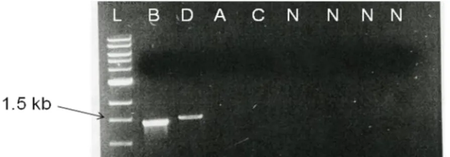
Three recombinant clones of E. faecalis V583 were obtained by cultivation on BHI/chloramphenicol. Further cultivation on BHI/tetracycline resulted in two resistant isolates, 1 and 2, possible single crossovers and one sensitive isolate, 3, a possible double crossover. All three clones were tested with PCR analysis. Four PCR reactions were carried out amplifying the possible junctions between pLR7 and the chromosome, see fig 3. Expected sizes of colony PCR products A-D for pLR7 crossovers were A – 2.2 kb, B – 1.6 kb, C – 2.4 kb, D – 1.6 kb. Clones 1 and 2 gave PCR products A (~2 kb) and B (~1.6 kb) indicating single crossovers. Analysis of clone 3 resulted in the expected products B and D of about 1.6 kb, which indicated that a double crossover had occurred and a chromosomal mutant containing the pheS*/cat cassette, V583PAI::pheS*cat, had been constructed, fig. 6.
Fig 6. Agarose gel electrophoresis showing results of colony PCR of three chloramphenicol resistant clones. Four PCR reactions, A-D, were carried out using primers to amplify the possible junctions of the pheS*/cat cassette and the chromosome. Both products B and D of about 1.6 kb were expected for a double crossover with pLR7. N = negative control and L = ladder.
30
pLR9
One chloramphenicol resistant clone of E. faecalis V583 was obtained after transformation with pLR9. Colony PCR amplification was performed according to the same procedure as for pLR7 using primers to amplify the junctions between the chromosome and pLR9 for products A-D. Products A or C indicates a single crossover. The clone gave products B and D of about 1.5 kb, see fig 7, as expected for a double crossover, thus chromosomal mutant V583cylM::pheS*cat was constructed.
Fig 7. Agarose gel electrophoresis showing results of colony PCR amplification for one chloramphenicol clone. Four PCR reactions, A-D, were carried out to amplify the possible junctions between the chromosome and the pheS*cat insertion. Products A or C indicates a single crossover while B and D confirms a double crossover. N = negative controls for each PCR reaction. L = ladder.
Excision experiments
One experiment was carried out using V583PAI::pheS*cat, and one using V583cylM::pheS*cat. The clones were cultivated for 20 h in different temperatures and V583cylM::pheS*cat was also cultivated in the presence of Mitomycin C. Cl-phe was used to screen for deletions of the Cl-pheS*/cat cassette. Colonies resistant to p-Cl-phe were evaluated for growth on BHI supplemented with chloramphenicol to check for chloramphenicol sensitivity to further confirm deletions.
31
V583PAI::pheS*cat
Growth of V583PAI::pheS*cat on MM9YEG plates supplemented with p-Cl-phe was heavy ranging from about 60-150 colonies depending on growth conditions. Patching colonies from MM9YEG/p-Cl-phe in duplicate on BHI and BHI supplemented with chloramphenicol resulted in a total of 13 chloramphenicol sensitive clones. Excision rate according to correct phenotype (p-Cl-phe resistant and chloramphenicol sensitive) ranged from 6*10-7 to 7*10-8, see table IIIb. Triplicate experiments were carried out but only one experiment at 42°C resulted in a chloramphenicol sensitive clone.
32
Table IIIa-b. Results of excision experiment using V583PAI::pheS*cat. RT = room temperature. The tables show total count (CFU/ml) in culture, total count of
p-Cl-pheR (CFU/ml) in culture, p-Cl-pheR /Total count is the frequency of excision based on phenotypical p-Cl-pheR. The average of triplicate experiments is presented i table IIIa. The adjusted excision rate based on chloramphenicol sensitivity is shown in table IIIb. The average of duplicate or triplicate experiments is presented. RT (n=3), 37°C (n=3), 42°C (n=1). Chloramphenicol sensitive/p-Cl-pheR shows the number of sensitive clones compared to number of tested p-Cl-pheR. Excision rate is the adjusted rate based on chloramphenicol sensitivity. Table IIIa. Growth Condition Total count (CFU/ml) p-Cl-pheR
(CFU/ml) p-Cl-pheR /Total count RT 2.56*109 ± 2.23*108 1.45*104 ± 2.50*103 5.64*10-6±5.15*10-7 37°C 1.88*109 ± 1.15*108 1.20*104 ± 2.14*103 6.33*10-6± 7.90*10-7 42°C 2.33*108 ± 8.08*107 6.13*103 ± 4.51*102 2.80*10-5± 7.50*10-6 Table IIIb. Growth Condition Chloramphenicol
Sensitive/p-Cl-pheR analysed Excision rate
RT 2/145 7.62*10-8 ±3.28*10-8
37°C 1/120 5.33*10-8 ± 3.37*10-9
33
V583cylM::pheS*cat
The growth of V583cylM::pheS*cat on MM9YEG media supplemented with p-Cl-phe was as for V583PAI::pheS*cat very heavy. A total of 108 chloramphenicol sensitive clones were obtained. Growth was less on all plates after cultivation in the presence of Mitomycin C compared to experiments using different temperatures. Excision rates of 1*10-6 to 9*10-7 were seen, see table IVb. No p-Cl-pheR and chloramphenicol sensitive clones were found using Mitomycin C. Triplicate experiments were carried out but due to over growth the average of two experiments is presented for room temperature (RT) and Mitomycin C.
Table IVa-b. Results of excision experiment using V583cylM::pheS*cat. The average is of duplicate or triplicate experiments presented, RT (n=2), 37°C (n=3), Mitomycin C (n=2). The tables show total count (CFU/ml) in culture, total count of p-Cl-pheR (CFU/ml) in culture. p-Cl-pheR/Total count is the frequency of excision based on p-Cl-pheR. Chloramphenicol sensitive shows the number of sensitive clones compared to number of tested p-Cl-pheR. Excision rate is the adjusted rate based on chloramphenicol sensitivity. RT = room temperature.
Table IVa. Growth condition Total count (CFU/ml) p-Cl-pheR
(CFU/ml) p-Cl-pheR /Total count
RT 2.45*109 1.96*104 8.09*10-6
37°C 1.88*109 ± 1.42*108 1.67*104 ± 7.43*102 8.88*10-6 ± 7.23*10-7
34 Table IVb.
Growth Condition
Chloramphenicol
Sensitive/p-Cl-pheR analysed Excision rate
RT 28/196 1.13*10-6
37°C 17/167 9.40*10-7 ± 3.94*10-7
42°C 0 -
Characterization of chloramphenicol sensitive isolates
V583PAI::pheS*cat
Colony PCR-amplification of pheS*, see fig 8a and b, and cat, fig. 8c (clones 12 and 13; data not shown), of the 13 isolated chloramphenicol sensitive clones showed that both genes had excised from the chromosome of V583PAI::pheS*cat. Untreated V583PAI::pheS*cat was used as positive control and showed amplification of both
35
Fig 8a. Fig 8b.
Fig 8c.
Fig 8a-c. Colony PCR amplification of pheS*, fig 8a, and cat, fig 8c, for p-Cl-phe resistant and chloramp-Cl-phenicol sensitive isolates 1-11. Fig. 8b. Amplification of pheS* for two additional clones, 12-13. L =ladder, P = positive control (V583PAI::pheS*cat), N = negative control.
Further PCR analysis of the 13 chloramphenicol sensitive clones obtained using primers amplifying the left (product A) and right junctions (product B) of the PAI in the chromosome of V583, see fig 4a on page 24, showed that the left junction was still present in 12 clones and the right junction was present in all 13 clones, see fig 9. Wildtype E. faecalis V583 (containing the PAI) was used as a positive control for the left and right junctions and E. faecalis OG1X (without PAI) was used as negative control. A third PCR reaction was carried out using primers FeoB1/OxoR for detection of complete PAI excisions, product C. For this reaction V583 was used as negative control and OG1 as positive. None of the clones show an amplification product in reaction C, indicating that large parts of the PAI are still present.
36
Fig. 9. Amplification of left junction using primers FeoB1/ EF0479R (A), right junction using primers EF0628F/OxoR (B) and FeoB1/OxoR (C) of 13 chloramphenicol sensitive clones (1-13). V583 was used as positive control for the presence of the left and right junctions, OG1X was used as negative control. For excision of the complete PAI OG1 was used as positive control and V583 as negative.
Further PCR analyses were carried out amplifying the aggregation substance (aggS), the integrase (int) and the excisionase (exc) genes for 13 p-Cl-phe resistant and chloramphenicol sensitive clones. It revealed that all 13 clones still had the aggS, see fig 10a, and int, see fig 10b, but only two clones still carried the exc gene, see fig 10c.
37 Fig. 10b.
Fig. 10c.
Fig 10a-c. Amplification of aggS, fig 10a, int, fig 10b and exc, fig 10c, for 13 p-Cl-phe resistant and chloramphenicol sensitive clones. V583 was used as positive control (P) and OG1 as negative (N).
The excised part of the genome at this position could not be specified exactly but seems to be very restricted to the area of the pheS*/cat cassette integration point, see fig 4a page 24.
V583cylM::pheS*cat
The pheS*, see fig. 11, and cat genes (data not shown) were amplified for 10 of the p-Cl-phe resistant and chloramphenicol sensitive clones of V583cylM::pheS*cat. As expected none of the analyzed clones carried the pheS* or cat gene.
38
Fig 11. PCR-amplification of pheS* for 10, 1-10, p-Cl-phe resistant and chloramphenicol sensitive clones of V583cylM::pheS*cat. L = ladder, P = positive control (V583cylM::pheS*cat), N = Negative control.
To investigate if the complete PAI had excised from any of the 10 isolates, PAI junctions were amplified and amplification using primers on each side of the PAI was carried out to detect excision of the PAI in the same way as for V583PAI::pheS*cat, see fig 4b. PCR amplification of the left PAI junction was positive for 9 of 10 isolates, see fig. 12a. Amplification of the right junction was positive for 8/10 isolates, see fig 12b. Amplification using primers FeoB1 and OxoR to detect excision of the complete PAI was negative for all 10 isolates, see fig 12c.
39 Fig 12a.
Fig. 12b.
Fig. 12c.
Fig. 12 a-c. Agarose gel electrophoresis showing the results of amplification of the left, 12a, and right, 12b, PAI junctions of 10, 1-10, p-Cl-phe resistant and chloramphenicol sensitive clones of V583cylM::pheS*cat. E. faecalis V583 was used as positive control and OG1X as negative. Fig 12c. Results of PCR-amplification using primers on each side of the PAI to detect complete excision of PAI. E. faecalis OG1X was used as positive control and V583 as negative control.
To characterize how much of the genome that had excised, PCR-amplifications of genes between the putative movable genetic elements were carried out. See fig 4b. The EF0503 PCR, the furthest away from the insert of pheS*, was positive for all 10 tested clone, see fig. 13a. Amplification of the EF0516 ORF closer to the pheS*/cat cassette was positive only for one clone, see fig 13b and cbh closest to the cassette was present in four out of ten tested p-Cl-phe resistant and chloramphenicol sensitive clones. See fig. 13c.
40 Fig. 13a.
Fig. 13b.
Fig. 13c.
Fig. 13a-c. Agarose gel electrophoresis showing the results of amplification ofEF0503 (Fig 13a) EF0516 ORF (Fig. 13b) and
cbh (Fig. 13c), for 10 p-Cl-phe resistant and chloramphenicol
sensitive clones of V583cylM::pheS*cat, 1-10. L = ladder, P = positive control (V583) and N = negative control (OG1X).
Amplification was carried out for ORFs downstream of the pheS*/cat cassette as well. EF0527/EF0528 closest to the pheS* insert were only positive for one of 10 tested clones. See fig. 14.
41
Fig. 14a.
Fig. 14b.
Fig. 14a-b. Amplification of the EF0527/EF0528 ORF:s, fig 14a and EF0530, fig 14b, in 10 p-Cl-phe resistant and chloramphenicol sensitive clones of V583cylM::pheS*cat, 1-10. V583 was used as positive control, P, and OG1X as negative, N.
Amplification of the ORF EF0541, furthest away from the pheS*/cat cassette was positive for three out of 10 tested clones (clones number; 1, 6 and 8) were positive. (Data not shown).
Characterization of chloramphenicol resistant isolates of V583PAI::pheS*cat Four chloramphenicol resistant clones of V583PAI::pheS*cat previously cultivated at room temperature, 37°C or 42°C were checked for the presence of pheS* by PCR-amplification, V583PAI::pheS*cat was used as positive control and V583 as negative. All four were still carrying the pheS* gene, see fig 15.
42
Fig. 15. Amplification of the pheS*/cat cassette in four chloramphenicol resistant clones of V583PAI::pheS*cat. V583PAI::pheS*cat was used as positive control and V583 as negative.
Sequencing of the four isolates revealed mutations in the inserted pheS* gene. In fig. 16 the protein sequences for the PheS* α-subunit of E. faecalis V583 wildtype (GenBank Accession: AAO80915), the mutated PheS* used as a counterselectable marker and the four sequenced clones are presented. The amino acid marked with red indicates the mutation introduced to cause relaxed substrate specificity. Isolate 4 has a truncated ORF while the other three clones carry amino acid exchanges. Clone 1 has reverted to the wildtype protein sequence.
PheS WT …VVGGFANEI…DMQDTFYIS…GKVFRRDTD…SHQFHQIEGL…SGFAFGL PheS* …VVGGFANEI…DMQDTFYIS…GKVFRRDTD…SHQFHQIEGL…SGFGFGL Isolate 1…VVGGFANEI…DMQDTFYIS…GKVFRRDTD…SHQFHQIEGL…SGFAFGL Isolate 2…VVGGFANEI…DMKDTFYIS…GKVLRRDTD…SHQFHQIEGL…SGFGFGL Isolate 3…VVGGFANEI…DMQDTFYIS…GKVLRRDTD…SHQLHQIEGL…SGFGFGL Isolate 4…VVGGLCK*
Fig 16. Protein sequences of the PheS α-subunit of E. faecalis V583 wildtype, that of the mutated counterselectable marker and four sequenced clones. Amino acids in bold causes the relaxed substrate specifity of pheS*.
43
DISCUSSION
The aim of this project was to study the PAI of E. faecalis V583 and evaluate a method to determine the rate at which the PAI or pieces of it excises. This was carried out by introducing the counterselectable marker pheS* into two different positions of the PAI, growing the bacteria on the selective p-Cl-Phe and looking at excision patterns.
The plasmids pLR7 and pLR9 used for incorporation of the pheS*/cat cassette at the two positions, an intergenic region in the beginning of the PAI and into the cylM gene of the E. faecalis V583 chromosome, were successfully constructed. This was proven by restriction enzyme digestion, to release the PAI sequences inserted in the plasmids, and following agarose gel electrophoresis. The inserts of the constructed plasmids were also sequenced and compared to the E. faecalis PAI genome by a BLAST search and proven to contain the correct flanking regions.
Incorporation of the pheS*/cat cassette into the PAI genome was successfully carried out with plasmids pLR7 and pLR9 by double homologous recombination between the plasmids and the E. faecalis V583 PAI resulting in the construction of chromosomal mutants V583PAI::pheS*cat and V583cylM::pheS*cat. To confirm the mutants, they were analyzed by amplifying the supposable junctions between the PAI sequence and the pheS*/cat insertion. The expected junctions were present and the mutants considered successfully constructed.
Excision experiments using V583PAI::pheS*cat and V583cylM::pheS*cat were carried out by cultivation in different conditions such as room temperature, 37°C, 42°C and at 37°C in the presence of Mitomycin C. Clones with possible deletions of
pheS* and consequently possible excisions of the PAI were selected using p-Cl-phe.
Growth on the selective media was heavy for both mutants, thus a high occurrence of phenotypical p-Cl-phe resistant clones was observed. Calculation of the frequency of excision based on the p-Cl-phe resistance gave values of 10-4 to 10-6 depending on the
44
mutant used and condition for cultivation. This is, compared to similar experiments in
E. coli, to be considered as high frequencies of excision. The p-Cl-phe resistant clones
were screened for chloramphenicol sensitivity which would be expected for excision clones considering the the pheS*-cat genes being incorporated as a cassette. This analysis showed that a majority of the clones were chloramphenicol resistant and therefore not the result from excisions of the PAI but from possible mutations of the
pheS* gene. If this aspect is taken in consideration when calculating the excision rates,
the frequency of excision was 10-6 to 10-8. However, the established frequencies are uncertain due to the low number of experiments carried out. Since single colonies were difficult to achieve when selecting for p-Cl-phe resistance the frequency of chloramphenicol resistant clones might be overrated. It is possible that the same colony was patched several times. It is also a possibility that chloramphenicol sensitive clones were not detected because clean isolates of the colonies were difficult.
The difference in excision frequencies between different cultivation conditions is insignificant in this experiment since the values obtained from the excision experiments are very uncertain. Mitomycin C might have an effect on p-Cl-phe resistance but probably through an increased mutation rate rather than an increase of excision rate based on the higher count of resistant clones, 105 compared to 104 at 37°C but a substantially lower count of chloramphenicol sensitive clones.
To confirm the hypothesis of mutations in pheS* for p-Cl-phe resistant and chloramphenicol resistant clones PCR-amplification of pheS* and cat were carried out on four clones which all turned out positive. Sequencing of the same isolates revealed additional mutations in the pheS* gene besides the one causing relaxed substrate specificity. The mutations differed for each of the four tested clones. Correlation between these mutations and the ability to grow on the p-Cl-phe was not proven in this study but is to be considered very likely. Since the wildtype pheS is present and functional in the E. faecalis chromosome, all mutations eliminating the function of the counterselectable pheS* marker will cause clones with resistance to p-Cl-phe and the
45
phenotype of an excision clone. The high frequency of mutations makes the pheS* method difficult for studies of excision patterns since a large number of clones have to be processed to get a significant number of excision clones.
A few p-Cl-phe resistant and chloramphenicol sensitive clones were obtained from both V583PAI::pheS*cat and V583cylM::pheS*cat. These were analyzed to study if the complete PAI, or parts of it, had excised from the chromosome. 13 p-Cl-phe resistant and chloramphenicol sensitive clones of V583PAI::pheS*cat were analyzed by PCR for the presence of pheS* and cat, which were excised for all 13 isolates as expected. PCR-amplification showed that in none of the clones had the complete PAI excised. Further PCR-analysis showed that the excisions were very limited to the close surroundings of the pheS*/cat cassette.
For V583cylM::pheS*cat 10 p-Cl-phe resistant and chloramphenicol sensitive clones were analyzed according to the same principle. In none of these clones the complete PAI had excised. However, in two clones, 7 and 9, the right PAI junction seemed to be missing. Considering the ORF EF0503 is positive for all tested clones it is likely that the PCR reaction did not work for these two clones. This problem could be eliminated using an internal positive control. Genes between putative movable genetic elements were chosen to be amplified in further PCR-analyses. The genes present differed between the 10 analyzed clones. Logically, genes close to the insert positions of the
pheS*/cat cassette seemed more likely to excise than genes further away from the
insert. For clones 1, 5 and 8 the ORF EF0516 was negative although cbh closer to the insertion of the pheS*/cat cassette was positive. This could as discussed earlier be a problem with the PCR amplification reaction. It could also be the result of rearrangements of genetic elements due to the insertion of the pheS*/cat cassette, which indicates instability in this region of the PAI. This could be likely considering that the strange excision patterns is only seen for the second insertion position of
pheS*/cat surrounded by many movable genetic elements. The exact point of excision
46
genetic element, could be defined for the 10 tested clones. However, the hypothesis of excision taking place between movable genetic elements could be supported by the fact that the frequency of excisions was higher for V583cylM::pheS*cat, where the cassette is inserted in a region surrounded by many movable genetic elements as compared to the first position, V583PAI::pheS*cat. The excisions seemed random, however, a more obvious pattern might have been discovered if more clones had been tested. To evaluate the pheS* method further another insert position of the pheS* was planned in a conserved region of the genome outside of the PAI to see if the excision frequency was, as would be expected much lower.
Conclusions
This study was a pilot study and much more work is needed to draw any conclusions about excision rates and patterns of the E. faecalis V583 PAI. The excisions seem to be random and limited to parts of the PAI and the excision rate is rather low. This might suggest that the PAI is stably inserted in the E. faecalis chromosome. The fact that the rate of mutation of the pheS* gene is much higher than the excision rate makes the method difficult to work with and the obtained results are uncertain. A large number of clones would have to be processed.
47
ACKNOWLEDGEMENT
I would like to thank the Gilmore lab at Schepens Eye Research Institute in Boston and especially Michael S. Gilmore and Janet Manson. A big thank you to Britt-Inger Marklund who has been helping me a lot with putting this text together. And a special thanks to all of my friends who has helped me to end this project.
48
REFERENCES
1. Abigail A. S., D.D.W., ed. Bacterial Pathogenesis A Molecular Approach. Second ed. 2002, p. 232-245. ASM Press: Washington DC.
2. Paulsen, I.T., et al., Role of mobile DNA in the evolution of
vancomycin-resistant Enterococcus faecalis. Science, 2003. 299(5615): p. 2071-4.
3. Jett, B.D., M.M. Huycke, and M.S. Gilmore, Virulence of enterococci. Clin Microbiol Rev, 1994. 7(4): p. 462-78.
4. Pillar, C.M. and M.S. Gilmore, Enterococcal virulence--pathogenicity island of
E. Faecalis. Front Biosci, 2004. 9: p. 2335-46.
5. MacCallum W. G. and Hastings T. W., A Case of Acute Endocarditis Caused
by Micrococcus Zymogenes (Nov. Spec.), with a Description of the Microorganism. J. Exp. Med., 1899(4): p. 13.
6. Low, D.E., et al., Clinical prevalence, antimicrobial susceptibility, and
geographic resistance patterns of enterococci: results from the SENTRY Antimicrobial Surveillance Program, 1997-1999. Clin Infect Dis, 2001. 32
Suppl 2: p. S133-45.
7. Shankar, N., A.S. Baghdayan, and M.S. Gilmore, Modulation of virulence
within a pathogenicity island in vancomycin-resistant Enterococcus faecalis. Nature, 2002. 417(6890): p. 746-50.
8. Sahm, D.F., et al., In vitro susceptibility studies of vancomycin-resistant
Enterococcus faecalis. Antimicrob Agents Chemother, 1989. 33(9): p.
1588-91.
9. Madigan M. T., M.J.M., Parker J., ed. Brock Biology of Microorganisms. Tenth ed. 2003, p. 276-278. Pearson Education, Inc: Upper Saddle River, New Jersey.
10. Middendorf, B., et al., Instability of pathogenicity islands in uropathogenic
49
11. Osborn, A.M. and D. Boltner, When phage, plasmids, and transposons collide:
genomic islands, and conjugative- and mobilizable-transposons as a mosaic continuum. Plasmid, 2002. 48(3): p. 202-12.
12. Hacker, J. and E. Carniel, Ecological fitness, genomic islands and bacterial
pathogenicity. A Darwinian view of the evolution of microbes. EMBO Rep,
2001. 2(5): p. 376-81.
13. Doublet, B., et al., The Salmonella genomic island 1 is an integrative
mobilizable element. Mol Microbiol, 2005. 55(6): p. 1911-24.
14. Turner, S.A., et al., Nested deletions of the SRL pathogenicity island of
Shigella flexneri 2a. J Bacteriol, 2001. 183(19): p. 5535-43.
15. Coburn, P.S., et al., Horizontal transfer of virulence genes encoded on the
Enterococcus faecalis pathogenicity island. Mol Microbiol, 2007. 63(2): p.
530-44.
16. Manson, J.M. and M.S. Gilmore, Pathogenicity island integrase cross-talk: a
potential new tool for virulence modulation. Mol Microbiol, 2006. 61(3): p.
555-9.
17. Manning, P.A., G. Morelli, and M. Achtman, traG protein of the F sex factor
of Escherichia coli K-12 and its role in conjugation. Proc Natl Acad Sci U S
A, 1981. 78(12): p. 7487-91.
18. Schmidt, H. and M. Hensel, Pathogenicity islands in bacterial pathogenesis. Clin Microbiol Rev, 2004. 17(1): p. 14-56.
19. Sakellaris, H., et al., Regulated site-specific recombination of the she
pathogenicity island of Shigella flexneri. Mol Microbiol, 2004. 52(5): p.
1329-36.
20. Bellanger, X., et al., Derepression of excision of integrative and potentially
conjugative elements from Streptococcus thermophilus by DNA damage response: implication of a cI-related repressor. J Bacteriol, 2007. 189(4): p.
1478-81.
21. Brown, T.A., ed. Genomes. Second ed. 2002, p. 108-109, 205. BIOS Scientific Publishers Ltd: Oxford.
50
22. Kristich, C.J., J.R. Chandler, and G.M. Dunny, Development of a
host-genotype-independent counterselectable marker and a high-frequency conjugative delivery system and their use in genetic analysis of Enterococcus faecalis. Plasmid, 2007. 57(2): p. 131-44.