UMEÅ UNIVERSITY MEDICAL DISSERTATIONS New Series No 1180 – ISSN 0346-6612 – ISBN 978-91-7264-567-7
On dopamine neurons. Nerve fiber
outgrowth and
L
-DOPA effects
Sara af Bjerkén
Umeå 2008
Department of Integrative Medical Biology, Section for
Histology and Cell Biology, Umeå University, Umeå,
Department of Integrative Medical Biology Section for Histology and Cell biology Umeå University
Cover illustration: Photomicrograph of dopamine nerve fibers (red) and astrocytes (green) in mice fetal ventral mesencephalic slice culture.
Copyright Sara af Bjerkén 2008 ISSN: 0346-6612
ISBN: 978-91-7264-567-7
TABLE OF CONTENTS
ABBREVIATIONS ...1
ABSTRACT ...2
ORIGINAL PAPERS...4
INTRODUCTION ...5
General history and background ...5
The neurotransmitter dopamine ...5
Clinical features and pathology ...6
Etiology ...7
The basal ganglia system...8
Treatment strategies ...11
Nerve fiber formation...13
Neurotrophic factors...14
6-OHDA model of Parkinson’s disease ...16
Animal model of L-DOPA induced dyskinesia ...17
AIMS OF THIS THESIS ...18
MATERIALS AND METHODS...19
Subjects, animals...19
Dissection of VM, frontal cortex, LGE, and raphe dorsalis ...19
Organotypic primary cultures ...20
Intracranial (ventricular) grafting (GDNF)...22
Enzyme-linked immunosorbent assay...22
Genotyping ...23
Dopamine lesions ...24
Behavioral testing...25
In vivo chronoamperometry ...26
Intraocular transplantation...27
Tissue preparation and brain sectioning...28
Immunohistochemistry...29
Image analysis...31
Statistics...32
RESULTS AND DISCUSSION...32
Characterization of rat fetal VM slice cultures, paper I ...32
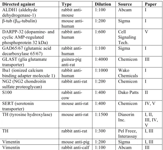
Different nerve fiber growth patterns depending on culture technique...35
Glial cells in rat fetal VM cultures...36
Effects of GDNF on mice fetal VM slice cultures, paper II...37
GDNF protein levels in culture medium and fetal brain...39
The effect of GDNF on astrocytes ...40
The importance of GDNF for midbrain dopaminergic neurons and nerve fiber formation ...40
Inhibition of astroglia in rat fetal VM cultures, paper III...41
Importance of fetal age upon grafting...42
Radial glia in VM cultures...43
Fetal VM and cortex co-cultures...43
Dopaminergic subpopulations in fetal VM cultures ...44
Grafting of fetal midbrain dopamine neurons ...44
Effects of L-DOPA in a rat model of dyskinesia, paper IV...45
AIMs ratings...45
In vivo chronoamperometry, electrochemical recordings...46
TH-positive nerve fiber density ...47
SERT-positive nerve fiber density ...48
The conversion of L-DOPA to dopamine in dopamine-depleted striatum, paper V...49
Different L-DOPA phenomena ...49
Effects of local striatal ejections of L-DOPA...49
Evaluation of the SERT-positive nerve fiber density...50
L-DOPA effects on SERT-positive nerve fibers...51
L-DOPA conversion to dopamine...51
CONCLUDING REMARKS ...53
Dopaminergic nerve fiber formation in organotypic slice cultures 53 Importance of GDNF for dopaminergic nerve fiber formation ...53
Importance of astrocytes for dopaminergic nerve fiber formation .54 Chronic and acute L-DOPA effects...54
ACKNOWLEDGEMENTS...57
ABBREVIATIONS
5-HT 5-hydroxytryptamine (serotonin) 6-OHDA 6-hydroxydopamine
AADC L- aromatic amino acid decarboxylase
AIM Abnormal involuntary movements ALDH1 Aldehyde dehydrogenase-1
Ara-C Cytosine β-D-arabinofuranoside
CNS Central nervous system CRL Crown-rump length
DAPI 4',6-diamidino-2-phenylindole DIV Days in vitro
DMEM Dulbecco’s modified Eagle´s medium
E Embryonic day
ELISA Enzyme-linked immunosorbet assay FCS Fetal calf serum
GABA Gamma-aminobutyric acid
GDNF Glial cell line-derived neurotrophic factor
GFRα1 Glycosylphosphatidylinositol-linked co-receptor 1 GLAST Glia glutamate transporter
GP Globus Pallidus
GPe Globus Pallidus pars externa GPi Globus Pallidus pars interna HCl Hydrochloric acid
i.p. Intraperitoneal
LGE Lateral ganglionic eminence
L-DOPA 3,4-dihydroxy-L-phenylalanine
NCAM Neural cell adhesion molecule PCR Polymerase chain reaction s.c. Subcutaneous
SERT Serotonin transporter SN Substantia nigra
SNc Substantia nigra pars compacta SNr Substantia nigra pars reticulata STN Subthalamic nucleus
TH Tyrosine Hydroxylase VM Ventral mesencephalon VTA Ventral tegmental area
ABSTRACT
Parkinson’s disease is a disorder mainly characterized by progressive degeneration of dopamine producing neurons in the substantia nigra of the midbrain. The most commonly used treatment strategy is to pharmacologically restore the lost function by the administration of the dopaminergic precursor L-DOPA. Another treatment strategy is to
replace the degenerated neurons with immature fetal ventral mesencephalic tissue, or ultimately stem cell-derived tissue. Grafting trials have, however, revealed poor reinnervation capacity of the grafts, leaving much of the striata dopamine-denervated. An additional drawback is the upcoming of dyskinesia (involuntary movements), a phenomenon also observed during L-DOPA treatment of Parkinson’s
disease patients. Attempts to characterize nerve fiber formation from dopamine neurons have demonstrated that the nerve fibers are formed in two morphologically diverse outgrowth patterns, one early outgrowth seen in the absence of astrocytes and one later appearing outgrowth seen in co-existence with astrocytes.
The overall objective of this thesis has been to study the dopaminergic outgrowth including guidance of nerve fiber formation, and to look into the mechanisms of L-DOPA-induced dyskinesia. The
first paper in this thesis characterizes the different outgrowth patterns described above and their relation to different glial cells. The study demonstrated the two different outgrowth patterns to be a general phenomenon, applying not only to dopamine neurons. Attempts of characterization revealed no difference of origin in terms of dopaminergic subpopulations, i.e. A9 or A10, between the outgrowth patterns. Furthermore, the “roller-drum” technique was found optimal for studying the dual outgrowth sequences.
The second and the third paper also utilized the “roller-drum” technique in order to promote both patterns of neuronal fiber formation. The effects of glial cell line-derived neurotrophic factor (GDNF) on the formation of dopamine nerve fibers, was investigated. Cultures prepared from gdnf knockout mice revealed that dopaminergic neurons survive and form nerve fiber outgrowth in the absence of GDNF. The dopaminergic nerve fibers exhibited an outgrowth pattern consistent with that previous observed in rat. GDNF was found to exert effect on the glial-associated outgrowth whereas the non-glial-associated was not affected. Astrocytic proliferation was
inhibited using cytosine β-D-arabinofuranoside, resulting in reduced
glial-associated outgrowth. The non-glial-associated dopaminergic outgrowth was on the other hand promoted, and was retained over longer time in culture. Furthermore, the non-glial-associated nerve fibers were found to target the fetal frontal cortex. Different developmental stages were shown to promote and affect the outgrowths differently. Taken together, these data indicate and state the importance of astrocytes and growth factors for neuronal nerve fiber formation and guidance. It also stresses the importance of fetal donor age at the time for transplantation.
The fourth and fifth studies focus on L-DOPA dynamics and
utilize in vivo chronoamperometry. In study four, 6-OHDA dopamine-depleted rats were exposed to chronic L-DOPA treatment and then
rated as dyskinetic or non-dyskinetic. The electrochemical recordings demonstrated reduced KCl-evoked release in the intact striatum after chronic L-DOPA treatment. Time for maximal dopamine
concentration after L-DOPA administration was found to be shorter in
dyskinetic animals than in non-dyskinetic animals. The serotonergic nerve fiber content in the striatum was evaluated and brains from dyskinetic animals were found to exhibit significantly higher nerve fiber density compared to non-dyskinetic animals. Furthermore, the mechanisms behind the conversion of L-DOPA to dopamine in
6-OHDA dopamine-depleted rats were studied. Local administration of
L-DOPA in the striatum increased the KCl-evoked dopamine release
in the intact striatum. Acute application of L-DOPA resulted
sometimes in a rapid conversion to dopamine, probably without vesicle packaging. This type of direct conversion is presumably occurring in non-neuronal tissue. Furthermore, KCl-evoked dopamine releases were present upon local application of L-DOPA in the
dopamine-depleted striatum, suggesting that the conversion to dopamine took place elsewhere, than in dopaminergic nerve fibers. In conclusion, these studies state the importance of astrocytes for neuronal nerve fiber formation and elucidate the complexity of L
ORIGINAL PAPERS
This thesis is based on the following papers, which will be referred to by their Roman numerals:
I. Berglöf E*, af Bjerkén S*, Strömberg I. Glial influence on
nerve fiber formation from rat ventral mesencephalic organotypic tissue cultures. J. Comp. Neurol. 501:431-442, 2007 *equal contribution
II. af Bjerkén S, Boger HA, Nelson M, Hoffer BJ, Granholm AC, Strömberg I. Effects of glial cell line-derived neurotrophic factor deletion on ventral mesencephalic organotypic tissue cultures. Brain Res. 1133:10-19, 2007 III. af Bjerkén S, Marschinke F, Strömberg I. Inhibition of
astrocytes promotes long-distance growing nerve fibers in ventral mesencephalic cultures. Submitted manuscript IV. Lundblad M, af Bjerkén S, Cenci MA, Pomerleau F,
Gerhardt GA, Strömberg I. Chronic intermittent L -DOPA treatment induces changes in dopamine release and 5-HT nerve fiber density. Manuscript
V. af Bjerkén S, Nevalainen N, Lundblad M, Pomerleau F, Gerhardt GA, Strömberg I. L-DOPA conversion to dopamine in the rat dopamine-depleted striatum. Manuscript
INTRODUCTION
General history and background
Parkinson’s disease is a progressive disorder of the nervous system primarily affecting the motor system of the body. The condition was described as early as 5000 B.C. by Indians practicing the medical system of Ayurveda (Vedas). They referred to the disorder as Kampavata, kampa meaning tremor and vata meaning lack of muscular movement. The Greek physician Galen also described symptoms of the disorder in year 175 A.D, referring to the disease as a “Shaking palsy” (Bendick, 2002). The formal documentation of the disease was, however, first accomplished in 1817 by James Parkinson, a physician in London. In “An Essay on the Shaking Palsy”, Parkinson describes the medical history of six persons displaying symptoms of the disease (Parkinson, 2002).
The neuropathology underlying Parkinson’s disease was first revealed in 1919 when Trétiakoff found a correlation between the symptoms of the disease and degeneration of dopamine producing neurons in the substantia nigra in the midbrain (Trétiakoff, 1919). Substantia nigra is Latin meaning the black substance, a name referring to the neuromelanin inclusions that are accumulated with age (Mann and Yates, 1983b). Neuromelanin is a non-organic substance thought to develop as a side/waste product of the catecholamine metabolism. In Parkinson’s disease, the inclusions disappear along with the dopaminergic neurons as the disease progresses. A pathologic hallmark of Parkinson’s disease is the presence of cytoplasmic eosinophilic inclusions termed Lewy bodies in the surviving dopaminergic neurons (Lewy, 1912). The primary component of these inclusions has been found to be the fibrillar form of α-synuclein (reviewed in (Eriksen et al., 2003)).
The neurotransmitter dopamine
Dopamine is an endogenous catecholamine synthesized from the amino acid tyrosine. Tyrosine is converted to dihydroxyphenylalanin (DOPA) by the rate-limiting enzyme tyrosine hydroxylase (TH).
DOPA is then further synthesized to dopamine with the help of the enzyme L- aromatic amino acid decarboxylase (AADC).
Dopamine was first considered as a precursor to adrenaline and noradrenaline, acting as an intermediate in the synthesis. Discoveries in the 1950’s did, however, come to change this view when Arvid Carlsson and colleagues demonstrated dopamine to be present in the brain in about similar concentrations as noradrenaline, but with a different distribution (Carlsson et al., 1958). Carlsson also showed that the drug reserpine caused a decrease in dopamine levels resulting in loss of motor control; a loss that could be counteracted by the dopamine precursor 3,4-dihydroxy-L-phenylalanine (L-DOPA)
(Carlsson et al., 1957). Reserpine was found to act on the dopamine terminals in the striatum, causing a decrease in dopamine levels by emptying of the terminals (Carlsson et al., 1958). Ehringer and Hornykiewics supported these results, demonstrating reduced dopamine levels in the striatum of patients with Parkinson’s disease (Ehringer and Hornykiewicz, 1960). In 2000, Arvid Carlsson was awarded the Nobel Prize for his discoveries, along with his co-laureates Erik Kandell and Paul Greengard.
The regional distribution of dopamine was described in 1964 when Dahlström and Fuxe mapped the dopamine cell groups in the brain (Dahlström and Fuxe, 1964), using the Falck-Hillarp immunohistochemical method (Falck et al., 1962). The dopaminergic nuclei in the ventral mesencephalon (VM) were named A8 (retrorubal area), A9 (substantia nigra; SN), and A10 (ventral tegmental area; VTA).
Clinical features and pathology
The cardinal symptoms of the disease are motor symptoms i.e. muscle rigidity, tremor, hypokinesia and bradykinesia (diminished and slow movements), and postural instability. However, many patients also develop non-motor symptoms as disturbances in mood, cognition, and sensation (reviewed in (Fahn, 2003)). Most of these symptoms are not responsive to dopaminergic replacement therapy; implicating that neurotransmitters other than dopamine are involved in the pathogenesis. In fact, neurodegeneration in Parkinson’s disease have been demonstrated in the noradrenergic locus coeruleus (Braak et al., 2003; Mann and Yates, 1983a; Zarow et al., 2003), the serotonergic raphe nuclei (Jellinger and Paulus, 1992), the cholinergic neurons in
the nucleus basalis (Nakano and Hirano, 1984), as well as a number of other nuclei.
Nowadays, the term parkinsonism is commonly used to describe conditions with any combination of at least two of the following features: tremors at rest, bradykinesia, rigidity, loss of postural reflexes, flexed posture, and freezing. There are four categories of parkinsonism - primary parkinsonism, secondary parkinsonism, parkinson-plus syndromes, and heredodegenerative disorders. Primary parkinsonism, or Parkinson’s disease, has an idiopathic or genetic etiology, whereas secondary parkinsonism has an environmental etiology. Environmental meaning insults to the brain such as toxins, drugs, trauma, and tumors etc. The third category, the Parkinson-plus syndromes, is characterized by a more extended neurodegeneration as well as poor response to L-DOPA therapy. Heredodegenerative
disorders are inherited conditions characterized by neuronal atrophy and dysfunction of neuronal systems. There are three findings that are helpful in distinguishing Parkinson’s disease from the other pakinsonisms, in order to set the correct diagnosis, i.e. asymmetrical onset of symptoms, symptomatic response to L-DOPA, and presence
of resting tremor (Fahn, 2003).
Etiology
The etiology of primary parkinsonism, i.e. Parkinson’s disease, is still unknown. It is has now become clear, however, that there are many different causes of Parkinson’s disease, and a number of factors implicated in the pathogenesis. These include oxidative stress, inflammation, mitochondrial dysfunction, excitotoxicity, apoptosis, and proteolytic stress (reviewed in (Olanow, 2007)). An increasing number of genes possibly involved in the pathogenesis have also been demonstrated e.g. α-synuclein, DJ1, Parkin, PTEN-induced kinase 1 (PINK1), ubiquitin carboxyl-terminal esterase L1 (UCHL1), and leucine-rich repeat kinase 2 (LRRK2) (reviewed in (Farrer, 2006)). One of the most recent factors taken under consideration is proteolytic stress. Proteolytic stress arises as a consequence of production of misfolded proteins. Considering the formation of Lewy bodies in Parkinson’s disease, it is not unlikely that it is involved in the pathogenesis.
The basal ganglia system
The basal ganglia are a subcortical group of interconnected nuclei consisting of input and output stations. There are four major nuclei of the basal ganglia, the striatum, the subthalamic nucleus (STN), the globus pallidus pars interna (GPi)/pars externa (GPe), and the SN. The substantia nigra is further divided into three subdivisions, the pars compacta (SNc), the pars reticulata (SNr), and the pars lateralis. The striatum and the STN are the major input nuclei, e.g. receiving excitatory glutamatergic input from the cortex and thalamus, dopaminergic input from the SNc, and serotonergic input from the dorsal raphe nucleus. The output nuclei of the basal ganglia include the GPi and the SNr. The striatum is mostly composed of gamma-aminobutyric acid (GABA) medium spiny neurons, but contains also GABAergic and cholinergic interneurons. In addition, the striatum can anatomically and functionally be divided into matrix and striosomes (Graybiel and Ragsdale Jr, 1978). There are two pathways connecting the striatum to the output nuclei, the direct and the indirect (striatopallidal) pathways. The direct pathway projects directly to the GPi and the SNr, whereas the indirect pathway projects to GPi through GPe and the STN. The present basal ganglia model is a development of the Albin-DeLong model (Albin et al., 1989; Alexander et al., 1986; Crossman, 1989; DeLong, 1990), a model stating excitatory input to the striatum from cortex and thalamus, and consequently activation of the striatal projecting neurons. The basic theory of the Albin-DeLong model is still today considered accurate, anatomical investigations have, however, revealed a more complex organization than originally proposed (Figure 1).
The nigrostriatal pathway
The dopaminergic neurons in the SNc project their axons to the striatum creating the nigrostriatal pathway (mesostriatal), part of the basal ganglia. The action of dopamine on striatal neurons depends on the type of dopamine receptors involved. There are two different groups of dopamine receptors, D1-like and D2-like receptors. D1 receptors stimulate adenylate cyclase activity and D2 receptors inhibit adenylate cyclase activity. The receptors are thought to be localized to different striatal output pathways, the direct and the indirect pathway (Gerfen and Wilson, 1996). This is however not fully confirmed since there are implications that D1 and D2 may be co-localized (Lester et
al., 1993; Surmeier et al., 1992), and many striatal neurons have been demonstrated to project simultaneously to the GPe, GPi, and SNr (Parent et al., 2000). The theory is, however, that the normal effects of the dopamine input to the striatum are excitation of the GABAergic medium spiny neurons that project directly to the GPi (direct pathway) and inhibition of the GABAergic medium spiny neurons that project to the GPe (indirect pathway). The direct and indirect pathways together balance the motor control of the basal ganglia. Thus, loss of dopamine in the SNc results in an increase in activity through the indirect pathway and a reduction in activity through the direct pathway, resulting in reduced activity in the output of the basal ganglia. The thalamic activation of the motor cortex is therefore inhibited, for illustration see Figure 1. The reduction in dopaminergic neurons in the SNc, thus, gives the typical manifestations of Parkinson’s disease, e.g. hypokinesia, tremor at rest, and rigidity. Other midbrain dopamine systems are the mesolimbic and the mesocortical which both arise from dopaminergic neurons in VTA. Both systems are involved in emotion-based behavior, such as motivation and award.
Development of the nigrostriatal system
In rat, the cells of the VM are born between embryonic days (E) E11 and E16 and, historically, peak ontogeny have been considered to occur around E13 (Altman and Bayer, 1981; Hanaway et al., 1971; Lauder and Bloom, 1974; Marchand and Poirer, 1983). A recent study suggests, however, the peak ontogeny to occur at E12 (Gates et al., 2006). The midbrain dopamine cells originate at the mesencephalic-diencephalic junction and then migrate to their final location in the VM around E18 (Hanaway et al., 1971; Marchand and Poirer, 1983; Olson and Seiger, 1972; Specht et al., 1981a; Tennyson et al., 1972; Voorn et al., 1988). In rat, immunoreactivity towards TH can be detected at E12.5 (Foster et al., 1988; Specht et al., 1981b) and approximately one day after, at E13-13.5, dopamine can be visualized (Olson and Seiger, 1972; Voorn et al., 1988). Dopaminergic fibers have been demonstrated to reach the striatal anlage at E13.5 (Olson et al., 1972; Specht et al., 1981b). At P14 the ventral mesencephalon has matured, exhibiting adult features (Kalsbeek et al., 1992).
The symptoms of the disease are first seen when the dopamine concentration in the striatum is reduced with 80%, and approximately 60% of the nigral dopamine neurons are lost (Fearnley and Lees,
1991; Marsden, 1990). The loss of dopamine in Parkinson’s disease patients is more prominent in the putamen compared to the caudate (Kish et al., 1988; Nyberg et al., 1983), which is a consequence of the more severe loss of dopamine neurons in the ventral tier of the SN (Fearnley and Lees, 1991). Thus, a reduction of the neurons that mainly projects to the putamen (Gibb and Lees, 1991).
Figure 1. Simplified schemes of the basal ganglia model. The left scheme illustrates the conditions in healthy individuals and the right scheme illustrates the conditions in Parkinson’s disease.
Treatment strategies
The discovery of the importance of dopamine in the pathogenesis of Parkinson’s disease, along with the effects of L
-DOPA seen in reserpine-induced hypokinesia (Carlsson et al., 1957), made researchers focus on L-DOPA as a possible treatment for
Parkinson’s disease. Unlike dopamine, L-DOPA can pass the
blood-brain-barrier further supporting its potential for dopamine replacement therapy. Clinical trials with L-DOPA were undertaken, yet with
relatively poor results due to low dosage and side effects such as cardiac arrhythmia and nausea (Barbeau, 1961; Birkmayer and Hornykiewicz, 1961). However, higher dosage along with a stepwise introduction of the drug improved the outcome and it became a useful treatment (Cotzias et al., 1967). This strategy along with peripheral decarboxylase inhibitors further increased the efficacy of the drug, yielding more L-DOPA in the brain (Bartholini and Pletscher, 1968;
Dunner et al., 1971; Tissot et al., 1969). L-DOPA therapy in
combination with peripheral decarboxylase inhibitors (benzerazid, carbidopa) is still today the most commonly used treatment for Parkinson’s disease. Interestingly, the ancient Indians referring to the condition as Kampavata (Vedas) treated the disorder with a tropical legume called Mucuna Pruriens, which seeds have been found to contain high concentrations of L-DOPA (Hussain and Manyam, 1997).
Although L-DOPA is effective to treat the symptoms of Parkinson’s
disease, there are some limitations. After long-term treatment, many patients develop motor fluctuations, such as dyskinesia (involuntary abnormal movements) and decreased drug response (known as “wearing-off”) (Granérus, 1978). The loss of drug response can sometimes be counteracted with higher doses of L-DOPA. High doses
may, however, result in increased dyskinesia.
Other drugs used to treat the symptoms are dopamine agonists, which act directly on postsynaptic dopamine receptors. The development of dyskinesia and upcoming of the wearing-off effect is less prominent using these drugs. However, side effects as confusion and psychosis are more prone to occur compared to L-DOPA therapy
(Rascol et al., 2000). Metabolic inhibitors, such as catechol-O-methyl transferase and monoamine oxidases-B, can also be used to treat the symptoms of Parkinson’s disease. The use of a metabolic inhibitor together with L-DOPA can extend its half-life, leaving higher levels in
-DOPA, preventing reuptake to the presynaptic terminal, leaving more
L-DOPA in the synaptic cleft.
In addition to drug therapy, surgical procedures can be used for treatment of symptoms in Parkinson’s disease. Deep brain stimulation is a method were electrical stimulation is utilized to adjust the activity in the basal ganglia. Targets for electrode implantation are, most commonly, either the thalamus, the GP or the STN. Deep brain stimulation was preceded by pallidotomy, a technique, where the GP is destroyed by heat (reviewed in (Narabayashi, 1990)).
A different approach to reduce the symptoms in Parkinson’s disease is to replace the lost dopaminergic neurons through transplantation of dopamine producing tissue. Grafting trials with fetal dopaminergic neurons to the striatum of dopamine-depleted rats were performed in the late 1970’s (Björklund and Stenevi, 1979; Perlow et al., 1979). The studies were a success, in that they showed a compensatory effect on the motor deficits caused by the dopamine-depletion. Further studies showed long-term survival of the grafts (Freed et al., 1980; Strömberg and Bickford, 1996) as well as an organotypic innervation pattern of the striatum (Björklund et al., 1981; Strömberg et al., 1992). The grafts were functional in that new synaptic connections were formed (Bolam et al., 1987; Clarke et al., 1988; Mahalik et al., 1985; Strömberg et al., 1988), the striatal firing rate was normalized (Fisher et al., 1991; Strömberg et al., 1985; van Horne et al., 1990), and dopamine could be released from the reinnervated striatum (Rose et al., 1985; Strömberg et al., 1988; Zetterström et al., 1986). The first grafting trials in humans were performed using catecholamine-producing cells from the adrenal medulla. Patients received grafts to the caudate (Backlund et al., 1985) or to the putamen (Lindvall et al., 1987), but with disappointing results. The first clinical attempts using human fetal ventral mesencephalon for grafting to parkinsonian patients was published in 1988 (Lindvall et al., 1988; Madrazo et al., 1988). Moreover, postmortem studies of these patients revealed graft survival and reinnervation of the putamen (Kordower et al., 1998). It is now known that transplanted fetal dopamine neurons can survive for over ten years after grafting (Piccini et al., 1999). To date, around 350 patients have received intrastriatal implants of fetal mesencephalic tissue, and a number of open-label trials have reported symptomatic improvement e.g. (Cochen et al., 2003; Hagell et al., 1999; Wenning et al., 1997). The first double-blind study reported less symptomatic improvement
compared to the open-label trials, no improvement was observed in the sham-operated patients (Freed et al., 2001). This study did, however, provide evidence that the improvement is not solely placebo based. A second double-blind trial also demonstrated poor clinical outcome, maybe as a consequence of more severely affected patients taking part in the study (Olanow et al., 2003). Histopathological examination has shown graft survival and reinnervation of the host brain (Kordower et al., 1995). The grafts can also provide functional effects, in that they release dopamine, and become integrated in the neural circuits (Piccini et al., 1999; Piccini et al., 2000). However, the main problem with grafting of fetal dopaminergic neurons is the incapacity of the grafts to sufficiently reinnervate the areas of dopamine depletion. A rat study has shown that the dopaminergic neurons halt in reinnervation shortly after implantation (Barker et al., 1996). Several implant sites are therefore necessary in order to cover the dopamine-depleted striatum. What induces the halt in reinnervation is not known but lack of attractants, such as neurotrophic factors and extracellular matrix molecules, may be a possible cause. Another shortcoming is the development of graft-induced dyskinesias (Freed et al., 2001; Hagell et al., 2002). Furthermore, a major drawback is the poor survival of grafted dopamine neurons, along with ethical concerns using fetal tissue. Therefore, the focus for tissue replacement is today, to a large extent, on the development of stem cells to be used in grafting (for review see e.g. (Correia et al., 2005; Parish and Arenas, 2007)).
Nerve fiber formation
As already mentioned, a way to enhance graft outcome is to improve reinnervation of the host brain. To study factors that influence nerve fiber formation, organotypic tissue cultures can be utilized. Dopaminergic nerve fiber formation has been suggested to occur in two temporally separate waves in organotypic slice cultures of fetal VM, one early and one later occurring wave of nerve fiber outgrowth. The early formed nerve fibers are seen without the presence of glia whereas the later is detected over glial cells (Johansson and Strömberg, 2002). Furthermore, fetal lateral ganglionic eminence (LGE) seems to attract the late occurring wave of dopaminergic nerve fiber outgrowth (Johansson and Strömberg, 2003). The mesencephalic tissue used for transplants and primary cell
cultures contain a mixture of A9 and A10 dopaminergic neurons, as well as A8 dopaminergic neurons (Dahlström and Fuxe, 1964). This might suggest different origins of the observed dopaminergic nerve fiber outgrowths, i.e. that they derive from different dopamine subpopulations.
Neurotrophic factors
Neurotrophic factors are a family of proteins found to support and nourish neurons and nerve fibers. They are responsible for growth and survival during development, but also of importance in the mature brain and nerve system. The first neurotrophic factor found and described was simply named nerve growth factor (Levi-Montalcini and Hamburger, 1953). It was found critical for survival and maintenance of sympathetic and sensory ganglia (Cohen, 1959; Cohen, 1960; Levi-Montalcini and Hamburger, 1953). In 1986, Stanley Cohen and Rita Levi-Montalcini were rewarded the Nobel Prize for their discovery. In the central nervous system (CNS), target neurons synthesize neurotrophic factors acting retrogradely on their afferent neurons (DiStefano et al., 1992; Mufson et al., 1999). Trophic factors can, however, be recruited from other sources as well, e.g. presynaptic neurons (Bartheld, 2004; Conner et al., 1998), and axon-ensheathing glial cells (Creange et al., 1997; Kinameri and Matsuoka, 2003). There are a number of neurotrophic growth factors that may be considered as promoting for ventral mesencephalic dopamine neurons, e.g. glial cell line-derived neurotrophic factor (Lin et al., 1993), brain-derived neurotrophic factor (Hyman et al., 1991), neurturin (Horger et al., 1998), neurotrophin-4 (Hynes et al., 1994), and fibroblast growth factor-2 (Date et al., 1993; Giacobini et al., 1993; Mayer et al., 1993).
Glial cell line-derived neurotrophic factor
In 1993, a novel neurotrophic factor for dopamine neurons was purified. It was found as a secretion product from the rat B49 glial cell line and termed glial cell line-derived neurotrophic factor (GDNF). It was characterized and found to promote the survival of embryonic SN dopamine neurons in vitro (Lin et al., 1993). High levels of GDNF mRNA have been detected in developing but not in adult rat striatum (Schaar et al., 1993; Strömberg et al., 1993). GDNF is, however, secreted in low concentrations in the adult striatum and transported retrogradely to the dopaminergic cells in the mesencephalon (Tomac
et al., 1995). This suggests a trophic role for GDNF in the adult brain as well. A few years after the discovery, GDNF was demonstrated to promote survival of dopamine neurons also in vivo, both after intracranial administration in the intact rat brain (Hudson et al., 1995) and after administration to parkinsonian rats (Granholm et al., 1997; Strömberg et al., 1993). GDNF gene delivery to the striatum, using viral vectors, has shown to be neuroprotective and promote regeneration in the lesioned nigrostriatal system (Georgievska et al., 2002; Kirik et al., 2000). Moreover, trials with encapsulated GDNF-producing cells to parkinsonian rat striatum revealed protection of both the SN neurons and the striatal fiber innervation (Ahn et al., 2005; Shingo et al., 2002; Yasuhara et al., 2005). A study showed increased density of striatal dopaminergic nerve fibers without changes in SN cell numbers, implicating neuroregenerative properties (Sajadi et al., 2006). GDNF administration into patients with Parkinson’s disease has been performed with various outcomes. GDNF injected into the cerebral ventricles did not result in symptomatic improvement (Nutt et al., 2003). A possible explanation is that the GDNF did not diffuse into the brain parenchyma (Kordower et al., 1999), but spread via the cerebrospinal fluid. Therefore, in following studies, the GDNF administration was infused directly into the striatum. Motor improvements in patients chronically infused with GDNF to the putamen (Gill et al., 2003; Slevin et al., 2005), as well as sprouting of dopaminergic fibers (Love et al., 2005) have been reported. In contrast, another study failed to demonstrate motor improvements after GDNF infusion into the putamen (Lang et al., 2006). The study was stopped due to lack of efficacy as well as for safety reasons, since some patients developed antibodies towards GDNF. In addition, a parallel high-dose study in monkeys showed degeneration of cells in the cerebellum.
GDNF signaling is mainly considered to occur through the tyrosine kinase receptor Ret (Durbec et al., 1996) by forming a complex with the glycosylphosphatidylinositol-linked co-receptor 1 (GFRα1) (Buj-Bello et al., 1997). However, studies have also revealed a Ret independent signaling of GDNF (Förander et al., 2001; Poteryaev et al., 1999; Trupp et al., 1999), and that neural cell adhesion molecule (NCAM), together with GFRα1, functions as an alternative signaling receptor for GDNF (Paratcha et al., 2003).
The GDNF knockout mice display deficits in primary sensory, sympathetic, and motor neurons and also fail to develop kidneys,
urethers and most of the enteric nervous system (Moore et al., 1996; Pichel et al., 1996; Sánchez et al., 1996). The GDNF knockout mice die shortly after birth, as a consequence of these deficiencies, limiting research to heterozygous and fetal tissue.
6-OHDA model of Parkinson’s disease
The 6-hydroxydopamine (6-OHDA) model (Ungerstedt, 1968) is a common used animal model of Parkinson’s disease. 6-OHDA is a hydroxylated analogue of dopamine (Blum et al., 2001), isolated in 1959 (Senoh et al., 1959; Senoh and Witkop, 1959). It was first demonstrated to induce noradrenergic depletion in sympathetic nerves of mice (Porter et al., 1963; Porter et al., 1965), and later found to cause a catecholaminergic specific neurodegeneration in the CNS (Tranzer and Thoenen, 1968). These findings led to the development of an animal model for Parkinson’s disease (Ungerstedt, 1968; Ungerstedt and Arbuthnott, 1970). Since 6-OHDA does not cross the blood-brain-barrier, the toxin has to be injected directly into the brain. Preferred injection sites are the SN, the medial forebrain bundle, and the striatum (Perese et al., 1989; Przedborski et al., 1995; Sauer and Oertel, 1994). Depending on the site of injection, different model characteristics are achieved. Following injection into SN or medial forebrain bundle, a rapid degeneration occurs with depleted striatal dopamine levels within 2-3 days (Faull and Laverty, 1969; Hökfelt and Ungerstedt, 1969), whereas an intrastriatal injection causes a slow progressive degeneration (Berger et al., 1991; Przedborski et al., 1995; Sauer and Oertel, 1994). 6-OHDA is most commonly injected unilaterally, causing a hemiparkinsonian model (Perese et al., 1989). Parkinsonian symptoms as bradykinesia and postural abnormalities can be observed already 3 days after lesion. The severity of the degeneration can be assed with drug-induced rotations using amphetamine or apomorphine. Amphetamine acts as an indirect dopamine agonist by increasing the extracellular concentration of striatal dopamine on the intact side, which induces ipsilateral rotational behavior in dopamine-depleted animals (Schwarting and Huston, 1996; Ungerstedt and Arbuthnott, 1970; Ungerstedt, 1971). Apomorphine is a receptor agonist stimulating both D1- and D2-like receptors, with slightly higher affinity for D2-receptors. Systemically administered apomorphine induces rotations contralateral to the
lesion, due to 6-OHDA evoked D2-receptor hypersensitivity in the lesioned striata (Herrera-Marschitz and Ungerstedt, 1984).
Animal model of
L-DOPA induced dyskinesia
The rat model of L-DOPA-induced dyskinesia is a development
of the 6-OHDA model described above. Chronic administration of low doses of L-DOPA to 6-OHDA hemiparkinsonian rats induces
dyskinetic movements contralateral to the lesion (Cenci et al., 1998; Lee et al., 2000). The severity of the dyskinesia is, as in patients, increased over time of treatment, and measured as abnormal involuntary movements (AIMs) (Cenci et al., 1998; Lundblad et al., 2002; Steece-Collier et al., 2003).
AIMS OF THIS THESIS
• To characterize the neuronal and glial cell interactions in fetal ventral mesencephalic slice cultures
• To characterize the two diverse TH-positive nerve fiber formations found in fetal ventral mesencephalic slice cultures, in respect to:
- glial-association and relation
- effects of neurotrophic factors, i.e. GDNF - fetal tissue age at plating
• To investigate the effects of chronic and acute L-DOPA
administration in normal and dopamine-depleted striatum, and in an dyskinetic animal model
MATERIALS AND METHODS
Subjects, animals
Female Spraque-Dawley adult rats (B&K Stockholm, Sweden) or fetuses were used in paper I, III, IV, and V. In paper II GDNF knockout mice generated from the C57BL/6 strain were used (Granholm et al., 1997; Pichel et al., 1996). Heterozygous mice were mated overnight, and the day following mating (day of vaginal plug) was considered day 0.5 of pregnancy. All animals were housed under a 12 h dark/light cycle and were given access to water and pellets ad
libitum. All experiments were in accordance with internationally
excepted guidelines and approved by the local ethics committee.
Dissection of VM, frontal cortex, LGE, and raphe
dorsalis
VM, frontal cortex, LGE, and raphe dorsalis (pontine raphe) were dissected from rat or mice fetuses at different developmental stages. In paper I and III, gentle palpation of the abdomen of the pregnant rats were used to decide developmental stage of the fetuses (Olson et al., 1983). Pregnant rats or mice were deeply anesthetized using 4% isofluran (Baxter Medical) before neck dislocation and opening of the abdominal cavity. The uterine horns were isolated and placed in a sterile Petri dish.
All dissections were made using a dissection microscope and performed under sterile conditions. Dulbecco’s modified Eagle´s medium (DMEM; Gibco, Grand Island, USA) was used for rinsing and conditioning of the tissue. VM was dissected bilaterally by coronal cuts at the levels of the mesencephalic junction and the mesencephalic flexure. Sagittal cuts were made to disattach the ventral mesencephalic tissue piece (Dunnett and Björklund, 1992; Seiger, 1985), now resembling the wings of a butterfly (connected by the midline). The dissected VM piece comprises besides the A9 (SN) dopamine neurons also the A10 (VTA) as well as the A8 (retrorubal area) neurons (Dahlström and Fuxe, 1964). Fetal frontal cortex was dissected bilaterally and cut into smaller tissue pieces used for culturing. For the LGE dissection, a paramedial cut was made in the
cortex and the cortical tissue piece was folded laterally, exposing the ganglionic eminences. The most lateral part of the striatal anlage was dissected (Pakzaban et al., 1993; Wictorin et al., 1989) and used as one tissue piece for in oculo grafting. Fetal pontine raphe was dissected bilaterally between the rhombencephalic isthmus and the pontine flexure. Sagital cuts were made approx. 0.5 mm above the ventral midline on both sides. The dissected tissue was separated at the midline resulting in two pieces used for in oculo grafting.
Organotypic primary cultures
VM “roller-drum” cultures
Primary cell cultures were performed using the “roller-drum” technique (Gähwiler et al., 1997; Stoppini et al., 1991). This method ensures a constant flow of medium over the slice culture, minimizing the direct contact with cell debris and other waste products. Cultures were prepared from VM tissue pieces dissected from E12 rat crown-rump length (CRL) = 6-7 mm (paper III), E14 rat CRL = 12-13 mm (paper I and III), E18 rat CRL = 25-28 mm (paper III), or E14 mice CRL = 12 mm (paper II) fetuses. The butterfly shaped tissue pieces were chopped in 300 µm thick coronal sections using a tissue chopper and separated at the midline, each small tissue piece constituting one culture. The GDNF mouse VM tissue, in paper II, was divided into 6 comparable pieces using a scalpel (3 pieces on each side of the midline). All instruments used were sterilized prior to dissection. In paper II, sterilization of the instruments was made in a glass bead sterilizer after each dissected fetus, to prevent DNA contamination. The tissue pieces prepared for culture were plated on 10x24 mm glass coverslips, pretreated with poly-D-lysine (Sigma, Stockholm,
Sweden), in a mixture of chicken plasma (Sigma) and thrombin (Sigma). The mixture was left to dry for approximately 20 min before insertion into 15 ml Falcon tubes containing 0.9 ml culture medium. The culture tubes were placed in a “roller drum”, turning at speed of 0.5 rpm, in an incubator at 37 ºC and in 5% CO2. The cultures were
VM and fetal frontal cortex “roller-drum” co-cultures
In paper III, E14 rat VM tissue pieces were isolated and co-cultured with pieces of E14 rat frontal cortex. The cultures were prepared as described above and placed adjacent to each other on poly-D-lysine treated coverslips. The cultures were cultivated
according to the parameters applying for the single cultures.
VM insert cultures
In paper I, isolated VM tissue pieces were placed and cultivated on membrane insert cultures, pore size 0.4 µM (Millipore, Bedford, MA, USA). The inserts were positioned in wells containing 1.7 ml culture medium and kept at 37 ºC in 5% CO2. Fresh medium was
supplied every 3-4 days.
The organotypic cultures were incubated over different time windows. Paper I: 1, 2, 3, 5, 7, 14, 21, and 28 days in vitro (DIV). Paper II: 12 DIV. Paper III: 5, 7, and 14 DIV.
Culture medium
Culture medium was composed of (volume percent) 55% DMEM, 32.5% Hanks’ balanced salt solution (Gibco), 10% fetal calf serum (FCS; Gibco), 1.5% glucose, and 1% Hepes (Gibco). The medium was sterile filtered using a 0.22 µm pore size filter (Sterivex; Millipore), and kept in small batches thawed before usage. Additionally antibiotics were added, to a final volume of 1% (10,000 units/ml penicillin, 10,000 µg/ml streptomycin and, 25 µg/ml amphotericin) to the medium at plating, and were thereafter excluded at the first medium change. The medium is well suited for administration of different substances to the tissue. In paper II, recombinant human GDNF (10 ng/ml; Promega, Madison, WI, USA) was added to the medium and in paper III the antimitotic agent cytosine β-D-arabinofuranoside (Ara-C; Sigma) was added in
Intracranial (ventricular) grafting (GDNF)
Fetal VM tissue was dissected, as described above, from gdnf knockout (n=3), gdnf heterozygous (n=1), or wildtype (n=1) E14 fetuses and used for intracranial transplantation. Adult female wildtype mice were deeply anesthetized using 4% isofluran and mounted in a stereotaxic frame for mice. The scalp was cut open, burr holes were made bilaterally over the lateral ventricles (stereotaxic coordinates in mm: AP=±0, ML=±0.8), and the dura mater was pierced using a needle. The dissected VM tissue pieces were divided in the midline rendering two pieces for grafting from each fetus. The tissue was pulled up using a cannula assembled to the stereotaxic frame and lowered into the ventricle (3.5 mm below dural surface) and implanted. The cannula was left in the brain for 1 min before retraction, in order to assure disattachment of the tissue. The scalp was then closed using steel agraffes. The animals were sacrificed 6 weeks postgrafting by intracardial perfusion. All surgical procedures were made under an operating microscope.
Enzyme-linked immunosorbent assay
In paper II, enzyme-linked immunosorbent assay (ELISA) was utilized to determine GDNF protein levels in brain tissue from E14
gdnf knockout, gdnf heterozygous, or wildtype fetuses as well as in
culture medium collected after 3-4 days of incubation (at medium change). The tissue was homogenized with lysis buffer and protease inhibitor, and the GDNF protein levels were determined using a GDNF assay kit (R&D Systems, Abingdon, U.K.). 96-well flat bottom plates were coated with the corresponding capture antibody over night. Duplicate samples of either brains or media were added to the wells and incubated for 2 h at room temperature. Incubation in detection antibody was performed for 2 h followed by incubation with a Streptavidin-HRP antibody. Wash steps removed all unbound conjugates. The plates were incubated with the chromagenic substrate Turbo TMP and an ELISA plate reader was used to monitor the color change. A standard curve using known amounts of GDNF protein was generated to determine sample concentrations.
Genotyping
Polymerase chain reaction (PCR) was performed in paper II and for the GDNF intraventricular grafts in order to genotype the fetal tissue for the presence or absence of the mutant allele.
DNA isolation
Genomic DNA was isolated from fetal tissue not used in culture. The tissue was lysed in 0.5 ml lysis buffer (100 mM Tris pH 8.5, 5mM EDTA pH 8.0, 0.2% SDS, 200 mM NaCl and, 200 mg/ml Proteinase K; Promega) at 56 ºC in a thermomixer (Thermomixer Compact, Eppendorf, Horsholm, Denmark) over night. After lysis, 1 µl/ml Ribonuclease A (RNaseA; Sigma) was added to the homogenates and incubated at 37 ºC for 30 min. Thereafter, 200 µl phenol/chloroform/isoamylalcohol 25:24:1 (Sigma) was added to the samples and the suspensions were centrifuged (14,000 rpm, 5 min). The supernatants were separated from the pellets and mixed with 100 µl chloroform before centrifugation (14,000 rpm, 5min). The supernatants were once more collected and DNA precipitation was performed using 1 ml of cold isopropanol. The DNA was stabilized for 30 min at -20 ºC followed by centrifugation at 4 ºC (14,000 rpm, 5 min). The DNA pellets were washed in 200 µl cold ethanol (70%) and centrifuged (14,000 rpm, 5 min). The purified DNA pellets were dissolved in 200 µl 1 x TE buffer (Promega) at 37 ºC for 2 h.
Polymerase chain reaction
The PCR was performed in a thermal cycler. The DNA was assayed for the presence of the GDNF wildtype or knockout allele in two separate PCR reactions, using primers specific to either the wildtype or the knockout allele. The amplification reaction mixtures, total volume of 20 µl, contained 1.5 µl genomic DNA (or dH2O for
negative controls), 30 µM sense primer: wildtype – 5’-CCA GAG AAT TCC AGA GGG AAA GGT C-3’ or knockout – 5’-CGG ACG CGG TTG CGC CTA CCG G-3’ (TAG Copenhagen, Copenhagen, Denmark), 30 µM antisense primer, wildtype – 5’-CAG ATA CAT CCA CAC CGT TTA GCG G-3’ or knockout – 5’-ACG ACT CGG ACC GCC ATC GGT G-3’ (TAG Copenhagen), 250 µM dNTP (Promega), 2 µl 10 x PCR buffer (Promega), 2.5 mM MgCl2, and 2
units of Taq polymerase (Promega). The DNA was amplified for a total of 40 cycles. Each cycle consisted of 1 min denaturation at 92 ºC,
1 min annealing at 56 ºC, and 2 min elongation at 72 ºC. The cycles were preceded by 4 min initial denaturation at 72 ºC.
PCR product analysis
The PCR products were analyzed on a 2% agarose gel (Agarose, LMP preparative grade for small fragments, Promega) using gel electrophoresis. The DNA bands were stained with Ethidium bromide and visualized under UV-light. A 100 bp DNA ladder was used as reference. Amplification of the wildtype allele gave a band of 344 bp, whereas the mutant allele gave a band of 255 bp (Granholm et al., 2000; Pichel et al., 1996).
Dopamine lesions
In paper IV and V, dopamine denervations were performed by unilateral injections of 6-OHDA into the medial forebrain bundle. Rats were anesthetized with 4% isofluran, mounted in a stereotaxic frame, the scalp cut opened and a burr hole was made over the injection site/s at the right hemisphere. 6-OHDA-HCl (Sigma) was dissolved in 0.02% ascorbic acid in 0.9% NaCl and injected into the brain using 10 µl Hamilton syringe, mounted to the stereotaxic instrument. The solution was injected at rate of 1 µl/min and the syringe was left in the brain for 3 min before retraction. After surgery, the scalp was sutured using steel agraffes. All surgical procedures were performed under an operating microscope.
In paper IV, rats weighting approx. 225 g were given two injections of a total of 13.5 µg 6-OHDA-HCl. The injections were performed at the following stereotaxic coordinates in relation to the bregma and the dural surface (in mm): AP = 4.4, ML = 1.2, DV = -7.8, tooth bar = -2.3 (7.5 µg) and AP=-4.0, ML = 0.8, V = -8.0, tooth bar = +3.4 (6 µg). In paper V, young female rats (160 g) were given one injection of a total of 8 µg 6-OHDA-HCl into the medial forebrain bundle at the following coordinates: AP = -4.4, ML = 1.2, V = -7.8, tooth bar = -2.3.
Behavioral testing
Rotational behavior
The efficacy of the 6-OHDA lesions in paper IV and V were assed by drug-induced rotations (Herrera-Marschitz and Ungerstedt, 1984; Ungerstedt and Arbuthnott, 1970).
The first rotational assessment was done approximately 2 weeks after lesion and then repeated on a regular basis. The rats were placed in plastic bowls and strapped into small leashes attached to the recording instrument. Their rotational behavior was then recorded, after drug onset, using a computer program.
In paper IV, i.p. injections of D-amphetamine (Sigma), 2.5
mg/kg in 0.9% NaCl, were used to asses rotations ipsilateral to the lesion. Animals exhibiting > 5 turns per min were selected for further experiments (Winkler et al., 2002). In paper V, rotational behavior was induced by s.c. injections of apomorphine (Apoteksbolaget, Stockholm, Sweden), 0.05 mg/kg in 0.9% NaCl. Animals rotating more than 450 turns contralateral to the lesion, over a time period of 70 min, were selected for further experiments (Heikkila et al., 1981; Herrera-Marschitz and Ungerstedt, 1984; Hudson et al., 1993; Ungerstedt and Arbuthnott, 1970).
Ratings of Abnormal Involuntary Movements (AIMs)
In paper IV, 6-OHDA depleted rats were chronically treated with L-DOPA in order to induce dyskinesia. L-DOPA (Sigma, PURE;
4 mg/kg) was administered i.p. in combination with the peripheral DOPA decarboxylase inhibitor benzerazide-HCl (Sigma; 15 mg/kg) for 14 days. AIMs ratings were done in accordance with the rating scale previously used (Lee et al., 2000; Lundblad et al., 2002) as well as with an amplitude rating scale (Carta et al., 2006). The ratings were performed on days 1, 4, 7, 10 and 14. Ratings were made every 20th min, starting 20 min post L-DOPA injection. The last observation was
done 180 min after L-DOPA injection. The animals were ranked either
as dyskinetic (global AIM score > 50) or non-dyskinetic. After chronic L-DOPA treatment, 2-3 L-DOPA injections/week were given
in order to maintain the dyskinetic behavior. Chronoamperometric measurements were made two days after the last L-DOPA injection in
Throughout this thesis, the pure L-DOPA form is being
consequently used. The pure form in contrast to the more commonly used L-DOPA methyl ester not electrochemically active. Hence, it is
better suited for the electrochemical recordings in paper IV and V.
In vivo chronoamperometry
In paper IV and V, in vivo electrochemical recordings were performed in rat brains using high-speed chronoamperometric measurement (5 Hz) (Kissinger et al., 1973; Stamford, 1990). The recordings were performed using the FAST-12 system (Quanteon, L.L.C Nicholasville, KY, USA) (Hoffman et al., 1998). Briefly, a carbonfiber electrode is held at a resting potential of 0.0 V vs. an Ag/AgCl reference electrode. The voltage is then instantly stepped to an oxidation voltage of +0.55 V. These square wave pulses are repeated at a rate of 5 Hz, resulting in extracellular oxidation and reduction of oxidizable substances, e.g. dopamine. The chemical reactions induce a rapid change in current recorded by the electrode. The current recorded is directly proportional to analyte concentration (Scatton et al., 1988). Rats were deeply anaesthetized with urethane (1.25-1.5 g/kg bodyweight) i.p. and placed on a heating pad to maintain normal body temperature. Tracheotomy was performed and the animals were allowed to breathe spontaneously. The skull was fixed in a stereotaxic frame, the scalp opened, and the skull and dura mater covering the striatum was bilaterally removed. A single burr hole for the reference electrode was maid on the left hemisphere, approximately 5 mm caudally from the bregma. Carbonfiber electrodes (Quanteon) coated with Nafion (Sigma) were used for the electrochemical recordings. Nafion is a teflon derivate that repells anions, such as ascorbic acid, and enhances the selectivity for cations, such as dopamine (Gerhardt et al., 1984; Nagy et al., 1985). The carbonfiber electrodes were dried at 200 °C for 3 min, swirled in Nafion followed by oven baking at 200 °C for 3 min. The coating procedure was repeated 2-3 times. Electrode calibration was performed to determine sensitivity and linearity. The calibration was assessed in 0.1 M phosphate buffered saline (PBS; pH 7.4) in a beaker using standardized solutions of ascorbic acid (20 mM) and dopamine (2 mM). Electrodes exhibiting high selectivity to dopamine, compared to ascorbic acid, and good linearity were selected for the experiments. The carbonfiber electrodes were assembled with one or two glass
micropipettes using sticky wax. The tip distance between electrode and micropipette was 130-160 µm. The micropipettes were made from glass capillaries (World Precision Instruments, Sarasota, FL, USA) using a pipette puller (PN-30, Narishige, Japan). One micropipette was filled with KCl (120 mM) to evoke potassium-induced dopamine releases, and the other, when used, was filled with L-DOPA (2-10
mM). An Ag/AgCl electrode was prepared in a reference-plating bath, 1 M HCl saturated with NaCl. A silver wire was coated with Cl- by hooking up the silver wire (future reference electrode) to the anode of a 12 V battery and the cathode to another silver or stainless steel wire. Both wires were then placed in the plating bath for 15-30 min. The Ag/AgCl reference was implanted in the brain, and the electrode/micropipette assembly was then lowered, in the brain, using a microdrive. The micropipettes were connected to a micropressure system allowing pressure ejection of KCl or L-DOPA. Ejections and
recordings were performed at different stereotaxic coordinates in the striatum.
Intraocular transplantation
In paper V, the in oculo grafting model (Olson et al., 1983) was used to study serotonergic nerve fiber innervation of LGE co-grafts. This is a technique that allows evaluation of the grafts nerve fiber growth, through the transparent cornea. It also provides an isolated in
vivo milieu, close to the normal authentic, were the graft/s are able to
expand without other tissue interference. Furthermore, the blood-brain-barrier of the grafted tissue has been demonstrated to close again approximately 2 weeks after grafting (Granholm et al., 1996).
Grafting of LGE and pontine raphe
LGE and pontine raphe from E14 rat fetuses were implanted under the cornea at different time points. The LGE was implanted and matured for 6 weeks prior to implantation of the pontine raphe. The tissue grafts were placed adjacent to each other on the iris, allowing potential fiber innervation. Briefly, atropine was dropped in the eyes of the graft receiving rats for pupil dilation prior to surgery. The rats were anesthetized with gas anesthesia (4% isofluran) and placed under an operating microscope. A cut was maid in the cornea using a sharp razor and the graft tissue was implanted with a glass pipette.
Chronic L-DOPA treatment
The host rats for the in oculo co-grafts were chronically treated with L-DOPA for 21 days, starting at 14 days after pontine raphe
implantation. L-DOPA (4 mg/kg) and benserazide-HCl (15 mg/kg)
were together dissolved in 0.9% NaCl and injected i.p. once a day.
Tissue preparation and brain sectioning
In paper I, II, and III, organotypic slice cultures were fixed for 1 h in 2% paraformaldhyde in 0.1 M phosphate buffer (pH 7.4). The cultures were, thereafter, rinsed in PBS 3x10 min.
In paper IV, urethane anesthetized rats used for electrochemical recordings, were sacrificed through intracardial perfusion with cold Ca2+-free Tyrode solution followed by 4% paraformaldehyde in 0.1 M phosphate buffer. The brains were dissected and postfixed in 4% paraformaldehyde for 1-2 h at 4 ºC, and then rinsed in 10% sucrose in 0.1 M phosphate buffer (with 0.1% sodium azide) for at least 72 h. The rinsing solution was changed 4-5 times to assure adequate removal of paraformaldehyde from the tissue.
In paper V, brains were dissected out following electrochemical recordings in urethane-anaesthetized rats. The brains were rapidly removed after neck dislocation and immersion fixed in 4% paraformaldehyde for 24 hours at 4 ºC. Rinsing in sucrose-solution was performed as described above.
Intraocular grafts, paper V, were fixed through intracardial perfusion described above. The rats were injected i.p. with an overdose of sodium pentobarbital prior to sacrifice. The grafts were dissected from the eyes together with the irides and postfixed in 4% paraformaldehyde in 0.1 M phosphate buffer. Glucose rinsing was performed according to the same proceedings as above.
The GDNF intraventricular grafts were fixed according to the same proceedings as described for the rat brains in paper IV.
Upon sectioning, the brains were rapidly frozen using gaseous CO2. Mice (GDNF intraventricular grafting, preliminary data) and rat
(paper IV and V) brains were sectioned in 14 µm thick sections using a cryostat. Intraocular grafts were assembled to, and frozen together with, brains for easier sectioning. The frozen sections were mounted on cleaned glass slides pretreated with chrome alun. After sectioning, the tissue was processed for indirect immunohistochemistry or mounted in 90% glycerol in PBS for storage at -20 ºC.
Immunohistochemistry
Cultures
Fixed and carefully rinsed organotypic cultures were incubated with primary antibodies for 48-72 h at 4 ºC in a humid chamber. After primary antibody incubation the cultures were rinsed in PBS for 3x10 min and incubated with secondary antibodies for 1 h at room temperature. The cultures were then again rinsed in PBS for 3x10 min before incubation with 4',6-diamidino-2-phenylindole (DAPI) staining (diluted 1:50) for 10 min at room temperature, for nuclei visualization. Final PBS rinsing for 3x10 min was performed and the cultures mounted on glass slides in 90% glycerol in PBS.
Sections
The slide-mounted sections were thoroughly rinsed in PBS in order to remove glycerol and/or paraformaldehyde. The sections were processed for immunohistochemistry according to the same protocol as for the organotypic cultures, except for DAPI staining which was excluded. Sections of rat SN and the pontine raphe were used for antibody evaluation (not shown).
In paper IV and V, sections of the striata from rats used for electrochemical recordings were immunohistochemically evaluated.
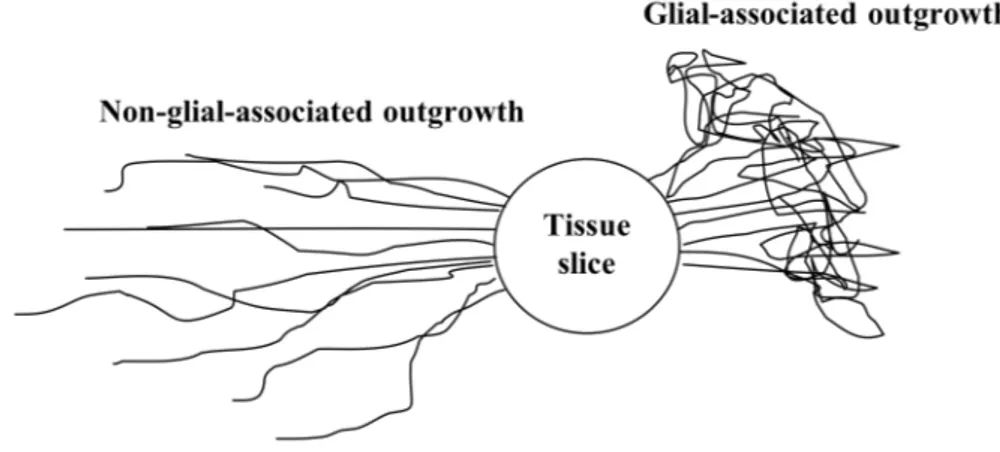
For antibodies and dilutions used see Tables 1 and 2. Antibody dilutions were generally performed in 1% Triton X-100 in PBS for cultures and 0.3% Triton X-100 in PBS for sections. However, the primary antibodies directed against ALDH1 (paper I) and SERT (paper IV and V) were found sensitive to Triton X-100. The primary antibody was therefore diluted in PBS when using these antibodies. Furthermore, these antibody incubations were preceded by a primary blocking step of 10% FCS diluted in 0.3% Triton X-100 in PBS for 1 h at room temperature. After blocking, the sections were quickly rinsed in PBS followed by incubation with primary antibodies.
A second serum block using 5% goat serum was performed before incubation with secondary antibodies in paper III, IV, and V. The goat serum was diluted in PBS and applied for 15 min at room temperature. The blocking solution was gently tipped off after incubation and secondary antibodies were applied.
Table 1. Primary antibodies
Directed against Type Dilution Source Paper
ALDH1 (aldehyde dehydrogenase-1)
rabbit anti-mouse
1:100 Abcam I β-tub (βIII-tubulin) mouse
anti-human 1:200 Sigma I DARPP-32 (dopamine- and
cyclic AMP-regulated phosphoprotein 32 kDa) rabbit anti-human 1:600 Cell Signaling Tech. V
GAD65/67 (glutamic acid decarboxylase 65/67)
rabbit anti-human
1:100 Sigma I GLAST (glia glutamate
transporter)
guinea-pig anti-rat
1:4000 Chemicon III Iba1 (ionized calcium
binding adapter molecule 1)
rabbit anti-human 1:1000 Wako Chemicals I NG2 (NG2 chondroitin sulfate proteoglycan)
rabbit anti-rat 1:200 Chemicon I S100 rabbit
anti-cow
1:400 Dako Patts II SERT (serotonin
transporter)
mouse anti-rat 1:400 Chemicon IV, V TH (tyrosine hydroxylase) mouse anti-rat 1:1500 Diasorin
Inc.
I, II, III, IV, V TH rabbit anti-rat 1:300 Pel Freez,
Interassay
I, III Vimentin mouse anti-pig 1:200 Sigma I, III Vimentin rabbit anti-calf 1:100 Abcam III
Tabel 2. Secondary antibodies. Alexa Fluor® conjugates (Molecular Probes Inc., Eugen, OR, USA) were used as secondary antibodies for detection.
Fluorophore Type Dilution Paper
Alexa 594 goat anti-mouse 1:500 I, II, III, IV, V Alexa 594 goat anti-rabbit 1:500 I, III
Alexa 488 goat anti-guinea pig 1:500 III Alexa 488 goat anti-mouse 1:200 I, III Alexa 488 goat anti-rabbit 1:500 I, II, IV, V
Image analysis
Immunohistochemically processed organotypic slice cultures and sections were analyzed using fluorescence microscopy. Photomicrographs were made with a digital camera (Hamamatsu, Solna, Sweden), connected to a computer, and analyzed using the Openlab software (Improvision, Coventry, U.K.). Identical areas were captured at different wavelengths and merged to achieve pictures of double and triple labeling.
Astrocytic migration and fiber outgrowth measurements
Astrocytic migration and nerve fiber outgrowth from fetal VM slice cultures were measured in paper I, II, and III using a scale mounted in one ocular of the microscope. All measurements were performed from the periphery of the tissue slice, defined by the density and depth of DAPI-positive cell nuclei. The astrocytic migration was measured in four perpendicularly placed directions from the periphery of the tissue slice. The nerve fiber outgrowth was measured from the periphery of the tissue slice to the distal end reached by the fibers. For the glial-associated outgrowth, the mean per culture was calculated from 3-4 measurements. The non-glial-associated outgrowth was determined by measuring the distance from the most distal nerve fibers to the periphery of the tissue slice.
Density measurements
In paper II, IV and V, optical density measurements were performed on cultures or sections using the NIH Image software. Images of the same magnitude were captured using a CCD camera (ProgRes C14, Jenoptic, Jena, Germany) and turned binary. The optical density was then measured in a set frame and expressed as gray values.
Cell counting
Cell counts on Iba1-positive cells were performed in paper I. The cells were counted in a set frame in the microscope, in areas over the slice culture. Counting was made over areas dense with TH-positive neurons and over areas with no TH-TH-positive neurons.
Statistics
Statistical analyses were performed using one- or two-way analysis of variance (ANOVA) followed by post hoc testing, or using Student’s t-test. Statistical significance was set at p<0.05. All data is expressed as group means ± standard error of the mean.
RESULTS AND DISCUSSION
Characterization of rat fetal VM slice cultures, paper I
In paper I, the TH-positive nerve fiber outgrowth and its relation to astroglia and other glial cells was investigated in rat E14 VM cultures. The organotypic cultures were incubated over a time course of 1, 2, 3, 7, 14, 21, and 28 DIV in order to map the nerve fiber outgrowths and the glial cell migration over time. The occurrence and characteristics of other neuronal nerve fiber formation was evaluated using β-tubulin immunohistochemistry. The results revealed nerve fibers positive for both β-tubulin and TH, as well as nerve fibers only exhibiting β-tubulin-immunoreactivity. This was an important finding revealing the presence of nerve fibers other than TH-positive. Indeed, antibodies directed against GAD65/67 showed the presence of GABA neurons in the VM slice cultures. These findings demonstrate the complexity of the VM cultures, as well as the use of fetal VM for transplantation. The glial content and migration from the cultures were evaluated using antibodies against markers of astrocytes, microglia, and oligodendroglia. The VM tissue slices were cultivated either using the roller-drum technique or on membrane inserts.Roller-drum cultures
In the organotypic cultures, β-tubulin-positive nerve fibers were observed evenly distributed within the tissue slice and thin fibers were projecting from the slice already at 1 DIV, demonstrating early attachment to the plating surface. Only a few of these nerve fibers displayed immunoreactivity against TH. Hence, TH-immunoreactivity was at this time point mainly found in the tissue slice. At 2 DIV, β-tubulin-positive nerve fiber outgrowth had become more abundant and