This is the author manuscript accepted for publication and has undergone full peer review but has not been through the copyediting, typesetting, pagination and proofreading process, which may lead to differences between this version and the Version of Record. Please cite this article as doi:
Does the time-point of orthodontic space closure initiation after tooth extraction affect the incidence of gingival cleft development? A randomized controlled clinical trial.
Kristina Bertl*,†, PhD, Hemma Neuner‡, DMD, Antonia Meran‡,•, DMD, Michael H Bertl‡, Docent, Ilse Reich‖,‣, DMD, Michael Nemec‡, DMD, Corinna Bruckmann‖, DMD, Andreas Stavropoulos*,‖, Professor, Hans-Peter Bantleon‡, Professor
* Department of Periodontology, Faculty of Odontology, University of Malmö, Malmö, Sweden † Division of Oral Surgery, University Clinic of Dentistry, Medical University of Vienna, Vienna, Austria. ‡ Division of Orthodontics, University Clinic of Dentistry, Medical University of Vienna, Austria
§ Private practice, Wolfsberg, Austria
‖ Division of Conservative Dentistry and Periodontology, University Clinic of Dentistry, Medical University of Vienna, Austria
¶ Private practice, Taiskirchen, Austria
Word count: 3976 Number of figures: 3 Number of tables: 3 Number of references: 31
Summary: The development of a gingival cleft is a frequent finding during orthodontic tooth movement and seems to occur more frequently with early and/or fast tooth movement towards an extraction site.
Authors contribution statement
Kristina Bertl contributed to Concept/Design, Data collection, Drafting article, Approval of article Hemma Neuner contributed to Data collection, Drafting article, Approval of article
Antonia Rezac contributed to Concept/Design, Data collection, Approval of article
Michael Bertl contributed to Concept/Design, Data collection, Drafting article, Approval of article Ilse Steiner contributed to Data collection, Drafting article, Approval of article
Michael Nemec contributed to Data collection, Drafting article, Approval of article Corinna Bruckmann contributed to Concept/Design, Drafting article, Approval of article Andreas Stavropoulos, Data interpetation, Drafting article, Approval of article
Hans-Peter Bantleon contributed to Concept/Design, Drafting article, Approval of article
Corresponding author: Andreas Stavropoulos, Professor & Chair, Department of Periodontology, Faculty of Odontology, University of Malmö, Carl Gustafs väg 34, 20506 Malmö, Sweden; phone: +46 040 66 58066, email: andreas.stavropoulos@mau.se
Abstract
Background
Gingival clefts (GC) develop frequently during orthodontic space closure and may compromise the treatment outcome. This study assessed whether the time-point of orthodontic space closure initiation, after permanent tooth extraction, affects the incidence of GC.
Methods
In 25 patients requiring bilateral premolar extraction due to orthodontic reasons, one premolar, chosen at random, was extracted 8 weeks before space closure initiation (“delayed movement”, DM),
while the contralateral premolar was extracted one week before (“early movement”, EM) (“treatment group”). Presence/absence of GC after 3 and 6 months (“time-point”) was recorded and any association with various parameters (i.e., treatment group, time-point, gender, jaw, craniofacial growth, gingival biotype, buccal bone dehiscence after extraction, space closure) was statistically assessed.
Results
Twenty-one patients contributing with 26 jaws were finally included in the analysis. Overall, GC were frequent after 3 (DM: 53.9%; EM: 69.2%) and 6 months (DM: 76.9%; EM: 88.5%). EM (p=0.014) and larger space closure within the study period (p=0.001) resulted in a significantly higher incidence of GC. Further, there was a tendency for GC development in the presence of buccal bone dehiscence (p=0.052) and thin gingival biotype (p=0.054). “Fast movers” (herein cases with a tooth movement ≥ 1mm per month) developed a GC in > 90% of the cases already after 3 months. “Slow movers” developed a GC only in 25 and 70% after 3m and FE, respectively.
Conclusions
GC development is a frequent finding during orthodontic space closure and seems to occur more frequently with early tooth movement initiation and in “fast movers”.
Keywords
Gingiva, gingival disease, orthodontic space closure, tooth extraction, tooth socket. Introduction
A frequent complication associated with orthodontic space closure, for example after permanent tooth extraction, is development of a gingival cleft (GC), often also called invagination / infolding / duplication / crease. In this context, extraction of permanent teeth is a frequent component of orthodontic treatment; specifically, tooth extraction (excluding third molars) is performed in approximately 25% of the patients, while in 10 to 15% of the patients all first premolars are extracted
1
. GC have been reported to develop in about 35 to 100% of the extraction cases and are more frequent in the mandible 2–6. In general, GC can appear on the buccal, lingual, and/or occlusal aspect
or they can extend across the alveolar ridge from the buccal to the lingual (Figure 1 A-C). The vertical and horizontal extent of infolded tissue indicates the severity of the GC. A GC may present as a minor crease in the gingiva, but may also extend into the alveolar ridge, i.e., presenting as a bone defect 7. A defect depth in the soft tissues of 1 5 or 2 mm 4, has been previously proposed as case-definition criterium of a GC.
Development of a GC can be problematic from both an orthodontic and periodontal aspect. Specifically, a GC can delay or prevent complete space closure, cause relapse (re-opening) after space closure, or impair the aesthetic outcome of treatment 2, 8–11. Further, presence of GC also impairs oral hygiene and is associated with increased probing pocket depths (PD) and/or increased attachment loss at the neighboring teeth 2, 4, 5, 11, 12. Two main mechanisms regarding GC development are considered in the literature: a) an increased amount of alveolar bone loss (in height and width) after tooth extraction might cause invagination of the soft tissue, or b) the mechanical compression of the gingiva in combination with reduced tissue remodeling leads to a piling-up of the soft tissue and GC development (for review on GC see: 13, 14).
In this context, the timepoint of orthodontic space closure initiation after tooth extraction has been discussed to have an impact on the risk of GC development. In a preclinical trial study in dogs, early tooth movement after extraction reduced the risk of GC development compared to delayed movement (i.e., 12 weeks after tooth extraction) 15. Additionally, in a very recently published clinical trial 3 a tendency for a somehow lower GC incidence with early vs. delayed movement for space closure (i.e., 2 to 4 weeks vs. ≥ 12 weeks after tooth extraction) was reported. Furthermore, in one retrospective evaluation of 30 patients, in sites developing GC, orthodontic space closure initiation was on average 7.5 months after extraction, while in sites not developing GC, orthodontic space closure initiation was on average 3.3 months after extraction 2. Yet, in another retrospective evaluation of 40 patients, of whom 14 had developed a GC, no relationship between presence or
severity of the GC and the time period between tooth extraction and orthodontic space closure was detected 5. Thus, the information on this topic is scarce and controversial.
The aim of the present split-mouth study was to assess whether the time-point of orthodontic space closure initiation, i.e., early vs. delayed, after permanent tooth extraction affects the incidence of GC development.
Materials and Methods Study population
The study protocol of the present randomized controlled clinical split-mouth trial was approved by the ethics committee of the Medical University of Vienna (EK-Nr. 105/2011) and the study was conducted in accordance with the Helsinki Declaration of 1975, as revised in 2013. Oral and written informed consent was obtained from all participants, or their legal representatives prior to any intervention. Reporting complies with the CONSORT guidelines 16 and the study was registered on clinicaltrials.gov (No.: NCT01402323). Patients attending the Division of Orthodontics (University Clinic of Dentistry, Medical University of Vienna, Austria) from 10/2011 until 03/2017 were recruited based on the following eligibility criteria: 1) orthodontic treatment plan including bilateral extraction of the first or second premolar in the upper or lower jaw (if bilateral premolar extraction was planned for both the upper and lower jaw, both jaws were included); 2) ≥ 10 years of age; 3) no systemic diseases; 4) no periodontitis; and 5) space closure performed by means of an open coil spring for reciprocal force on the adjacent teeth.
Study design
After alignment of the posterior segment, one of the premolars (i.e., right or left premolar) was randomly chosen by coin toss for extraction 8 weeks before space closure initiation (i.e., delayed movement group, DM). The contralateral premolar was extracted only one week before space closure
initiation (i.e., early movement group, EM). Thus, tooth movement was performed either towards a relatively healed or fresh extraction socket (“treatment group”). The time-point of space closure initiation was defined as baseline (BL), and an assessment was scheduled after 3 months (3m) and 6 months (final evaluation; FE), respectively (Figure 2 A).
Orthodontic treatment and tooth extraction
Orthodontic treatment started with alignment of the posterior segments with .022” self-ligating brackets and nickel-titanium archwires. Upon insertion of an .017 x .025 stainless steel archwire, one of the premolars was extracted with forceps specifically designed for minimally traumatic tooth extraction#. This was done by rotation and traction movements without flap elevation or any digital compression of the socket after tooth extraction. No orthodontic forces were applied for the following 8 weeks, and the space at the extraction site was kept stable by means of a closed coil spring. After 7 weeks, the contralateral premolar was extracted in the same fashion as above. One week after the second tooth extraction space closure was initiated by attaching nickel-titanium coil springs** with 200 cN of reciprocal force to the brackets adjacent to the extraction sites. Regular check-up and, if required, re-activation were scheduled every 4 to 6 weeks.
Evaluated parameters
Standard patient information (i.e., age, gender, jaw, and craniofacial growth prior to starting orthodontic treatment) were recorded. Craniofacial growth was diagnosed on lateral cephalograms by calculating Jarabak's angular sum of Björk's polygon (i.e., nasion-sella-articulare + sella-articulare-constructed gonion + articulare-sella-articulare-constructed gonion-menton) 17. The craniofacial growth was divided into 3 categories based on the angular sum (i.e., horizontal growth: < 394°; regular growth: 394 to
#Golden Line extraction forceps; American Dental Systems, Vaterstetten, Germany **Sentalloy® coil springs, GAC International, Inc., Gräfelfing, Germany
398°; vertical growth: > 398°). Further, the following parameters were registered at time-point of tooth extraction by a blinded examiner:
Gingival biotype, measured before tooth extraction, with an endodontic needle, at the center of the base of the mesial and distal papilla; gingival biotype was judged as “thin” if both values were ≤ 2 mm, and as “thick” if at least one of the values was > 2 mm.
Buccal bone dehiscence, judged as “present” if the buccal alveolar crest was ≥ 2 mm lower compared to the lingual bone level.
And at BL, 3m, and/or FE, also by a blinded examiner:
Gap extent, i.e., the mesio-distal distance (in mm) of the gap measured at the height of the gingiva with a manual caliper on cast models made from alginate impressions.
Space closure, i.e., the difference in gap extent a) between BL and 3m and b) between BL and FE.
PD (mm), absence/presence of bleeding on probing (%; BoP), and absence/presence of plaque (%; PI) were assessed at BL and FE at 3 sites each. Specifically, at the distal aspect of the mesial adjacent tooth and at the mesial aspect of the distal adjacent tooth (i.e., disto-lingual, distal, disto-buccal at the mesial tooth, and mesio-disto-lingual, mesial, mesio-buccal at the distal tooth).
GC, absence/presence at 3m and FE; a GC was classified as present when it was ≥ 1 mm deep or wide.
GC severity, at 3m and FE; i.e., depth of the GC in mm (vertically in 90° angle to the occlusal plane with a periodontal probe), and width of the GC in mm (horizontally and parallel to the occlusal plane with a periodontal probe from the buccal and/or lingual aspect and the deeper value was recorded) (Figure 1 D-E).
Statistical analysis
No clinical data from previous studies were available at the time-point of initiation of the present study. Thus, no sample size calculation could be performed based on the primary outcome parameter “incidence of GC”; hence, 25 patients were recruited. Normal distribution of all data was confirmed graphically by Q-Q-plots of the residuals. Periodontal parameters (i.e., PD, BoP, PI) were compared among treatment groups (i.e., DM and EM) as well as between BL and FE by dependent t-test. Further, gap extent and space closure were compared between treatment groups (i.e., DM and EM) by dependent t-test. Any differences in the frequency distribution of various parameters (i.e., incidence of GC development, gingival biotype, buccal bone dehiscence, GC type) between the treatment groups (i.e., DM and EM) were assessed by McNemar’s test. By means of random effects logistic regression models any associations between the primary outcome parameter (“incidence of GC”) and various secondary parameters *i.e., “treatment group” (DM, EM), “time-point” (BL, 3m, FE), “gender”, “jaw”, “craniofacial growth” (horizontal, regular, vertical), “gingival biotype” (thin, thick), “buccal bone dehiscence” (present, absent), “space closure”+ were assessed in 2 steps. First, “treatment group” and “time-point” were tested together with one of the following parameters: “gender”; “jaw”; “craniofacial growth”; “gingival biotype”; “buccal bone dehiscence”; “space closure”. Thereafter, “treatment group” and “time-point” and all parameters being significant on a 0.20-level in the separate analyses were combined in the final model. Additionally, the average space closure of all extraction sites (i.e., independent of the treatment group) after 3 and 6 months, respectively, was calculated. Extraction sites were grouped as “slow movers” and “fast movers” when being below or above the mean average space closure. Any difference in the frequency distribution in terms of GC development between “slow movers” and “fast movers” was assessed by χ2-test. Statistical analysis was performed using statistical software†† and p-values < 0.05 were considered as statistically significant.
Results
Study population
In total, 36 patients, whose orthodontic treatment plan included bilateral extraction of a premolar, were assessed for eligibility. Eleven patients were not included due to various reasons (e.g., insufficient compliance, change of treatment plan, denied participating), while 25 were included and allocated to receive treatment. Four patients were lost to follow-up (i.e., 2 did not want to continue, 2 lacked compliance). Hence, 21 patients were finally analyzed 6 months after space closure initiation; i.e., 21 patients (6 male, 15 female; mean age: 19.8 years; horizontal/regular/vertical craniofacial growth: 13/5/3 patients) contributed with 26 jaws (9 upper, 17 lower) to the analysis (Figure 2 B).
Periodontal parameters
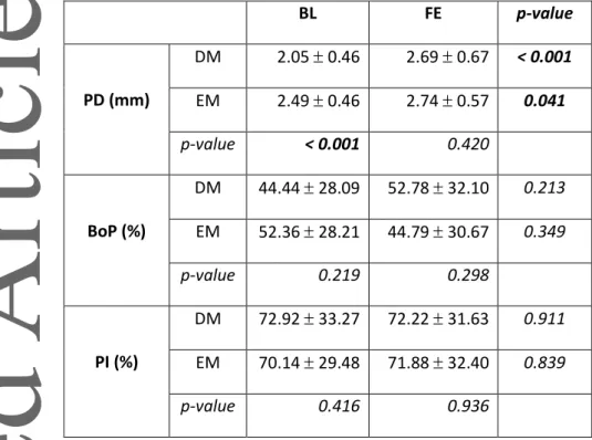
The periodontal parameters (i.e., PD, BoP, PI) are summarized in Table 1. At BL, average PD at the teeth adjacent to the EM extraction sites was about 0.5 mm higher compared with those adjacent to the DM extraction sites. Average PD at the teeth adjacent to the gap increased significantly over time until FE in both DM and EM groups, but there was no inter-group difference at FE. Nevertheless, average PD stayed below 3 mm and only 2 patients presented a PD ≥ 5 mm, at only one site each (both at FE, one each of the DM and EM group). Differences in BoP and PI between treatment groups and between BL and FE were not statistically significant, but BoP and PI presented generally relatively high average values (i.e., ≥ above 40 and 70%, respectively).
GC development and possible predictor variables
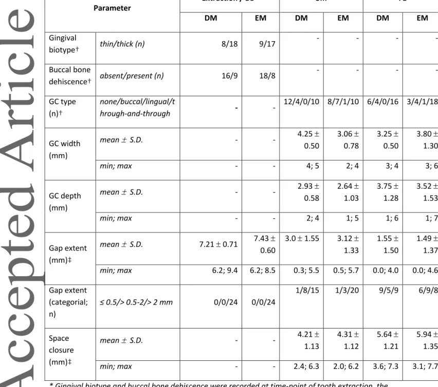
The characteristics of the extraction sites, GC, and tooth gaps are summarized in Table 2. In general, BL characteristics (i.e., gingival biotype, presence of a buccal bone dehiscence, and gap extent) were comparable between the DM and EM groups. Further, space closure did not differ between DM and EM at 3m and FE. GC development was a frequent finding in both groups at 3m (DM: 53.9%; EM:
69.2%) with approximately 3 mm remaining gap extent and at FE (DM: 76.9%; EM: 88.5%) with approximately 1.5 mm remaining gap extent, but no significant difference was observed regarding the incidence of GC at 3m and FE between DM and EM (Figure 3 A). All of the GC diagnosed at 3m were still present at FE. The majority of GC were complex (i.e., through-and-through clefts), with no statistically significant differences between DM and EM; i.e., at 3m 71.4 and 55.6% and at FE 80.0 and 78.3% at DM and EM sites, respectively. A representative patient case developing a GC at both extraction sites is presented in Figure 1 F-L.
Craniofacial growth could not be considered for the random effects logistic regression model, because all 3 patients with a vertical craniofacial growth presented at both 3m and at FE with a GC, which impeded to include this parameter due to perfect prediction. Further, the results of the first step of the random effects logistic regression model showed that only gender (coefficient: -2.16, 95% CI: -6.51 to 2.20, p = 0.331; increased risk for males) should not be included in the final model. Jaw (coefficient: -2.68, 95% CI: -5.53 to 0.17, p = 0.065; increased risk for upper jaws), gingival biotype (coefficient: -3.62, 95% CI: -7.48 to 0.24, p = 0.066; increased risk for thin biotype), buccal bone dehiscence (coefficient: 2.00, 95% CI: -0.28 to 4.28, p = 0.085; increased risk in the presence of a buccal bone dehiscence), and space closure (coefficient: 11.28, 95% CI: 4.93 to 17.63, p < 0.001; increased risk per mm) appeared relevant and should be included in the final model. Treatment group (p = 0.014) and space closure (p = 0.001) remained statistically significant in the final model (Table 3). Specifically, EM and a larger space closure within the study period resulted in a higher incidence of GC. The risk for GC development was increased 7.5-times by EM and 12.3-times per mm of space closure. The model also indicated a tendency for increased risk of GC development in the presence of a buccal bone dehiscence (p = 0.052) and of a thin gingival biotype (p = 0.054). Further, average space closure at 3m was 4.3 mm and at FE 5.8 mm, respectively. “Fast movers” had a statistically significant higher incidence of GC at both 3m and FE compared with “slow movers” (Figure 3 B). In “fast
movers”, a GC developed already after 3 months in > 90% of the cases, while in “slow movers” a GC developed in 25 and 70% after 3m and FE, respectively.
Discussion
Development of a GC during orthodontic space closure, for example after extraction of permanent teeth, is a frequent finding (35 to 100%) 2–6 and GC are associated with orthodontic, periodontal, and aesthetic concerns 2, 8–112, 4, 5, 11, 12. Excision of a GC is recommended shortly before or right after space closure is achieved 18, while resolution over time without any intervention cannot be expected in all cases (i.e., GC can persist for years) 4, 5, 19. Thus, measures to avoid or minimize the risk for GC development are welcome.
It is known that marked dimensional changes of the alveolar ridge occur during the first 6 months after tooth extraction. These dimensional changes are amounting to a horizontal bone loss of 29 to 63% (corresponding to 2.5 to 4.5 mm) and a vertical bone loss of 11 to 22% (corresponding to 0.8 to 1.5 mm), mostly affecting the buccal aspect of the ridge 20, 21. One of the mechanisms potentially leading to GC development is the advanced alveolar ridge resorption before initiating space closure. Hence, interventions, that reduce the significant post-extraction resorption modelling of the alveolar ridge, may also reduce the incidence of GC. Indeed, a few clinical reports, including a limited number of patients (i.e., 3 to 10) 22–24 described in general a reduced tendency for development and/or severity of GC when a socket preservation technique was used (i.e., guided tissue regeneration or grafting) prior to orthodontic space closure initiation. In this context, results from a preclinical in vivo study have indicated that early orthodontic space closure initiation may reduce this significant resorption modelling. Specifically, bilateral extraction of the second incisors was performed in 3 dogs, and orthodontic space closure was initiated either immediately or 12 weeks after extraction. This represents a timepoint, where extraction socket healing and post-extraction resorption modelling is completed in the dog 15. After an 8-week bodily tooth movement period and a 2-month retention
period, clinical and histological parameters were recorded. The results revealed, that sites involving immediate tooth movement, showed a broader alveolar process with higher bone density – but less mature bone – and a reduced tendency for GC development, compared with sites involving delayed tooth movement. These latter sites showed reduced bone density and more mature bone, pronounced horizontal atrophy of the alveolar process, and increased tendency towards gingival invagination. Thus, the present study was designed expecting that early orthodontic space closure initiation after tooth extraction – an easy, low-cost, and low-morbidity alternative to ridge preservation procedures – would reduce the incidence of GC, compared with a delayed orthodontic space closure initiation. Indeed, another study with a parallel arm design, published just after the present study was concluded, showed a tendency for lower incidence of GC with early vs. delayed movement for space closure (i.e., 2 to 4 weeks vs. ≥ 12 weeks after tooth extraction) 3. However, the present study showed that early tooth movement initiation increased the risk of GC development, seemingly contradicting the currently available scarce evidence on the topic. A possible explanation for the observed differences between the current and the above-mentioned study can be that the timeframe corresponding to the early and delayed groups was different (1 vs. 2 to 4 weeks and 8 vs. ≥ 12 weeks, respectively), thus representing histologically different stages of healing of the post-extraction socket. Specifically, in a histological report of trephine biopsies from human post-extraction sockets, woven bone occupied between 2 to 34% of the biopsy after 2 to 4 weeks of healing. While after 6 to 8 weeks in most of the cases woven bone was ≥ 20%, and after 12 to 21 weeks it was ≥ 33% in most of the cases 25. In contrast, it is expected that 1 week after extraction the socket is largely filled with granulation tissue and there is no woven bone 26. In that perspective, it seems reasonable to suggest waiting for a few weeks before starting to move the tooth, so that some amount of woven bone has formed in the extraction socket in order to reveal the benefits of orthodontically induced bone remodeling and/or modeling.
An interesting/important finding in this study was that, among the assessed parameters, the strongest predictor for GC development was space closure. Specifically, the risk for GC development was increased about 12-times per mm additional space closure. This resulted in a significantly higher incidence of GC at 3 and 6 months in sites with space closure above vs. sites with space closure below the average; i.e., “fast movers” presented a higher incidence of GC (Figure 3 B). The concept of “slow movers” and “fast movers” in orthodontic treatment describes, that there is a distinct variation in the distance (shorter vs. longer, respectively) the teeth move among individuals within a given timeframe despite application of the same orthodontic force. This has been described previously in a few preclinical in vivo experiments 27–29 and in one clinical trial 30. Indeed, in the above-mentioned pre-clinical in vivo study showing a reduced tendency for GC development in the EM group compared with the DM group, teeth in the DM group moved approximately 1.4-times faster than in the EM group 15. Similar, in the above-mentioned clinical study, where a tendency for lower incidence of GC with early vs. delayed space closure initiation was observed, space closure in patients in the DM group was complete on average after 7.5 months, while space closure in those in the EM group took on average 9.4 months 3. Thus, it appears that there was an accumulation of “fast movers” in the DM group in that parallel arm study. This in turn could also explain the discrepancy in the results compared with the present study; such interindividual differences are better controlled herein due to the split-mouth design. It can thus be assumed that in “fast movers”, tissue remodeling is slower compared to the speed of tooth movement, and thus mechanical compression of the gingiva leads to the piling-up of the soft tissue and to GC development. In this context, the relative importance of another still not explored factor that may influence GC development, is the degree of socket corticalization, i.e., the formation of the hard tissue bridge covering the socket entrance. In a recent cross-sectional radiographic study, corticalization 6 to 9 months after extraction was concluded in 40% of the sockets, and in about 80% of the sockets after 9 to 12 months 31. Hence, it may be that the tooth can move faster towards an extensively modelled yet not corticalized socket, compared with tooth movement towards a site that has already corticalized. Future studies could focus on the
possible impact of corticalization of the extraction socket as well as of controlled speed of space closure on GC development; e.g., whether a slow movement, for example based on the present results < 1 mm per month, which in turn would give the soft tissue sufficient time for remodeling, might reduce incidence of GC.
Several other parameters have been considered herein possibly associated with GC development. However, only presence of the buccal crest at a level ≥ 2 mm lower than that of the lingual crest and presence of a thin gingival biotype were marginally not statistically significant. One may speculate that both parameters are associated with larger post-extraction resorption of the alveolar ridge, compared with sites where the buccal and lingual alveolar crest are on the same level or with sites with thick gingival biotype. This is compatible with the notion that advanced alveolar ridge resorption before initiating space closure is one of the mechanisms leading to GC development. Another potential parameter – craniofacial growth – could not be included in the analysis herein due to the limited number of patients with a vertical craniofacial growth (n=3). However, as all 3 patients developed a GC already after 3 months, it might be an interesting parameter for future studies with a larger sample size.
Herein, periodontal parameters (i.e., plaque and bleeding indices, PD) had been recorded at BL and at FE, but not consistently over time. Plaque and bleeding values appear high at BL (i.e., above 40 and 70%, respectively) in the present group of patients, despite the fact that all patients regularly received oral hygiene instructions prior to initiating orthodontic treatment. This might be, at least partly, due to the fact that BoP and PI values were recorded only on the tooth aspects next to the tooth gap, i.e. they are not full-mouth scores. One may thus consider that the specific orthodontic appliances, including a coil spring, at that specific part of the arch, were obviously challenging for young adults. Indeed, mean PD increased statistically significant during the course of the study, but average values remained < 3 mm and only 2 patients presented a PD ≥ 5 mm at only one site each at FE. As
mentioned earlier, previous studies have reported that GC are associated with impaired oral hygiene, increased PD and/or increased attachment loss at the neighboring teeth2, 4, 5, 11, 12 , but gingival health has not been associated with GC development 4. Lack of BOP and PI values during the course of the study, and the high BL values do not permit any meaningful evaluation of the possible impact of overall gingival health on GC formation herein.
Conclusion
GC development in the present study was indeed frequent (i.e., > 75% of the sites) and was associated with early tooth movement initiation, herein 1 week after extraction, and with “fast movers”, herein cases where teeth moved ≥ 1 mm per month.
Acknowledgments
The authors thank Prof. Dr. N. Pandis (School of Dental Medicine, Department of Orthodontics and Dentofacial Orthopedics, University of Bern, Switzerland) for statistical advice. The forceps supporting atraumatic tooth extraction (Golden Line extraction forceps) were kindly provided by American Dental Systems (Vaterstetten, Germany).
Conflicts of interest
The authors declare no conflict of interest and no external funding was obtained for performing this study.
References
1. Jackson TH, Guez C, Lin FC, Proffit WR, Ko CC. Extraction frequencies at a university orthodontic clinic in the 21st century: Demographic and diagnostic factors affecting the likelihood of extraction. Am J Orthod Dentofacial Orthop 2017;151:456-462.
2. Reichert C, Gölz L, Dirk C, Jäger A. Retrospective investigation of gingival invaginations : Part I: Clinical findings and presentation of a coding system. J Orofac Orthop 2012;73:307-316.
3. Reichert C, Kutschera E, Plötz C et al. Incidence and severity of gingival invaginations associated with early versus late initiation of orthodontic space closure after tooth extraction : A multicenter pilot and randomized controlled trial. J Orofac Orthop 2017;78:415-425.
4. Rivera Circuns AL, Tulloch JF. Gingival invagination in extraction sites of orthodontic patients: their incidence, effects on periodontal health, and orthodontic treatment. Am J Orthod 1983;83:469-476.
5. Robertson PB, Schultz LD, Levy BM. Occurrence and distribution of interdental gingival clefts following orthodontic movement into bicuspid extraction sites. J Periodontol 1977;48:232-235. 6. Stappert D, Geiman R, Zadi ZH, Reynolds MA. Gingival clefts revisited: Evaluation of the
characteristics that make one more susceptible to gingival clefts. Am J Orthod Dentofacial
Orthop 2018;154:677-682.
7. Pinheiro ML, Moreira TC, Feres-Filho EJ. Guided bone regeneration of a pronounced gingivo-alveolar cleft due to orthodontic space closure. J Periodontol 2006;77:1091-1095.
8. Edwards JG. The prevention of relapse in extraction cases. Am J Orthod 1971;60:128-144. 9. Parker GR. Transseptal fibers and relapse following bodily retration of teeth: a histologic study.
Am J Orthod 1972;61:331-344.
10. Ronnerman A, Thilander B, Heyden G. Gingival tissue reactions to orthodontic closure of extraction sites. Histologic and histochemical studies. Am J Orthod 1980;77:620-625.
11. Wehrbein H, Fuhrmann R, Andreas A, Diedrich P. [The significance of gingival invagination in orthodontic space closure. A clinico-radiological study (in German)]. Fortschr Kieferorthop 1993;54:231-236.
12. Golz L, Reichert C, Dirk C, Jager A. Retrospective investigation of gingival invaginations: Part II: microbiological findings and genetic risk profile. J Orofac Orthop 2012;73:387-396.
13. Gölz L, Reichert C, Jäger A. Gingival invagination--a systematic review. J Orofac Orthop 2011;72:409-420.
14. Bertl K. [Gingival cleft development during orthodontic space closure (in German)] wissen
kompakt 2015;9:139-146.
15. Diedrich P, Wehrbein H. Orthodontic retraction into recent and healed extraction sites. A histologic study. J Orofac Orthop 1997;58:90-99.
16. Schulz KF, Altman DG, Moher D, CONSORT G. CONSORT 2010 Statement: updated guidelines for reporting parallel group randomised trials. BMC Med 2010;8:18.
17. Björk A. The face in profile - an anthropological X-ray investigation of Swedish children and conscripts (in Swedish). Svensk Tandläkare Tidskrift 1947;40:5b.
18. Malkoc S, Buyukyilmaz T, Gelgor I, Gursel M. Comparison of two different gingivectomy techniques for gingival cleft treatment. Angle Orthod 2004;74:375-380.
19. Kurol J, Ronnerman A, Heyden G. Long-term gingival conditions after orthodontic closure of extraction sites. Histological and histochemical studies. Eur J Orthod 1982;4:87-92.
20. Araújo MG, Silva CO, Misawa M, Sukekava F. Alveolar socket healing: what can we learn.
Periodontol 2000 2015;68:122-134.
21. Tan WL, Wong TL, Wong MC, Lang NP. A systematic review of post-extractional alveolar hard and soft tissue dimensional changes in humans. Clin Oral Implants Res 2012;23 Suppl 5:1-21. 22. Reichert C, Wenghofer M, Gotz W, Jager A. Pilot study on orthodontic space closure after
guided bone regeneration. J Orofac Orthop 2011;72:45-50.
23. Reichert C, Wenghoefer M, Kutschera E, Götz W, Jäger A. [Ridge preservation with synthetic nanocrystalline hydroxyapatite reduces the severity of gingival invaginations-a prospective clinical study (in German)]. J Orofac Orthop 2014;75:7-15.
24. Tiefengraber J, Diedrich P, Fritz U, Lantos P. Orthodontic space closure in combination with membrane supported healing of extraction sockets (MHE) a pilot study. J Orofac Orthop 2002;63:422-428.
25. Trombelli L, Farina R, Marzola A, Bozzi L, Liljenberg B, Lindhe J. Modeling and remodeling of human extraction sockets. J Clin Periodontol 2008;35:630-639.
26. Cardaropoli G, Araújo M, Lindhe J. Dynamics of bone tissue formation in tooth extraction sites. An experimental study in dogs. J Clin Periodontol 2003;30:809-818.
27. Danz JC, Bibby BM, Katsaros C, Stavropoulos A. Effects of facial tooth movement on the periodontium in rats: a comparison between conventional and low force. J Clin Periodontol 2016;43:229-237.
28. Pilon JJ, Kuijpers-Jagtman AM, Maltha JC. Magnitude of orthodontic forces and rate of bodily tooth movement. An experimental study. Am J Orthod Dentofacial Orthop 1996;110:16-23. 29. van Leeuwen EJ, Maltha JC, Kuijpers-Jagtman AM. Tooth movement with light continuous and
discontinuous forces in beagle dogs. Eur J Oral Sci 1999;107:468-474.
30. Giannopoulou C, Dudic A, Pandis N, Kiliaridis S. Slow and fast orthodontic tooth movement: an experimental study on humans. Eur J Orthod 2016;38:404-408.
31. Bertl K, Kukla EB, Albugami R, Beck F, Gahleitner A, Stavropoulos A. Timeframe of socket cortication after tooth extraction: A retrospective radiographic study. Clin Oral Implants Res 2018;29:130-138.
Table legends
Table 1. Periodontal parameters at baseline and final evaluation (mean S.D.).
BL FE p-value PD (mm) DM 2.05 0.46 2.69 0.67 < 0.001 EM 2.49 0.46 2.74 0.57 0.041 p-value < 0.001 0.420 BoP (%) DM 44.44 28.09 52.78 32.10 0.213 EM 52.36 28.21 44.79 30.67 0.349 p-value 0.219 0.298 PI (%) DM 72.92 33.27 72.22 31.63 0.911 EM 70.14 29.48 71.88 32.40 0.839 p-value 0.416 0.936
Statistically significant p-values are indicated in bold.
BL, baseline; BoP, bleeding on probing; DM, delayed movement; EM, early movement; FE, final evaluation; PD, probing pocket depth; PI, plaque index; S.D., standard deviation.
Table 2. Characteristics of the extraction sites, gingival clefts, and tooth gaps. Parameter Extraction / BL* 3m FE DM EM DM EM DM EM Gingival biotype† thin/thick (n) 8/18 9/17 - - - - Buccal bone dehiscence† absent/present (n) 16/9 18/8 - - - - GC type (n)† none/buccal/lingual/t hrough-and-through - - 12/4/0/10 8/7/1/10 6/4/0/16 3/4/1/18 GC width (mm) mean S.D. - - 4.25 0.50 3.06 0.78 3.25 0.50 3.80 1.30 min; max - - 4; 5 2; 4 3; 4 3; 6 GC depth (mm) mean S.D. - - 2.93 0.58 2.64 1.03 3.75 1.28 3.52 1.53 min; max - - 2; 4 1; 5 1; 6 1; 7 Gap extent (mm)‡ mean S.D. 7.21 0.71 7.43 0.60 3.0 1.55 3.12 1.33 1.55 1.50 1.49 1.37 min; max 6.2; 9.4 6.2; 8.5 0.3; 5.5 0.5; 5.7 0.0; 4.0 0.0; 4.6 Gap extent (categorial; n) ≤ 0.5/> 0.5-2/> 2 mm 0/0/24 0/0/24 1/8/15 1/3/20 9/5/9 6/9/8 Space closure (mm)‡ mean S.D. - - 4.21 1.13 4.31 1.12 5.64 1.21 5.94 1.35 min; max - - 2.4; 6.3 2.0; 6.2 3.6; 7.3 3.1; 7.7
* Gingival biotype and buccal bone dehiscence were recorded at time-point of tooth extraction, the other parameters at BL.
† Statistically insignificant differences in the frequency distribution between treatment groups (i.e., DM and IM) according to McNemar’s test at all available time-points (p > 0.05).
‡ Statistically insignificant differences between treatment groups (i.e., DM and IM) according to dependent t-test at all available time-points (p > 0.05).
BL, baseline; DM, delayed movement; EM, early movement; FE, final evaluation; GC, gingival cleft; max, maximum; min, minimum; S.D., standard deviation.
Table 3. Final random effects logistic regression model on the evaluation of possible predictor variables on the development of a gingival cleft.
Parameter Coefficient 95% CI p-value lower upper Jaw Upper 1.00 0.069 Lower -6.74 -13.99 0.52 Gingival biotype Thin 1.00 0.054 Thick -6.27 -12.64 0.11
Buccal bone dehiscence
Absent 1.00 0.052 Present 6.85 -0.07 13.77 Space closure mm 12.29 5.23 19.34 0.001 Time-point 3m 1.00 0.088 FE 4.59 -0.69 9.87 Treatment group DM 1.00 0.014 EM 7.52 1.51 13.54
Statistically significant p-values are indicated in bold.
3m, 3 months; CI, confidence interval; DM, delayed movement; EM, early movement FE, final evaluation.
Figure legends
Figure 1. (A) A through-and-through gingival cleft between the canine and second premolar, which developed during space closure after extraction of the first premolar. The cleft is extending from the lingual (B) to the buccal (C) indicated by the white arrows. By means of a periodontal probe the depth of a gingival cleft can be probed (D) vertically in a 90° angle to the occlusal plane and (E) the width horizontally and parallel to the occlusal plane. (F-L) Representative patient case developing gingival clefts during orthodontic space closure after extraction of the first premolars in the lower jaw; (F) before starting orthodontic treatment, (G) after alignment of the posterior teeth, (H) after extraction of both teeth (healed extraction socket on the right and the fresh extraction socket on the left side), (I) after 3 months, and (J) at final evaluation. Gingival clefts developed at the right (K) and left (L) side between the canine and second premolar indicated by the white arrows.
Figure 2. (A) Study design (3m, 3 months evaluation; BL, baseline; DM, delayed movement; EM, early movement; FE, final evaluation; GC, gingival cleft; PM, premolar). (B) CONSORT flowchart of the study.
Figure 3. (A) Frequency distribution of gingival cleft (GC) development at 3 months (3m) and final evaluation (FE) between the treatment groups [i.e., delayed (DM) and early movement (EM)]; corresponding p-values are presented above the bars. (B) Frequency distribution of gingival cleft (GC) development at 3 months (3m) and final evaluation (FE) between extraction sites with space closure below or above the average space closure; corresponding p-values are presented above the bars.


![Figure 3. (A) Frequency distribution of gingival cleft (GC) development at 3 months (3m) and final evaluation (FE) between the treatment groups [i.e., delayed (DM) and early movement (EM)]; corresponding p-values are presented](https://thumb-eu.123doks.com/thumbv2/5dokorg/3952077.75654/24.892.85.783.107.580/frequency-distribution-gingival-development-evaluation-treatment-corresponding-presented.webp)
