Emulsion Electrospinning as an Approach to
Fabricate PLGA/Chitosan Nanofibers for
Biomedical Applications
Fatemeh Ajalloueian, Hossein Tavanai, Jons Hilborn, Olivier Donzel-Gargand, Klaus Leifer,
Abeni Wickham and Ayyoob Arpanaei
Linköping University Post Print
N.B.: When citing this work, cite the original article.
Original Publication:
Fatemeh Ajalloueian, Hossein Tavanai, Jons Hilborn, Olivier Donzel-Gargand, Klaus Leifer,
Abeni Wickham and Ayyoob Arpanaei, Emulsion Electrospinning as an Approach to
Fabricate PLGA/Chitosan Nanofibers for Biomedical Applications, 2014, BIOMED
RESEARCH INTERNATIONAL, 475280.
http://dx.doi.org/10.1155/2014/475280
Copyright: Hindawi Publishing Corporation
http://www.hindawi.com/
Postprint available at: Linköping University Electronic Press
Research Article
Emulsion Electrospinning as an Approach to Fabricate
PLGA/Chitosan Nanofibers for Biomedical Applications
Fatemeh Ajalloueian,
1,2Hossein Tavanai,
1Jöns Hilborn,
2Olivier Donzel-Gargand,
3Klaus Leifer,
3Abeni Wickham,
4and Ayyoob Arpanaei
51Department of Textile Engineering, Center of Excellence in Applied Nanotechnology, Isfahan University of Technology,
Isfahan 84156-83111, Iran
2Department of Materials Chemistry, Uppsala University, 75121 Uppsala, Sweden
3Department of Engineering Sciences, Applied Materials Science, Uppsala University, 75121 Uppsala, Sweden
4Department of Physics, Chemistry and Biology, Division of Molecular Physics, Linkoping University, 58183 Linkoping, Sweden 5Department of Industrial and Environmental Biotechnology, National Institute of Genetic Engineering and Biotechnology,
Tehran 14965-161, Iran
Correspondence should be addressed to Hossein Tavanai; tavanai@cc.iut.ac.ir
Received 16 April 2013; Revised 19 December 2013; Accepted 27 December 2013; Published 13 February 2014 Academic Editor: Ulrich Kneser
Copyright © 2014 Fatemeh Ajalloueian et al. This is an open access article distributed under the Creative Commons Attribution License, which permits unrestricted use, distribution, and reproduction in any medium, provided the original work is properly cited.
Novel nanofibers from blends of polylactic-co-glycolic acid (PLGA) and chitosan have been produced through an emulsion electrospinning process. The spinning solution employed polyvinyl alcohol (PVA) as the emulsifier. PVA was extracted from the electrospun nanofibers, resulting in a final scaffold consisting of a blend of PLGA and chitosan. The fraction of chitosan in the final electrospun mat was adjusted from 0 to 33%. Analyses by scanning and transmission electron microscopy show uniform nanofibers with homogenous distribution of PLGA and chitosan in their cross section. Infrared spectroscopy verifies that electrospun mats contain both PLGA and chitosan. Moreover, contact angle measurements show that the electrospun PLGA/chitosan mats are more hydrophilic than electrospun mats of pure PLGA. Tensile strengths of 4.94 MPa and 4.21 MPa for PLGA/chitosan in dry and wet conditions, respectively, illustrate that the polyblend mats of PLGA/chitosan are strong enough for many biomedical applications. Cell culture studies suggest that PLGA/chitosan nanofibers promote fibroblast attachment and proliferation compared to PLGA membranes. It can be assumed that the nanofibrous composite scaffold of PLGA/chitosan could be potentially used for skin tissue reconstruction.
1. Introduction
Tissue engineering as a means of functional tissue fabrication or repair requires three-dimensional (3D) scaffolds that vide the structural base (matrix) for cell attachment and
pro-liferation [1]. Among different methods to produce 3D
scaf-folds, electrospinning has received considerable interest [2,
3]. This technique allows fabrication of porous nanofibrous
webs with specific characteristics like three dimensional mor-phology, large surface area to volume ratio, and high porosity. In addition, the structure of electrospun nanofibrous webs mimics the size range of natural extracellular matrix (ECM) fibrous components. ECM provides a natural base for cell
adhesion, proliferation, migration, and metabolism [4–6].
Electrospinning has been used to fabricate nanofibrous scaffolds from synthetic and natural polymers. The most frequently electrospun synthetic polymers include
glycolide-and lactide-based linear aliphatic polyesters [7,8]. Although
such synthetic polymers are biodegradable and have good mechanical properties, they suffer from poor cell-scaffold attachment interactions due to their hydrophobic structure as
a result of lower surface energy [1]. Additionally, their
nega-tive surface charge and acidity cause product degradation [9].
These shortcomings have restricted the application of linear aliphatic polyesters in pure form for scaffold purposes.
Collagen, elastin, chitosan, and alginate in the pure form are examples of naturally derived polymers that have been electrospun for the production of nanofibrous webs
Volume 2014, Article ID 475280, 13 pages http://dx.doi.org/10.1155/2014/475280
[2,10–14]. Among these polymers, chitosan, a naturally occurring polysaccharide, and its derivatives have been widely explored for biomedical applications, thanks to their
biocompatibility and biodegradability [15,16]. A number of
studies have been performed to evaluate the cytocompatibil-ity of chitosan using a variety of cell types such as osteoblasts, fibroblasts, hepatocytes, and neural cells. These studies have shown that chitosan is nontoxic and can support growth of
the aforementioned cell types [17, 18]. Like many natural
polymers, chitosan suffers from poor mechanical properties
[19]. In addition, it is difficult to electrospin chitosan in its
pure form. This is mainly due to the limited number of solvents for chitosan as well as high viscosities at its low
concentrations [20–22].
Recently, mixture of polymers has attracted a great deal of attention for producing polyblend nanofibers, exhibiting a combination of characteristics related to each polymer
employed in the so-called polyblend [2]. Basically, a
poly-blend nanofibrous scaffold containing synthetic and natural polymers is expected to exhibit physicochemical properties of both components like hydrophilicity/hydrophobicity, surface charge, and mechanical strength of synthetic polymers as
well as the biochemical signatures of natural fibers [1, 23].
Considering different properties of PLGA and chitosan, a nanofibrous scaffold of the polyblend of PLGA/chitosan should show interesting applications in tissue engineering.
Literature reports several efforts aiming to produce elec-trospun scaffolds containing both PLGA and chitosan. For example, some researchers have used two sets of separate syringe pumps and power supplies with a common collector to produce a web consisting of a mixture of individual PLGA
and chitosan nanofibers [24,25] or a coaxial electrospinning
setup to produce a mat of core-sheath type nanofibers to electrospin with PLGA as the core and chitosan as the sheath
[26]. Other researchers have used a common solvent for both
the polymers [27]. This approach restricts the fraction of one
polymer in the solution due to limitations of simultaneous solubility of the two polymers. A recent report also introduces electrospinning of PLGA/chitosan nanofibers in which PLGA
is grafted by chitosan [28].
The present study aimed at developing an approach for preparation of nanofibrous scaffolds of PLGA/chitosan through an emulsion electrospinning technique. In this way, there is no need to use specially designed electrospinning setups as already explained. Moreover, there is no need for a common solvent for both polymers or grafting one of the polymers onto the other one. Such a scaffold is expected to be mechanically strong, because of PLGA, as well as hydrophilic as a result of the presence of chitosan. The hydrophilicity bestows the mat a surface prone to cell attachment. The novelty of this approach is applying the method of emulsion
electrospinning [29] to produce polyblend nanofibers from
synthetic and natural polymers with varying ratios. The challenge of trying to have an electrospinnable mixture of water insoluble PLGA and water soluble chitosan made us choose an emulsion system. The selected emulsifier was polyvinyl alcohol (PVA). Not only is PVA a rather easy electrospinnable polymer, but it also dissolves in water and
hence it can, if required, be extracted from the polyblend nanofibers.
2. Experiment
2.1. Materials. Chitosan (MW ≈ 200,000 g/mol, degree of
deacetylation≈ 90%) and PLGA (MW 40,000–70,000 g/mol,
L-lactide/glycolide, 50 : 50) were purchased from Aldrich
(Germany). PVA (MW ≈ 72,000 g/mol), chloroform
(ana-lytical grade), dimethylformamide (DMF) (ana(ana-lytical grade), glacial acetic acid, and hexamethyldisilazane (HMDS) were obtained from Merck (Germany). DMEM (Dulbecco’s Mod-ified Eagle Medium 1x), trypsin/EDTA, and phosphate-buffered saline (PBS) were purchased from GIBCO Invit-rogen, USA. Fetal bovine serum (FBS Hyclone research grade) was bought from Perbio Scientific, Sweden. CellTiter 96 AQueous One solution was purchased from Promega, Madison, WI, USA. 3T3 fibroblast cell line was purchased from American Type Culture Collection (ATCC). All solvents were used as received without further purification.
2.2. Preparation of the Electrospinning Emulsion. PLGA
solu-tions (12%, 16%, 20% w/v) were prepared by dissolving PLGA
in chloroform/DMF (volume ratio of 90 : 10) at 50∘C under
magnetic stirring for 3 h. Chitosan solutions (4%, 6% w/v) were prepared by dissolving chitosan in acetic acid (14%) at room temperature. Also, a PVA solution (8% w/v) in double
distilled water was prepared at 90∘C.
To obtain optimum conditions for emulsion electrospin-ning, a series of emulsions containing different ratios of
PLGA and chitosan were prepared (Table 1). The emulsions
were prepared by simultaneous adding of PVA and chitosan solutions to PLGA solution, followed by mixing with mag-netic stirrer. The volume ratio of the 3 solutions was 1 : 1 : 1 in all the emulsions. The final mixtures were stirred at room temperature for 12 h to obtain a homogenous emulsion.
2.3. Electrospinning. Electrospinning of the emulsions was
carried out, with a voltage of 14–16 kV applied to the blunt needle (21 gauge) tip of the 1 mL syringe (filled with the emulsion) and the grounded aluminum foil collector. The feed rate was 0.25 mL/h. The needle tip to collector distance was 15 cm. The same electrospinning conditions were used to electrospin pure PLGA (16% w/v in chloroform/DMF (volume ratio of 90 : 10)) as a reference for electrospun mats of PLGA/chitosan.
2.4. Surface Morphology of Electrospun Nanofibers. The
sur-face morphology of electrospun PLGA/chitosan nanofibers was investigated with the help of scanning electron micro-scope (SEM) (Seron Technology AIS 2100) micrographs. The average fiber diameter of the electrospun fibers was measured by applying Image J software to the SEM micrographs. Sta-tistical analysis was done using SPSS 8.0. (SPSS Inc. Chicago, USA). Transmission electron microscopy (TEM) (JEOL 2000 FXII (2401)) was employed to investigate the miscibility of PLGA and chitosan. To obtain TEM micrographs of the cross section of nanofibers, electrospun samples were
Table 1: The content of emulsion electrospinning solutions and the electrospun nanofibers. Sample number PLGA conc. (w/v) Chitosan conc. (w/v) PVA conc. (w/v)
Total emulsion conc. (w/v)
PLGA (%W) after and before PVA removal
Chitosan (%W) after and before PVA removal
1 20 4 8 10.6 84 (62.5) 16 (12.5) 2 16 4 8 9.3 80 (57) 20 (14.3) 3 12 4 8 8 75 (50) 25 (16.7) 4 20 6 8 11.3 77 (60) 23 (17.6) 5 16 6 8 10 73 (53) 27 (20) 6 12 6 8 8.6 67 (46) 33 (25)
embedded directly in EPON 812 (epoxy resin) first and then
polymerized at 60∘C. Sections were cut with the diamond
knife of microtome, floated on water, and collected on Cu-grids. The sections were contrasted with 2% uranyl acetate and Reynolds lead solution afterwards. TEM pictures were converted with Gatan software Digital Micrograph 3.6.5.
2.5. PVA Extraction. To extract the PVA from the
elec-trospun nanofibers with the aim of obtaining nanofibers containing only PLGA and chitosan, five samples of
PLGA/chitosan/PVA (sample Number 2 in Table 1) with
a thickness of about 100𝜇m were cut (20 mm × 20 mm),
weighed (±0.1 mg), and then immersed in an aqueous solution containing ethanol (50% v/v) at room temperature for 8 h. The samples were finally weighed after drying overnight in a vacuum oven under room temperature.
2.6. Water Contact-Angle Measurement. The degree of hydrophilicity of PLGA, PLGA/chitosan/PVA, and PLGA/chitosan electrospun samples was determined with the help of a video-based optical contact angle meter
(DataPhysics OCA 15EC). Small pieces (4 × 1 cm) of the
samples were cut and placed on a glass microscope slide using double-sided tape to ensure uniform viewing of the
surface. The glass slide was placed on the stage and a 6𝜇L
drop was placed on the sample surface. The contact angle on the left and right sides of the drop was measured by SCA software. The average of ten angles is reported for each sample.
2.7. Mechanical Properties. The tensile properties of the
electrospun samples with a thickness of 50–100𝜇m (60 mm ×
10 mm) were determined by the ZWICK Z005 testing machine (ZWICK, Germany) equipped with a 50 N load-cell. The cross-head speed and the gauge length were 10 mm/min and 40 mm, respectively. The average of five measurements is reported. To compare the strength of samples in wet condition with that of dry ones, wet specimens were prepared by soaking the samples in phosphate buffered saline (PBS) for
8 h at 37∘C.
2.8. In Vitro Degradation. The degradability tests were
per-formed in phosphate buffered saline solution (PBS) adjusted
to pH 7.4 at 37∘C. Electrospun PLGA and PLGA/chitosan
mats were cut into 1 cm× 1 cm pieces, vacuum-dried at room
temperature, and weighed accurately. They were then placed into closed vials containing 10 mL of PBS and were kept in
water bath (37∘C) under mild shaking. At each time point
(1, 4, 7, 14, 21, 28, 42, and 56 days), three samples of each type of PLGA or PLGA/chitosan were removed from PBS solution, rinsed with distilled water, and completely dried under vacuum at room temperature. The degradation rate was reported as the loss in the mass of electrospun mat using
the formula “mass loss(%) = ((𝑀0−𝑀𝑡)/𝑀0) × 100,” where
𝑀0and𝑀𝑡are the mass of electrospun mat before and after
incubating in PBS solution, respectively.
2.9. FT-IR Analysis. FT-IR spectrophotometer (Perkin Elmer
Spectrum 100) was used to record the FT-IR spectra of pure chitosan, electrospun mats of PLGA, PLGA/chitosan/PVA,
and PLGA/chitosan nanofibers. The range of 650–4000 cm−1
with a resolution of 4.0 cm−1 and 32 scans in transmission
mode was employed.
2.10. Differential Scanning Calorimetry (DSC) Analysis. To
evaluate the state of PLGA/chitosan miscibility in the emulsion-electrospun nanofibers, DSC (TA instruments, Q1000, TA Instruments Ltd., USA) was employed. DSC analysis was carried out for pure chitosan powder, pure PVA powder, and electrospun mats of PLGA, PLGA/chitosan/PVA and PLGA/chitosan. A cool-heat-cool cycle was applied at a
rate of 5∘C/min, starting from 30∘C and increasing to 200∘C,
and then cooling to −50∘C. The given DSC thermograms
were obtained during reheating the samples to 200∘C under
nitrogen atmosphere.
2.11. Cell Culture of 3T3 Fibroblasts. NIH 3T3 mouse
fibrob-lasts were cultured in DMEM (Dulbecco’s Modified Eagle Medium, Gibco, USA) containing 10% fetal bovine serum (FBS Hyclone research grade, Perbio Scientific, Sweden), 50 U/mL penicillin, and 50 U/mL streptomycin (Sigma-Aldrich, Sweden). The medium was replaced every 3 days and
cultures were maintained in a tissue culture incubator at 37∘C
with 5% CO2. After reaching about 80% confluence, cells were
detached by 0.05% trypsin/0.05% EDTA. Electrospun mats (PLGA/chitosan and PLGA) were sterilized by ultraviolet irradiation for both top and bottom surfaces in a laminar flow hood (each side for 20 min); cells were seeded onto scaffolds
under density of 5000 cells/cm2and were placed in 24-well
2.12. Metabolic Activity and Proliferation of 3T3 Fibroblasts.
Cell viability and metabolic activity in response to different substrates of tissue culture polystyrene cover slip (TCP) and electrospun samples of PLGA and PLGA/chitosan were mea-sured using MTS cytotoxicity assay (CellTiter 96 AQueous Non-Radioactive Cell Proliferation Assay, Promega, Madi-son, WI, USA), according to the manufacturer’s instructions.
Cells were seeded at a density of 5000 cells/cm2and the cell
activity was evaluated during a 7-day period. On days 1, 4, and 7, the cell seeded electrospun samples were washed with
PBS and transferred to new well plates containing 500𝜇L of
culture medium in each well. Then, 100𝜇L of MTS solution
was added to each well. Cells were maintained for an
addi-tional 4 hours in a humidified incubator at 37∘C and 5% CO2.
The absorbance was read at 450 nm in an ELISA plate reader (Multiskan Plus, LabSystem, Finland) and the response was
defined as (cell-seeded scaffolds A450− mean blank)/(mean
TCP 1 day A450− mean blank) ∗ 100%, where mean blank is
culture medium absorbance mean. In this way, cell metabolic activity on each substrate in different time points was com-pared with TCP on day 1. This was done in triplicates followed by calculations of mean values and standard deviations. Cell proliferation was determined by counting the number of DAPI-stained cell nuclei that were associated with the PLGA or PLGA/chitosan nanofibers. Cell nuclei were counter-stained with 4,6-diamidino-2-phenylindole dihydrochloride
(DAPI, Sigma-Aldrich; 1𝜇g/mL) in three separate samples
(PLGA or PLGA/chitosan) per time points of 1, 4, and 7 days. A minimum of 5 randomly selected visual fields (at 20 × magnification in a fluorescent confocal microscope, Zeiss LSM 510, Germany) were studied.
2.13. Cell Morphology. Morphological study of 3T3 fibroblasts
grown on electrospun PLGA and PLGA/chitosan nanofibers was performed after 1 and 7 days of cell culture by SEM. Cell-seeded nanofiber constructs were harvested and washed by PBS and subsequently fixed in 2.5% glutaraldehyde for 3 h. The scaffolds were dehydrated with increasing concentrations of ethanol (30%, 50%, 70% 90%, and 100%) for 10 min each. Finally the cell-scaffold constructs were treated with hexamethyldisilazane (HMDS) to further water extraction. The dehydrated, cell-seeded constructs were maintained in desiccators equipped with a vacuum for overnight air dry-ing. After sputter-coating with Platinum, SEM was used to observe cell and scaffold morphology and cell attachment on nanofiber scaffolds.
2.14. Statistical Analysis. All the data presented are expressed
as mean± standard deviation (SD) of the mean. Statistical
analysis was performed using a one-way analysis of variance (ANOVA) using Student’s t-test to determine the statistical
significance between the two means evaluated at𝑃 < 0.05.
3. Results and Discussion
To study the possibility of having nanofibrous scaffolds of PLGA/chitosan with different ratios of chitosan to PLGA in final mats from a single electrospinning setup, 6 emulsions
were tested (Table 1). Having a water soluble polymer
(chi-tosan) and a water insoluble polymer (PLGA), an emulsion mixture could be the key to do successful electrospinning. PVA was used as emulsifier and our preliminary tests revealed that the best concentration of PVA is 8% (w/v) which was kept constant for all emulsions.
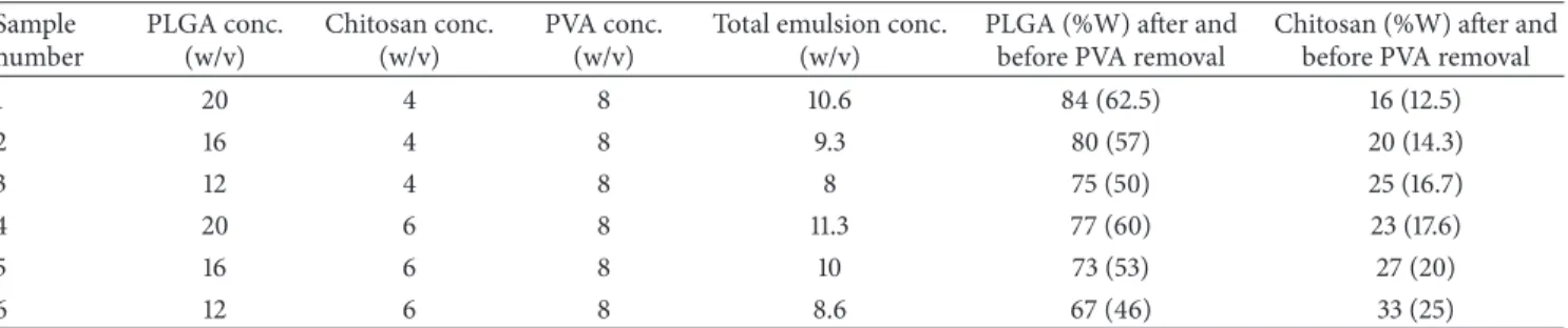
3.1. Electrospinning. To find optimum electrospinning
con-ditions, several factors of emulsion concentration, applied voltage, feed rate, and tip to collector distance were tried. The
SEM images inFigure 1show the morphology of nanofibers
electrospun from the 6 emulsions shown in Table 1 with
the electrospinning conditions mentioned in the caption. The variation of the average diameter of the electrospun nanofibers (prior to PVA extraction) versus the total con-centration of each emulsion (containing PLGA, chitosan,
and PVA) is shown inFigure 2. As can be seen, the average
diameter of electrospun nanofibers decreases with lower total concentration of polymers in the emulsion. This can be related to the lower viscosity of emulsions as a result
of lower polymer concentration. Images inFigure 1and the
plot inFigure 2also lead to the conclusion that decreasing
the concentration of PLGA in the emulsion from 16% to 12% decreases the average diameter sharply. This is related to the higher share of chitosan in the emulsion. Similar results have been reported by Meng et al. who electro-spun PLGA/gelatin and observed a sharp decrease in the diameter of nanofibers as a result of lowering the ratio
of PLGA relative to gelatin [30]. Our work showed that
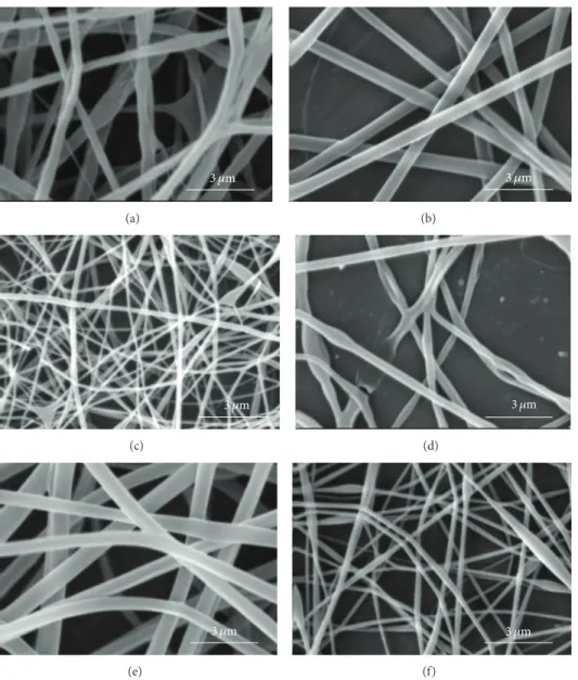
concentration of PLGA in the range of 12% to 16% and the concentration of chitosan in the range of 4% to 6% in the electrospinning emulsions led to optimum electrospinning. Higher concentrations of PLGA and chitosan produced emulsions which were too viscous for electrospinning. SEM images of PLGA (16%)/chitosan (4%)/PVA (8%) nanofibers electrospun at voltages of 8, 12, and 16 kV are shown in
Figure 3. Other electrospinning conditions are mentioned in
the corresponding caption. Evidently, at 8 kV (Figure 3(a)),
beads are formed along the electrospun nanofibers. Increas-ing the voltage to 12 kV leads to the disappearance of beads,
but the nanofibers are less uniform (Figure 3(b)). However,
voltage of 16 kV produces uniform nanofibers (Figure 3(c)).
It can be concluded that a stronger electrical field resulting from higher voltage induces a higher draw in the emulsion jet trajectory leading to the formation of bead-free, more uniform, and finer PLGA/chitosan/PVA nanofibers. As far as needle tip to collector distance during electrospinning of PLGA (16%)/chitosan (4%)/PVA (8%) is concerned, the results show that, with voltage of 16 kV and feed rate of 0.25 mL/h, increasing the needle tip to collector distance from 15 cm to 25 cm decreases the average nanofiber diameter
from 519 ± 70 nm to 502 ± 84 nm which is practically
insignificant. Literature also shows that needle tip to collector distance in the range of 8–15 cm has no significant effect on
the average diameter of electrospun PVA [31] and chitosan
[32] nanofibers. Electrospinning of the emulsion of PLGA
(16%)/chitosan (4%)/PVA (8%) nanofibrous mats with feed rates of 0.15 mL/h and 0.25 mL/h (needle tip to collector distance = 15 cm, voltage = 16 kV) led to the fabrication of
3 𝜇m (a) 3 𝜇m (b) 3 𝜇m (c) 3 𝜇m (d) 3 𝜇m (e) 3 𝜇m (f)
Figure 1: SEM images of emulsion electrospun PLGA/Chitosan/PVA nanofibrous mats ((a), (b), (c), (d), (e), and (f) are SEM images of
samples numbered as 1, 2, 3, 4, 5, and 6 described inTable 1, respectively. Electrospinning parameters are voltage of 16 Kv, feed rate of 0.25 mL/h,
and tip-collector distance of 15 cm).
0 200 400 600 800 11.3 10.6 10 9.3 8.6 8 Total concentration (w/v) A verag e dia m et er (nm)
3 𝜇m (a) 3 𝜇m (b) 3 𝜇m (c)
Figure 3: SEM images of PLGA (16%)/chitosan (4%)/PVA (8%) nanofibrous mats prepared using various voltages of (a) 8 kV, (b) 12 kV, and (c) 16 kV (feed rate = 0.25 mL/h, tip to collector distance = 15 cm).
nanofibers with average diameters of369 ± 120 nm and 519 ±
70 nm, respectively. As expected, lowering the feed rate led to a lower average diameter, but a lower uniformity of the electrospun nanofibers was observed.
3.2. PVA Extraction. Considering the purpose of this work,
that is, the fabrication of a nanofibrous mat of PLGA/ chitosan, PVA was removed from electrospun mats. This increases the share of the natural polymer (chitosan) in the
final mat as well as leading to a more porous structure [33,34].
AsTable 1shows, the share of chitosan in the final electrospun nanofibers (after PVA extraction) varies from 16% to 33%. To remove PVA from the electrospun polyblend nanofibrous mats, an aqueous solution containing 50% ethanol was used. Mass reduction measurements showed that 8 h was sufficient for the PVA removal. We found that 100% water is not a suitable solvent, as chitosan has a water solubility of
about 10% [35]. However, PVA dissolves slightly in ethanol
and chitosan solubility in water decreases almost linearly with increasing the share of ethanol in a water-ethanol mixture, so that the solubility of chitosan in the solution of
water-ethanol (50-50) is less than 0.1% [35].Figure 4shows
the electrospun sample of PLGA (16%)/chitosan (4%)/PVA (8%) before and after PVA extraction. This sample (sample
number 2 inTable 1) has been used for further analyses of
FT-IR, contact angle measurement, tensile testing, in vitro degradation, DSC, TEM, and cell seeding and is referred hereafter to as PLGA/chitosan/PVA prior to PVA removal and PLGA/chitosan after PVA removal.
3.3. Hydrophilicity. Water contact angles of pure PLGA,
PLGA/chitosan/PVA, and PLGA/chitosan mats are shown
in Figure 5. The average contact angle of PLGA (105.5 ±
5.2∘), PLGA/chitosan/PVA (48 ± 7.1∘), and PLGA/chitosan
(24 ± 8.7∘) reveals the hydrophobic nature of PLGA mat
(Figure 5(a)) as well as the role of chitosan in bestowing hydrophilicity to the PLGA/chitosan nanofibrous mat. Also, it can be seen that the removal of PVA from the electrospun
nanofibers leads to higher hydrophilicity (Figures5(b)and
5(c)), which has been attributed to the generation of more
pores in the composite membrane after PVA dissolution [34].
The improvement in hydrophilicity of the PLGA/chitosan scaffold is expected to lead to higher cell affinity of the hybrid
scaffold compared to PLGA [36,37].
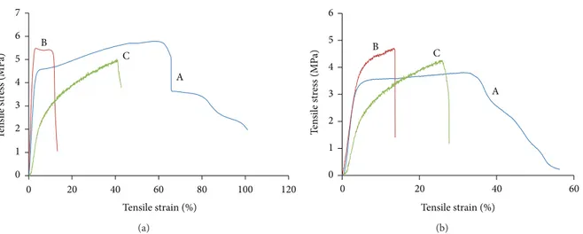
3.4. Tensile Properties. Figure 6compares the tensile charac-teristics of PLGA, PLGA/chitosan/PVA, and PLGA/chitosan
in dry (Figure 6(a)) and wet (Figure 6(b)) conditions. For
dry condition, addition of chitosan to PLGA leads to a strong reduction in elongation at break, whereas there are no significant differences in Young’s modulus or tensile strength. It is also observed that PVA removal has no effect on tensile strength but results in a sharp decrease in Young’s
modulus from 274.3 ± 17.2 MPa in PLGA/chiotsan/PVA
to 64 ± 11.8 MPa in PLGA/chitosan. However, the PVA
removal has led to an increase in extensibility of the hybrid structure, leading to more flexibility of the PLGA/chitosan, compared to PLGA/chitosan/PVA. It could be due to the PVA extraction which causes a longitudinal shrinkage in PLGA/chitosan construct (around 30%). In the wet condi-tion, there is an overall decline in tensile characteristics of all three samples as expected, whilst pure PLGA exhibits a higher reduction.
10 𝜇m
(a)
10 𝜇m
(b)
Figure 4: PLGA (16%)/chitosan (4%)/PVA (8%) electrospun mats: (a) before and (b) after PVA extraction in ethanol 50%-water 50% for 8 hours.
(a) (b) (c)
Figure 5: Contact angle of (a) PLGA, (b) PLGA/chitosan/PVA, and (c) PLGA/chitosan electrospun mats.
3.5. In Vitro Degradation. To simulate the in vivo
degrada-tion, we have studied the hydrolysis in vitro under physiologi-cal conditions at pH 7.4 using PBS. We observed a higher mass loss for the polyblend nanofibrous mat (PLGA/chitosan) in
all time points in comparison with PLGA (Figure 7(a),). The
morphological changes of both the mat types, after two weeks of incubation in PBS during in vitro degradation, are shown inFigure 7(b)(PLGA/chitosan) andFigure 7(c)(PLGA). It is seen that the fibrous morphology in the PLGA mat was almost unchanged, whilst the fibers in the composite mat were somehow swollen. However, no obvious morphological changes in both scaffolds were observed up to 8 weeks. These results suggest that both the PLGA and PLGA/chitosan nanofibers can be considered as potential scaffolds for biomedical applications like skin tissue engineering where the rate of tissue formation can be around several weeks
[38,39].
3.6. FT-IR Spectra. The FT-IR spectra of pure chitosan,
pure PLGA, PLGA/chitosan/PVA and, PLGA/chitosan are
shown in Figure 8. The characteristic absorption bands of
PLGA lie at about 2922 cm−1 (asymmetric CH2stretching),
2853 cm−1 (symmetric CH2 stretching), 1752 cm−1 (C=O
stretching), 1452 cm−1 (C–H stretching), 1182 cm−1 (C–O–
C stretching), and 1130 cm−1 (C–O stretching) [38].
Con-versely, the characteristic absorption bands of chitosan lie at
1154 cm−1and 893 cm−1(saccharide groups), 1535 cm−1(N–H
bending), 1600 cm−1(amide I stretching), 1647 cm−1(amide
II bending), 2977 cm−1 (C–H stretching), and 3425 cm−1
(N–H stretching) with the broad peak between 3400 and
3700 cm−1, corresponding to the stretching of O–H [40,41].
Comparing the above mentioned spectra with those of the PLGA/chitosan/PVA and PLGA/chitosan nanofibers leads to the conclusion that both PLGA and chitosan are present in the polyblend emulsion electrospun nanofibers. Moreover, extraction of PVA leads to a narrower and shorter peak in
the range of 3400–3700 cm−1 which originates from fewer
hydroxyl groups after PVA removal.
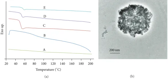
3.7. Issue of Miscibility or Immiscibility. DSC and TEM were
employed to assess the phase miscibility of PLGA/chitosan nanofibers produced by electrospinning. DSC thermograms of the second heat run for pure chitosan, PVA pow-der, electrospun mats of PLGA, PLGA/chitosan/PVA and.,
PLGA/chitosan are shown in Figure 9(a). As can be seen,
the glass transition temperature(𝑇𝑔) of chitosan is around
153∘C which is in accordance with that reported by other
researchers [42–44]. The additional inclination of DSC curve
appearing at about 92∘C for chitosan can be explained by
the water-induced relaxation [44]. The 𝑇𝑔 of PLGA and
PVA are 45.2∘C and 74.8∘C, respectively. As can be seen,
the DSC curves of PLGA/chitosan/PVA and PLGA/chitosan
mats show a 𝑇𝑔 of 47∘C and 48.2∘C, respectively.
More-over, no additional peaks are observed when compared with the thermograms of pure PLGA, PVA, and chitosan.
0 1 2 3 4 5 6 7 0 20 40 60 80 100 120 T en si le st re ss (MP a) Tensile strain (%) A B C (a) 0 1 2 3 4 5 6 0 20 40 60 T ensi le st re ss ( M P a) Tensile strain (%) A B C (b)
Figure 6: Tensile strength curves of (A) PLGA, (B) PLGA/chitosan/PVA, and (C) PLGA/chitosan electrospun mats under (a) dry and (b) wet conditions. 0 20 40 60 80 100 0 7 14 21 28 35 42 49 56 63 M ass loss (%)
Degradation time (days) PLGA/chitosan PLGA (a) 1 𝜇m (b) 20 𝜇m (c)
Figure 7: Degradation of PLGA/chitosan and PLGA nanofibers: (a) comparing the mass loss percentage at different time points, (b) and (c)
SEM image of PLGA/chitosan and PLGA nanofibers, respectively, after 14 days of incubation in PBS solution at 37∘C.
PLGA/Chitosan blends relative to the𝑇𝑔 of pure materials
indicates nonmiscible blends as declared by Wang et al. [45].
However the absence of any considerable inclination around
𝑇𝑔of PVA, in the DSC thermogram of PLGA/chitosan/PVA,
suggests miscibility of PVA and chitosan [46]. These DSC
results imply that PLGA and chitosan have poor miscibil-ity. In spite of the conclusions reached from DSC ther-mograms, TEM cross-sectional image of PLGA
(16%)/chi-tosan(4%)/PVA(8%) fibers (Figure 9(b)) indicates a
650 1150 1650 2150 2650 3150 3650 D T ra n smi tt an ce (a.u .) A B C and OH Amide I and
amide II Saccharidegroup
C=O
Wavenumber (cm−1) NH2
Figure 8: FTIR spectra of (A) pure chitosan and electrospun mats of (B) PLGA/chitosan, (C) PLGA/chitosan/PVA, and (D) PLGA.
C D E B A Ex o u p . 20 40 60 80 100 120 140 160 180 200 Temperature (∘C) (a) 200 nm (b)
Figure 9: Miscibility evaluation of PLGA and chitosan in electrospun polyblend of PLGA/chitosan based on (a) DSC thermograms of (A) pure chitosan, (B) pure PVA, and electrospun mats of (C) PLGA, (D) PLGA/chitosan/PVA, and (E) PLGA/chitosan; and (b) TEM image of the cross section of PLGA/chitosan/PVA electrospun sample embedded in EPON.
and chitosan which suggests some miscibility between PLGA and chitosan. It is worth mentioning that, although a clear contrast originating from the segregation of the two polymers
cannot be observed inFigure 9(b), the image indicates that
the system is multiphase. Considering the results of DSC and TEM, the system is better described as microphase separated. Hence, the term described by Utracki as “immiscible but compatible” blends can be applied to the situation. Such a blend conforms to definitions given for compatibility (a visibly homogeneous mixture) and immiscibility (a blend that does not conform to the thermodynamic conditions of
phase stability) [45].
3.8. Cell Metabolic Activity and Proliferation. We used
murine cell lines (NIN 3T3 fibroblast) to evaluate the bio-compatibility of our scaffolds. Considering the fact that 99% of mouse genes have an equivalent in humans, mice are
widely used as a model [47] for function of human cells/genes
in different studies [48, 49]. The metabolic activity of 3T3
fibroblasts on the scaffolds prepared by electrospinning was
determined by an MTS assay after culturing the cells on
the nanofibers over a period of 7 days (Figure 10(a)). It
was found that the metabolic activity of cells on PLGA and PLGA/chitosan nanofibers increases with culture time, similar to the trend observed on tissue culture plates (TCP). PLGA/chitosan nanofibers demonstrated higher number of viable cells in all time points compared to PLGA nanofibers and TCP. A significant increase was also found from first day of culture onto both PLGA/chitosan and PLGA nanofibers to four and seven days after culturing. Cell proliferation studies were performed by counting the number of adhered cells (cell nuclei stained by DAPI) on PLGA and PLGA/chitosan nanofibers. It was found that there is a progressive trend in number of cells adhered to both PLGA and PLGA/chitosan
scaffolds after 1, 4, and 7 days of culturing (Figures 10(b)
and 10(c)). As the rate of proliferation on PLGA/chitosan
scaffold was significantly higher in all time points, com-pared to PLGA, we conclude that PLGA/chitosan represents better cell interactions and would be a good candidate for skin regeneration applications. As mentioned earlier, the
0 2 4 6 8 10 12 1 4 7 M et ab o lic ac ti vi ty
Culture time (day) PLGA/chitosan PLGA PLGA TCP 0 40 80 120 160 1 4 7
Culture time (day)
C ell n u m b er ( ×1 0 3 ) PLGA/chitosan PLGA
1 day 4 days 7 days
PL GA/c hi to sa n (a) (c) (b)
Figure 10: Viability and proliferation of PLGA/chitosan and PLGA nanofibers: (a) metabolic activity of 3T3 fibroblasts on PLGA/chitosan and PLGA nanofibers and TCP measured by an MTS assay, (b) DAPI-stained nuclei of 3T3 fibroblasts on PLGA/chitosan, and PLGA nanofibers
after 1, 4, and 7 days; scale bar representing 100𝜇m, and (c) proliferation of 3T3 fibroblasts on PLGA/chitosan and PLGA nanofibers measured
by counting number of cell nuclei stained by DAPI.
PLGA/chitosan composite scaffold is expected to provide the necessary mechanical support to the tissue to be replaced (here skin) and offer cellular cues to assist in the regeneration of the target tissue. Incorporating the hydrophilic polymer of chitosan into PLGA has successfully triggered the biological responses such as cellular proliferation and metabolic activity in PLGA/chitosan compared to PLGA scaffold, which is
con-sistent with previous reports [18,50]. Rather than higher cell
adhesion and viability observed with the polyblend nanofiber
of PLGA/chitosan, antibacterial properties of chitosan [51,52]
as well as its potential to initiate fibroblast proliferation [53]
(due to gradual release of N-acetyl-𝛽-D-glucosamine during the degradation procedure) make it an appropriate candidate for applications like wound healing and skin regeneration. It should also be considered that chitosan is a hemostat and can lead to natural blood clotting, which restricts its application in several fields of regenerative medicine. However, it is a positive point for wound healing, since the formation of a clot serves as a temporary shield which protects the denuded wound tissues and provides a provisional matrix over and
20 𝜇m 20 𝜇m 10 𝜇m 10 𝜇m 2 𝜇m PLGA 1 day 7 days PL GA/c h it os an
Figure 11: SEM images of 3T3 fibroblasts after 1 and 7 days culturing onto PLGA/chitosan and PLGA nanofibers.
through which cells can migrate during the repair process
[54].
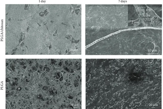
3.9. Cell Morphology. The SEM micrographs of fibroblasts
cultured on PLGA and PLGA/chitosan nanofibers for 1 and
7 days are represented in Figure 11. It can be seen that
fibroblasts attached on both the membranes (after 1 day) and changed their original round shape to elongated shape. However, they have covered the surface of scaffolds after 7 days, making a multilayer in PLGA/chitosan scaffold (the image on the corner of PLGA/chitosan 7 days), confirming the higher proliferation rate of fibroblasts on PLGA/chitosan nanofibers.
4. Conclusion
A method for producing electrospun PLGA/chitosan mat from a single setup has been presented which alleviates the need for two sets of separate electrospinning systems or a coaxial electrospinning setup. Moreover, the possi-bility of having different PLGA to chitosan ratios in the PLGA/chitosan nanofibers has been confirmed by SEM images of electrospun mats obtained from homogenous emulsions of PLGA/chitosan/PVA. The polyblend structure is much more hydrophilic than pure PLGA which is a key point for improved cell-scaffold interactions. FT-IR spectra show no chemical interaction between PLGA and chitosan. Mechanical tests show that the nanofibrous mats of PLGA/chitosan enjoy enough strength in both dry and wet conditions for many biomedical applications. DSC, SEM, and TEM results indicate that the emulsion system has been capable of introducing a compatible, homogenously distributed mixture of PLGA and chitosan in PLGA/chitosan
nanofibers. We observed that both the electrospun PLGA and PLGA/chitosan scaffolds promote the fibroblast attachment and proliferation. Favorable interaction between cell-cell and cell-matrix was also demonstrated by cell morphology seen in SEM images. Considering higher rate of viability and proliferation in PLGA/chitosan nanofibers, it can be assumed that such a composite scaffold fabricated through emulsion electrospinning would be an appropriate candidate for skin tissue engineering applications.
Conflict of Interests
The authors declare there is no conflict of interests regarding the publication of this paper.
References
[1] C.-Y. Wang, K.-H. Zhang, C.-Y. Fan, X.-M. Mo, H.-J. Ruan, and F.-F. Li, “Aligned natural-synthetic polyblend nanofibers for peripheral nerve regeneration,” Acta Biomaterialia, vol. 7, no. 2, pp. 634–643, 2011.
[2] Q. P. Pham, U. Sharma, and A. G. Mikos, “Electrospinning of polymeric nanofibers for tissue engineering applications: a review,” Tissue Engineering, vol. 12, no. 5, pp. 1197–1211, 2006. [3] S. G. Kumbar, R. James, S. P. Nukavarapu, and C. T. Laurencin,
“Electrospun nanofiber scaffolds: engineering soft tissues,” Biomedical Materials, vol. 3, no. 3, Article ID 034002, pp. 1–15, 2008.
[4] K. S. Rho, L. Jeong, G. Lee et al., “Electrospinning of col-lagen nanofibers: effects on the behavior of normal human keratinocytes and early-stage wound healing,” Biomaterials, vol. 27, no. 8, pp. 1452–1461, 2006.
[5] X. M. Mo, C. Y. Xu, M. Kotaki, and S. Ramakrishna, “Electro-spun P(LLA-CL) nanofiber: a biomimetic extracellular matrix
for smooth muscle cell and endothelial cell proliferation,” Biomaterials, vol. 25, no. 10, pp. 1883–1890, 2004.
[6] K. Ma, C. K. Chan, S. Liao, W. Y. K. Hwang, Q. Feng, and S. Ramakrishna, “Electrospun nanofiber scaffolds for rapid and rich capture of bone marrow-derived hematopoietic stem cells,” Biomaterials, vol. 29, no. 13, pp. 2096–2103, 2008.
[7] M. Li, M. J. Mondrinos, X. Chen, M. R. Gandhi, F. K. Ko, and P. I. Lelkes, “Co-electrospun poly(lactide-co-glycolide), gelatin, and elastin blends for tissue engineering scaffolds,” Journal of Biomedical Materials Research A, vol. 79, no. 4, pp. 963–973, 2006.
[8] E. D. Boland, G. E. Wnek, D. G. Simpson, K. J. Pawlowski, and G. L. Bowlin, “Tailoring tissue engineering scaffolds using electrostatic processing techniques: a study of poly(glycolic acid) electrospinning,” Journal of Macromolecular Science A, vol. 38, no. 12, pp. 1231–1243, 2001.
[9] H. Nie, L. Y. Lee, H. Tong, and C.-H. Wang, “PLGA/chitosan composites from a combination of spray drying and supercrit-ical fluid foaming techniques: new carriers for DNA delivery,” Journal of Controlled Release, vol. 129, no. 3, pp. 207–214, 2008. [10] N. Bhattarai, D. Edmondson, O. Veiseh, F. A. Matsen, and
M. Zhang, “Electrospun chitosan-based nanofibers and their cellular compatibility,” Biomaterials, vol. 26, no. 31, pp. 6176– 6184, 2005.
[11] S. A. Sell, P. S. Wolfe, K. Garg, J. M. McCool, I. A. Rodriguez, and G. L. Bowlin, “The use of natural polymers in tissue engi-neering: a focus on electrospun extracellular matrix analogues,” Polymers, vol. 2, no. 4, pp. 522–553, 2010.
[12] J. A. Matthews, G. E. Wnek, D. G. Simpson, and G. L. Bowlin, “Electrospinning of collagen nanofibers,” Biomacromolecules, vol. 3, no. 2, pp. 232–238, 2002.
[13] Z.-M. Huang, Y.-Z. Zhang, M. Kotaki, and S. Ramakrishna, “A review on polymer nanofibers by electrospinning and their applications in nanocomposites,” Composites Science and Tech-nology, vol. 63, no. 15, pp. 2223–2253, 2003.
[14] L. Huang, R. A. McMillan, R. P. Apkarian, B. Pourdeyhimi, V. P. Conticello, and E. L. Chaikof, “Generation of synthetic elastin-mimetic small diameter fibers and fiber networks,” Macromolecules, vol. 33, no. 8, pp. 2989–2997, 2000.
[15] H. Onishi and Y. Machida, “Biodegradation and distribution of water-soluble chitosan in mice,” Biomaterials, vol. 20, no. 2, pp. 175–182, 1999.
[16] Y. M. Yang, W. Hu, X. D. Wang, and X. S. Gu, “The controlling biodegradation of chitosan fibers by N-acetylation in vitro and in vivo,” Journal of Materials Science, vol. 18, no. 11, pp. 2117–2121, 2007.
[17] M. N. V. Ravi Kumar, “A review of chitin and chitosan applica-tions,” Reactive and Functional Polymers, vol. 46, no. 1, pp. 1–27, 2000.
[18] X.-H. Chu, X.-L. Shi, Z.-Q. Feng, Z.-Z. Gu, and Y.-T. Ding, “Chitosan nanofiber scaffold enhances hepatocyte adhesion and function,” Biotechnology Letters, vol. 31, no. 3, pp. 347–352, 2009. [19] A. Wang, Q. Ao, W. Cao et al., “Porous chitosan tubular scaf-folds with knitted outer wall and controllable inner structure for nerve tissue engineering,” Journal of Biomedical Materials Research A, vol. 79, no. 1, pp. 36–46, 2006.
[20] J.-H. Jang, O. Castano, and H.-W. Kim, “Electrospun materials as potential platforms for bone tissue engineering,” Advanced Drug Delivery Reviews, vol. 61, no. 12, pp. 1065–1083, 2009. [21] K. Ohkawa, D. Cha, H. Kim, A. Nishida, and H. Yamamoto,
“Electrospinning of chitosan,” Macromolecular Rapid Commu-nications, vol. 25, no. 18, pp. 1600–1605, 2004.
[22] S. Haider, W. A. Al-Masry, N. Bukhari, and M. Javid, “Prepa-ration of the chitosan containing nanofibers by electrospinning chitosan-gelatin complexes,” Polymer Engineering and Science, vol. 50, no. 9, pp. 1887–1893, 2010.
[23] N. Bhattarai, Z. Li, J. Gunn et al., “Natural-synthetic polyblend nanofibers for biomedical applications,” Advanced Materials, vol. 21, no. 27, pp. 2792–2797, 2009.
[24] B. Duan, L. Wu, X. Yuan et al., “Hybrid nanofibrous membranes of PLGA/chitosan fabricated via an electrospinning array,” Journal of Biomedical Materials Research A, vol. 83, no. 3, pp. 868–878, 2007.
[25] B. Duan, X. Yuan, Y. Zhu et al., “A nanofibrous composite mem-brane of PLGA-chitosan/PVA prepared by electrospinning,” European Polymer Journal, vol. 42, no. 9, pp. 2013–2022, 2006. [26] L. Wu, H. Li, S. Li et al., “Composite fibrous membranes of
PLGA and chitosan prepared by coelectrospinning and coaxial electrospinning,” Journal of Biomedical Materials Research A, vol. 92, no. 2, pp. 563–574, 2010.
[27] Z. X. Meng, W. Zheng, L. Li, and Y. F. Zheng, “Fabrication, char-acterization and in vitro drug release behavior of electrospun PLGA/chitosan nanofibrous scaffold,” Materials Chemistry and Physics, vol. 125, no. 3, pp. 606–611, 2011.
[28] J. D. Schiffman and C. L. Schauer, “Cross-linking chitosan nanofibers,” Biomacromolecules, vol. 8, no. 2, pp. 594–601, 2007. [29] H. Qi, P. Hu, J. Xu, and A. Wang, “Encapsulation of drug reser-voirs in fibers by emulsion electrospinning: morphology char-acterization and preliminary release assessment,” Biomacro-molecules, vol. 7, no. 8, pp. 2327–2330, 2006.
[30] Z. X. Meng, Y. S. Wang, C. Ma, W. Zheng, L. Li, and Y. F. Zheng, “Electrospinning of PLGA/gelatin randomly-oriented and aligned nanofibers as potential scaffold in tissue engineer-ing,” Materials Science and Engineering C, vol. 30, no. 8, pp. 1204–1210, 2010.
[31] C. Zhang, X. Yuan, L. Wu, Y. Han, and J. Sheng, “Study on morphology of electrospun poly(vinyl alcohol) mats,” European Polymer Journal, vol. 41, no. 3, pp. 423–432, 2005.
[32] X. Geng, O.-H. Kwon, and J. Jang, “Electrospinning of chitosan dissolved in concentrated acetic acid solution,” Biomaterials, vol. 26, no. 27, pp. 5427–5432, 2005.
[33] Y. You, J. H. Youk, S. W. Lee, B.-M. Min, S. J. Lee, and W. H. Park, “Preparation of porous ultrafine PGA fibers via selective dissolution of electrospun PGA/PLA blend fibers,” Materials Letters, vol. 60, no. 6, pp. 757–760, 2006.
[34] M. N. Kim, J. Koh, Y. Lee, and H. Kim, “Preparation of pva/pan Bicomponent nanofiber via electrospinning and selective dis-solution,” Journal of Applied Polymer Science, vol. 113, no. 1, pp. 274–282, 2009.
[35] M. Sano, O. Hosoya, S. Taoka et al., “Relationship between solubility of chitosan in alcoholic solution and its gelation,” Chemical and Pharmaceutical Bulletin, vol. 47, no. 7, pp. 1044– 1046, 1999.
[36] N. T. Hiep and B.-T. Lee, “Electro-spinning of PLGA/PCL blends for tissue engineering and their biocompatibility,” Jour-nal of Materials Science, vol. 21, no. 6, pp. 1969–1978, 2010. [37] D. Liang, B. S. Hsiao, and B. Chu, “Functional electrospun
nanofibrous scaffolds for biomedical applications,” Advanced Drug Delivery Reviews, vol. 59, no. 14, pp. 1392–1412, 2007. [38] F. Sabol, L. Dancakova, P. Gal et al., “Immunohistological
changes in skin wounds during the early periods of healing in a rat model,” Veterinarni Medicina, vol. 57, no. 2, pp. 77–82, 2012.
[39] A. F. Laplante, L. Germain, F. A. Auger, and V. Moulin, “Mechanisms of wound reepithelialization: Hints from a tissue-engineered reconstructed skin to long-standing questions,” The FASEB Journal, vol. 15, no. 13, pp. 2377–2389, 2001.
[40] J. Brugnerotto, J. Lizardi, F. M. Goycoolea, W. Arg¨uelles-Monal, J. Desbri`eres, and M. Rinaudo, “An infrared investigation in relation with chitin and chitosan characterization,” Polymer, vol. 42, no. 8, pp. 3569–3580, 2001.
[41] T. S. George, K. Samy, S. Guru, and N. Sankaranarayanan, “Extraction, purification and characterization of chitosan from endophytic fungi isolated from medicinal plants,” World Journal of Science and Technology, vol. 1, pp. 43–48, 2011.
[42] Y. Dong, Y. Ruan, H. Wang, Y. Zhao, and D. Bi, “Studies on glass transition temperature of chitosan with four techniques,” Journal of Applied Polymer Science, vol. 93, no. 4, pp. 1553–1558, 2004.
[43] K. Ogura, T. Kanamoto, M. Itoh, H. Miyashiro, and K. Tanaka, “Dynamic mechanical behavior of chitin and chitosan,” Polymer Bulletin, vol. 2, no. 5, pp. 301–304, 1980.
[44] E. A. El-Hefian, E. S. Elgannoudi, A. Mainal, and A. H. Yahaya, “Characterization of chitosan in acetic acid: rheological and thermal studies,” Turkish Journal of Chemistry, vol. 34, no. 1, pp. 47–56, 2010.
[45] L. Wang, Z. Zhang, H. Chen, S. Zhang, and C. Xiong, “Prepa-ration and characterization of biodegradable thermoplastic Elastomers (PLCA/PLGA blends),” Journal of Polymer Research, vol. 17, no. 1, pp. 77–82, 2010.
[46] I. Arvanitoyannis, I. Kolokuris, A. Nakayama, N. Yamamoto, and S.-I. Aiba, “Physico-chemical studies of chitosan-poly(vinyl alcohol) blends plasticized with sorbitol and sucrose,” Carbohy-drate Polymers, vol. 34, no. 1-2, pp. 9–19, 1997.
[47] D. Nguyen and X. Tian, “The expanding role of mouse genetics for understanding human biology and disease,” DMM Disease Models and Mechanisms, vol. 1, no. 1, pp. 56–66, 2008. [48] B. Ma, J. Xie, J. Jiang, and J. Wu, “Sandwich-type fiber scaffolds
with square arrayed microwells and nanostructured cues as microskin grafts for skin regeneration,” Biomaterials, vol. 35, pp. 630–641, 2014.
[49] E. Ranzato, M. Patrone, M. Pedrazzi, and B. Burlando, “Hmgb1 promotes wound healing of 3T3 mouse fibroblasts via rage-dependent ERK1/2 activation,” Cell Biochemistry and Biophysics, vol. 57, no. 1, pp. 9–17, 2010.
[50] C.-H. Jou, W.-C. Chen, M.-C. Yang et al., “In vitro biocompati-bility of three-dimensional chitosan scaffolds immobilized with chondroitin-6-sulfate,” Polymers for Advanced Technologies, vol. 19, no. 5, pp. 377–384, 2008.
[51] N. Charernsriwilaiwat, P. Opanasopit, T. Rojanarata, and T. Ngawhirunpat, “Lysozyme-loaded, electrospun chitosan-based nanofiber mats for wound healing,” International Journal of Pharmaceutics, vol. 427, no. 2, pp. 379–384, 2012.
[52] M. Ignatova, Z. Petkova, N. Manolova, N. Markova, and I. Rashkov, “Non-woven fibrous materials with antibacterial properties prepared by tailored attachment of quaternized chi-tosan to electrospun mats from maleic anhydride copolymer,” Macromolecular Bioscience, vol. 12, no. 1, pp. 104–115, 2012. [53] R. Jayakumar, M. Prabaharan, P. T. Sudheesh Kumar, S. V. Nair,
and H. Tamura, “Biomaterials based on chitin and chitosan in wound dressing applications,” Biotechnology Advances, vol. 29, no. 3, pp. 322–337, 2011.
[54] P. Martin, “Wound healing—aiming for perfect skin regenera-tion,” Science, vol. 276, no. 5309, pp. 75–81, 1997.
Submit your manuscripts at
http://www.hindawi.com
Scientifica
Hindawi Publishing Corporation http://www.hindawi.com Volume 2013Corrosion
International Journal ofHindawi Publishing Corporation
http://www.hindawi.com Volume 2013
Hindawi Publishing Corporation
http://www.hindawi.com Volume 2013
Polymer Science
International Journal ofISRN Corrosion
Hindawi Publishing Corporation
http://www.hindawi.com Volume 2013
Hindawi Publishing Corporation
http://www.hindawi.com Volume 2013
Composites
Journal ofAdvances in
Materials Science and Engineering
Hindawi Publishing Corporation
http://www.hindawi.com Volume 2013
International Journal of
Biomaterials
Hindawi Publishing Corporation
http://www.hindawi.com Volume 2013
ISRN Ceramics
Hindawi Publishing Corporation
http://www.hindawi.com Volume 2013
Hindawi Publishing Corporation
http://www.hindawi.com Volume 2013
Materials
Journal ofNanotechnology
Hindawi Publishing Corporation
http://www.hindawi.com Volume 2013
Journal of
ISRN
Materials Science
Hindawi Publishing Corporation
http://www.hindawi.com Volume 2013
Hindawi Publishing Corporation
http://www.hindawi.com Volume 2013 Hindawi Publishing Corporation
http://www.hindawi.com Volume 2013
The Scientific
World Journal
ISRN
Nanotechnology
Hindawi Publishing Corporation
http://www.hindawi.com Volume 2013
Nanoparticles
Journal ofHindawi Publishing Corporation
http://www.hindawi.com Volume 2013
Research
Hindawi Publishing Corporation
http://www.hindawi.com Volume 2013
Hindawi Publishing Corporation
http://www.hindawi.com Volume 2013
Metallurgy
Journal ofInternational
Hindawi Publishing Corporation
http://www.hindawi.com Volume 2013
ISRN
Polymer Science
Hindawi Publishing Corporation
http://www.hindawi.com Volume 2013
N
a
no
ma
te
ria
ls
Hindawi Publishing Corporation
http://www.hindawi.com Volume 2013 Journal of