Characterization of
adventitious root formation
in Populus species and
Norway spruce
Characterization of
adventitious root formation
in Populus species and
Norway spruce
Sanaria Abbas Jaafar Alallaq
Umeå Plant Science Centre (UPSC) Department of Plant Physiology
Umeå University Umeå 2021
This work is protected by the Swedish Copyright Legislation (Act 1960:729) Dissertation for PhD
© Sanaria Alallaq, 2021 ISBN:978-91-7855-538-3
ISBN digital version: 978-91-7855-539-0 Cover design: Sanaria Alallaq
This thesis is dedicated to:
The soul of my father, the man who taught me
perform all of life's tasks, no matter how big or small.
My mother
My sisters and my brother
My husband and my two daughters
Who always encouraged me to go on every adventure
Especially this one
Their support and drive are what has raised me to be the person
Table of Contents
Abstract ... iii Sammanfattning ... v Abbreviations ... vii List of chapters ... ix 1. General Introduction ... 11.1. Why is it important to study adventitious rooting? ... 1
1.2. Anatomy and histology analysis of adventitious roots ... 4
1.3. Role of phytohormones in the control of adventitious root formation ... 6
1.3.1. The key role of auxin in the control of adventitious root formation ... 6
1.3.2. Role of other phytohormones in the control of adventitious root development .... 13
1.3.2.1.Jasmonic acid has a controversial role in the control of adventitious root development ... 13
1.3.2.2.The role of cytokinins in the control of adventitious root development ... 19
1.3.2.3.The role of salicylic acid during adventitious root development ... 21
1.3.2.4.The role of abscisic acid during adventitious root development ... 21
1.3.2.5.The role of ethylene during adventitious root development ... 22
1.3.2.6.Role of gibberellins (GAs), strigolactones (SL) and brassinosteroids (BRs) in the control of adventitious root development ... 23
1.4. Environmental factors influencing adventitious root formation ... 26
1.4.1. Mineral nutrition ... 26
1.4.2. Temperature ... 27
1.4.3. Light: an environmental cue that controls adventitious root development ... 27
1.4.3.1.The role of light in adventitious root formation ... 31
1.5. Adventitious rooting in trees ... 33
2. Aim of Thesis ... 40
3. Results and discussion ... 41
Paper I: Red light controls adventitious root regeneration by modulating hormone homeostasis in Picea abies seedlings ... 41
Paper II: Genome wide comparative transcriptomic analysis of the cambium tissue from easy-to-root and difficult-to-root Populus genotypes ... 55
Paper III: Characterization of adventitious root formation in aspen clones from the Swedish Aspen collection ... 65
4. Conclusion and perspectives ... 69
5. Acknowledgement ... 72
Abstract
Adventitious root (AR) formation is a form of post-embryonic development and is a key adaptive trait in plants. De novo adventitious root regeneration represents an elegant evolutionary innovation that allows many plant species to multiply through vegetative propagation; it is widely used in forestry and horticulture to multiply elite genotypes. However, several tree species with high economic and ecological value are difficult to root, and the genetic and molecular bases underlying AR regeneration remain largely elusive. Recently our laboratory showed that jasmonate (JA) and cytokinins (CK) act cooperatively to repress AR initiation (ARI) in Arabidopsis hypocotyls, while auxin positively controls ARI by repressing this negative effect. With the recent availability of the reference genomes of Populus spp. and Norway spruce (Picea abies), the aim of this thesis is to explore the molecular and mechanistic foundations of AR formation in woody species and check whether or not there is conservation of the molecular mechanisms identified in Arabidopsis. First, physiological, molecular and hormonic approaches coupled with extensive anatomical analysis were combined to explore the role of light spectral quality in the control of ARI in P. abies de-rooted seedlings. We showed that constant red light (cRL) promotes ARI by reducing the content of the wound-induced phytohormones JA and JA-isoleucine and repressing the accumulation of the isopentyl-adenine-type cytokinins. These results suggest that the cooperative role of JA and CK signaling in the repression of ARI is evolutionarily conserved. Next we compared transcriptomic data from the cambium tissue of woody stem cuttings of the hybrid aspen T89, which is difficult-to-root, and from the hybrid poplar OP42, which is easy-to-root, under hydroponic conditions. The analyses revealed high transcriptional activity in OP42, with twice as many transcription factors differentially expressed in OP42 24 hours after cutting compared to T89. Although we did not observe significant differences in the expression of Auxin response factor (ARF) genes between the two genotypes, the production of transgenic plants downregulating or over-expressing ARF6, 8 or 17 confirmed that PtARF6 and PtARF8 positively and PtARF17 negatively regulate AR development in transgenic hybrid aspen T89. Interestingly, the expression of MYC2 orthologs as well as the expression of several genes involved in JA signaling increased more in T89 than in OP42, suggesting that JA could be a negative regulator of ARI in Populus spp. We also showed that overexpressing PtMYC2 led to a reduced number of ARs in hybrid aspen T89 cuttings. In addition, many genes encoding ROS scavenging proteins such as peroxidases or GSTs were significantly differentially expressed in OP42 24 h after cutting but not in T89, which is interesting since peroxidase activity has often been positively correlated with ARI. In parallel to this research, we characterized the rooting phenotype of clones from the Swedish Aspen (SwAsp)
rooting ability as well as different root system establishment between the clones. We analyzed the expression of some genes known to be involved in AR development in selected clones with contrasting AR phenotypes but could not identify any correlation between gene expression and rooting phenotype. A transcriptomic analysis of selected clones, with contrasting AR phenotypes, could be a useful tool in the identification of marker genes, which can be used for future selection of the best rooting clones of Populus or other economically important trees in breeding programs.
Key words
Adventitious root, Conifers, Picea abies, auxin, cytokinins, jasmonate, red light, Populus spp., hybrid poplar, hybrid aspen, cambium, stem cuttings, P. tremula, Swedish Aspen (SwAsp) collection.
Sammanfattning
Adventivrötters (AR) bildning är ett post-embryonalt utvecklingsprogram och en viktigt anpassningsegenskap hos växter. De novo-generering av AR representerar en elegant evolutionär innovation som möjliggör vegetativ propagering hos många växtarter. Detta används frekvent inom skogsindustri och hortikultur för att föröka önskevärda genotyper. Flera arter av träd av högt ekonomiskt och ekologiskt intresse är däremot svåra att propagera, och de underliggande molekylära grunderna bakom ARs regenerering har i stort förblivit okända. Nyligen visade vårt forskningsgrupp att jasmonat (JA) och cytokiner (CK) agerar kooperativt för att hämma AR-initiering (ARI) i hypocotylen hos Arabidopsis thaliana, meda auxin kontrollerar AR-initiering positivt genom att hämma denna hämmande effect. I och med nylig tillgång till referensgenom i Populus spp. och gran (Picea abies), så ämnar denna anvhandling att undersöka den molekylära och mekanistiska grunden som ligger bakom AR-bildning i vedbildande arter, och att undersöka huruvida de molekylära mekanismer identifierade i Arabidopsis är evolutionärt konserverade. Först har jag kombinerat fysiologiska, molekylära och hormonella metoder tillsammans med extensiv anatomisk analys för att utforska rollen hos ljus spektralkvalitet för AR-initiering i avrotade P. abies-groddar. Vi visade att konstant rött ljus (cRL) främjar AR-initiering genom att minska halterna av de skadeinducerade hormonerna JA och JA-isoleucine, samt genom att hämma ackumulering av isopentyl-adenine-typer av cytokiner. Dessa resultat tyder på att den kooperativa rollen hos JA och CK-signalering för hämmande av AR-initiering är evolutionärt konserverad. Efter detta jämförde vi transkriptom-data från cambium-vävnad i vedstamsnitt hos hybridasp T89, som är svår att rota samt hybridasp OP42 som är enkel att rota, under hydroponiska förutsättningar. Analyserna visade hög transkriptionell aktivitet I OP42, med två gånger fler transkriptionsfaktorer differentiellt uttryckta I OP42 24 timmar efter snitt jämfört med T89. Även om vi inte observerade signifikanta skillnader i uttrycksnivåer hos auxin-responsfaktorer (ARF)-gener mellan de två genotyperna så såg vi att transgena växter med ned- eller uppreglering av ARF6, 8 eller 17 bekräftade att PtARF6, PtARF8 positivt reglerar, samt PtARF17 negativt reglerar AR-utveckling i transgena hybridaspar. Intressant var att uttryck av MYC2-ortologer samt uttryck av flera gener involverade I JA-signalering ökade mer I T89 än I OP42. Detta indikerar att JA möjligen reglerar AR-initiering negativt I Populus spp. Vi visade också att överuttryck av PtMYC2 ledde till reducerat antal AR I hybridasp-snitt. Dessutom observerade vi att flera gener som kodar för ROS-rensande proteiner som t.ex. peroxidaser eller GSTs uppvisade significant ändrade uttrycksnivåer I OP42 24 timmar efter snitt, vilket ej skedde I T89. Detta är intressant eftersom peroxidas-aktivitet ofta har visat sig positivt relaterat med AR-initiering. Parallelt med dessa undersökningar så
kollektionen hos in vitro-snitt. Vi observerade en significant variation i rotningsförmåga samt rotsystemsetablering mellan klonerna. Vi analyserade uttrycksnivåer av gener kända för att reglera AR-utveckling i utvalda kloner med kontrasterande AR-fenotyper, men kunde inte finna någon korrelation mellan genuttryck of rotningsfenotyp. Transkriptomanalys av utvalda kloner med kontrasterande AR-fenotyp skulle kunna utgöra ett användbart redskap för identification av markörgener, vilka kan användas för framtida selection av bästa rotningskloner i Populus eller andra ekonomiskt viktiga trädarter i förädlingsprogram.
Abbreviations
AR Adventitious root
ARF Auxin response factor
AOS allene oxide synthase
AOC allene oxide cyclase
BAP 6-benzylaminopurine
cWL Constant white light
cRL Constant red light
CK Cytokinin
cis-OPDA cis-12-oxo-phytodienoic acid
DAC Days after cutting
DEG differentially expressed genes
dnOPDA dinor-oxo-phytodienoic acid
4,5ddh-JA 4,5-didiehydrjasmonate
GO gene ontology
IAA Indole -3-acetic acid
IBA Indole butyric acid
iPR iP ribosides
iPRMP iP riboside 5′-monophosphate
iP-types isopentyl-adenine-types
JA Jasmonic acid
JA-Ile Jasmonoyl-isoleucine
JAR1 jasmonate resistent1/GH311
JMT JA carboxyl methyltransferase
LOX lipoxygenase
LCM Laser capture Microdissection
LED Light emitting diodes
MeJA methyl jasmonate
NAA 1-Naphtalene acetic acid
OPR3 OPDA reductase
OPC6 6 -(3-oxo-2-(pentl-2-enyl) cyclopentyl) hexanoic acid
OPC8 8-(3-oxo-2-(pentl-2-enyl) cyclopentyl) octanoic acid
12-OPDA 12-oxo-phytodienoic acid
PAT Polar auxin transport
ROS Reactive oxygen species
SwAsp The Swedish Aspen collection
tnOPDA tetranor-OPDA
18:3 α-linolenic acid
List of chapters
This thesis is a summary of the following three chapters (papers):
I. Sanaria Alallaq, Alok Ranjan, Federica Brunoni, Ondřej Novák,
Abdellah Lakehal, and Catherine Bellini. Red light controls adventitious root regeneration by modulating hormone homeostasis in Picea abies seedlings.2020. Frontiers Plant Science. 11, 1–14. doi:10.3389/fpls.2020.586140.
II. Alok Ranjan*, Irene Perrone*, Sanaria Alallaq*, Rajesh Singh, Federica Brunoni1 Annegret Kohler, Francis Martin, Rishi Bhalerao,
Valérie Legué, and Catherine Bellini. Genome wide comparative transcriptomic analysis of the cambium tissue from easy-to-root and difficult-to-root Populus genotypes.
* These authors contributed equally to this work. (manuscript).
III. Sanaria Alallaq, Florencia Bannoud, and Catherine Bellini.
Characterization of AR formation in aspen clones from the Swedish Aspen collection. (manuscript).
Additional publication not included in this PhD thesis
Abdellah Lakehal, Asma Dob, Zahra Rahneshan, Ondřej Novák, Sacha Escamez,
Sanaria Alallaq, Miroslav Strand, Hannele Tuominen, and Catherine Bellini.
2020. ETHYLENE RESPONSE FACTOR 115 integrates jasmonate and cytokinin signaling machineries to repress adventitious rooting in Arabidopsis. New Phytologist. nph.16794. doi:10.1111/nph.16794.
1. General Introduction
Land plants play a vital role in everyday human activity. They provide us with food, oxygen, medicine, fuel, fibers materials for tools and shelter and they are also essential to the world’s wildlife (White et al., 2013). Plants have a unique feature of being able to reproduce in two ways: sexual reproduction through seeds and asexual propagation also called vegetative propagation. The latter is possible thanks to plants’ ability to develop adventitious roots (ARs) from non-root tissues such as stems, leaves or hypocotyls (Bellini et al., 2014; Steffens & Rasmussen, 2016). Adventitious root formation is a complexquantitative trait regulated by multiple endogenous factors such as phytohormones, phenolic compounds, polyamines or mechanisms related to the aging process, and environmental factors like light, temperature or nutrients (reviewed in Geiss et al., 2018). For many species, AR formation is intrinsically part of development and occurs post-embryonically. This is the case for monocots, for which ARs represent the main root system, but also for many naturally vegetatively propagated dicotyledonous plants like strawberries (Fragaria spp.), African violets (Saintpaulia spp.) or blackberries (Rubus spp.) (Figure 1A and B). Moreover, AR may be induced as a stress response to, for example, darkness, flooding or wounding (Figure 1C and D). These stress situations are not only caused by changes in the environment, but can be induced mechanically by wounding during tissue culture techniques (Figure 1C and D) (reviewed in Geiss et al., 2018; Bellini et al., 2014; Steffens & Rasmussen, 2016).
1.1. Why is it important to study adventitious rooting?
The importance of studying adventitious root formation lies in the fact that the ability of plants to undergo vegetative propagation from cuttings has been extensively used in breeding programs to multiply elite genotypes and fix interesting agronomic traits at relatively low cost (Stenvall, 2006; Mauriat et al., 2014).
This process is economically important for forest trees such as Populus spp., Pinus spp., Picea spp. and Eucalyptus spp. as well as horticultural species. One major limitation in clonal propagation of woody species is the highly reduced or rapid loss of ability to form ARs in a number of genotypes (Ragonezi et al., 2010; Legué et al., 2014).
The ability to form adventitious roots varies between plant species, which are generally characterized as easy-to-root or difficult-to-root plants. The former have the ability to form ARs without any special treatment of the cuttings most of the time, while the latter require special treatments of either the mother plant or the cuttings, involving application of phytohormones and/or modifications to their environment (reviewed in Lovell & White, 1986).
In some woody plants, the rooting capacity may decrease after a phase change, from juvenile to mature. Researchers working with woody plants such as Ficus pumila, Prunus avium and Eucalyptus grandis have found that cuttings from young plants readily form ARs but when cuttings are taken from the same adult plant the ability has been lost (Davies et al., 1982; Dick & Leakey, 2006; Abu-Abied et al., 2014).
In conclusion, adventitious rooting is a key step in clonal propagation of economically important horticultural and woody species, therefore it is important to gain a better understanding of the mechanisms that regulate AR initiation (ARI) and development in order to improve its application.
Figure 1: Examples of different types of adventitious roots.
This figure illustrates some examples of the developmental aspect of adventitious rooting. (A) and (B) show types of AR that are intrinsically part of plant development.
(A) Crown roots as an example of AR in monocots (scale bar: 2cm) from https://www.pinterest.com/pin/203295370653221607/.
(B) Adventitious roots during vegetative propagation of strawberries (scale bar: 1cm) from https://growingfruit.org/t/grin-usda-stolon/23666.
(C) and (D) show stress-induced ARs.
(C) AR induced by dark-light transition in Arabidopsis thaliana (scale bar: 0.5 cm) from Gutierrez et al. (2009).
(D) AR induced by wounding in a Populus mico-cutting during in vitro vegetative propagation (scale bar: 0.5 cm) Photo: Sanaria Alallaq.
1.2. Anatomy and histology analysis of adventitious roots
For all plants, the primary root meristem is established during embryogenesis, but lateral and AR meristems are formed post-embryonically (Casson and Lindsey, 2003). While lateral roots are commonly formed from mature pericycle cells of the main roots, ARs develop from different tissues and consequently from different cell types. ARs are also formed after tissue culture regeneration of shoots with or without hormone applications. From the literature, it appears that there is a debate about the number, the nature and the terminology of the histology stages characterizing AR formation (Haissig, 1974; Lovell & White, 1986; Altamura, 1996; da Costa et al., 2013; Guan et al., 2015).
According to Kevers et al. (1997) this process can be distinguished by three phases (Figure 2B). The first phase is the induction phase, which precedes any anatomical event; the second, the initiation phase (cell divisions leading to the formation of internal root meristems); and the third, the expression or
extension phase is characterized by the internal growth of root-primordia and
root emergence. However, according to De Klerk et al. (1999) and Pijut et al., (2011) a fourth stage exists and occurs before the induction phase and consists of cell dedifferentiation, it is followed by the induction phase during which no anatomical changes can be observed; thereafter, cells near the vascular bundles become meristematic and divide. This is followed by the development of dome-like root primordia, and finally root emergence. At the stage when the organized root primordium starts to differentiate and elongate, the vascular tissues also form and connect to the vascular system of the cutting.
Anatomical processes of AR formation have been analyzed in various species thanks to studies performed on cuttings, from vegetative portions of the plant, such as stems (rhizomes, tubers, corms, and bulbs), leaves or roots. It has been shown that ARs can arise from pericycle cells, parenchyma cells, cambium cells, or phloem initials. However, in all cases, the cells are located close to the vascular system.
Figure 2: The process of adventitious root formation in stem cuttings of a dicot tree.
(A) Organization of the tissue layers in the immature stem of a typical woody plant.
(B) The three progressive physiological stages of AR formation (induction, initiation and extension). The five steps indicated by Arabic numbers describe primordium development. AR primordia arise from deep ray cells adjacent to the cambium region, primordia lead to establishment of the main adventitious root (AR) and subsequently grow out and emerge by pushing out epidermal cells. Cell types are coloured as indicated in the key (modified from Guan et al., 2015).
For both herbaceous and woody plants there are two patterns of AR initiation. The indirect pattern consists of the formation of a callus, which is a mass of proliferating undifferentiated cells that often forms at the base of a cutting or after another type of mechanical damage, then root primordia initiate from the newly formed callus tissue. In contrast, in the case of the direct pattern, AR primordia form directly from the cells near the vascular system, without formation of a callus.
These two patterns of ARs can occur in the same species, but in general the indirect pattern is more often observed in difficult-to-root species while the direct pattern is characteristic of easy-to-root species (reviewed in Altamura, 1996; Guan et al., 2015). The localization of AR initiation in tissues may vary from species to species.
The length of the developmental stages and the cellular origin of ARs have been shown to be species- and genotype-dependent (reviewed in Bellini et al., 2014; Geiss et al., 2018). This illustrates the complexity and the variability of the process and the consequent difficulties in identifying early events in complex structures such as stem cuttings.
1.3. Role of phytohormones in the control of adventitious root formation
1.3.1. The key role of auxin in the control of adventitious root formation
The plant hormone auxin or indole acetic acid (IAA) has been considered the master player in the initiation and development of ARs (Haissig, 1974; De Klerk et al., 1999; Bellini et al., 2014). Its exogenous application has a consistent effect across plant taxa in inducing root formation (Pacurar et al., 2014b). Besides IAA, other types of auxins such as Indole-3-butyric acid (IBA), 1-naphthalen acetic acid (NAA) and 2,4-dichlorophenoxyacetic acid (2,4-D) have been used commercially to induce rooting from cuttings of many species because of their efficacy in stimulating ARs (De Klerk et al., 1999; Pandey et al., 2011).
In particular, IBA has been reported to be more effective than the other auxins in a wide range of species. This efficiency of IBA may be due to its stability upon light exposure and higher root-inducing capacity (Epstein & Ludwig- Müller , 1993; Ludwig-Müller et al., 2005; Bellini et al., 2014; Lakehal & Bellini, 2019). IBA is also used in combination with other auxins like 2,4-D or NAA, to stimulate more efficiently ARs in recalcitrant species of economic value (Oinam et al., 2011; Pijut et al., 2011). It is known that cuttings from many species have the ability to form ARs without using exogenous auxin, e.g. Populus spp. (Rigal et al., 2012). In this context, wounding stimulated ARs at the base of cuttings through the accumulation of endogenous auxin via polar auxin transport (PAT) at the site of cutting (Rigal et al., 2012). A high level of free IAA is required to induce ARs especially during the induction phase (Caboni et al., 1997; Gaspar et al., 2003). Bellamine and collaborators confirmed the important role of free IAA in the induction and expression phases at the base of Populus tremula × Populus tremuloides cuttings by using anti-auxins such as 2-phenoxy-2-methyl propionic acid (PBA) (Bellamine et al., 1998). In Eucalyptus globulus, Negishi and collaborators found that the free IAA content was twice as high in easy-to-root cuttings compared to recalcitrant cuttings (Negishi et al., 2011). The levels of auxin are tightly regulated (reviewed in Normanly et al., 2010; Ljung, 2013) and the contributions of transport and biosynthesis to auxin homeostasis have been identified as being essential for AR formation (reviewed in Gonin et al., 2019; Lakehal & Bellini, 2019).
In the model plant Arabidopsis, superroot2-1 (sur2-1) overproduces auxin due to the accumulation of indole-3-acetaldoxime (IAOx), a common intermediate in the IAA and the indole-glucosinolate biosynthesis pathways (Barlier et al., 2000; Mikkelsen et al., 2004). This IAA-overproducing mutant develops an abnormally high number of ARs along the hypocotyl (Barlier et al., 2000; Mikkelsen et al., 2004). Similarly, the activation tagged yuc1-D mutant, which also overproduces auxin, spontaneously forms many ARs along the hypocotyl. The YUCCA1 gene is reported to be directly involved in tryptophan-dependent auxin biosynthesis via the indole-pyruvic acid pathway (Zhao, 2001; Mashiguchi et al., 2011; Stepanova et al., 2011).
Pacurar and collaborators showed that the loss of function of several genes involved in auxin biosynthesis such as ANTHRNILATE SYNTHASE ALPHA 1/WEAK ETHYLENE INSENSITIVE 2 (ASA1/WE12), ANTHRANILATE SYNTHASE b 1/WEAK ETHYLENE INSENSITIVE 7 (ASB1/WE17 ) and TRYP-TOPHAN SYNTHASE BETA1 (TSB1) resulted in a reduced number of ARs in the sur2-1 mutant background (Pacurar et al., 2014a).Chen et al. (2016) showed that the expression levels of both YUC1 and YUC4 increased in the mesophyll cells of leaf explants within four hours of cutting. The same authors showed that the double mutants yuc1yuc4 and yuc2yuc6 were partially unable to produce ARs, whereas the quadruple mutant yuc1yuc2yuc4yuc6 was unable to produce ARs from leafy cuttings. All these results confirm the important role of auxin biosynthesis in adventitious root formation.
Polar auxin transport (PAT) plays an important role in the distribution of IAA and the establishment of IAA gradients (reviewed in Teale et al., 2006; Takahashi, 2013; Lakehal & Bellini, 2019). The surgical removal of the shoot apex, which is the major source of endogenous auxin, results in a reduction in endogenous IAA at the base of cuttings, causing a reduction in rooting (Liu & Reid, 1992). By using inhibitors of PAT, such as naphthylphtalamic acid (NPA) or 1,3,5 triiodobenzoic acid (TIBA), researchers observed a reduction in the development of ARs for many species, including Helianthus annuus, Syringa vulgaris, Petunia hybrida and Oryza sativa (Liu & Reid, 1992; Ford et al., 2002; Ahkami et al., 2013; Lin & Sauter, 2019). These experiments confirmed the importance of auxin biosynthesis at the shoot apex and the pivotal role of PAT in AR formation.
It is well known that IAA moves from cell to cell thanks to transporter proteins such as the influx carrier proteins AUXIN RESISTANT 1 (AUX1) and LIKE AUX1 (LAX), and the efflux carrier proteins such as PIN FORMED (PIN) or ATP BINDING CASSETTE B / MULTI DRUG RESISTANCE / P. GLYCOPROTEIN (ABCB/MDR/PGP) (reviewed in Takahashi, 2013). Li and collaborators found that cotyledon segments of mango (Mangifera indica L.) can form more ARs due to the increasing auxin concentration at the proximal cut surface via auxin influx carriers (Li et al., 2012). Sukumar et al. (2013) showed that excision of the root
cutting due to a 4-fold increase in auxin transport. The role of auxin polar transport was then confirmed by the characterization of mutants. Sukumar et al. (2013) showed that Arabidopsis mutants defective in IAA efflux transport (pin1, pin3, pin7 and abcb19) had a significant reduction in ARI in de-rooted seedlings compared with the wild type. In addition, lines over expressing ABCB19 had enhanced ARI in intact hypocotyls due to increased auxin transport (Sukumar et al., 2013). Simon and collaborators demonstrated that the PIN6 gene had a complex role in the control of auxin transport and homeostasis during AR and lateral root (LR) formation. They showed that the pin6 knock-out mutant produced more ARs in both intact and de-rooted Arabidopsis seedlings compared to the wild type, while the PIN6 overexpressing line developed fewer ARs compared to the wild type even after excision of the primary root (Simon et al., 2016). In rice (Oryza sativa L.), Xu and collaborators found that the OsPIN-FORMED1 (OsPIN1) gene was expressed in root primordia and AR emergence was significantly inhibited in the OsPIN1 RNA-interference lines (Xu et al., 2005). Similarly, Lin and Sauter (2019) found that the OsPIN2 gene is expressed in epidermal cells above AR primordia and its activation controls AR emergence. In addition to auxin polar transport, the homeostasis of auxin is controlled by conjugation with other molecules such as sugars, amino acids, or peptides or through degradation. In Arabidopsis, it has been shown that several members of the GRETCHEN HAGEN3 (GH3) family of acyl amido synthetases mediate conjugation of IAA with amino acids (Staswick et al., 2005; Westfall et al., 2010). Certain IAA conjugates can be hydrolyzed enzymatically and produce free IAA. This is the case when IAA is conjugated with amino acids such as alanine, leucine or phenylalanine (Kowalczyk & Sandberg, 2001; Le Clere et al., 2002). In contrast, IAA conjugation with amino acids such as aspartate or glutamate produces intermediates in the oxidative degradation pathway of IAA (Östin et al., 1998; Tam et al., 2000). The degradation process of auxin is important for the maintenance of the auxin homeostasis in the plant (Pěnčík et al., 2013; Peer et al., 2013). Modification of this pathway can alter AR formation. Butler and Gallagher (2000) showed that, in stem cuttings of apple (Malus domestica), the expression of ADVENTITIOUS ROOTING RELATED OXYGENASE 1 (ARRO-1)
was rapidly upregulated after IBA or IAA treatments to induce AR. This result suggested that this putative auxin oxidase gene could play a role in the regulation of auxin levels during AR formation in stem cuttings of apple.
In Arabidopsis, two genes that encode auxin oxidases have been identified (Mellor et al., 2016; Porco et al., 2016). DIOXYGENASE FOR AUXIN OXIDATION 1 and 2 (AtDAO1 and 2) act in concert with GH3 genes to control auxin levels during plant growth and development (Mellor et al., 2016; Porco et al., 2016). Recently, Lakehal and collaborators (2019) showed that AtDAO1 plays an essential role in auxin-jasmonate crosstalk during ARI in intact Arabidopsis hypocotyls (Lakehal et al., 2019b).
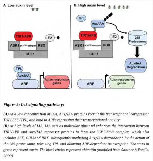
The auxin signaling begins with the interaction between the endogenous IAA, which acts as a molecular glue between the TRANSPORT INHIBITOR1/AUXIN-SIGNALING F-BOX (TIR1/AFB) receptor proteins and the auxin-induced AUXIN/INDOLE-3-ACETIC ACID (AUX/IAA) proteins that are transcriptional repressors in the auxin signaling pathway. Once a specific co-receptor complex is formed, the AUX/IAA proteins are ubiquitylated and targeted for degradation through the 26S proteasome machinery (Dharmasiri et al., 2005; Parry et al., 2009; Salehin et al., 2015).
In the cell, when there is a low concentration of auxin, AUX/IAA repressors bind to members of the AUXIN RESPONSE FACTOR (ARF) transcription factor family and inhibit their transcriptional activity. At high concentrations of auxin, IAA acts as a molecular glue which triggers degradation of the Aux/IAA proteins, releasing the activity of ARFs which induce the expression of auxin-responsive genes (Figure 3) (Santner & Estelle, 2009).
In the model plant Arabidopsis, parts of the auxin signaling network controlling AR formation have been unraveled. Sorin and collaborators found that the AUXIN RESONSE FACTOR 17 (ARF17) gene, negatively regulates AR formation by repressing the expression of three auxin-inducible GH3 genes (GH3.3, GH3.5 and GH3.6) (Sorin et al., 2005). Subsequently, Gutierrez and collaborators found that ARF6 and ARF8 transcription factors are positive regulators of ARI. They were shown to positively regulate the expression of GH3.3, GH3.5 and GH3.6 which, in the context of AR initiation, controls the homeostasis of jasmonate, which negatively controls ARI. Gutierrez et al., (2009) also showed that the three
Figure 3: IAA signaling pathway:
(A) At a low concentration of IAA, Aux/IAA proteins recruit the transcriptional corepressor TOPLESS (TPL) and bind to ARFs repressing their transcriptional activity.
(B) At high levels of IAA, IAA acts as molecular glue and enhances the interaction between
TIR1/AFB and Aux/IAA repressor proteins to form the SCF TIR1/AFB complex, which also
includes ASK, CUL1and RBX, subsequently mediating Aux/IAA degradation by the action of
the 26S proteasome, releasing TPL and allowing ARF-dependent transcription.The stars in
green represent auxin. The black circles represent ubiquitin (modified from Santner & Estelle, 2009).
ARFs regulate each other's expression at the transcriptional level and at the posttranscriptional level by modulating the abundance of their respective regulatory microRNA. The microRNA miR167 controls the transcript amount of the positive regulators ARF6 and ARF8, while the negative regulator ARF17 is regulated by miR160 (Gutierrez et al., 2009, 2012). These transcription factors do not only act in Arabidopsis but probably also in other species. Recently it was shown that the expression of ARF6 and ARF8 genes increased in phloem parenchyma cells in black walnut (Juglans nigra L.) stem cuttings during the early stages of AR primordia formation whereas the expression of ARF17 decreased (Stevens et al., 2018).Lakehal and collaborators showed that the F-box proteins TIR1 and AFB2 control JA homeostasis by promoting the degradation of at least three AUX/IAA (IAA6, 9 and 17) proteins that repress the transcriptional activity of ARF6 and ARF8 (Lakehal et al., 2019a).Overall, auxin-related genes play a central role in regulating AR formation (Pacurar et al., 2014b; Zhang et al., 2020).
Recently, it was shown that several members of the WUSCHEL-related homeobox (WOX) family, including WOX11, WOX12 and WOX5, are induced by auxin and are involved in adventitious rooting in herbaceous and woody plants (Liu et al., 2014). For example, Liu and collaborators found that auxin accumulation activates the expression of WUSCHEL RELATED HOMEBOX 11 and 12 genes (WOX11 and WOX12) in leaf cuttings of Arabidopsis (Liu et al., 2014). WOX11 responds to wounding-induced auxin accumulation in and surrounding the procambium. This gene, redundantly with its homolog WOX12, acts to control the transition of competent cells (procambium or its nearby parenchyma cells) into adventitious root founder cells by upregulating BOUNDARY LATERAL DOMAIN 16 and 29 (LBD16 and LBD29) genes at the base of leaf blade cuttings. In Populus cuttings, Xu et al., (2015b) found that the overexpression of PeWOX11a or PeWOX11b increased the number of ARs per cutting. Li and collaborators confirmed the involvement of PeWOX11a/b in adventitious rooting in hybrid poplar. They also showed that the overexpression of PtoWOX5a in the hybrid P. alba × P. glandulosa, increased the number of ARs but decreased their length (Li et al., 2018).
1.3.2. Role of other phytohormones in the control of adventitious root development
Several studies performed in different model plants and systems have reported the role of different classes of phytohormones as well as their interaction with each other and with the environment during AR development. Auxin appears as the central player which interacts with the other phytohormones in complex networks during the different stages of AR formation (reviewed in da Costa et al., 2013; Bellini et al., 2014b; Lakehal & Bellini, 2019).
1.3.2.1. Jasmonic acid has a controversial role in the control of adventitious root development
The plant hormone Jasmonic acid and its derivatives such as Methyl ester jasmonate (MeJA) or Jasmonoyl-isoleucine (JA-Ile), which is the active form, are collectively called jasmonates (JAs) and are oxylipin-derived hormones. JAs are very important molecules that regulate many genes involved in the control of many physiological processes in plant responses to biotic and abiotic stresses as well as plant growth and development (Wasternack & Strnad, 2018).
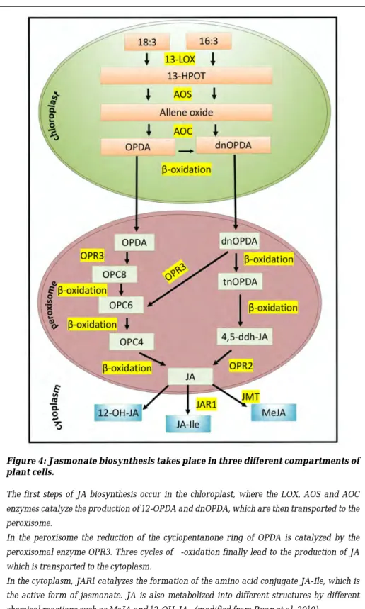
The biosynthesis of JAs has been extensively studied in many varieties of plants but mostly in the model plants Arabidopsis thaliana and Lycopersicon esculentum (tomato). In Arabidopsis, three pathways for the synthesis of JAs have been identified. They include the octadecane pathway starting from α-linolenic acid (18:3) and the hexadecane pathway starting from hexadecatrienoic acid (16:3) (Chini et al., 2018; Ruan et al., 2019). The biosynthesis of JA takes place in three cell compartments (Figure 4). In the chloroplast, the 13-LIPOXYGENASE (LOX) enzymes convert the a-linolenic acid (18:3) (a-LeA)(18:3) and the hexadecatrienoic acid (16:3) into 13-hydroperoxy-octadecatrienoic acid (13-HPOT), then then 13-HPOT is oxidized by the ALLENE OXIDE SYNTHETASE (AOS) enzyme to form the allene oxide which is then converted into12-oxo-phytodienoic acid (12-OPDA) and its 16-carbon homolog the dinor-oxo-phytodienoic acid (dnOPDA) by the ALLENE OXIDE CYCLASE (AOC) enzymes. The 12-OPDA and the dnOPDA are converted to JA in the peroxisome by the 12-OXOPHYTODIENOIC ACID REDUCTASE 3 (OPR3) enzyme, giving rise to formation of the final JA (Feussner & Wasternack, 2002;
Gfeller et al., 2010; Wasternack & Strnad, 2018). In the cytoplasm, JA is converted into active, inactive and partially active structures such as MeJA, JA-Ile, cis-jasmone (CJ) and 12-hydroxyjasmonic acid (12-OH-JA) by different chemical reactions (Reviewed in Ruan et al., 2019) (Figure 4).
The conjugation of JA with the amino acid isoleucine (Ile) by the JASMONATE RESISTANT 1/GRETCHEN HAGEN3.11 (JAR1/GH3.11) enzyme produces the bioactive form jasmonoyl-isoleucine (JA-Ile). JAR1/GH3.11 belongs to group I of the auxin-inducible GH3 family (Staswick et al., 2002; Staswick & Tiryaki, 2004). Interestingly, it was recently shown that three enzymes belonging to group II of the GH3 family contribute to the maintenance of JA homeostasis (Gutierrez et al., 2012). Indeed, GH3.3, GH3.5 and GH3.6 enzymes conjugate free JA with other amino acids such as tryptophan, methionine or aspartate, thereby inactivating it. In this way they contribute to diminishing the JA pool in the intact hypocotyl of Arabidopsis and control AR initiation downstream of auxin (Gutierrez et al., 2012).
Figure 4: Jasmonate biosynthesis takes place in three different compartments of plant cells.
The first steps of JA biosynthesis occur in the chloroplast, where the LOX, AOS and AOC enzymes catalyze the production of 12-OPDA and dnOPDA, which are then transported to the peroxisome.
In the peroxisome the reduction of the cyclopentanone ring of OPDA is catalyzed by the peroxisomal enzyme OPR3. Three cycles of β-oxidation finally lead to the production of JA which is transported to the cytoplasm.
In the cytoplasm, JAR1 catalyzes the formation of the amino acid conjugate JA-Ile, which is the active form of jasmonate. JA is also metabolized into different structures by different chemical reactions such as MeJA and 12-OH-JA. (modified from Ruan et al. 2019).
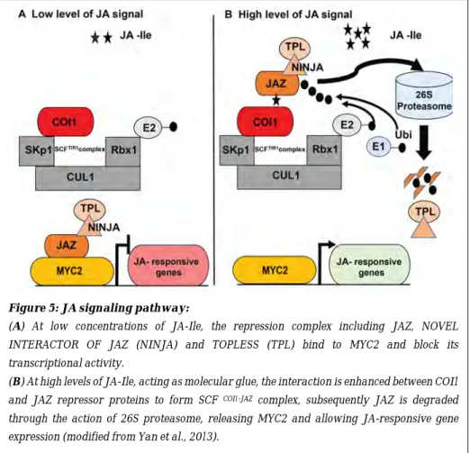
Similar to auxin, the bioactive JA-Ile acts as a molecular glue or ligand necessary for the formation of the coreceptor complexes between JASMONATE ZIM DOMAIN (JAZ) transcriptional repressors and the Skp/Cullin/F-box CORONATINE INSENSITIVE 1 (SCFCOI1) receptor (Hoo & Howe, 2009; Pauwels
& Goossens, 2011). In cells with sufficient bioactive JA-Ile, the JAZ repressor proteins bind to the COI1 receptor to form the Skp/Cullin/F-box CORONATINE INSENSITIVE1-JAZ complex (SCFCOI1-JAZ) (Sheard et al., 2010). This results in
the poly-ubiquitination and degradation of the JAZ repressor proteins through the 26S proteasome pathway, releasing the transcriptional activity of the master regulator MYC2/JASMONATE INSENTIVE1 (MYC2/JIN1) and other MYC transcription factors to trigger the expression of JA-responsive genes (Figure 5). In contrast, when there is a low level of JA-Ile in the cells, JAZ repressor proteins
Figure 5: JA signaling pathway:
(A) At low concentrations of JA-Ile, the repression complex including JAZ, NOVEL INTERACTOR OF JAZ (NINJA) and TOPLESS (TPL) bind to MYC2 and block its transcriptional activity.
(B) At high levels of JA-Ile, acting as molecular glue, the interaction is enhanced between COI1
and JAZ repressor proteins to form SCF COI1-JAZ complex, subsequently JAZ is degraded
bind physically to transcription factors such as MYC2, MYC3 or MYC4, repressing their transcriptional activity (Figure 5) (Yan et al., 2013).
JA plays a role in the control of primary root growth, lateral and adventitious root formation (Staswick et al., 1992; Vellosillo et al., 2007; Gutierrez et al., 2012; Gasperini et al., 2015; Fattorini et al., 2018). However, it appears that the role of JA in the control of ARI depends on the species, the organ and the growth conditions (reviewed in Lakehal & Bellini, 2019).
It has been shown by several researchers that exogenous application of JA inhibits AR formation in various species. For example, Chen and collaborators found that exogenous application of MeJA inhibits AR formation in Bupleurum kaoi cuttings (Chen et al., 2007). Lischweski et al. (2015) showed that leafy stem cuttings of petunia (Petunia hybrida) produced significantly fewer ARs compared to controls after treatment with JA, JA-Ile or OPDA. They also showed that exogenously applied JA repressed the positive effect of auxin (Lischweski et al., 2015).
Guttierrez and collaborators showed that very low concentrations of JA significantly reduced the average number of ARs in Arabidopsis etiolated hypocotyls (Gutierrez et al., 2012). Recently, Fattorini and collaborators found that expression of the negative regulator ARF17 was very quickly induced by exogenously applied MeJA (10 µM) (Fattorini et al., 2018). All these findings support the hypothesis that JA is an inhibitor of AR formation. This hypothesis has been corroborated in Arabidopsis etiolated hypocotyls, by genetic approaches (Gutierrez et al., 2012). For example, the loss-of-function mutants coi1-16, myc2-1, myc2myc3myc4 and also the knockout mutant jar1-12/gh3.1myc2-1, all altered JA signaling, resulting in the development of more ARs compared to the wild type, while the overexpressing 35S:MYC2 and 35S:JAR1 lines developed significantly fewer ARs than the wild type (Gutierrez et al., 2012). These genetic data indicate that the COI1-dependent JA signaling pathway negatively regulates AR formation through MYC2, MYC3 and MYC4 transcription factors (Gutierrez et al., 2012). More recently, Lakehal and collaborators found that the loss-of-function mutants ninja-1 and ninja-2 produced slightly fewer ARs than the wild type, and that the double mutants ninja-1myc2-322B and ninja-2myc2-322B, in which myc2-322B is a gain-of-function mutant, exhibit constitutively upregulated JA signaling with
a very strong reduction in the number of ARs compared to the wild type (Lakehal et al., 2020) Transcriptomic analysis of the ninja-1myc2-322B double mutant showed that many genes involved in JA biosynthesis as well as most JAZ genes were upregulated. Hormone quantification in this mutant confirmed that the levels of cis-OPDA, JA and JA-Ile were significantly higher compared to the wild type (Lakehal et al., 2020).
Nevertheless, some studies present the opposite theory, namely that JA is a positive regulator of ARI. Ahkami et al. (2009) found that excision of petunia cuttings led to rapid accumulation of JA at the wounding site as well as to an accumulation of soluble and insoluble carbohydrates, associated with increased transcriptional and metabolomic reprogramming at the base of the leafy stem cuttings, and an induction of AR formation (Ahkami et al., 2009). They concluded that JA could be the inducer of AR initiation in petunia cuttings. Lischweski and collaborators also proposed that JA acts as a positive regulator for ARI in petunia leafy stem cuttings. They showed that the downregulation by RNA interference of the PhAOC gene, involved in JA biosynthesis, reduced the number of ARs in the cuttings of the AOC-RNAi lines (Lischweski et al., 2015). A positive role for MeJA in promoting ARs in tobacco (Nicotiana tabacum) and Arabidopsis thin cell layers (TCLs) has also been shown (Fattorini et al., 2009, 2018). These authors observed the positive effect of MeJA only when the TCLs were cultured in a rooting medium containing a high (10 µM) concentration of IBA and a low concentration of cytokinin (0.1 µM kinetin) but they did not observe it when the TCLs were kept on hormone-free medium. Zhang and collaborators showed that leaf explants of Arabidopsis treated with coronatine-O-methyloxime (COR-MO) could not develop ARs (Zhang et al., 2019). The COR-MO acts as a JA-Ile competitive antagonist because it exhibits strong activity in preventing COI1-JAZ interaction (Monte et al., 2014), and this resulted in inhibition of the JA signaling machinery (Zhang et al., 2019).
In conclusion, the role of JA in the control of adventitious rooting could depend on the species and/or on the growth conditions. More investigation is needed to better understand the role of JAs during AR development.
1.3.2.2. The role of cytokinins in the control of adventitious root development
Cytokinins (CKs), a group of plant growth regulators, are involved in the regulation of many plant growth and development processes such as cell division, leaf senescence and caulogenesis, including adventitious shoot formation and rhizogenesis (lateral and adventitious root formation). CKs are mainly produced in the roots (Aloni et al., 2005; Hwang & Sakakibara, 2006; Agulló-Antón et al., 2014) although all organs can produce them (Hwang & Sakakibara, 2006; Chickarmane et al., 2012; Kieber & Schaller, 2014). Trans-zeatin riboside (tZR) is considered the major form in the xylem sap, while iso-penthenyl-adenine (iP) type cytokinins are the major form found in the phloem sap (Corbesier et al., 2003; Hirose et al., 2008).Kudo and collaborators proposed a model for long-distance CK transport through the plant vascular system (Kudo et al., 2010); in this model tZR is considered a long-distance messenger for shootward transport while iP is involved in rootward transport.
The role of CKs in AR formation has emerged from studies in various species and systems at different development stages of adventitious rooting. For example, trans-zeatin riboside present in the xylem sap acts as an inhibitor of AR formation in cucumber (Cucumis sativus) hypocotyls (Kuroha, 2002). Recently, Mao and collaborators found that the exogenous application of CK inhibited the development of adventitious root primordia in apple (Malus domestica) stem cuttings (Mao et al., 2019), while Werner and collaborators showed that the overexpression of the CYTOKININ OXIDASE/DEHYDROGENASE (CKX1) gene involved in the degradation of CKs in tobacco (Nicotiana tabacum) and Arabidopsis reduced the endogenous cytokinin content and resulted in increased AR formation (Werner et al., 2001, 2003). In line with this, Avalbaev et al. (2016) found that MeJA induced the accumulation of CKs by repressing the expression of the CKX1 gene (Avalbaev et al., 2016). These data suggest a probable link between JA and CKs in the control of AR formation. Recently, Lakehal and collaborators confirmed this link between these two inhibitory hormones in intact Arabidopsis hypocotyls. They showed that CK signaling was induced by JA which, resulted in the repression of ARI (Lakehal et al., 2020).
encoding genes such as PIN1 and thus modulate the auxin distribution and gradient during LR formation (Laplaze et al., 2007; Růzǐčka et al., 2009). In Arabidopsis, Della Rovere et al. (2013) found that CKs regulate the expression of PIN1 and LAX3 in such a way that this could regulate the establishment of ARs (Della Rovere et al., 2013). In 2014, Agulló-Antón et al. showed that auxin negatively affected CK biosynthesis and/or transport in carnation (Dianthus caryophyllus) stem cuttings during the initial steps of adventitious rooting (Agulló-Antón et al., 2014).
It is well known that the interaction between auxin and cytokinin plays a key role during plant organogenesis. There are several reports showing that auxin/cytokinin concentration ratio is a critical and important factor in regulating the cell fate acquisition in in vitro systems (De Klerk et al., 2001; Falasca et al., 2004; Kareem et al., 2016). In apple microcuttings, low CK levels are required at the early induction stage of AR formation in order to trigger cell divisions. But at later stages, CKs become inhibitors of AR formation (De Klerk et al., 1999; De Klerk, 2002). Histological analysis has determined that cytokinins inhibit the differentiation of AR primordia, mostly during the early stage of their development (Bollmark & Eliasson, 1986). In Populus cuttings, Ramirez-Carvajal et al. (2009) showed that the type-B CK response regulator (PtRR13) negatively controls the formation of AR primordia (Ramírez-Carvajal et al., 2009). More recently, Bustillo-Avendaño et al. (2018), confirmed the dual role of CKs in de novo organogenesis processes in Arabidopsis leaf explants including the petiole. They found that CKs could be positive regulators of cell division in the vasculature during the first stage of ARI but negative regulators of root primordia initiation (Bustillo-Avendaño et al., 2018). Hormone quantification at the base of cuttings from different species showed that auxin and cytokinin have opposite content levels during the 48 hours after cutting. Auxin levels are always high during the early stages (induction stage) whereas CK contents are low (Maldiney et al., 1986; Bollmark et al., 1988; Berthon et al., 1989; Kevers et al., 1997; De Klerk et al., 1999; Dong et al., 2012). All these results confirm that auxin and cytokinin play antagonistic roles during AR formation.
1.3.2.3. The role of salicylic acid during adventitious root deve-lopment
Salicylic acid (SA), is a stress-related hormone which has been reported to be a positive regulator for AR formation in different species. Arabidopsis mutants defective in SA biosynthesis eds5-1 and eds5-2 developed fewer ARs compared to the wild type (Gutierrez et al., 2012), and treatment of mung bean hypocotyl cuttings with SA significantly increased AR numbers in a dose- and time-dependent manner (Yang et al., 2013). Yang and collaborators suggested that SA promotes AR formation by stimulating the differentiation of cells at the origin of a new apical meristem. They observed that, after 48 hours of SA treatment, explants developed more root primordia than the control hypocotyls treated with water only (Yang et al., 2013). Agulló-Antón and collaborators (2014) analyzed the endogenous content of SA at the base of carnation stem cuttings, treated or untreated with auxin. They observed that endogenous SA levels were high after the excision and dropped during cold storage and rehydration, both in non-treated and auxin-non-treated cuttings. Once the cuttings were transferred to rooting conditions, with or without auxin treatment, the SA level remained constant in non-treated cuttings whereas it was highly induced 12 hours after transfer to rooting conditions in auxin-treated cuttings. The SA level rapidly came back to the steady state level 12 hours later (Agulló-Antón et al., 2014). Recently, Pasternak et al. (2019), showed that exogenous SA promoted AR formation but inhibited primary and lateral root growth in a dose-dependent manner in Arabidopsis. They showed that the different tested concentrations of SA could activate auxin synthesis in a similar way, but affected auxin transport in a concentration-dependent manner (Pasternak et al., 2019). All these findings indicate that SA plays a positive role in AR formation and interacts with auxin at different levels.
1.3.2.4. The role of abscisic acid during adventitious root devel-opment
Abscisic acid (ABA) is another class of stress-related hormone but in contrast to SA, it has been shown to negatively regulate AR formation. For example, the ABA-deficient tomato (Lycopersicon esculentum) mutant notabilis (not) developed
prolific adventitious roots (Thompson et al., 2004). Still in tomato, McAdam et al. (2016) suggested that the shoot-derived ABA inhibited the development of both ARs and LRs through ethylene- and auxin-mediated pathways (McAdam et al., 2016). In flooded rice plants, ABA also negatively affected AR emergence, probably via the altered balance between ethylene (ET) and gibberellic acid (GA) (Steffens et al., 2006). In a recent study, Vaičiukynė and collaborators (2019) showed that exogenous ABA application to aspen cuttings significantly reduced the number of ARs per explant (Vaičiukynė et al., 2019).
1.3.2.5. The role of ethylene during adventitious root development
Ethylene (ET) is also a stress-related hormone that has been shown to have a positive effect on AR formation in a variety of plants such as apple, rice, tomato, sunflower, petunia and mung bean (reviewed in Lakehal & Bellini, 2019; Gonin et al., 2019). In tomato, Negi and collaborators found that the Never ripe (Nr) mutant, which is insensitive to ethylene and delayed in ripening, developed fewer ARs than the wild type (Negi et al., 2010).
Transcriptomic analyses that have been performed with petunia cuttings suggest that ethylene plays the role of a stimulator of AR formation (Druege et al., 2014). Veloccia et al. (2016) showed that ET enhanced the formation of ARs when combined with IBA in dark-grown Arabidopsis thaliana seedlings. It was suggested that ET would enhance the conversion of IBA into active free IAA (Veloccia et al., 2016). Recently Bai and collaborators showed that IBA stimulated ET production during AR development in stem cuttings of apple (Malus domestica (Bai et al., 2020). These data suggest that ET acts in synergy with auxin in promoting AR formation; however, it interacts not only with auxin but also with other phytohormones during AR formation. For example, in deep water rice (Oryza sativa), AR development is induced when the plants are submerged. The addition of paclobutrazol, an inhibitor of GA biosynthesis, inhibits root emergence, demonstrating that it depends on GA activity (Steffens et al.; 2006). On the other hand, root growth rate depends on GA concentration and exogenous ABA acts as a potent inhibitor possibly of GA but also of ethylene signaling. On its own, GA is inefficient in promoting AR but acts in synergy with the ET which
emergence and elongation are distinct phases of AR growth that are regulated through different networking between ethylene, GA and ABA signaling pathways (Steffens et al., 2006). Ethylene has also been shown to stimulate rooting of hypocotyls of difficult-to-root Norway spruce cuttings by accelerating the breakdown of CKs (Bollmark & Eliasson, 1990b). All these findings suggest that ET is a positive regulator of AR formation, acting either in synergy with other positive regulators or by stimulating the degradation of repressors.
1.3.2.6. Role of gibberellins (GAs), strigolactones (SLs) and brassinosteroids (BRs) in the control of adventitious root development
The roles of GA, SLs and BRs in AR formation are still not clearly understood, but some studies have shown that they participate in this process in different species. For example, in stem cuttings of tobacco (Nicotiana tabacum), Niu et al. (2013) found that the exogenous application of GAs reduced the number of ARs (Niu et al., 2013). Similarly, in stem cuttings of the hybrid aspen clone T89 (P.tremula × P.tremuloides) and in etiolated Arabidopsis hypocotyls, Mauriat et al. (2014) found that GA treatment negatively affected AR formation, suggesting that the inhibitory effect of GAs is mediated by the perturbation of polar auxin (more precisely auxin efflux in Populus and both efflux and influx in Arabidopsis), and is independent of the JA signaling pathway and SL biosynthesis and signaling pathways (Mauriat et al., 2014). Recently, Moriconi and collaborators showed that GAs appear to be involved in inhibition of AR development in barley (Hordeum vulgare) (Moriconi et al., 2019). In contrast, in deep water rice, Steffens et al. (2006) found that GAs promote AR formation via interaction with ET signaling (Steffens et al., 2006). This interaction between GAs and ET may be specific to flooded species but this is still uncertain and awaiting more investigation (Bellini et al., 2014).
Strigolactones (SLs) repress AR formation in Arabidopsis and pea (Pisum sativum) (Rasmussen et al., 2012). The cited authors found that AR formation was enhanced in the SL-deficient and SL-response mutants in both species. In addition, SLs repress AR formation independently from IAA, ET and CK
pathways (Rasmussen et al., 2012, 2017). Kohlen and collaborators showed that SLs inhibit AR formation in tomato (Solanum lycopersicum). They found that CAROTENOID CLEAVAGE DIOXYGNASE 8 (SlCCD8) knock-down transgenic lines with different levels of strigolactone reduction produced more ARs compared to control plants (Kohlen et al., 2012). Despite their negative effect, SLs were shown to promote AR formation in rice through modulating auxin transport (Sun et al., 2015).This discrepancy in the effect of SLs suggests that their role in control AR formation may be species-specific and requires further investigation.
Brassinosteroids (BRs) have been shown to have a positive effect on AR formation in most of the published works that describe the effects of exogenously supplied BRs during AR development. For example, in a recent study, Uzunoğlu & Gökbayrak reported positive effects of BRs on rooting of hard-to-root grape (Vitis spp.) cuttings (Uzunoğlu & Gökbayrak, 2018). The stimulation of AR formation by the application of BRs was also observed in geranium (Pelargonium sp. ), Coleus (Plectranthus forskohlii) stem cuttings (Swamy & Seeta Ram Rao, 2006, 2010) and Norway spruce adult cuttings (Ronsch et al., 1993).
In Arabidopsis thaliana hypocotyls, Maharjan et al. (2014) showed that exogenously applied BRs stimulated ARI in the hypocotyl of the gulliver1/sur2-7 mutant, a weaker allele than the auxin overproducer mutant sur2-1, which accumulates less auxin and therefore does not normally develop ARs. Maharjan et al. (2014) observed that BR treatment stimulates auxin biosynthesis. These data suggest that the positive effect of BRs during AR formation is probably dependent on auxin biosynthesis (Maharjan et al., 2014).
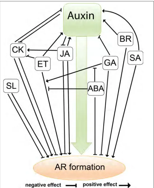
In conclusion, all the results described above demonstrate the complexity of the interactions between the phytohormones that control AR formation (Figure 6). Further detailed investigation is required to clarify the discrepancy in the effects on adventitious rooting of some important hormones.
Figure 6: Interactions between phytohormones during AR formation in different species
Auxin is the central player which interacts with the other phytohormones in complex networks during AR formation. The effects of the different classes of phytohormones: Jasmonic acid JA, cytokinin CK, ethylene ET, gibberellin GA, salicylic acid SA, strigolactone SL, abscisic acid ABA and brassinosteroids BRs are shown during this process. Their effects are either direct or via interactions with other phytohormones. Note that the model presented here is based on the results described above relating to different species.
1.4. Environmental factors influencing adventitious root formation
It is known that AR formation is controlled by many endogenous and environmental factors including nutrients, temperature and light conditions (reviewed in Bellini et al., 2014; Geiss et al., 2018).
1.4.1. Mineral nutrition
Mineral nutrients classified as macronutrients (e.g., nitrogen, phosphorus, potassium, magnesium, calcium and sulfur) and micronutrients (e.g., iron, boron, copper, chloride, molybdenum, manganese and zinc), are essential for plant growth and have specific functions in plant metabolism. These nutrients are considered to be key factors determining root morphogenesis (Bellini et al., 2014; Geiss et al., 2018). The adventitious rooting process and mineral nutrition are intimately related (reviewed in Bellini et al., 2014). For example, both number and length of ARs are positively correlated with the initial total nitrogen (N) concentration in the cuttings of pelargonium (Pelargonium× hortorum) (Druege et al., 2004), Chrysanthemum indicum (Druege et al., 2000), Euphorbia pulcherrima (Zerche & Druege, 2009) and petunia (Zerche et al., 2016). The effect of external nitrogen application in favoring AR formation by cuttings has also been shown for Eucalyptus globulus (Schwambach et al., 2005, 2015) and petunia (Hilo et al., 2017). In a recent study, Yang and collaborators (2019) found that limitation of nitrogen in cuttings of petunia inhibited AR formation. They suggested that the nitrogen limitation in these cuttings attenuated auxin signaling by modifying the expression levels of specific ARFs, GH3 and SAUR genes, thereby suppressing the auxin dose–response of ARI (Yang et al., 2019). Besides nitrogen, other minerals such as phosphorus, potassium, magnesium, calcium, iron and manganese also influence rooting of cuttings (reviewed in Li et al., 2009; Bellini et al., 2014; Geiss et al., 2018; Druege et al., 2019; Gonin et al., 2019).
1.4.2. Temperature
Temperature is another environmental factor that can impact many aspects of the adventitious rooting process starting from the growth rate of the donor plant up to root development, including root initiation, growth, orientation and rooting time (Kristiansen et al., 2005). Temperature may influence adventitious rooting capacity by interacting with several aspects such as, water and nutrient uptake, enzymatic activity and phytohormone responses (reviewed in De Almeida et al., 2017; Geiss et al., 2018). For example, Da Rocha Corrêa & Fett-Neto (2004) showed that subjecting the donor plants of Eucalyptus saligna cuttings, an easy-to-root species, to a moderate heat shock at 40 °C increased the root density and the root length in the cuttings thus obtained. In contrast, in the case of the difficult-to-root E. globulus cuttings, lower temperatures were more effective with the best rooting response observed with day/night cycles of 30 °C /20°C. In pelargonium cuttings, Druege & Kadner (2008) found that lowering the air temperature during cutting cultivation under low light, increased sugar levels in the cuttings as well as repressed leaf senescence and contributed to improved rooting at the base of the cuttings.
Based on the findings summarized above, we can conclude that there is an interaction network between environmental factors and AR formation and these factors seem to be very important parameters when considering rooting in vegetative propagation practices. Hereafter, we will discuss the effect of light, which is considered the most significant environmental factor.
1.4.3. Light: an environmental cue that controls adventitious root development
Among environmental factors light is, perhaps, the most important one that controls the photo-biological processes in plants (Alabadí & Blázquez, 2009). Plants have the ability to perceive different light signals, which regulate different aspects of development during their life cycle, for example seed germination, shade avoidance, de-etiolation, phototropism and flowering (Figure 7) (Quail, 2002a; Schepens et al., 2004; Fittinghoff, 2008; Alabadí & Blázquez, 2009; Kozai et al., 2016; Paik & Huq, 2019) . Plants have at least five different classes of
photoreceptors which are responsible for perceiving different light qualities and intensities (Figure 7). In Arabidopsis, five phytochromes (PHYA, PHYB, PHYC, PHYD and PHYE) have been identified that detect and respond to red light (RL) or far red (FR) light (600-750 nm). Blue/UV-A light (320-500 nm) is perceived by the cryptochromes CRY1, CRY2 and CRY3, the phototropins PHOT1 and PHOT2, and the F-box containing Flavin binding proteins such as the three LOV domain proteins, ZETLUPE (ZTL), KELCH REPEAT F-BOX1 (FKF1) and LOV KELCH protein2 (LKP2). Finally, UV-B (280-320nm) is perceived by UVB-RESISTANCE 8 (UVR8 ) (Schepens et al., 2004; Bae & Choi, 2008; Xu et al., 2015a; Paik & Huq, 2019) (Figure 7). Recent advances in plant photoreceptor research have identified novel roles of the receptors other than photoperception. For example, PHYB has been shown to act as a thermosensor and to integrate light and temperature signaling pathways (Jung et al., 2016; Legris et al., 2016). This supports the suggestion that photoreceptors are involved not only in light perception but also in the perception of a wide range of environmental cues suggesting a role as “multisensors”.
Phytochromes are present in the form of two interconvertible isoforms – the biologically inactive form Pr and the biologically active Pfr – in response to FR and R light respectively (Sager et al., 1988; Galvão & Fankhauser, 2015). The active Pfr forms of phytochromes translocate from the cytoplasm to the nucleus, where they interact directly with a class of basic helix-loop-helix (bHLH) transcription factors called PHYTOCHROME INTERACTING FACTORs (PIFs) to trigger a transcription cascade that leads to light-regulated gene expression. Among the transcription regulators that control light signaling pathways, PIFs have been characterized as key players in transducing light signals perceived by phytochromes (Sakamoto & Nagatani, 1996; reviewed in Quail, 2002b; Leivar & Monte, 2014; Paik et al., 2017).
Figure 7: Simplified functions of photoreceptors during the plant life cycle
Plants have the ability to utilize the different light signals perceived by wavelength-specific photoreceptors to regulate different aspects of development during their life cycle
Phytochromes in the model plant Arabidopsis, perceive RL (650-670 nm) and FRL (705- 740 nm). Phototropins, cryptochromes and F-box proteins (ZEL, FKF1and LKP2) can perceive blue light /UV-A (320-500 nm) and UVR8 perceives UV-B light (280-320nm).
All these photoreceptors adjust the growth and development of plants, affecting, for example, stomatal opening, de-etiolation, phototropism, shade avoidance, flowering and seed germination. (modified from Paik & Huq, 2019).