Electrochemistry of Chemically Trapped
Dimeric and Monomeric Recombinant
Horseradish Peroxidase
O.V. Ignatenko1, A. Sjölander2, D.M. Hushpulian*3, S.V. Kazakov4, I.V. Ouporov5, T.A. Chubar6, A.A. Poloznikov7, T. Ruzgas8, V.I. Tishkov9, L. Gorton10, N.L. Klyachko11 and I. G. Gazaryan12
1,3,5,6,7,9,11,12Department of Chemical Enzymology, Moscow State University, Moscow 119992, Russia 2Novo Nordisk A/S, Novo Allé, 2880 Bagsvaerd, Denmark
4Department of Chemistry and Physical Sciences, Pace University, Pleasantville, NY 10570, U.S.A. 3,7Peroxo LLC, Yaroslavskoe shosse 117, Moscow 129347, Russia
8Department of Biomedical Science, Malmö University, Malmö 205 06, Sweden
9A.N.Bach Institute of Biochemistry, Russian Academy of Sciences, Leninsky pr. 33-2, Moscow 119071, Russia 9Innovations and High Technologies MSU Ltd., Tsymlyanskaya ul, 16, of.96, Moscow 109559, Russia
10Department of Analytical Chemistry, Lund University, PO Box 124, Lund SE 22100, Sweden 12Burke Medical Research Institute, 785 Mamaroneck Ave, White Plains, NY 10605, U.S.A.
1ovig@enz.chem.msu.ru; 2annika.sjolander@novonordisk.dk; *3hushpulian@gmail.com; 4skazakov@pace.edu; 5iuporov@gmail.com; 6tchubar@enz.chem.msu.ru; 7andrey.poloznikov@gmail.com; 8tautgirdas.ruzgas@mah.se; 9vitishkov@gmail.com; 10lo.gorton@analykem.lu.se; 11klyachko@enzyme.chem.msu.ru; 12igg2002@med.cornell.edu
Abstract
Native horseradish peroxidase (nHRP) exists in the aggre-gated form in concentrated water solutions as shown by dy-namic light scattering (DLS). This is in contrast to recombi-nant horseradish peroxidase (recHRP) which mainly exists as a dimer. The native enzyme aggregates could be broken into the particles of nm-size only under the conditions of high ionic strength (0.5-1 M NaCl). Chemical cross-linking of recHRP with glutaraldehyde in water solutions yields 40% of the dimer. The chemically trapped dimeric and monomer-ic forms of recHRP were separated by gel-filtration, their substrate specificity towards a number of organic substrates compared. Parameters of direct and mediated electron trans-fer on graphite electrodes catalyzed by both preparations were analyzed. The difference in behavior of the monomeric and dimeric enzyme forms observed in electrochemical ex-periments was interpreted as a result of a “double” coverage of the electrode surface with the molecules of cross-linked dimeric enzyme, in contrast to both modified monomeric and original, unmodified recHRP providing “monolayer” coverage. In addition to the stabilization effects achieved due to enzyme surface modification with glutaraldehyde, the “double” coverage doubles the enzyme activity per sur-face unit.
Keywords
Dimeric Recombinant Peroxidase; Light Scattering; Direct Elec-tron Transfer; Electrochemical Kinetics; Monolayer; Bilayer; Gra-phite Electrode
Introduction
Horseradish peroxidase HRP (EC 1.11.1.7) is one of the enzymes most widely used for applied purposes and in biosensors in particular. The enzyme is usually im-mobilized on electrode surfaces by adsorption from concentrated solutions (10 mg/mL). Despite a lot of enzyme has been utilized, immobilization procedures yield rather low activity, commonly explained by en-zyme aggregation upon interaction with the electrode surface. Native HRP needs covalent immobilization on electron-conducting polymer films to catalyze direct electron transfer reactions (Ran et al, 2011), whereas recHRP lacking oligosaccharides (Gazaryan et al., 1994; Gazaryan et al., 1997) catalyzes direct electron transfer (DET) reactions (Gorton et al, 1999, Lindgren et al., 1999, Shipovskov et al., 2004, Ferapontova et al., 2005, Kartashov et al., 2010). The principal improvement in bioelectrochemical properties of recHRP is explained by the absence of oligosaccharide chains screening the active site from direct contact with the electrode ma-terial.
The oligomeric state of both nHRP and recHRP in wa-ter solutions has never been the focus of research pa-per. It was found earlier that recHRP exhibited a ten-dency to form dimeric structures in the system of re-versed micelles (Gazaryan et al., 1997). RecHRP upon
binding its inhibitor, benzohydroxamic acid, forms a kind of a dimer in the crystal cell (Henriksen et al., 1998). Both enzymes, nHRP and recHRP, have never been studied by dynamic light scattering (DLS) tech-niques in water solutions. This paper presents a com-prehensive study answering the question on different oligomeric state of both enzymes in water solutions and includes DLS of native and recHRP, chemical trapping of dimeric and monomeric recHRP forms and their comparison in terms of substrate specificity and electrochemical behavior on graphite electrodes. The discovery of heavily aggregated native enzyme in water solutions may explain the advantages of using recHRP for the purposes of construction of biosensors based on DET.
Materials and Methods
Chemicals and Reagents
Guaiacol was from Fluka (Buchs, Switzerland). 2, 2’-azino-bis(3-ethylbenzthiazoline-6-sulfonic acid) (ABTS), o-phenylenediamine (o-PD), o-dianisidine (o-DA), py-rogallol, glutaraldehyde, sodium bihydride, Tris were from Sigma (St. Louis, MO). p-Cresol, hydrogen perox-ide (30% H2O2), buffers and salts were from Merck (Darmstadt, Germany). All aqueous solutions were prepared using water purified with a Milli-Q system (Millipore, Bedford).
Enzyme Preparation
Native horseradish peroxidase (HRP), RZ=3.0, was purchased from Biozyme (UK). Wild-type recombi-nant HRP was produced with a scaled-up protocol based on the method developed earlier (Gazaryan et al., 1994). The refolding of 1 g of inactive protein solu-bilized from inclusion bodies (obtained from 60 g of BL21(DE3)/pSA261 biomass) was performed in 10 L refolding medium containing 1 mM DTT, 0.5 mM oxi-dized glutathione, 5 mM CaCl2, 5 µM hemin and 2 M urea in 50 mM Tris-HCl buffer, pH 8.0, for 4 days at 4oC, in accord with the solubilization protocol de-scribed in (Gazaryan et al., 1994). The refolding me-dium was concentrated up to 20-fold and the concen-tration of urea was reduced to 0.2 mM using a flow-through membrane concentrator. Then, the pH value was adjusted to 4.5 to precipitate the unfolded, aggre-gated protein. The supernatant solution obtained after centrifugation was purified by gel-filtration on a Se-phacryl S-200 column (Pharmacia Biotech, Germany). The procedure yielded a homogeneous preparation of wild-type recombinant HRP (ca. 250 mg, RZ> 3.0). This
preparation was used in this work and others pub-lished before (Gazaryan et al., 1997; Gazaryan et al., 1999; Lindgren et al., 1999, Abad et al., 2002, Shipovs-kov et al., 2004, Ferapontova et al., 2005).
Samples for Light Scattering
50 mM Na-acetate buffer, pH 5.0, K-phosphate and Tris-HCl buffers, pH 8.0, were used to prepare the 0.1-0.5 mg/ml enzyme solutions. The solutions were fil-tered through 0.2 µm Sartorius Minisart filter into a ∅10 mm quartz cuvette.
Dynamic Light Scattering
Dynamic Light Scattering DLS measurements were carried out on a Malvern 4700c System equipped with a 128-channel digital correlator. An argon ion laser op-erated at a 488 nm wavelength was used as the light source. The scattering angle was kept constant at 90o. The quality of measurements was tested over the sig-nal-to-noise ratio, the range of the correlation function, and the difference between the measured and calcu-lated baselines. The particle size distributions were calculated by means of the optimized Laplace inver-sion methods (CONTIN), whereas a contribution of different modes of molecular motion into the integral size distribution and characteristic particle sizes were determined using cumulant analysis for different por-tions of the correlation function. Both software pro-grams were supplied by the manufacturer. All mea-surements were performed at (25.0±0.1) °C.
Cross-Linking
The cross-linking procedure was performed using a 20-fold excess of glutaldehyde with respect to the en-zyme (10-4 M enzyme, 2x10⁻³ M glutaraldehyde) in 50 mM K-phosphate buffer, pH 8.0, by incubation (3h) at room temperature. Then the mixture was treated with 25 µL of NaBH4 solution (4.8 mg/ml). The prod-ucts of cross-linking were analyzed by SDS-PAGE.
Separation of Chemically Trapped Dimeric and Mo-nomeric RecHRP
Separation of chemically trapped dimeric and mono-meric recHRP was performed by FPLC gel-filtration on a Superose-12 column (Pharmacia, Uppsala, Swe-den) in 0.01 K-phosphate buffer, pH 7.5, containing 0.15 M NaCl.
Specific Activity Measurements
and pyrogallol was measured on a Shimadzu UV-1602 spectrophotometer (Japan) in accordance with the fol-lowing protocols. ABTS (0.5 mM) oxidation was car-ried out in 0.1 M Na-citrate/phosphate buffer, pH 4.8, at 414 nm, using an extinction coefficient of 31.1 mM -1cm-1 (Arnao et al., 1990). o-Phenylenediamine (2.2 mM) oxidation was studied in 0.1 M Na-citrate/phosphate buffer, pH 5.5, at 445 nm, using an extinction coefficient of 11.1 mM-1cm-1 (Porstmann et al., 1985).o-Dianisidine (0.1 mM) oxidation was stu-died in 0.1 M Na-citrate/phosphate buffer, pH 5.5, at 460 nm using an extinction coefficient of 30 mM-1cm-1 (Ugarova et al., 1979). Guaiacol (3 mM) oxidation was studied in 0.05 M potassium-phosphate buffer, pH 7.0, at 470 nm using an extinction coefficient of 5.7 mM -1cm-1 (Hosoya et al., 1967). Pyrogallol (1 mM) oxidation was studied in 0.025 M Na-citrate/phosphate buffer, pH 6.5, at 420 nm using an extinction coefficient of 4.4 mM -1cm-1 (Cioprada et al., 1978). The concentration of hydrogen peroxide used was kept equal to 1 mM. The extinction coefficient for hydrogen peroxide was taken as 72.7 M-1cm-1 at 230 nm (George, 1953). The concen-tration of the recombinant enzymes was equal to 10-9 M. Recombinant HRP concentration was calculated from the Soret band absorbance (ε403 = 100,000 M-1cm-1 (Smith et al., 1990)).
Steady-State Kinetics of ABTS Oxidation
Steady-state kinetics was measured on a plate reader (Molecular Devices, USA). Rate constants for ABTS oxidation were calculated from double-reciprocal plots by varying concentrations of both substrates, i.e. hy-drogen peroxide and ABTS, in the range of 0.008-0.4 mM and 0.1-0.5 mM, respectively. Enzyme concentra-tions were equal to 0.26 nM.
Computer Modeling
Dimer modeling and structure optimization were per-formed as previously described (Gazaryan et al., 1997) using the crystal structure coordinates of the recombi-nant HRP (PDB code 1ATJ).
Electrode Preparation
Peroxidase electrodes were prepared using rods of solid spectroscopic graphite (SGL Carbon, Werke Ringsdorff, Bonn, Germany, type RW001, 3.05 mm di-ameter). The graphite rods were first polished on wet fine-structured emery paper (grit size: P1200) and then additionally polished on paper to obtain a mirror-like surface. The electrode rods were carefully rinsed with
deionized water and allowed to dry at room tempera-ture. 8 µL aliquot of a peroxidase solution in 20 mM phosphate buffer, pH 7, was added to each of the po-lished ends of the graphite rods and the adsorption was allowed to proceed for 1 h at 4°C. The enzyme electrodes were then thoroughly rinsed with 0.1 M so-dium phosphate buffer, pH 7.0, and if not immediately used, they were stored in the same buffer at 4°C. Weakly adsorbed peroxidase was desorbed before measurements, by rotating the electrode in buffer for at least 30 min.
Amperometric Measurements
The peroxidase modified electrode was fitted into a ro-tating disk electrode holder (RDE; EG&G Model 636, Princeton Applied Research, Princeton, NJ), which was placed in a three-electrode cell with an Ag|AgCl (3 M KCl) reference electrode (EG&G) and a platinum wire auxiliary electrode. The electrodes were con-nected to a BAS 100W potentiostat (Bioanalytical Sys-tems, West Lafayette, IN). All measurements were per-formed at an applied potential of 0 mV versus Ag|AgCl (Ruzgas et al., 1995; Lindgren et al., 1998) with an electrolyte of 0.1 M phosphate buffer, pH 7.0. The current was registered using various rotation speeds (200–1000 rpm) and H2O2 concentrations. For mediated electron transfer, a p-cresol concentration of 60 µM was used. Prior to experiments, the solution was bubbled with argon for 15 min. Argon was passed over the solution during the experiments.
Calculation of Kinetic Parameters from Electrochemi-cal Measurements
The reduction current for H2O2 at a peroxidase-modified electrode is limited by mass-transfer of H2O2 to the electrode and by the kinetics of the enzymatic reaction. The measured current, I, is a combination of both the mass-transfer limited current, Ilim and the ki-netically limited current, Ikin, according to the follow-ing equation: 1/I = 1/Ilim + 1/Ikin, which holds for the ro-tating disk electrode used in this study. Ikin is calcu-lated by extrapolating the dependence of the regis-tered current (I) on the rotation speed, ω, in Koutecky– Levich plots (I-1 vs. ω-1/2): the intercept of the Kou-tecky–Levich plot is equal to the inverse of Ikin. In the absence of an electron donor other than the trode, the process is usually referred to as direct elec-tron transfer (DET) (Gorton et al., 1999). In this case (see Fig.1A) the peroxidase catalytic cycle can be pre-sented as follows:
SCHEME 1
E + H2O2 → EI + H2O (k1) EI + 2e → E (ks)
where E and EI are ferric and 2e-oxidized states of en-zyme, respectively. 6H2O2 6H2O e- e -2H2O2 2H2O S* S S*S S* S S* S S S* S S* e -A B
FIG. 1 SCHEMATIC REPRESENTATION OF DIRECT (A) AND MEDIATED (B) ELECTRON TRANSFER IN THE CASE OF PEROXIDASE-GRAPHITE ELECTRODES ILLUSTRATING THE
STATEMENT THAT ONLY A PART OF THE IMMOBILIZED PEROXIDASE MOLECULES CAN ACCEPT ELECTRONS DIRECTLY FROM THE ELECTRODE, IN CONTRAST TO
DIFFUSING SUBSTRATE RADICALS (S*)
The dependence of the kinetic current on H2O2 in this case is described by the following equation (Gorton et al., 1999; Lindgren et al., 1998; Ruzgas et al., 1995):
1/Ikin= (1/k1c* + 1/ks)/nFEDET,
where n is the number of electrons transferred per H2O2 molecule (n = 2), c* is the bulk concentration of H2O2, EDET is the amount of adsorbed enzyme active in direct ET and F is the Faraday constant (96 484 C mol-1). The dependence of Ikin vs. [H2O2] in double re-ciprocal plots allows the kinetic constants k1 and ks to be evaluated from the slope and intercept, respectively (Lindgren et al., 1998; Ruzgas et al., 1995).
In the presence of a peroxidase electron donor in a sa-turating concentration sufficient to engage all perox-idase molecules into mediated ET as shown in Fig. 1B, the peroxidase reaction cycle can be presented as:
SCHEME 2 E + H2O2 → E I + H2O (k1) EI + S → EII + S• (k 2) EII+S → E + S• (k 3) 2S• + 2e → 2S (k s,m)
where E, EI and EII are ferric peroxidase and its oxi-dized compounds I and II, respectively, S and S• are one-electron peroxidase electron donor and its oxi-dized form.
Among all rate constants controlling the reduction steps, k3 is usually considered to be rate-limiting. In this case Ikin can be expressed as follows:
1/Ikin= (1/k1c* + 1/k3[S])/2n1FE,
where n1 is the number of electrons transferred per donor molecule (n1=1), E is the total amount of active enzyme on the electrode surface, k3 is the rate-limiting
constant for the enzyme reduction by the peroxidase substrate (Gorton et al., 1999; Lindgren et al., 1998). By presenting the experimental data in double-reciprocal plots 1/Ikin vs. 1/[H2O2], the kinetic constants k1 and k3 can be evaluated. The basic assumption is that in the presence of saturating concentrations of a soluble elec-tron donor, acting as an ET mediator, all peroxidase molecules work in mediated ET, whereas in its ab-sence only a portion of the molecules is active in direct ET. The ratio of the slopes obtained in the presence and absence of a saturating concentration of an elec-tron donor (p-cresol) is the ratio between the enzyme active in direct ET (EDET) and the total amount of active enzyme (E) (Gorton et al., 1999; Lindgren et al., 1998). It should be emphasized that the ratio between the en-zyme active in direct ET and the total enen-zyme is not dependent on the amount of enzyme on the electrode surface and thus, various peroxidase forms can easily be compared on this basis.
The rate constants (ks, k1 and k3) can be calculated, if the enzyme concentration on the electrode is known. The latter is a serious and still unsolved problem for peroxidases adsorbed on graphite surfaces (Lindgren et al., 1998). Peroxidases are not expected to adsorb on solids with a higher coverage than a monolayer (An-drade et al., 1986). In our calculations, we therefore as-sume for monolayer coverage of adsorbed peroxidase on the electrode surface (with a roughness factor of 5 (Jaegfeldt et al., 1983)) to be equal to 40 pmol cm-2 (Paddock et al., 1989). Although the absolute values of the rate constants obtained from electrochemical kinet-ics are not precise (since the true surface concentration of peroxidase remains unknown) this method can be used to compare various forms of recHRP as a first approach, assuming their equal concentration on the electrode surface.
Results and Discussion
Light Scattering
The results of dynamic light scattering are represented as particles size distributions obtained by CONTIN program. In the case of recHRP (Fig. 2a) an asymme-tric size distribution supposes a multimodal molecular motion. Cumulant analysis of the shape of the time correlation function allows us to suggest that there have three modes of molecular motion in the solution, corresponding to the sizes of monomeric (~3.2 nm), dimeric (~6.4 nm), and possibly tetrameric recHRP (Table 1). The native HRP in the same buffer (Fig. 2b) exhibits a size distribution different from that of
recHRP. Large characteristic sizes of the scattered par-ticles (Table 1) and significant polydispersity (the width of distribution) testify that the native enzyme forms aggregates. The addition of salt crucially changes the size distribution for native HRP (Fig. 2c) shifting it to smaller sizes similar to those observed in the case of recHRP (cf. Fig. 2a). The value of this z-average hydrodynamic diameter is ca. twice larger than that of the monomer. The higher hydrodynamic radius can be attributed to the oligosaccharide “fur” surrounding the native enzyme.
0
5
10
15
20
25
0
5
10
15
20
c
d
h, nm
% , in c la s s0
50
100
150
200
250
0
5
10
15
20
b
d
h, nm
% , in c la s s0
5
10
15
20
25
0
5
10
15
a
d
h, nm
% , in c la s sFIG. 2 PARTICLE SIZE DISTRIBUTIONS FOR ENZYMES SOLUTIONS IN ACETIC BUFFER AT pH 8: a) recHRP, b) NATIVE
HRP, c) NATIVE HRP CONTAINING 0.5 M NaCl
TABLE 1 CHARACTERISTIC SIZES (Z-AVERAGE HYDRODYNAMIC DIAMETERS) OF SCATTERED PARTICLES FOR THE SOLUTIONS OF
RECOMBINANT AND NATIVE HRP
Enzyme RecHRP Native HRP Native HRP 0.5 M NaCl
dH (Mode 1), nm 3.2±0.2 - -
dH (Mode 2), nm 6.4±0.5 - 6.7±0.5
dH (Mode 3), nm 13.1±1.0 13.3±1.0 9.1±1.0
dH (Mode 4), nm - 167±8.0 - The light scattering data unequivocally prove that the
oligomeric states of recombinant and native enzyme in solution are strikingly different. Presumably, it is this difference that has effect on the integral characteristics of the enzyme such as stability in solution and in the course of physical immobilization on surfaces.
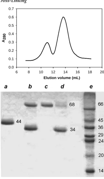
Cross-Linking Elution volume (mL) 6 8 10 12 14 16 18 20 A280 0.0 0.1 0.2 0.3 0.4 0.5 0.6 0.7
FIG. 3 GEL-FILTRATION PROFILE (UPPER) AND SDS-PAGE OF HRP PREPARATIONS AFTER CROSS-LINKING AND SEPARATION (LOWER): (a) NATIVE HRP AFTER A LINKING ATTEMPT; (b) THE PRODUCT OF recHRP CROSS-LINKING; (c) DIMERIC, AND (d) MONOMERIC recHRP PREPARATIONS AFTER GEL-FILTRATION; (E) MW MARKERS An attempt to obtain dimeric recHRP by cross-linking in the system of reversed micelles yields no more than 10% of dimeric enzyme (results not shown). In con-trast, in water solutions, the very first attempt resulted in a 40% yield of the dimeric form, which was easily separated from the monomeric one by gel-filtration (Fig. 3). Of note, both enzyme preparations were sepa-rated from unreacted glutaraldehyde by this proce-dure. The dimeric fraction was homogeneous, while the monomeric fraction still contained ca. 10% of the dimer (Fig. 3). The cross-linking of native HRP (Fig. 3)
was inefficient: no enzyme dimers or aggregates were observed. Glutaraldehyde is a rather small linker and requires two a.a. residues to be at a close distance (6-7 angstrem) to be cross-linked. Heavily glycosylated and aggregated nHRP does not expose candidate a.a. resi-dues on the surface, and shows no cross-linking prod-ucts: there is a single band on SDS-PAGE (Fig.3). Con-trary to nHRP, recHRP has Lys 232 residues sufficient-ly close in a dimer structural model, and these sufficient-lysines can be considered as potential candidates for intramo-lecular cross-linking in recHRP dimer (Fig. 4). It is clear from this structural model that glycosylated Asn 268 and Asn198 in nHRP will interfere with the forma-tion of the optimized dimeric structure, and thus, modification of nHRP lysines with glutaraldehyde does not yield any cross-linked oligomeric structure, despite that it is known to increase the enzyme stabili-ty (Ugarova et al., 1979).
Changes in Substrate Specificity
The specific activity of monomeric and dimeric recHRP compared to the original preparation of the wild-type recHRP was changed as shown in Fig. 5. In all cases modification with glutaraldehyde causes a drop in the enzyme specific activity. This drop in ac-tivity is similar to monomeric and dimeric enzyme in the case of phenolic substrates, and may reflect the fact that extensive modification of the enzyme surface with glutaraldehyde interferes with the access of perox-idase substrates to the active site. In the case of organic amines such as ABTS, o-PD, and o-DA, the activity of the dimeric enzyme is ca. 50% of that of the monomer-ic (Fig. 5). The distinct effects in the case of phenolmonomer-ic and amine substrates may indicate principal differenc-es in the mechanism of their oxidation. To evaluate the effects of cross-linking on the individual steps of the peroxidase catalytic cycle, we have studied ABTS oxi-dation (Table 2). The reaction of ABTS oxioxi-dation with hydrogen peroxide catalyzed by wild-type recHRP in-cludes a rate-limiting monomolecular step, which is ascribed to the product dissociation step by Smith et al. (Smith et al., 1992) as shown in Scheme 3.
SCHEME 3 E + H2O2 → EI + H2O (k1) EI + S → EII + S• (k 2) EII+S → E-S• (k 3) E-S• → E + S• (k 4)
As evident from the values of the rate constants pre-sented in Table 2, cross-linking has almost no effect on the properties of the monomeric enzyme, while it has
effect on the enzyme reduction steps in the case of the dimer, in particular, by decreasing the monomolecular rate-limiting constant k4 corresponding to the product dissociation step. It can be speculated that cross-linking results in a dimeric structure, where the active sites are almost facing each other (Fig. 4) and therefore, interaction with the oxidation product may interfere with the access of the substrate to the adjacent subunit. Alternatively, it is inferred that all amine substrates are oxidized via electron transfer through the protein molecule, and therefore, the decrease in the available surface per active center in the dimer results in the lower rate constant for the substrate oxidation step. TABLE 2 RATE CONSTANTS FOR ABTS OXIDATION BY DIFFERENT FORMS OF
RECHRP Enzyme form k1/107, M-1s-1 k3/106,M-1s-1 k4/103,s-1 Wild-type recHRP 1.2±0.2 2.5±0.5 2.6±0.5 Monomeric recHRP 1.0±0.2 1.6±0.3 2.4±0.5 Dimeric recHRP 1.0±0.2 0.8±0.2 1.3±0.3
As it was previously reported (Gazaryan et al., 1997) the presence of a particular substrate in the experi-ments with reversed micelles shifted the equilibrium between monomeric and dimeric recHRP forms. The most pronounced effect was observed for o-DA, which decreased the amount of the dimeric form up to 2-fold. A lesser effect was observed for pyrogallol, where the amount of the dimer was decreased by 20% (Gazaryan et al., 1997). Guaiacol had no effect on the equilibrium at all. These observations are in agreement with the data obtained in this work and support the first expla-nation, i.e. steric hindrance for the access of bulky sub-strates to the active site in recHRP dimer.
FIG. 5 SUBSTRATE SPECIFICITY OF recHRP, AND ITS MONOMERIC AND DIMERIC FORMS OBTAINED IN THE COURSE OF CROSS-LINKING WITH GLUTARALDEHYDE
FIG. 4 COMPUTER MODEL OF DIMERIC recHRP: ASN RESIDUES GLYCOSYLATED IN NATIVE PEROXIDASE ARE SHOWN IN GREY TO DEMONSTRATE THAT OLIGASACCHARIDE CHAINS ATTACHED TO ASN 268 AND ASN 198 WILL INTERFERE WITH ENZYME
DIMERIZATION AND INTRAMOLECULAR CROSS-LINKING OF LYS 232
On the other hand, o-PD was shown to increase the amount of dimeric recHRP by 20% in the reversed mi-celles (Gazaryan et al., 1997), while in this work the substrate specificity of the dimer towards o-PD exhi-bits a 30% drop compared to the monomeric enzyme. The distinct results obtained with o-PD for the ferric form of recHRP in reversed micelles and for recHRP dimer in the reaction course may reflect the high affin-ity of the enzyme for both substrate and oxidation product, and thus, the decreased rate constant for the product dissociation, as in the case of ABTS. Overall results obtained in this work unequivocally demon-strate that cross-linking imposes more restrictions for substrate access to the active site in dimeric recHRP compared to the modified monomeric enzyme and es-pecially unmodified recHRP.
Electrochemical Kinetics
The method developed by us earlier (Gorton et al., 1999; Lindgren et al., 1998; Ruzgas et al., 1995) was applied to estimate the kinetic parameters for the mo-nomeric and dimeric recHRP immobilized on graphite electrodes and compared with previously reported values for the unmodified recHRP and native enzyme (Table 3). First, the amount of enzyme available for di-rect electron transfer is 2 times lower in the case of the dimeric enzyme than that in the case of the monomeric one. This observation can be rationalized if one im-agines that the dimer actually forms a kind of a
“double” layer as shown in Fig. 6B. In this case, the ac-tual concentration of active centers on the electrode surface is doubled compared to that for the monomer-ic enzyme, and so, we must divide the kinetmonomer-ic parame-ters by 2 to account for the double concentration of ac-tive sites in the case of dimeric recHRP, leading to the conclusion on equal rate constants for all catalytic steps for the monomeric and dimeric enzyme within the experimental error. This conclusion is in agree-ment with the substrate specificity data for both en-zyme forms in the case of phenolic substrates (it was reminded that p-cresol, a phenolic substrate, was used in the electrochemical experiments). Second, a com-parison of the amount of enzyme in DET for the mod-ified monomeric and unmodmod-ified recHRP with that for native HRP shows that modification with glutaralde-hyde has the same effect as the presence of oligosac-charide chains in the native enzyme, i.e. screens a sig-nificant portion of enzyme surface and/or active sites available for direct electron transfer. The dramatic im-provement in all kinetic constants observed for the recHRP forms compared to the native enzyme has now to be interpreted as the enzyme structural stabili-zation providing maximum activity yield in the course of physical immobilization on the graphite surface. The hydrophilic native enzyme, heavily aggregated in water solutions, undergoes major inactivation in the course of interaction with graphite surface. This is the main reason for the unbelievably low, at least 2 orders of magnitude, values for the rate constants of the
indi-vidual steps of the peroxidase catalytic cycle estimated from electrochemical experiments for the native en-zyme (Table 3) compared to those in water solutions (Table 2). The recombinant monomeric HRP shows almost an order of magnitude increase in all rate con-stants compared to the native enzyme (Table 3), and a 2-fold increase compared to the unmodified recHRP. Thus, additional hydrophobization of the recHRP sur-face has an extremely positive effect in terms of mini-mizing the enzyme inactivation in the course of its immobilization on electrodes. However, the achieved values for k1 and k3 are still lower than those in solu-tion by 20- and 5–fold, respectively.
e- e -H2O2 H2O
A
e -H2O2 H2OB
e-FIG. 6 SCHEMATIC REPRESENTATION OF “DOUBLED” CONCENTRATION OF ACTIVE SITES IN THE CASE OF EQUAL
MOLECULAR COVERAGE OF THE ELECTRODE SURFACE IN THE CASE OF MONOMERIC (A) AND DIMERIC (B) RECHRP
This difference actually points to the fact that the H2O2 cleavage site and the substrate binding site are struc-turally different because they exhibit different stability with respect to immobilization by physical absorption. It is concluded that the peroxidase active site featuring a unique disposition of catalytic histidine and arginine toward the heme iron is much more sensitive to struc-tural damage than the substrate binding sites in the peroxidase molecule.
TABLE 3 KINETIC CHARACTERISTICS OF DIFFERENT FORMS OF HRP IMMOBILIZED ON GRAPHITE ELECTRODE. AVERAGE OF 3 ELECTRODES,
95% CONFIDENCE LIMITS GIVEN.0.1 M NA-PHOSPHATE, PH 7.0. THE RATE CONSTANTS ARE CALCULATED USING A MONOLAYER COVERAGE
(NUMBER OF HRP ACTIVE SITES/AREA) Native HRP (Lindgren et al., 1998) RecHRP (Lindgren et al., 1999) Mono-meric recHRP Dimeric recHRP % in DET 48.0±4.0 63.0±7.0 42.0±5.0 19.0±5.0 k1/105 M-1s-1 1.3±0.2 2.8±0.6 6.0±3.1 11.6±2.5 ks s-1 1.9±0.3 7.6±2.5 13.0±6.0 21.0±9.0 k3/105 M-1s-1 0.3±0.1 1.4±0.7 2.3±1.4 5.0±4.0 Number of electrons 2.1±0.2 1.8±0.2 1.94±0.06 1.90±0.06
In conclusion, the revealed tendency of the native gly-cosylated peroxidase to form aggregates in water
solu-tions is of key importance for all protocols based on physical adsorption of the enzyme on surfaces, and in particular immobilization of electrodes. Immobiliza-tion by adsorpImmobiliza-tion is known to aggregate and inacti-vate proteins (Kondo et al., 1993), and obviously, for the protein already aggregated in solution, the inacti-vation effect in the course of immobilization will be even stronger. It is interesting to note that the recent study aimed at production of HRP encapsulated in hollow gold nanoparticles was successful due to the use of HRP micelles which provided the entrapment of individual molecules first into the micelle and then used as a basic structure to make a 100 nm nanopar-ticle (Kumar et al., 2005).
The results obtained in this study allow us to demon-strate that formation of dimeric structures by the re-combinant wild-type HRP is the inherent property of the enzyme molecule, which is preserved both in wa-ter solutions and in micellar systems. The demonstrat-ed increase in operational characteristics of immobi-lized recHRP by chemical cross-linking in water solu-tions can be used when studying enzyme applicasolu-tions to biosensors and in enzyme immunoassay. The major practical conclusion from our studies is that for immobilization on an electrode one needs monomeric HRP (native or recombinant), no matter how this will be achieved, i.e. reversed micelles, cross-linking and separation for recHRP, high salt for nHRP, or entrapment into hollow nanospheres (Xie et al., 2011).
ACKNOWLEDGMENT
The authors thank the following organizations for the financial support: The Swedish Research Council (VR), The Swedish Royal Academy of Science (KVA), The European Commission (INTAS 991-1768 and BIO4-CT97-2199) and Russian Foundation for Basic Re-search (RFBR 13-04-02013-а).
REFERENCES
Abad, J.M., Vélez, M., Santamaría, C., Guisán, J.M., Matheus, P.R., Vázquez, L., Gazaryan, I., Gorton, L., Gibson, T., Fernández, V.M. 2002. Immobilization of peroxidase gly-coprotein on gold electrodes modified with mixed epoxy-boronic Acid monolayers.J Am Chem Soc. 124, 12845-53.
Andrade, J.D., Hlady, V., 1986. Protein adsorbtion and ma-terial biocompatibility: A tutorial review and suggested hypotheses. Adv. Polym. Sci. 79, 1-63.
Arnao, M.B., Acosta, M., del Rio, J.A., Garcia-Canovas, F., 1990. Inactivation of peroxidase by hydrogen peroxide and its protection by a reductant agent. Biochim. Biophys. Acta 1038, 85-89.
Cioprada, J., Nokulescu, S., and Marinesku, M., 1978. Spec-trophotometric method for the determinaton of perox-idase activity. Reuve Roumaine de Biochimie 4, 259-63. Ferapontova, E.E., Castillo, J., Hushpulian, D., Tishkov,V.,
Chubar, T., Gazaryan, I., Gorton, Lo. 2005 Direct electro-chemistry of recombinant tobacco peroxidase on gold. Electrochemistry Communications 7, 1291.
Gazaryan, I.G., Doseeva, V.V., Galkin, A.G., Tishkov, V.I., 1994. Effect of single-point mutations Phe41-->His and Phe143-->Glu on folding and catalytic properties of re-combinant horseradish peroxidase expressed in E. coli. FEBS Letters 354 248-50.
Gazaryan, I.G., Klyachko, N.L., Dulkis, Y.K., Ouporov, I.V., Levashov, A.V., 1997. Formation and properties of di-meric recombinant horseradish peroxidase in a system of reversed micelles. Biochem. J. 328 643-7.
George, P., 1953. The chemical nature of the second hydro-gen peroxide compound formed by cytochrome c perox-idase and horseradish peroxperox-idase. I. Titration with re-ducing agents. Biochem. J. 54 267-76.
Gorton, L., Lindgren, A., Larsson, T., Munteau, F.-D., Ruzgas, T., Gazarian, I.G., 1999. Direct electron transfer between heme-containig enzymes and electrodes as basis for third generation biosensors. Analytica Chimica Acta 400, 91-108.
Henriksen, A., Schuller, D.J., Gajhede, M. 1998. Structural in-teractions between horseradish peroxidase C and the substrate benzhydroxamic acid determined by X-ray crystallography. Biochemistry37, 8054-8060.
Hosoya, T., M. Morrison., 1967. A study of the hemoproteins of thyroid microsomes with emphasis on the thyroid pe-roxidase. Biochemistry 6, 1021-26.
Jaegfeldt, H., Johansson, G., and Kuwana, T., 1983. Electro-chemical Stability of Catechols with a Pyrene Side Chain Strongly Adsorbed on Graphite Electrodes for Catalytic Oxidation of Dihydronicotinamide Adenine Dinucleoti-deJ. Am. Chem. Soc. 105, 1805-143.
Kartashov, A.V., Serafini, G., Dong, M., Shipovskov, S., Ga-zaryan, I., Besenbachera, F., Ferapontova, E.E. 2010
Long-range electron transfer in recombinant peroxidases aniso-tropically orientated on gold electrodes. Phys. Chem. Chem. Phys. 12, 10098-10107.
Kumar, R., Maitra, A.N., Patanjali, P.K., Sharma, P., 2005. Hollow gold nanoparticles encapsulating horseradish peroxidase. Biomaterials 26, 6743-53.
Lindgren, A., Munteau, F.-D., Gazarian, I.G., Ruzgas, T., Gorton, L., 1998. Comparison of roating disk and wall-jet electrode systems for studying the kinetics of direct elec-tron transfer for horseradish peroxidase on a graphite electrode. J.Electroanal.Chem. 458, 113-20.
Lindgren, A., Tanaka, M., Ruzgas, T., Gorton, L., Gazaryan, I., Ishimori, K., Morishima, I., 1999. Direct electron trans-fer catalysed by recombinant forms of horseradish perox-idase: insight into the mechanism. Electrochem. Com-mun. 1, 171–175.
Paddock, R.M. and Bowden, E.F., 1989. Electroanalytic re-duction of hydrogen peroxide via direct electron transfer from pyrolytic graphite electrodes to irreversible ad-sorbed cytochrome C peroxidase. J. Electroanal. Chem. 260, 487-94.
Porstmann, T., Porstmann, B., Wietschke, R., von, Baehr R., Egger, E., 1985. Stabilization of the substrate reaction of horseradish peroxidase with o-phenylenediamine in the enzyme immunoassay. J. Clin. Chem. Clin. Biochem. 23 41-44
Ran, Q., Peng, R., Liang, C., Ye, S., Xian, Y., Zhang, W., Jin, L. 2011. Direct electrochemistry of horseradish peroxidase immobilized on electrografted 4-ethynylphenyl film via click chemistry. Anal Chim Acta 697,:27-31
Ruzgas, T., Gorton, L., Emneus, J., and Marko-Varga, G., 1995. Kinetic models of horseradish peroxidase action on a graphite electrode . J. Electroanal. Chem. 391, 41-49. 1995.
Shipovskov, S., Ferapontova, E.E., Gazaryan, I., Ruzgas, T. 2004. Recombinant horseradish peroxidase - and cytoch-rome c-based two-electrode system for detection of supe-roxide radicals. Bioelectrochemistry 63, 277-80.
Smith, A.T., Sanders, S.A., Thorneley, R.N., Burke, J.F., Bray, R.R., 1992 . Characterisation of a haem active-site mutant of horseradish peroxidase, Phe41Val, with altered reac-tivity towards hydrogen peroxide and reducing sub-strates. Eur. J. Biochem. 207, 507-19.
Smith, A.T., Santama, N., Dacey, S., Edwards, M., Bray, R.C., Thorneley, R.N., Burke, J.F., 1990. Expression of a syn-thetic gene for horseradish peroxidase C in Escherichia coli and folding and activation of the recombinant en-zyme with Ca2+ and heme. J. Biol. Chem. 265 13335-43. Ugarova, N. N., Rozhkova, G. D., Berezin, I. V., 1979.
Chem-ical modification of the epsilon-amino groups of lysine residues in horseradish peroxidase and its effect on the
catalytic properties and thermostability of the enzyme. Biochim. Biophys. Acta 570, 31-42.
Xie, Q., Zhao, Y., Chen, X., Liu, H., Evans, D.G., Yang, W., 2011. Nanosheet-based titania microspheres with hollow core-shell structure encapsulating horseradish peroxidase for a mediator-free biosensor. Biomaterials. 32(27), 6588-94.