birpublications.org/dmfr
distributed under the terms of the Creative Commons Attribution 4.0 Unported License http:// creativecommons. org/ licenses/ by/ 4. 0/, which permits unrestricted use, distribution and reproduction in any medium, provided the original author and source are credited.
ReseaRch aRticle
evaluation of a low- dose protocol for cone beam computed
tomography of the temporomandibular joint
1,2Durer Iskanderani, 1,3Mats Nilsson, 4Per Alstergren, 1,5,6Xie- Qi Shi and 1Kristina Hellen- halme
1Department of Oral and Maxillofacial Radiology, Faculty of Odontology, Malmö University, Malmö, Sweden; 2Department of
Oral and Maxillofacial Radiology, Faculty of Dentistry, King Abdulaziz University, Jeddah, Saudi Arabia; 3Department of Medical
Radiation Physics, Skåne University Hospital, Malmö, Sweden; 4Department of Orofacial Pain and Jaw Function, Faculty of
Odontology, Malmö University, Malmö, Sweden; 5Division of Oral Maxillofacial Radiology, Department of Clinical Dentistry,
Faculty of Medicine, University of Bergen, Bergen, Norway; 6Section of Oral Diagnostics and Surgery, Department of Dental
Medicine, Karolinska Institutet, Huddinge, Sweden
Objective: Evaluation of cone beam CT (CBCT) examination with a low- dose scanning
protocol for assessment of the temporomandibular joint (TMJ).
Methods: 34 adult patients referred for CBCT imaging of the TMJ underwent two
examina-tions with two scanning protocols, a manufacturer- recommended protocol (default) and a low- dose protocol where the tube current was reduced to 20% of the default protocol. Three image stacks were reconstructed: default protocol, low- dose protocol, and processed (using a noise reduction algorithm) low- dose protocol. Four radiologists evaluated the images. The Sign test was used to evaluate visibility of TMJ anatomic structures and image quality. Receiver oper-ating characteristic analyzes were performed to assess the diagnostic accuracy. κ values were used to evaluate intraobserver agreement.
Results: With the low- dose and processed protocols, visibility of the TMJ anatomical
struc-tures and overall image quality were comparable to the default protocol. No significant differ-ences in radiographic findings were found for the two low- dose protocols compared to the default protocol. The area under the curves (Az) averaged for the low- dose and processed protocols, according to all observers, were 0.931 and 0.941, respectively. Intraobserver agree-ment was good to very good.
conclusion: For the CBCT unit used in this study, the low- dose CBCT protocol for TMJ
examination was diagnostically comparable to the manufacturer- recommended protocol, but delivered a five times lower radiation dose. There is an urgent need to evaluate protocols for CBCT examinations of TMJ in order to optimize them for a radiation dose as low as diagnos-tically acceptable (the as low as diagnosdiagnos-tically acceptable principle recommended by NCRP). Dentomaxillofacial Radiology (2020) 49, 20190495. doi: 10.1259/dmfr.20190495
cite this article as: Iskanderani D, Nilsson M, Alstergren P, Shi X- Q, Hellen- halme K.
Evalu-ation of a low- dose protocol for cone beam computed tomography of the temporomandibular joint. Dentomaxillofac Radiol 2020; 49: 20190495.
Keywords: Cone- beam CT; Radiation dosage; TMJ introduction
In recent decades, cone beam computed tomography (CBCT) has become an essential examination tool in oral and maxillofacial radiology. CBCT produces multi-planar images of high spatial resolution, which for
many diagnostic tasks give information that is unattain-able with two- dimensional projection radiography. In the diagnosis and treatment planning of patients with temporomandibular disorders (TMD), CBCT plays an effective role in assisting the clinicians when performing bony changes assessments with high diagnostic accu-racy.1 In comparison to medical CT, no significant
Correspondence to: Durer Iskanderani, E-mail: durer. iskanderani@ mau. se
difference in diagnostic accuracy was found between the two techniques; moreover, it is a cost- and dose- effective alternative.2,3 Hence, it is considered as an appro-priate imaging technique for evaluating bony changes of the temporomandibular joint (TMJ).4 However, the growing use of CBCT, partly because of its avail-ability and easiness of use, demands a critical evalua-tion of the relatively high patient radiaevalua-tion dose. Many different scanning protocols are available, and some-times the urge from clinicians to have more or less noise free images can increase the radiation dose to patients without adding more information to the specific aim of the investigation. Thus, with the many CBCT scanners on the market today, clinicians together with medical physicists should ensure that the scanner they are using is being set for the lowest possible radiation dose, yet capable of providing acceptable image quality for the particular diagnostic task.5
In current practice, most clinicians use the manufacturer- recommended exposure settings for the scanner in all clinical applications. This prac-tice usually produces acceptable image quality, but might raise questions concerning the associated radi-ation dose. Attempts to optimize the patient radiradi-ation dose without sacrificing diagnostic outcome include approaches, such as optimizing tube current (mA), tube voltage (kV), and voxel size, selecting field of view (FOV) suitable to the diagnostic task, and using 180° or 360° of rotation.6–11
One practical approach of dose optimization is to adjust the scanner’s tube current to a level that the dentist may obtain enough diagnostic information of a given task at the expense of acceptable loss of image quality.11,12 The structures in the TMJs have the advan-tage of appearing in relatively high contrast on CBCT scans, and are thus less susceptible to a reduction in exposure than for low- contrast structures. The possi-bility to establish if a degenerative disease is present or not may be easier to establish, in comparison to a thin periodontal ligament for endodontic questions. Radiographic images of the TMJ do not have to be very sharp with a high signal to noise ratio. Moreover, advanced denoising algorithms may further improve the image quality of scans made using a low- dose protocol.
There is clearly a need for a broader understanding of reduced- dose protocols related to subjective image quality in specific dental CBCT applications. Such protocols have already been assessed for some dental situations but not in the context of TMJ imaging in vivo. The aim of this study was thus to evaluate a low- dose protocol with and without image processing in CBCT examination of the TMJ.
Our hypothesis was that the low- dose protocol differed significantly compared to the default, manufacturer- recommended protocol in regard to the radiographic diagnostics on TMJ.
Methods and materials
The regional ethics review board in Lund, Sweden, approved this study (Dnr 2017/434). The study enrolled 34 adult patients (5 males and 29 females with a mean age of 57 years, ranged from 20 to 74 years) who had been referred for CBCT examination of the TMJ from the Department of Orofacial Pain and Jaw Function at Malmö University, Malmö, Sweden. For a power of 0.8 with a significant level of 0.05, around 60 TMJs were required. The indication to perform a CBCT investigation of TMJ was related to the clinicians’ questions after thorough history and clinical investiga-tion. The main reason for the CBCT investigation was when the performed treatment failed. All participants underwent two consecutive CBCT examinations with different exposure settings. All study participants signed informed- consent forms. This form included informa-tion about the addiinforma-tional radiainforma-tion dose, 20% extra, which is equivalent to less than one week of background exposure in Sweden.
Cone beam CT examination
Two CBCT examinations (Veraviewepocs 3D F40, J. Morita Corp., Kyoto, Japan) were performed both TMJs of each patient which resulted in a total number of 68 CBCT volumes. The applied exposure protocols were: the default, manufacturer- recommended protocol (exposure settings: 90 kV, 5 mA, and 9.4 s) and a low- dose protocol (90 kV, 1 mA and 9.4 s). All other param-eters were kept constant throughout the examinations, i.e. FOV of 40 × 40 mm, 180° rotation, and head posi-tion with the Frankfurt plane paralleled and the mid- sagittal plane perpendicular in relation to the floor of the mouth. Patients were informed to sit still during exposure. Multiplanar data were reconstructed with a pixel size of 0.125 mm, 1 mm slice thickness, and 1 mm slice interval.
Image evaluation
68 reconstructed sagittal volumes were saved in DICOM format. These volumes were grouped into three stacks for display and processing using Image J (National Institutes of Health, Bethesda, MD): a default protocol stack, a low- dose protocol stack, and a processed low- dose stack with noise reduction (Figure 1). The processed stacks were created using an advanced noise reduction algorithm.13
We anonymized, randomly coded, and stored 204 data sets using Microsoft Excel Worksheet (Office Professional Plus 2016, Microsoft Corporation, Redmond, WA). Four (three senior and one junior) oral and maxillofacial radiologists evaluated all images. Before the study began, observer calibration was done on CBCT sagittal volumes of the first 25 stacks with a consensus on the instructions for assessing the images. The observers were blinded to any clinical or radio-graphic information and independently assessed the
visibility of five anatomical structures: the outlines of the condyle, the articular eminence, and the articular fossa and the trabecular patterns of the condyle and the temporal bone. All observers used the same type of viewing monitor, (Barco MDCC-6430, 8500 Kortrijk, Belgium) under the same viewing conditions with no time limitations. The observers were allowed to adjust brightness and contrast when necessary. A 3- point scale was applied to assess the visibility of each anatomical structure: 1 = definitely visible, 2 = questionably visible, and 3 = not visible. Furthermore, the observers were asked to an overall impression on subjective image quality ranked as: 1 = diagnostically acceptable, 2 = diagnostically questionable, and 3 = not diagnosti-cally acceptable. To assess the radiographic finding, the observers were asked to record their level of confidence about the presence of degenerative joint disease (DJD) and were stated as 1 = definitely not, 2 = probably not, 3 = questionable, 4 = probably and 5 = definitely according to Diagnostic Criteria of TMD (DC/TMD).14 Intraobserver agreement was determined by asking each observer to re- evaluate 40 TMJs at an interval of at least 14 days and under the same conditions.
Statistical analysis
The five anatomical structures were pooled together in all settings for each observer and compared pairwise. For instance, the amount of “definitely visible” in the default protocol in relation to the number of “definitely visible” in the low- dose protocol. The overall image quality was evaluated in the same way. The results were
analyzed using the Sign test15 with the level of signifi-cance at 0.05.
The default protocol of the CBCT unit was consid-ered to be the reference standard to establish if DJD was present or not, by two observers (XS, KHH). A consensus was reached in cases of disagreement. Receiver operating characteristic (ROC) curves were used to analyze the radiographic findings concerning the presence of DJD for all the observers.16 The area under the curves (Az) were calculated. Intraobserver agreement was estimated using κ (κ) statistics.17 The values were interpreted according to the guidelines of Landis and Koch18 adapted by Altman.15
Results
Visibility of the five anatomical structures and overall image quality, according to all observers, showed no difference (p = 0.00), for the two low- dose protocols compared to the default protocol. Only one observer found improvement in processing the images for both structures’ visibility and image quality between low- dose protocol and processed protocol, respectively (p = 0.79 (visibility), p = 1.00 (image quality)).
The evaluation of the default protocol for the 68 TMJ cases showed that half of them were sound while the rest had DJD with varying osseous changes. When establishing the reference standard, the two observers disagreed in one of the cases, which had minor erosions on the superior surface of the condyle.
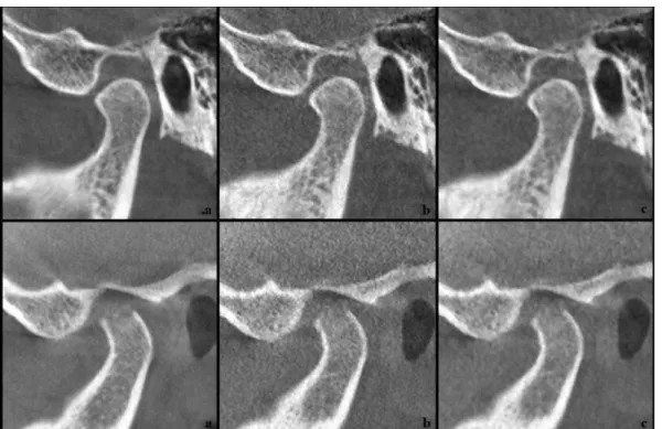
Figure 1 Examples of cone beam CT images for two temporomandibular joints using the three protocols (a) default, manufacturer
recom-mended, (b) low- dose and (c) processed low- dose. 1 (a, b, c): case with no signs of degenerative joint disease; 2 (a, b, c) case with signs of degen-erative joint disease.
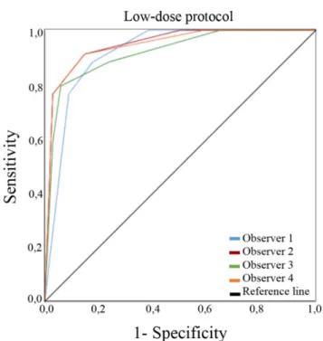
ROC curves for the low- dose and processed proto-cols assessed by all the observers according to DJD presence are presented in Figures 2 and 3, respectively.
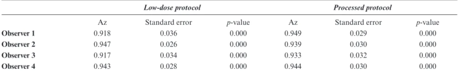
Table 1 presents the different Az for each observer. The differences in the radiographic findings for the two
low- dose protocols compared to the default one, as well as between them, were not significant. The average Az for the observers were 0.931 for low- dose protocol and 0.941 for the processed protocol. Intraobserver agree-ment among the radiographic finding were good to very good for all observers (κ values = 0.75, 0.80, 0.80, 0.85).
Discussion
Several studies in the literature have demonstrated that CBCT imaging exposed with low- dose protocol may provide diagnostically acceptable image quality for various dental indications.6–11 The present study assessed a low- dose CBCT protocol for TMJ imaging, where tube current was lowered to just 20% of the manufacturer- recommended level in a clinical setting. In this study, the ethics review board approved that the potential benefit to future clinical practice for CBCT examination of the TMJ overcomes the increased radi-ation burden (equivalent to less than 1 week of back-ground exposure in Sweden) for the patients included in the study. Therefore, we used an ethically approved study design that has been tested with different radio-graphic examinations.19–22
It is well known that manufacturer- recommended exposure settings for the CBCT scanners designed for dentistry vary widely, which results in varying amounts of radiation doses. Tube current (mA) and exposure time (s) control the number of X- ray photons emitted; thus, higher mAs increases the measured signal from the sensor and decreases image noise. The mA setting is linearly proportional to radiation dose.23 Hence, when tube voltage and exposure time are constant, lowering the mA setting by 50% reduces the delivered dose by half and consequently the quantum noise in the resultant image will be higher. Pauwels et al12 reported minimal loss of image quality when reducing tube current compared to when reducing tube voltage. Another study by Sur J et al,11 confirmed the potential to reduce dose through a reduction in tube current.
In line with the recommendations24,25 for the use of any radiographic investigation, optimization should be done whenever possible in order to expose the patient to the lowest radiation dose according to the As Low As Diagnostically Acceptable (ALADA) principle, which is proposed as a variation of the acronym ALARA (as low as reasonably achievable) to emphasize the importance of optimization according to a given diagnostic task in medical imaging.25 High image quality in the sense of being noise free and having high resolution is not necessary in all clinical situations. Structural changes in the few anatomical structures that comprise the region of the TMJ are quite visible at exposure levels well below those giving the highest image quality. Usually, we try to find osseous changes when any form of DJD is suspected; thus, lower radiation dose protocols are possible. A high resolution might be needed for assessing fine pathological changes, however. Thus, the diagnostic Figure 2 ROC curves for the low- dose protocol assessed by four
observers according to the radiographic finding of degenerative joint disease. ROC, receiver operating characteristic.
Figure 3 ROC curves for the processed protocol assessed by four
observers according to the radiographic finding of degenerative joint disease. ROC, receiver operating characteristic
task determines the level of image quality needed and, in turn, the degree of exposure parameter optimization.5 In our faculty, the demand for CBCT investigation of TMJ has decreased over time. Only patients where no explanation of their pain and no effect of treatment for TMD were found, and for those where other questions that possibly could be answered would be subject to a CBCT investigation. Thus, the CBCT examination of the TMJ were considered justified.
Our results demonstrated the ability of a low- dose protocol to image the TMJ sufficiently well without significant difference in diagnostic accuracy. Thus, this protocol is preferable, since the radiation dose was 20% of the dose delivered using the default protocol. Kadesjö et al26 reported a 50% potential for dose reduc-tion compared with the manufacturer’s recommenda-tion for the CBCT examinarecommenda-tion of TMJ. Comparable results were found for reduced dose protocols with other diagnostic tasks.6,10,11
Reduction in dose is associated with degradation of image quality, principally due to the increased noise level. Noise influences both contrast resolution and spatial resolution and consequently, a representation of an object. However, in a sense of visual perception, the radiologist may still be able to see the object details and maintain diagnostic performance. When the diag-nostic performance is influenced by a high level of noise, a noise reduction algorithm may be used to improve the image quality. Such programs filter out noise to varying degrees while preserving texture, contour, and fine details.13 The denoising method that we used was reported to be the technique of choice when very fine details are not required.27 We found that processing the images did not significantly improve the diagnostic accuracy (Table 1).
We used the default settings of the CBCT as a refer-ence standard, although no “truth” can be obtained in this kind of clinical studies. We calculated the Az in order to determine the validity of the two reduced- dose protocols for detecting the presence of DJD. Our find-ings revealed that both low- dose and processed proto-cols showed comparable diagnostic accuracy in relation to the reference standard (average Az = 0.931 and 0.941, respectively), implying that the observers performance on detection of possible degenerative changes were
comparable. A retrospective practice- based study found a similar result for pre- surgical implant assessment.9
It should be noted that these results were obtained in a real clinical setting, including factors such as motion artifacts that affect image quality. However, the limita-tions of this study could be that the results cannot be generalized to all CBCT machines. The number of observers chosen could be another limitation. However, the interobserver agreement was good, and therefore, further observers would, in our opinion, probably not contribute to any significant difference. A previous study by Hintze et al28 reported that the statistical power is dependent on the total number of evaluations, including the number of observers and surfaces evaluated for carious lesions, not on each separately. However, this kind of evaluation is known to show lower interobserver agreement due to the fact that these lesions are low contrast lesions. In comparison, TMJ structures appear with relatively high contrast.
As previous studies on low- dose protocols have also found adequate diagnostic image quality, our results could be considered together with these; it should be concluded that dose reduction achieved using a tube current below the recommendations of the manufacturer should always be investigated, in order to improve compliance with the ALADA principle.25 More important, a low- dose protocol should be the self- evident choice for younger patients, as the developing tissues of young patients are more radio-sensitive and thus at a higher risk from X- ray exposure than adult tissues.29
conclusion
The hypothesis that low- dose protocol would affect the radiographic diagnostics on TMJ could be rejected, which means that for the CBCT unit used in this study, the low- dose CBCT protocol for TMJ examination was diagnos-tically comparable to the manufacturer- recommended protocol but delivered a five times lower radiation dose. There is an urgent need to evaluate protocols for CBCT examinations of TMJ in order to optimize them for a radiation dose as low as diagnostically acceptable (the ALADA principle recommended by NCRP).
table 1 Area under the receiver operating characteristic curves (Az) for assessing the presence of the degenerative joint disease, according to four observers as evaluated on cone- beam computed tomography images produced by three protocols: the low- dose (1 mA tube current) protocol, the processed (low- dose with noise reduction) protocol, and the reference standard (“default” manufacturer- recommended, 5 mA tube current) protocol
Low- dose protocol Processed protocol
Az Standard error p- value Az Standard error p- value
Observer 1 0.918 0.036 0.000 0.949 0.029 0.000
Observer 2 0.947 0.026 0.000 0.939 0.030 0.000
Observer 3 0.917 0.034 0.000 0.933 0.032 0.000
acknowledgment
The authors acknowledge Karin Näsström and Xin Feng for their participation in image observation. This study was financially supported by King Abdulaziz University.
Funding
This study was financially supported by King Abdulaziz University.
human and animal rights statement:
All procedures followed were in accordance with the ethical standards of the responsible committee on human experimentation (institutional and national) and with the Helsinki Declaration of 1964 and later versions. Informed consent was obtained from all patients for being included in the study.
ReFeReNces
1. Ma R- H, Yin S, Li G, Ma Ruo- han, Yin Shuang LG. The detec-tion accuracy of cone beam CT for osseous defects of the tempo-romandibular joint: a systematic review and meta- analysis. Sci
Rep 2016; 6: 34714. doi: https:// doi. org/ 10. 1038/ srep34714
2. Honda K, Larheim TA, Maruhashi K, Matsumoto K, Iwai K. Osseous abnormalities of the mandibular condyle: diagnostic reliability of cone beam computed tomography compared with helical computed tomography based on an autopsy material.
Dentomaxillofac Radiol 2006; 35: 152–7. doi: https:// doi. org/ 10. 1259/ dmfr/ 15831361
3. Sirin Y, Guven K, Horasan S, Sencan S. Diagnostic accuracy of cone beam computed tomography and conventional multislice spiral tomography in sheep mandibular condyle fractures.
Dento-maxillofac Radiol 2010; 39: 336–42. doi: https:// doi. org/ 10. 1259/ dmfr/ 29930707
4. Tsiklakis K, Syriopoulos K, Stamatakis HC. Radiographic examination of the temporomandibular joint using cone beam computed tomography. Dentomaxillofac Radiol 2004; 33: 196– 201. doi: https:// doi. org/ 10. 1259/ dmfr/ 27403192
5. Lofthag- Hansen S, Thilander- Klang A, Gröndahl K. Evaluation of subjective image quality in relation to diagnostic task for cone beam computed tomography with different fields of view. Eur J
Radiol 2011; 80: 483–8. doi: https:// doi. org/ 10. 1016/ j. ejrad. 2010. 09. 018
6. Liljeholm R, Kadesjö N, Benchimol D, Hellén- Halme K, Shi X- Q. Cone- Beam computed tomography with ultra- low dose protocols for pre- implant radiographic assessment: an in vitro study. Eur J
Oral Implantol 2017; 10: 351–9.
7. Hidalgo Rivas JA, Horner K, Thiruvenkatachari B, Davies J, Theodorakou C. Development of a low- dose protocol for cone beam CT examinations of the anterior maxilla in children. Br
J Radiol 2015; 88: 20150559.. doi: https:// doi. org/ 10. 1259/ bjr. 20150559
8. Pauwels R, Seynaeve L, Henriques JCG, de Oliveira- Santos C, Souza PC, Westphalen FH, et al. Optimization of dental CBCT exposures through mAs reduction. Dentomaxillofac Radiol 2015;
44: 20150108. doi: https:// doi. org/ 10. 1259/ dmfr. 20150108
9. Dawood A, Brown J, Sauret- Jackson V, Purkayastha S. Optimiza-tion of cone beam CT exposure for pre- surgical evaluaOptimiza-tion of the implant site. Dentomaxillofac Radiol 2012; 41: 70–4. doi: https:// doi. org/ 10. 1259/ dmfr/ 16421849
10. Neves FS, Souza TdeC, de- Azevedo- Vaz SL, Campos PSF, Bóscolo FN. Influence of cone- beam computed tomography milliamperage settings on image quality of the mandibular third molar region. Oral Radiol 2014; 30: 27–31. doi: https:// doi. org/ 10. 1007/ s11282- 013- 0132-6
11. Sur J, Seki K, Koizumi H, Nakajima K, Okano T. Effects of tube current on cone- beam computerized tomography image quality for presurgical implant planning in vitro. Oral Surg Oral Med Oral
Pathol Oral Radiol Endod 2010; 110: e29–33. doi: https:// doi. org/ 10. 1016/ j. tripleo. 2010. 03. 041
12. Pauwels R, Silkosessak O, Jacobs R, Bogaerts R, Bosmans H, Panmekiate S. A pragmatic approach to determine the optimal kVp in cone beam CT: balancing contrast- to- noise ratio and radiation dose. Dentomaxillofac Radiol 2014; 43: 20140059. doi:
https:// doi. org/ 10. 1259/ dmfr. 20140059
13. Rudin LI, Osher S, Fatemi E. Nonlinear total variation based noise removal algorithms. Physica D 1992; 60(1-4): 259–68. doi:
https:// doi. org/ 10. 1016/ 0167- 2789( 92) 90242-F
14. Ahmad M, Hollender L, Anderson Q, Kartha K, Ohrbach R, Truelove EL, et al. Research diagnostic criteria for temporoman-dibular disorders (RDC/TMD): development of image analysis criteria and examiner reliability for image analysis. Oral Surg
Oral Med Oral Pathol Oral Radiol Endod 2009; 107: 844–60. doi: https:// doi. org/ 10. 1016/ j. tripleo. 2009. 02. 023
15. Altman DG. Practical statistics for medical research (1st edn. London, UK: Chapman and Hall; 1991.
16. Metz CE. Basic principles of ROC analysis. Semin Nucl Med 1978;
8: 283–98. doi: https:// doi. org/ 10. 1016/ S0001- 2998( 78) 80014-2
17. Cohen J. A coefficient of agreement for nominal scales. Educ
Psychol Meas 1960; 20: 37–46. doi: https:// doi. org/ 10. 1177/ 001316446002000104
18. Landis JR, Koch GG. The measurement of observer agreement for categorical data. Biometrics 1977; 33: 159–74. doi: https:// doi. org/ 10. 2307/ 2529310
19. Phexell E, Åkesson A, Söderberg M, Bolejko A. Intra- and inter- rater reliability in a comparative study of cross- sectional and spiral computed tomography pelvimetry methods. Acta Radiol
Open 2019; 8: 205846011985518. doi: https:// doi. org/ 10. 1177/ 2058460119855187
20. Meyer E, Labani A, Schaeffer M, Jeung M- Y, Ludes C, Meyer A, et al. Wide- volume versus helical acquisition in unenhanced chest CT: prospective Intra- patient comparison of diagnostic accuracy and radiation dose in an ultra- low- dose setting. Eur
Radiol 2019; 29: 6858–66. doi: https:// doi. org/ 10. 1007/ s00330- 019- 06278-6
21. Lee NK, Kim S, Hong SB, Kim TU, Ryu H, Lee JW, et al. Low- Dose CT with the adaptive statistical iterative reconstruction V technique in abdominal organ injury: comparison with routine- dose CT with filtered back projection. AJR Am J Roentgenol 2019; 213: 659–66. doi: https:// doi. org/ 10. 2214/ AJR. 18. 20827
22. Pan S, Su JJ, Syed J, Moore C, Israel G, Singh D. Reduced dose computed tomography: the effects of voltage reduction on density measurements of urolithiasis. J Endourol 2019; 33: 682–6. doi:
https:// doi. org/ 10. 1089/ end. 2019. 0149
23. Qu X- min, Li G, Ludlow JB, Zhang Z- yan, Ma X- chen, Qu X, Zhang Z, Ma X. Effective radiation dose of ProMax 3D cone- beam computerized tomography scanner with different dental protocols. Oral Surg Oral Med Oral Pathol Oral Radiol Endod 2010; 110: 770–6. doi: https:// doi. org/ 10. 1016/ j. tripleo. 2010. 06. 013
24. The I. The 2007 recommendations of the International Commis-sion on radiological protection. ICRP publication 103. Ann ICRP 2007; 37: 1–332. doi: https:// doi. org/ 10. 1016/ j. icrp. 2007. 10. 003
25. Bushberg JT. Science, radiation protection, and the NCRP: building on the past, looking to the future. In: Fiftieth Annual
Meeting Program of NCRP: Achievements of the Past 50 Years and Addressing the Needs of the Future ; : Bethesda, MD; 2014. pp.
5. -–7.
26. Kadesjö N, Benchimol D, Falahat B, Näsström K, Shi X- Q. Eval-uation of the effective dose of cone beam CT and multislice CT for temporomandibular joint examinations at optimized exposure levels. Dentomaxillofac Radiol 2015; 44: 20150041. doi: https:// doi. org/ 10. 1259/ dmfr. 20150041
27. Shahmoradi M, Lashgari M, Rabbani H, Qin J, Swain M. A comparative study of new and current methods for dental micro- CT image denoising. Dentomaxillofac Radiol 2016; 45: 20150302. doi: https:// doi. org/ 10. 1259/ dmfr. 20150302
28. Hintze H, Frydenberg M, Wenzel A. Influence of number of surfaces and observers on statistical power in a multiobserver ROC radiographic caries detection study. Caries Res 2003; 37: 200–5. doi: https:// doi. org/ 10. 1159/ 000070445
29. Demeter S, Applegate KE, Perez M. Internet- Based ICRP resource for healthcare providers on the risks and benefits of medical imaging that uses ionising radiation. Ann ICRP 2016; 45(1 Suppl): 148–55. doi: https:// doi. org/ 10. 1177/ 0146645316637786