-PARTICULARLY THE ILIOPSOAS
by
Eva A. Andersson
Stockholm Sweden
EMG AND STRENGTH IN TRUNK AND HIP MUSCLES
- PARTICULARLY THE ILIOPSOAS
by
Eva A. Andersson
- PARTICULARLY THE ILIOPSOAS
Eva A. Andersson
Dissertation from the Department of Neuroscience, Karolinska Institute, and the Department of Human Biology, University College of Physical Education and Sports,
Box 5626, S-114 86, Stockholm, Sweden.
ABSTRACT. The overall aim of this thesis was to study the myoelectric activity of all major muscles involved in the movements and stabilization of the trunk, pelvis and hips during training exercises, postures, motor tasks, maximal strength performance and locomotion. By the use of ultra-sound, EMG electrodes could be guided safely and accurately into muscles situated even close to the spinal column, such as psoas, quadratus lumborum and deep parts of erector spinae.
A task specific variation in activation levels were seen between muscle synergies, as well as between individual muscles within a synergy. Selective engagement of the abdominal muscles could be achieved in trunk flexion sit-ups, that is lifting only the upper trunk from the floor. An even higher activation of abdominal muscles was needed for static stabilization of the trunk and pelvis during hip flexion sit-ups, whereas single leg lifts were performed without involvement of the abdominal muscles. A selective activation of either the iliacus or psoas muscles was observed, for example in certain types of training exercises and in walking and running. Applying bending moments to the spine, resulted in a grading of the muscle activation response according to mechanical advantage, that is highest in the quadratus lumborum in lateral loading and in the superficial erector spinae in ventral loading. An exception was the most forward flexed position in standing where the superficial erector spinae "relaxed" but quadratus lumborum remained active. In general, the level of EMG in maximal efforts was maintained at the same high level irrespective of position in the range of motion, despite a marked variation in strength output. This position-dependency, as well as the strength values as such, varied in a specific way in groups of athletes, related to previous training background.
These data contribute to the understanding of muscle function and motor control of the trunk, pelvis and hips. They are also of relevance when evaluating and designing tests and training programs in rehabilitation and sports contexts as well as for improving biomechanical models of spinal loading.
Key words: abdominal muscles, biomechanics, electromyography, erector spinae, iliacus, iliopsoas, hip flexor muscles, locomotion, motor control, normalization, posture, psoas, quadratus lumborum, rehabilitation, running, sit-ups, strength, training exercises, ultra-sound, walking.
will also be presented.
I Andersson, E., L. Sward and A. Thorstensson. (1988) Trunk muscle strength in athletes. Med Sci Sports Exerc 20, 587-593.
II Andersson, E.A., J. Nilsson, Z. Ma and A. Thorstensson. (1997) Abdominal and hip flexor muscle activation during various training exercises. Eur J Appl Physiol (In press).
III Andersson, E.A., Z. Ma and A. Thorstensson. (1997) Relative EMG levels in training exercises for abdominal and hip flexor muscles. Scand J Rehab Med (Submitted).
IV Andersson, E., L. Oddsson, H. Grundstrom and A. Thorstensson. (1995) The role of the psoas and iliacus muscles for stability and movement of the lumbar spine, pelvis and hip. Scand J Med Sci Sports
5, 10-16.
V Andersson, E.A., J. Nilsson and A. Thorstensson. (1997)
Intra-muscular EMG from the hip flexor muscles during human locomotion. Acta Physiol Scand (Submitted).
VI Andersson, E.A., L.I.E. Oddsson, H. Grundstrom, J. Nilsson and A. Thorstensson. (1996) EMG activities of the quadratus lumborum and erector spinae muscles during flexion-relaxation and other motor tasks. Clin Biomech 11, 392-400.
Chapter 1
Introduction ... ..
9Thesis objectives ...
10Chapter 2
Methods ...
13 Subjects ... 13 Electromyography ... ... 14Intra-muscular EMG with ultra-sound guidance... . . 14
Muscles ... 14
Recording sites. . . 14
Fine-wire electrodes ... 15
Surface EMG ... ... 17
Muscles and recording sites ... 17
Electrodes ... 18
EMG analysis and normalization... . . 18
Signal processing ... 18
Signal analysis. . . 18
Amplitude normalization. . . 19
Timing and duration normalization ... 19
Strength measurements ... ... 20
Movement recordings ... ... 21
Training exercises for the trunk and hip flexor muscles ... .... 22
Dynamic exercises ... ... 22
S tatzc exerczses ... ... _.) . . ?~ Other postures and motor tasks ... ... 23
Trunk and pelvis tasks ... 23
Standing ... 23 Sitting ... 24 Lying ... 24 Leg tasks ... ... 24 Standing ... 24 Sitting ... 24 Lying ... 25 Locomotion ... ... 25 Statistics ... ... 25
Training exercises ... ... 26
General aspects ... ... 26
Exercises for the trunk and hip jlexor muscles ... ... 27
Exercise type and selectivity ... 27
Abdominal muscle involvement ... ... 28
Muscle action types in different exercises ... 28
Effects of position ... 29
Effects of leg modifications and support ... 31
Consequences for training ... 3 2 Hipflexor muscle involvement ... 33
Activity levels and specificity ... 33
Effects of position ... 3 3 Effects of leg modifications and support ... 34
Consequences for training ... 3 5 Exercises for tile lumbar back muscles ... ... 36
Other motor tasks ... ... 40
General aspects ... 40
Flexion - relaxation ... 40
Erect standing and sitting ... .41
Holding and lifting loads ... 42
Stabilization in the frontal plane ... 42
Locomotion ... ... 42
Strength measurements ... ... 45
Acknowledgements ...
49Chapter 1
Introduction
Many everyday activities require the involvement of the trunk segment. A wide range of demands are put on the motor control and strength of the muscles surrounding the lumbar spine, pelvis and hips. These include the subtle activation to adjust standing balance ( cf. Oddsson 1990), controlling the multitude of forces during locomotion ( cf. Nilsson 1990), and counteracting the extreme loads exerted in heavy lifting ( cf. Cresswell 1993). These demands are accentuated in many sports, such as rowing, gymnastics and weight lifting. At the other extreme, pathological conditions affecting the functional capacity of the trunk muscles can be deleterious for the patient. Thus, there are incentives over the entire range of functionality and among individuals with widely varying proficiencies to regain, maintain, or further gain the strength and coordination of these muscles via training.
Specific strength tests can help in diagnosing the demand for trunk and hip muscle strength in certain activities, as well as the presence of special weaknesses, by comparing with healthy subjects. In sports, one way of achieving this is via a characterization of the strength profiles of the top athletes in a certain event. In a clinical situation, the analogous procedure would be to characterize the strength in a specific group of patients, such as those with low back pain. However, in the former case it is at times hard to differentiate between effects of training and heredity, and in the latter between cause and effect of the disorder.
Electromyography (EMG), that is recording the electrical activity associated with muscle contraction, is a hitherto unsurpassed way of establishing which muscles actually participate in a certain motor task. The EMG technique offers many possibilities, but is also having its limitations. One is the restricted accessibility of certain muscles, due to their small size, closeness to other muscles, or deep location in the body. In the lumbar region of the trunk, several of the key muscles are situated close to the spine, which necessitates intra-muscular recordings, preferably with thin flexible wire electrodes. Another reason for using intra-muscular electrodes, and not the simpler surface ones placed on the skin, is to reduce the effects of movements of the muscles, which are particularly prominent in vigorous activities such as locomotion. Inserting these wires into the muscles with minimum discomfort and risk for the subject, as well as verifying the correct placement, become a major concern.
One such deep muscle, which has been rarely investigated, is the psoas. Psoas constitutes, together with iliacus, the two main parts of the iliopsoas muscle, and is the only muscle that has the anatomical prerequisites to simultaneously and directly
contribute to stability and movement of the trunk, pelvis and leg. The psoas originates from the lumbar spine and the iliacus from the il1ac bone, respectively, and the muscles join in a common insertion on the trochanter minor on the femur. The two muscles have separate innervation, which makes selective recruitment possible. The involvement of psoas is often considered as undesirable in training exercises, since a strengthening, and a possible concomitant shortening, of this muscle is thought to increase forward pelvic tilt and hyperlordosis of the lumbar spine. Many hypotheses have been advanced on how to modify the training exercises to "uncouple" the psoas, such as varying the degree of leg flexion and support in sit-ups. Verification of such hypotheses requires EMG measurements.
Thus, EMG recordings make it possible to establish if a muscle is activated in a certain situation as assumed from its anatomy. When it comes to evaluating the level of EMG activity, and especially when attempting to compare it between muscles and individuals, one has to consider the fact that recording conditions are difficult to standardize. Generally, a normalization of the EMG signal is warranted. Any recorded EMG can be used for normalization, but most often the EMG in a certain situation is related to that during a maximal voluntary effort. Maximal, as well submaximal, EMG outputs are known to vary with muscle length, joint angle action type, velocity and fatigue (cf. Westing 1990, Seger 1994, Loscher 1995). Ideally, these factors should be matched when comparing submaximal to maximal efforts. In
addition, when evaluating the effects of one factor, the others should be controlled for.
Thesis objectives
The overall aims were to establish an experimental procedure which allowed safe and accurate placement of EMG electrodes in muscles situated close to the spinal column, such as the iliopsoas muscles, and to use this technique to study the activation of the major muscles involved in the movements and stabilization of the
trunk, pelvis and hips during common training exercises, postures and motor tasks, including locomotion.
In
addition, the maximal muscle activation and strength output of these muscle groups were to be investigated. The aims of the individual studies are summarized on the next page.The aims of the individual studies were to:
I. Characterize the maximal static and dynamic (concentric) muscle strength capacity of agonist and antagonist muscle groups at the trunk and hip; and to compare these strength
performance characteristics for normal, untrained, subjects with those of elite athletes in events, with widely varying strength requirements.
II: Compare the activity levels between and within the abdominal and hip flexor muscle synergies in different common static and dynamic training exercises for the trunk and hip muscles, such as sit-ups and leg lifts, and to document effects of systematically varying starting positions, for example with respect to leg angles and support.
III: Relate the EMG levels of trunk and hip flexor muscles during sit-up and leg
lift
training exercises to those attainable during maximal voluntary efforts in corresponding motor tasks, matched for body position, average movement velocity and range of motion, and to compare alternative techniques of normalizing EMG.
IV: Establish the actual involvement of the individual muscle portions of the iliopsoas, the iliacus and psoas, in a variety of common motor tasks
in
standing, sitting and lying, including training exercises, for the trunk, pelvis and legs.
V: Document the co-ordination between five individual muscles in the hip flexor synergy, including psoas and iliacus, during locomotion, its relation to different phases of the stride and hip movement cycle as well as its adaptation to speed, particularly the transition between walking and running.
VI: Provide new information on the activation patterns of the quadratus lumborum, the deep lateral and superficial medial erector spinae, as well as the iliacus and psoas muscles in various motor tasks in lying, sitting and standing, including the flexion-relaxation phenomenon.
Subjects (Study I-VI)
Chapter 2
Methods
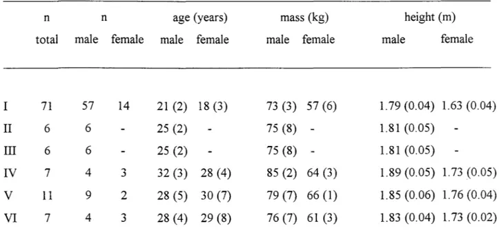
The characteristics of the subjects are summarized in Table 1. In Study I, the athletes belonged to the national elite of Sweden. In Study II-VI, all subjects were healthy, whereas in Study I some of the athletes had suffered episodes of low back pain, particularly among the gymnasts and wrestlers (cf. Sward 1990). In total, 94 different subjects have been investigated. Six subjects were common for Study II and III, two subjects for Study II, III and V, four subjects for Study IV and V, and one subject for Study IV-VI. The studies were approved by the Ethics Committee of the Karolinska Institute and all subjects gave their informed consent to participate.
Table 1. The number of subjects in Studies I-VI and their average (SD) age, body mass and height.
n n age (years)
total male female male female
I 71 57 14 21 (2) 18 (3) II 6 6 25 (2) III 6 6 25 (2) IV 7 4 3 32 (3) 28 (4) V 11 9 2 28 (5) 30 (7) VI 7 4 3 28 (4) 29 (8) mass (kg) male female 73 (3) 57 (6) 75 (8) -75 (8) -85 (2) 64 (3) 79 (7) 66 (1) 76 (7) 61 (3) height (m) male female 1.79 (0.04) 1.63 (0.04) 1.81 (0.05) 1.81 (0.05) 1.89 (0.05) 1.73 (0.05) 1.85 (0.06) 1.76 (0.04) 1.83 (0.04) 1.73 (0.02)
Electromyography (Study II-VI)
Intra-muscular EMG with ultra-sound guidance (Study II-VI)
Prior to the intra-muscular EMG measurements, extensive studies of the anatomy were performed, using both dissection of cadavers and magnetic resonance imaging, to assure that the wire-electrodes were accurately placed in the desired muscles. The method of using ultra-sound for guidance during insertion of fine-wire electrodes into deep pelvis and trunk muscles, was first employed for iliacus (Andersson et al. 1989) followed by simultaneous recordings from the vertebral psoas and the iliacus muscles (Andersson et al. 1991). The same method was subsequently used for the abdominal muscles (Cresswell et al. 1992). Before the start of all experiments with intra-muscular EMG accompanied by ultra-sound guidance, a needle insertion into the psoas muscle was made, as well as an ultra-sound inspection of various trunk muscles, at the Radiology Department, Soderhamn Hospital, Sweden. In the thesis experiments all the deep needle insertions with simultaneous guidance of ultra-sound was performed by Dr. Helen Grundstrom at the Radiology Department, Danderyd Hospital.
Muscles
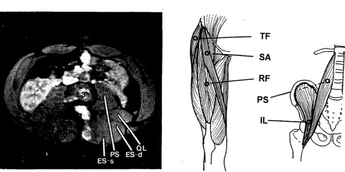
The investigated muscles and the insertion sites of the fine-wire electrodes ( cf. below) for the intra-muscular EMG recordings are shown in Figures 1-3. EMG activity was recorded unilaterally from the following muscles: iliacus, IL (Study II-VI), psoas, PS (Study IV-II-VI), sartorius, SA, tensor fascia latae, TF, and rectus femoris, RF (Study V), quadratus lumborum, QL, the deep lateral portion of lumbar erector spinae, ES-d, and the superficial medial part of lumbar erector spinae, ES-s (Study VI). Ultra-sound was utilized for guidance during the electrode insertions into the PS, QL and ES-d muscles in all subjects, and to control the routinely performed placement of the IL electrodes in one subject.
Recording sites
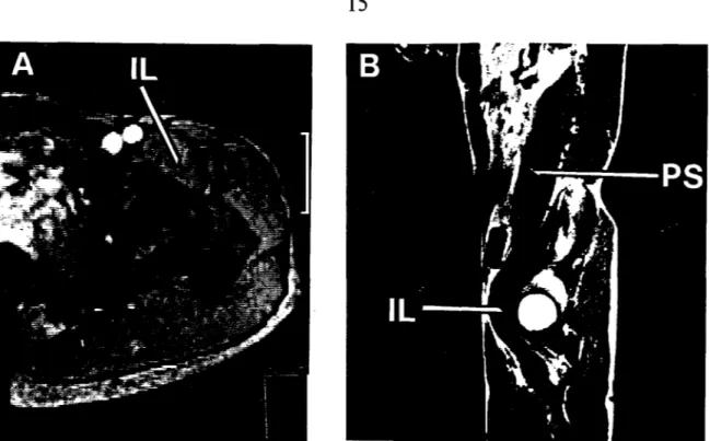
Insertion into IL was made at a point about 3 cm lateral to the femoral artery, 1 cm medial to the sartorius muscle and 1 cm inferior to the inguinal ligament, 3-4 cm deep from the skin (Fig. 1-3). Care was taken not to place the electrodes in the sartorius muscle, which happened occasionally in preliminary trials, prior to the start of the experiments included in the thesis.
The insertion of the needle carrying the electrodes (cf. below) into the PS, QL, ES-d and ES-s muscles were made at the L3-L4 level. The needle into PS was directed obliquely from lateral to medial, starting 5-8 cm lateral to the spinal processes, to a depth from the skin of between 8.5 and 12.5 cm (Fig. 1 and 2, cf. Andersson et al. 1991). Initially in the series of PS experiments, the insertion in one
Figure 1. MRI sections showing where the intra-muscular wire electrodes were· placed via a needle, in the iliacus (IL) and the psoas (PS, Study IV-VI). A) is a cross-section at the level of the hip joirit and B) is an oblique section taken at an approximate angle of 15 degrees to the sagittal plane, through the center line of the iliopsoas muscle.
subject was not made deep enough, the electrodes ending up in QL instead of PS. This caused erroneous conclusions about PS function ( cf. Nachemson 1966). One example thereof was the finding of activity in the most forward flexed position of a relaxed trunk in standing, where later correct recordings showed PS to be inactive (Andersson et al. 1995, Study VI). The insertions into the PS were performed without complications in all but one subject. For this subject the testing had to be prematurely terminated due to a decrease of skin sensation of pain and pressure around the anterior and superior part of the thigh. A normal sensory function was completely recovered after one year.
The insertion into the QL muscle was made into its mid-portion and for ES-d 0.5-1 cm posterior to the middle of the dorsal fascia of QL (corresponding to the iliocostalis muscle according to Bogduk 1980). The ES-s insertion was made 3 cm lateral to the mid-line and 1 cm into the muscle (multifidus, Bogduk 1980). The tips of the electrodes in SA, TF and RF (insertion sites showed in Fig. 2B) were placed at an approximate depth from the skin of 1-1.5 cm (Study V).
Fine-wire electrodes
The bipolar, multi-stranded, fine-wire electrodes consisted of seven thin stainless steel wires, each with a diameter of 0.035 mm. The electrodes were teflon insulated,
Figure 2. A) An MRI cross-section at the L3-L4 level with insertion sites for the . wire electrodes in the quadratus lumborum (QL), the deep lateral portion of erector spinae (ES-d), the superficial medial erector spinae (ES-s) (Study VI), and the psoas (PS) (Study IV-VI). B) Schematic drawings showing the approximate insertion sites for the wire electrodes into the tensor fascia latae (TF), sartorius (SA), rectus femoris (RF), psoas (PS) and iliacus (IL) (Study V).
except for 3 mm at the tips. The outer diameter of each electrode, including the teflon insulation, was 0.22 mm. The two fine-wires, twisted together, were placed outside and along a needle with their tips, kept 5 mm apart, hooked into the lumen at the top of the needle. At the base of the needle the wires were fixed to the needle with adhesive tape. The needle with the wires was sterilized in an autoclave (Tuttnauer 234 OE) with distilled water steamed at 134 degrees Celcius during 7 minutes, followed by a drying time of 15 minutes. The sizes of the needles were 0.9xl50/200 mm for PS, QL and ES-d and 0.7x50 mm for IL, ES-s, SA, TF and RF.
Prior to the electrode insertions, the skin was cleaned with 95% ethyl alcohol followed by local anesthesia (Xylocain 5mg/ml) given subcutaneously. An ultra-sound equipment (Acuson 128, Acuson Corp., USA; 5-7.5 MHz) was used to facilitate guidance of the electrodes to the desired area and provide assurance of placement into each muscle portion. The needle with its lumen is seen with a contrast echo in the ultra-sound image, as well as the various fascias separating the muscles. Upon reaching the desired region in the muscle, the electrodes were detached from the needle, which then was carefully withdrawn.
Before and after all experiments the length of the free ends of the wires outside the skin was controlled and found to be unchanged. A re-examination of one subject with ultra-sound prior to removing the fine-wires from the abdominal muscles after
place (Cresswell 1993). ·
Evidently, intra-muscular electrodes are required for EMG recordings from deeply situated muscles, such as PS, QL and the deep portions of ES.
It
is, in our opinion, not possible to obtain any reliable recording from PS with surface electrodes in the groin region. Furthermore, as the present data will clearly show, it is not valid to use surface recordings from other muscles as "surrogates", e.g. external oblique for psoas and lower erector spinae for quadratus lumborum, as suggested by Cholewicki and McGill (1996). Wire electrodes were preferred before needle electrodes since they have the distinct advantage of being flexible and thus causing minimal discomfort even during the extreme efforts performed here. The size of the wire electrodes is small which increases the specificity and minimizes the risk of "cross-talk", i.e. that the electrodes should pick up activity from adjacent muscles. On the other hand, the small pick-up volume of the electrodes may put restrictions on how well the recorded activity will represent that of the whole muscle. This seems to be less of a problem in muscles which appear homogenous, e.g. PS, than in complex muscles such as the ES.In
the present studies, the selection, preparation and insertion of the wire electrodes were deliberately made to allow for a relatively large recording volume. Any systematic study comparing simultaneous recordings from multiple intra-muscular sites or from intra-muscular and surface electrodes was not performed.--...__.,,
OE
~-~----RA
01
Surface EMG (Study II-IV) Muscles and recording sites
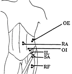
Figure 3. Positions of the surface electrodes on three abdominal muscles: obliquus externus (OE), rectus abdominis (RA), obliquus internus (01) and the two hip flexor muscles: sartorius (SA) and rectus femoris (RF) (Study II and III). The site for the insertion of the intra-muscular fine-wire electrodes into the iliacus (IL) is also indicated.
Surface electrodes were used to record EMG from the left hip flexor muscles: sartorius (SA) and rectus femoris (RF) and from the abdominal muscles: rectus abdominis (RA), obliquus extemus (OE) and obliquus intemus (01) (Study II and III;
recording sites are shown in Fig. 3) and from superficial medial erector spinae (ES-s) at the L3-L4 level on the right side in Study IV.
Electrodes
The sruface electrodes were Beckman miniature silver/silver chloride (Beckman Instruments, USA), diameter of pick-up area 4 mm, with a fixed inter-electrode distance of 8 mm. One common reference (ground) electrode was placed over the iliac crest in all experiments. Before applying the sruface electrodes, filled with electrode paste, the skin was prepared by shaving, gentle abrading with sandpaper and cleaning with 95% ethyl alcohol.
Sruface electrodes are less selective than intra-muscular electrodes. On the other hand, they may give a more representative picture of the overall activation of a muscle. On big, superficially located muscles, e.g. the thigh muscles, there is a possibility to choose either type of electrode. In the locomotion study (Study V), intra-muscular electrodes were used primarily to avoid the effects of the movement of the sruface electrodes on the skin in relation to the underlying muscles during the vigorous movements. The above-mentioned problem with "cross-talk" is evident in the case of recording with sruface electrodes from the muscles of the ventro-lateral abdominal wall. The present placement of the sruface electrodes on OE and OI has been shown to result in the least contamination from adjacent muscle layers ( cf. Cresswell 1993).
EMG analysis and normalization (Study II-VI) Signal processing
All EMG signals were differentially pre-amplified (100 times) close to the site of the electrodes, using small customized amplifiers attached to the skin. Signals were then band-pass filtered at 10-1000 Hz (Neurolog NL-125 filters, Digitimer Ltd, England), further amplified (10-50 times) and collected on magnetic tape for subsequent analogue to digital conversion (500 Hz) and computer analysis (APAS System, Ariel Dynamics, USA). During the experiments the signals were displayed in parallel, for direct visual inspection, on a Mingograf 803 ink-writer with a straight frequency response up to 1200 Hz (Siemens-Elema, Sweden).
Signal analysis
Prior to normalization, the average EMG amplitude was calculated from the rectified and filtered EMG signals (Study II-VI). The mean EMG amplitude for all the static tasks was calculated during the first two seconds after a stable level had been reached (Study II-IV and VI). For the dynamic submaximal and maximal sit-up and leg lift exercises (Study II and III), the mean EMG amplitude was calculated during the whole motion cycle, beginning 200 ms before the start of the motion ( cf. below).
exercises were analyzed separately (Study II).
Amplitude normalization
The EMG amplitude values for each muscle and task in Study II, IV and VI were normalized to, i.e. expressed as a percentage of, one value, the highest activity level observed during all the measurements. This normalization procedure was done separately for the static and the dynamic training exercises in Study II.
In Study III, two different normalization techniques for the mean EMG amplitude were performed and compared. First, the absolute activity level in each specific muscle and training exercise was expressed as a percentage of the activity level obtained in each corresponding maximal strength measurement, matched for position and average movement speed (30 degrees/s). Further, the activity levels in all static and dynamic training exercises were expressed as a percentage of only one EMG value, the highest activity level observed in any of all recorded maximal efforts for each muscle. The consequences of using various normalization techniques are discussed in detail in Study III. In the dynamic sub-maximal training exercises in Study III, only the EMG amplitude for the upward phase of each movement cycle was analyzed (0-30 degrees in TF, 0-60 degrees in HF and LL). During the maximal dynamic strength measurements, the mean EMG amplitude was calculated over the corresponding ranges of motion.
For each task the means (± standard error, SE) of the EMG percentage values for all included subjects were calculated for each muscle (Study II-VI), and in Study II and III also for the abdominal (RA, OE, OI) and the hip flexor synergy (IL, RF, SA).
In the locomotion study (V), band-pass filtering between 50-1000 Hz was applied before calculating the mean EMG amplitude for PS and IL, to eliminate movement artifacts. Amplitude analysis was made between the onset and termination of each main period of activity. Analysis of time of onset and termination of the EMG activity periods was performed by two experienced investigators who together made a decision based on visual inspection. The mean EMG amplitude for each individual muscle was expressed as a percentage of the highest mean EMG value for that particular muscle observed at any of the recorded velocities of walking and running. A mean of three stride cycles was calculated for each muscle (IL and PS) and subject (n=4). Finally, the mean values for all subjects (± standard error of the mean, SE) were calculated for each muscle and locomotion velocity.
Timing and duration normalization
Timing and duration values were only obtained in the locomotion study (Study V). The different phases of the stride cycle are defined under a separate heading below.
All events were presented in relation to a normalized stride cycle (Tc), i.e. the time between consecutive foot strikes of the right foot, set to 100% (cf. below). The onset of the main activity period for each muscle relative to the start of the stride cycle, end of the support phase and onset of hip flex.ion, as well as the total duration of each period, were measured. Means were calculated as for EMG amplitudes ( cf. above).
Strength measurements (Study I and 111)
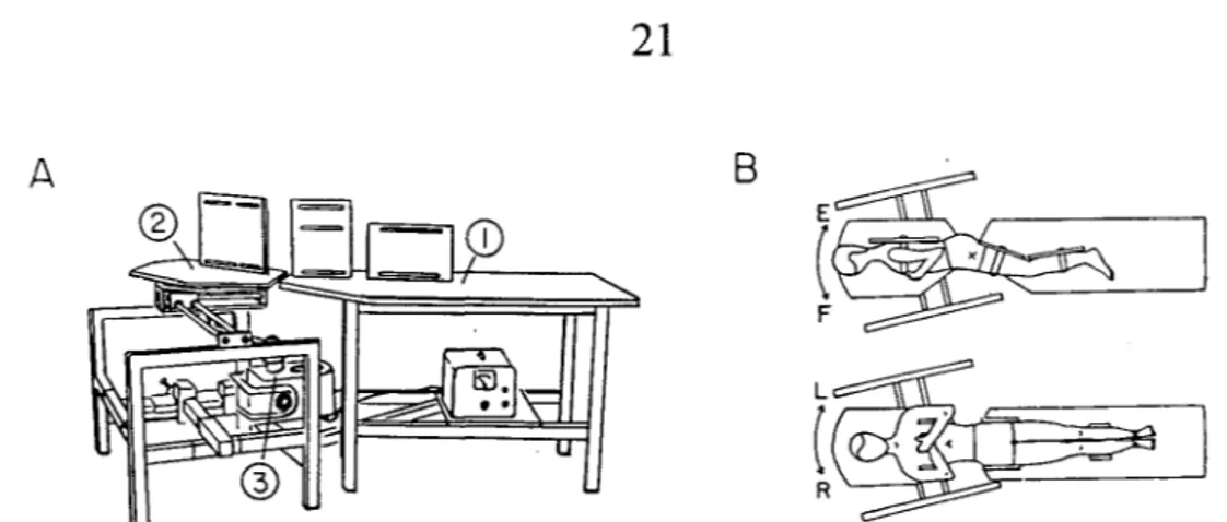
Maximal voluntary muscle strength was measured in trunk flex.ion, extension and lateral flex.ion to either side (Study I) with a method introduced by Thorstensson and Nilsson (19 82 ), utilizing an isokinetic ( constant velocity) technique ( Cybex II). The experimental set-up (also used in Study III) is shown schematically in Fig. 4. The subject was placed in a horizontal position to avoid direct influence of gravity on the measurements. The lower part of the body was strapped to a rigid table, (1) in Fig. 4~ and the upper part to a swivel table, (2) in Fig. 4A, which could be moved in an arc covering the whole movement range of motion for the trunk. The subject was placed in a right lateral supine position (flex.ion and extension, Study I and III) or a supine position (lateral flex.ion, Study I) (Fig. 4B). In Study III, an additional body position was measured, i.e. with the legs placed on the swivel and the upper body on the rigid table.
The strength, measured as produced torque, was transmitted to the lever arm of a Cybex-dynamometer, (3) in Fig. 4A, positioned underneath the table. The center of rotation of the body was placed above the connection between the tables. In
flex.ion/extension movements, the subjects were tested with the pivot point either at the hip joint or at the L2-L3 level, the latter referred to as trunk flex.ions and extensions. The pivot point in lateral flex.ion was at the L5-S 1 level (Fig. 4B). All these tasks were measured in Study I, whereas in Study III only flex.ion around the hip joint and the Tl2-Ll level were recorded. The additional hip flex.ion task in Study
ill, with the legs on the swivel table and the hip joint as pivot point, was referred to as "leg lift". The strength tasks in Study III were selected to be equivalent to training exercises for the abdominal and hip flexor muscles ( cf. above).
Torque was recorded at the constant velocities of 15 and 30 degrees/sin Study I, and only at the latter speed in Study III. Readings from the torque curves were made in five different trunk positions for flex.ion/extension and in six positions for lateral flex.ion movements. Also the peak torque value and the position at which peak torque occurred in the range of motion were recorded. In addition, static recordings were made in a straight body position (0 degrees) in Study I. In Study III, static strength was obtained at the following angles: 10, 20, 30 degrees and maximal angle in trunk flex.ion (TF), 10, 30 and 60 degrees in hip flex.ion attempting to move the upper body (HF) as well as in attempted leg lifts (LL). In Study I, both absolute strength (Nm) and strength normalized to body mass were compared between groups.
A B
( ~
F
t ~
Figure 4. A) Experimental set-up for measurements of maximal trunk and hip muscle strength (Study I) and EMG during maximal efforts (Study III). B) Body position in extension (E) - flexion (F) movements (pivot: hip joint) and in lateral flexion movements (pivot: L5-Sl; L = left, R = right). For further description - see Methods.
Strength ratios were calculated for antagonist muscles ( extension/flexion and nondominant/dominant side) as well as for agonist muscles, i.e. hip/trunk strength in extension and flexion movements, respectively.
It
may be argued that the maximal strength capacity could vary due to differences in overall body position and that a comparison between submaximal training exercises in a supine position and strength measurements during maximal efforts in a side-lying position therefore may be misleading. Possible reasons for this could be varying peripheral input to the central nervous system, such as from skin receptors and the vestibular apparatus. Up till now, no systematic comparison of the maximal trunk strength performance in different body positions, such as standing, sitting and lying, has been published. Comparisons with strength values in the literature are of limited value since they were obtained on different groups of subjects using different experimental paradigms. One frequent source of variation 1s an inability to standardize the contribution from hip and trunk muscles.Movement recordings (Study I-VI)
An electrogoniometer (high precision potentiometer, plus to levers) was attached over trochanter major (a in Fig. 5B) to record angular displacement in the sagittal plane at the hip joint (Study II-VI). The two levers of the goniometer (10 and 15 cm long, respectively) were firmly taped to the skin. The lower lever was pointing to the lateral midpoint of the knee joint and the upper one to the midpoint of the crista iliaca. In Study II and III, an additional goniometer was used to record movements of the whole upper body during the sit-up exercises. This goniometer was positioned under the bench, on which the subject was placed supine, and connected via a string taped to the skin over the spinous process of C7 (bin Fig. 5B). To obtain the specific
angles in the static tests a manual goniometer was used, _which also was set at the upper limit for each movement in the training exercises. During the measurements of maximal strength in Study I and III, the goniometer on the Cybex equipment was utilized to measure angular displacement of the swivel table ( cf. above).
Training exercises for the trunk and hip flexor muscles (Study II-IV, VI)
In Study II and III the primary intention was to systematically measure various training exercises for the abdominal and hip flexor muscles. In Study IV and VI, some of these tasks were repeated with additional modifications, and training exercises for the back extensor and trunk lateral flexor muscles were also recorded ( cf. below).
Dynamic exercises
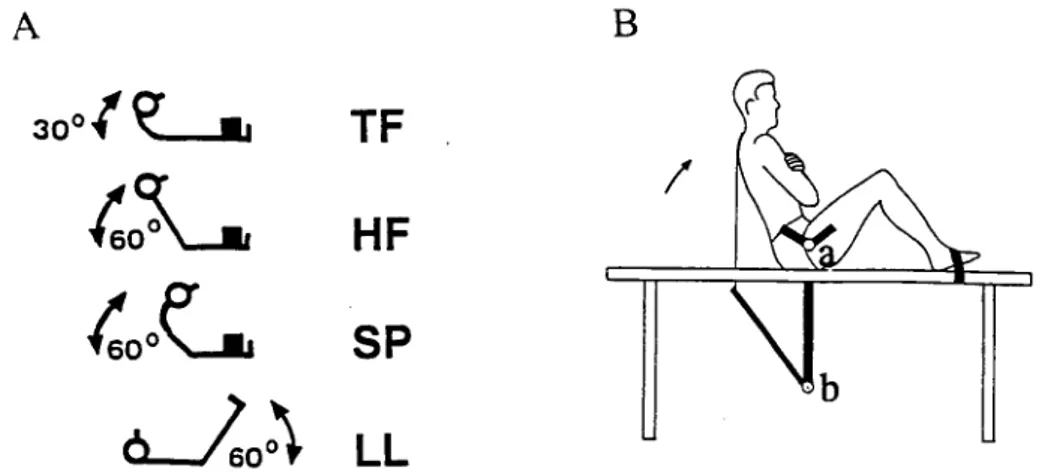
Four different dynamic exercises for the abdominal and hip flexor muscles were investigated in Study II (Fig. 5A): (1) trunk flexion (TF), i.e. "a shoulder lift" or "trunk curl", pivot point approximately at Tl2-Ll level, (2) hip flexion (HF), i.e. lifting the whole upper body held straight, (3) spontaneous sit-up (SP), without any specific instruction, carried out as a combination of trunk and hip flexion and ( 4) leg lift (LL) with straight knee joints, either bi- or unilaterally, with the pelvis in a neutral position. Each of exercises (1) - (3) was performed in four different ways: with the legs straight supported ( ss ), straight unsupported ( s ), bent (knees at 90 and hips at 13 5 degrees) supported (bs ), and bent unsupported (b).
A B
30°(~
.,
TF
tsL
HF
/
fso
0e:__.
SP
J.o:\
LL
Figure 5. A) Tested training exercises and their overall angular excursions (Study II and III): trunk flexion sit-up (TF), hip flexion sit-up (HF), spontaneous sit-up (SP) and leg lift (LL). B) Experimental set-up for studying training exercises, with hip goniometer (a), indicating movements at the hip, and bench goniometer (b) recording both trunk and hip angular movements (Study II and III).
representative cycle was selected for analysis.
In
total, 15 different dynamic tasks were measured in Study II.In
Study III, the 3 dynamic exercises utilized were TF and HF, with straight and supported legs, and bilateral LL.The movement ranges were from 0-30 degrees in TF and 0-60 degrees in HF, SP and LL (0 degrees= straight body). The angles were taken between the horizontal and a line from the pivot point (Tl2-Ll level in TF and hip joint in HF, SP and LL) to the C7 level when lifting the upper body, and to the lateral malleolus in leg lifts. The exercises were performed to the rhythm of a metronome set at 1 Hz. The duration of the entire movement, including upward and downward phases, was 2 s in TF and 4 s in HF, SP and LL, respectively. Thus, all the dynamic exercises were performed with a mean angular velocity of approximately 30 degrees/s.
Static exercises
TF, HF and bi- and unilateral LL were also studied in different static positions. TF and HF were performed either with straight or bent supported legs. The static angles were, in TF: 10, 20, 30 degrees and maximal (approximately 40-45 degrees) and in HF and LL: 10, 30 and 60 degrees (angles defined as in dynamic exercises). A total of 23 static positions were measured in Study II.
In
Study III, static TF and HF with straight and supported legs, as well as bilateral LL, were utilized at the above mentioned angles, i.e. a total of 10 static positions were investigated.Other postures and motor tasks ( Study IV and VI)
In
Study IV and VI various static and dynamic tasks were performed for the trunk, pelvis and legs in standing, sitting and lying positions, including some previously described exercises (Study II). A few of the static positions are illustrated in the photo sequence (Fig 10) in Chapter 3.Trunk and pelvis tasks Standing
In
standing, the following trunk and pelvis tasks were measured: standing at ease on both legs, with the whole upper body flexed 30, 60 and 90 degrees at the hip with thetrunk
held straight, as well as with the trunk held relaxed and kyphotic at the 90 degrees hip angle.In
addition, a dynamic test was performed from upright standing to full forward flexion of the wholetrunk,
held relaxed and kyphotic. The duration of the movement was approximately 4 s. Additional positions were: maximal flexion of thetrunk
to either side, maximal hyperlordosis of the lumbar back, maximal forward pelvic tilt, maximal lifting (tilting) of the pelvis at either side, erect standing carryingloads of 34 and 20 or I 0kg, either in two hands in front or in one hand along either side of the body. In addition, dynamic trunk flexions · from side to side were performed at a slow angular velocity. Slow dynamic lifts with the 34 kg load were done, either laterally with each hand separately or with two hands in front of the body, keeping the back straight and bending and extending the legs.
Sitting
The following trunk positions were tested in sitting: straight, relaxed kyphotic, extended 30 degrees at the hip, flexed 30 and 60 degrees at the hip, and maximally flexed laterally to the ipsi- and contralateral side. Additionally, positions with a combination of maximal hyperlordosis of the lmnbar spine and a forward tilted pelvis, and with the pelvis lifted maximally at either side, were measured. Also dynamic lateral flexions of the trunk were performed in sitting, either as fast flexions to the ipsi- or contralateral side from an upright position or as slow movements from side to side.
Lying
In lying, different static and dynamic training exercises were executed. In a supine position, a trunk flexion ("trunk curl") to a maximally flexed position was performed with the legs placed on a stool (90 degrees in the hips and knees) and the feet unsupported. In the same leg position, but with supported feet, the whole trunk was held elevated via a hip flexion at I 0, 30 and 60 degrees. In addition, the latter exercises were recorded with the legs resting straight and supported on the floor. The hip flexion sit-up was also performed dynamically with both leg positions. The movements were guided by a metronome set to give an average angular velocity of 30 degrees/s ( cf. above).
In a prone position maximal trunk extension was performed with the arms held straight in front of the body. Maximal lateral flexion of the trunk was carried out in a lateral supine position with supported feet.
Leg tasks Standing
In standing, the following static leg positions were tested ipsi- and contralaterally: standing on both legs but with the body weight shifted onto one leg, and standing on one leg on a box with the other leg hanging freely or flexed 30, 60 and 90 degrees at the hip. Additional static tasks were: with either leg extended 30 degrees and maximally, or maximally abducted at the hip.
Sitting
In lying, the leg tasks included: straight leg lift at 10, 30 and 60 degrees of hip flex.ion from a supine position with both legs and one at a time, bi- and unilateral maximal hip extension in a prone position, and maximal ipsilateral hip abduction in a lateral supine position.
Locomotion (Study V)
Level walking (1.0, 1.5, 2.0 and 3.0 m/s) and running (1.5, 2.0, 3.0, 4.0, 5.0 and 6.0 m/s) were performed on a motor-driven treadmill (treadmill belt size: 2.7 · 0.8 m). All subjects had previous experience of treadmill locomotion. The recordings were made during approximately 30 s of "steady state" walking and running at each velocity.
The instances of foot strike and toe off were recorded with a pressure sensitive device mounted to the sole of the right shoe (Nilsson et al. 1985b) and used to calculate temporal aspects of the stride cycle, e.g. durations of the stride cycle, support and swing phases. The device consisted of a pressure transducer (National Semiconductor LX-0503A) connected to a flexible silicone tube which was glued to the outer perimeter of the sole. Deformation of the tubing from foot - belt contact caused a change of pressure inside the tube, which was converted to a voltage change via the transducer. Stride cycle duration (Tc) was defined as the time from foot strike (onset of support) of the right foot to the subsequent foot strike of the same foot. The support phase duration (Tsu) was defined as the time from right foot strike to right toe off. The swing phase duration (Tsw) was taken as Tc minus Tsu for the right foot.
Statistics (Study I-VI)
Differences between exercises in EMG activity levels for each muscle (Study II-VI) and whole muscle synergies (Study II and III), in duration or onset of muscle activity periods (Study V), and in strength values (Study I), were tested for significance with a one-way ANOVA. Differences (p<0.05) between specific exercises (study I-VI) and between muscles (Study V), were detected with a Studentized range test (Study I) corresponding to the post-hoe Tukey test-HSD, or with the Neuman-Keuls (Study II), Duncan (Study III and V), and Tukey-HSD (Study VI) post hoe tests or with non-overlapping 95% confidence intervals (Study IV). The significant differences reported in the latter study (IV) were present also when re-tested with the post-hoe Duncan-test.
Chapter 3
Results and Discussion
The emphasis in this chapter will be on the activation levels of the trunk and hip muscles during various training exercises, body positions and motor tasks (Study II-IV and VI). Only brief accounts will be given of the analysis of locomotion (Study V) and the strength measurements (Study I).
The definition of the term "training exercise" as opposed to other motor tasks is somewhat arbitrary. Any of the motor tasks studied may, of course, be used as a training exercise to improve a specific function, such as sitting or walking. Likewise, the tasks named training exercises here may well be part of everyday activities, such as "sit-ups", which are commonly used to get out of bed.
Given that the exact stimulus for training induced gains in muscle strength is unknown, much of the present discussion is resting on the assumption that the higher the activation of a muscle during an exercise, the more likely the muscle is to benefit from using that particular exercise in training.
Training exercises (Study II-IV, VI)
General aspects
The present training exercises represent common exercises used in sport, rehabilitation, and general health and fitness programs to strengthen the trunk and hip musculature. Naturally, there exists a myriad of other variants of, for example, sit-ups, whose activation patterns are not covered by the present results. Further studies like these are needed, since, due to the high degree of task specificity of the trunk
and hip muscles demonstrated here, one can not simply extrapolate from one exercise to another.
When evaluating the results, some general characteristics regarding training and training effects should be kept in mind. A key word in this context is specificity. This specificity encompasses muscle engagement, which means that training effects are only reached to any substantial degree in muscles that are actually engaged in the training exercise. This is where the present studies give an important contribution. This is true also with respect to the values presented for the relative degree of activation. However, the percentage values serve mainly the purpose of making possible comparison between exercises. An attempt to compare the EMG levels in
rule of thumb in strength training is that the training load should correspond to at least 60% of the maximum strength to be effective.
The training effects are also thought to be specific with respect to muscle action type. Types of muscle actions are generally divided into: concentric, that is dynamic muscle actions where the muscle is allowed to shorten and attempts to increase the speed of motion; eccentric, which means dynamic muscle actions in which the active muscle undergoes lengthening, attempting to decrease the speed of motion; and lastly, static (isometric) muscle actions, where the overall length of the muscle is kept constant. In addition, during dynamic actions, there may be speed-specific training effects and in static actions the response to training has been shown to be position-specific. (For overviews on strength training, see Enoka 1988, Komi
1992.)
Exercises for the trunk and hip flexor muscles
Exercise type and selectivity
The activity levels of individual muscles and muscle synergies could be varied substantially by modifying the training exercises with respect to type of exercise, body position, range of motion, and leg support. The effects of some of these modifications are illustrated by the recordings shown in Fig. 6 and 9 and further quantified in Fig. 7 and 8.
A selective involvement of either the abdominal or hip flexor muscles could be achieved by varying the type of exercise. In trunk flexion sit-ups, both static and dynamic, essentially only the abdominal muscles were activated. The involvement of hip flexor muscles was particularly low for the trunk flexion exercises with straight legs and at small trunk flexion angles. The reversed pattern, that is a selective engagement of hip flexor muscles while the abdominals were silent, was attained in unilateral leg lifts. Noteworthy was the high abdominal and low hip flexor EMG when dynamic hip flexion or spontaneous sit-ups were to be performed with bent legs and no leg support ( cf. below).
Also, the activation levels of individual muscles could vary markedly with type of exercise. One of the most conspicuous differences was seen for the external oblique muscle whose activation was lower than that of the other abdominal muscles, specifically in trunk flexion exercises. Interestingly, a selective activation of the psoas muscle was present in a sit-up exercise when the hip flexor muscles , including the iliacus, were silent. This was the case for a sit-up with bent legs placed on a stool with no leg support. The influence of varying conditions on the relative activation of different trunk and hip muscles will be further outlined below. The task specificity that will be shown, can be one reason for the discrepancies between results of earlier studies, where standardization and objective quantification have been less rigorous.
BG
~
_ _ . . - - _ HG(~----~-RALJl__
tti~L
OE 01 IL RF SAl
\:ft t:tH, I__.,-tn-•-"-- ...MltYrl
+dl•w►,+◄sf:Jblfb
I illillih1111.,ll!i ..
.JWli#
11w, 11!"6 1ttl!1:
l
t ik+r'fll:t+ ttlt t:1+: --trMii~+:al ♦~\ftriotlb!ltdli!NIWi·: JIM :'t\ttu~...+hlUJ,◄IWW1tb' ' 1 II J Hlit
,w..,, . . . tt .. 1L1M.r.Wd1AA+i,»+ I I
2 s 1 2 s 4s 4s , - - - 4 s - - ~
TFss TFbs HFss HFbs Llli
30° ( ~ 30° ( ~
l ~
t,~
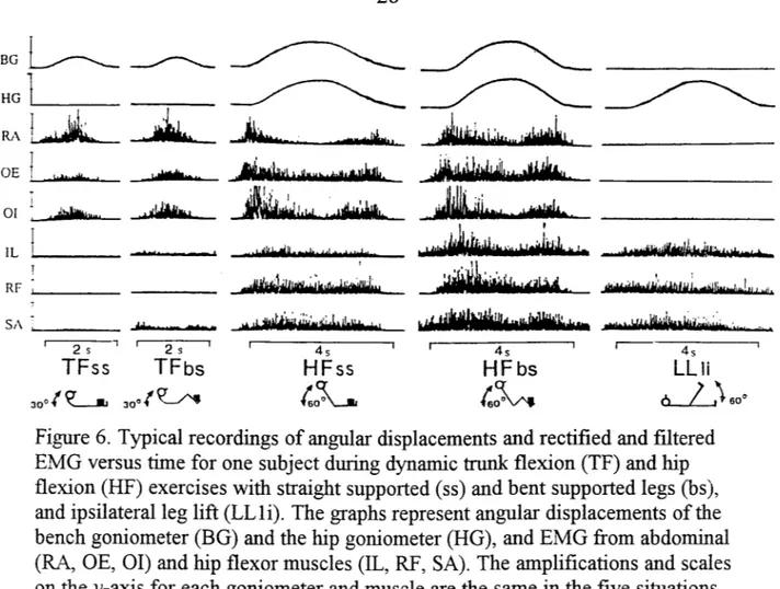
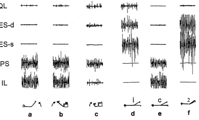
6 / )60°Figure 6. Typical recordings of angular displacements and rectified and filtered EMG versus time for one subject during dynamic trunk flex.ion (TF) and hip flex.ion (HF) exercises with straight supported ( ss) and bent supported legs (bs ), and ipsilateral leg lift (LLli). The graphs represent angular displacements of the bench goniometer (BG) and the hip goniometer (HG), and EMG from abdominal (RA, OE, 01) and hip flexor muscles (IL, RF, SA). The amplifications and scales on the y-axis for each goniometer and muscle are the same in the five situations. ("Ipsi-" always refers to the side where the EMG was recorded.)
For example, concerning the abdominal muscle involvement in trunk flex.ion sit-ups (shoulder lifts) as compared to hip flex.ion sit-ups, lifting the whole trunk, studies can be found reporting equal (Walters and Partridge 1957, Partridge and Walters 1959) as well as higher (Halpern and Bleck 1979), and even lower values (Noble 1981.)
Abdominal muscle involvement
Muscle action types in different exercises
As mentioned above, the abdominal muscles were activated during most of the training exercises tested, except the unilateral leg lifts. The function of the abdominal muscles differs, however, between exercises. It is only in the dynamic trunk flex.ion sit-up and initially in the spontaneous sit-up that the abdominal muscles are actually undergoing a length change, that is acting dynamically ( concentrically up and eccentrically down). In all the other dynamic exercises, the abdominal muscles have static actions, stabilizing the upper body and pelvis. This has to be remembered when comparing and evaluating the EMG levels, even though no extrapolation from EMG to force output of a muscle is done
in
the current studies.% 60 50 40 30 2
tttt
10 ■- ■ '~·
' 0 I ss s bs b TF 3 0 ° ( ~Dynamic
I I I I ss s bs b HFtsA_.
Abdominal synergye
Hip flexor synergy ■
•
I I I I ss s bs b SPfso
0~ \ \ \ \ \~
2 1i 1c LLJ.o~
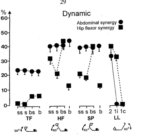
Figure 7. Mean EMG amplitudes (and SE) in dynamic exercises (up and down, Study II, n=6) for the abdominal (RA, OE, 01, circles) and hip flexor muscle synergies (IL, RF, SA, squares). The values are expressed as a percentage of the single highest activity value observed for each muscle among all maximal tests in Study III. The exercises were: trunk flexion (TF), hip flexion (HF), spontaneous sit-up (SP) and leg lift (LL). The first three exercises were carried out in four different ways: with legs straight and supported (ss), straight and unsupported (s), bent and supported (bs) and bent and unsupported (b), respectively. Three types of LL were performed: bilateral (2), ipsilateral (li) and contralateral (le).
E.ffects of position
The position in the range of motion during trunk an hip flexion sit-ups had a profound effect on the activation levels of all abdominal muscles. This is evident from the recordings of dynamic exercises in Fig. 6, and is shown more clearly for the abdominal muscle synergy in different static angles in Fig. 8.
In trunk
flexion sit-ups, the activation of the abdominal muscles increased with increasing flexion of thetrunk,
in spite of a decreasing external moment due to a shortening of the lever arrn for the gravitational force. A possible reason for the need for an increased activation could be the decreased force producing capability of the muscles at shorter lengths (e.g. Inmanet al.
1952, Bigland and Lippold 1954). Another contributing factor may be an increased "inner resistance" due to a compression of the abdominal content.%
Static
60
50
Abdominal synergy
e
Hip flexor synergy ■
40
·i
,,
,f
''
30 '~
' ' I 2If'
10•
, 0...
I I I I I I I.,
'
,
__
,
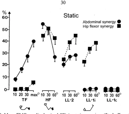
10 20 30 max0 10 30 60° 10 30 60° 10 30 60° 10 30 60° TF HF LL-2 LL-1i LL-1c ~½
J
Figure 8. Mean EMG amplitudes (and SE) in static exercises (Study II, n=6) for the abdominal (RA, OE, OI, circles) and hip flexor muscle synergies (IL, RF, SA, squares). Values are expressed as a percentage of the single highest activity value observed for each muscle among all maximal tests in Study III. In trunk flexion (TF) the positions were: 10, 20, 30 degrees and maximal angle, and in hip flexion (HF) and leg lifts (LL): 10, 30 and 60 degrees. The TF and HF were performed with bent and supported legs (bs) (cf. Fig. 7, legend). Three types of LL were performed: bilateral (2), ipsilateral (li) and contralateral (le).
The opposite pattern, that is a decrease of abdominal EMG with angle, was present with increasing degrees of hip flex.ion in the hip flex.ion sit-ups (Fig. 6 and Fig. 8).
It
should be recalled that the abdominal muscle action now is static throughout the motion. Therefore, the mechanical conditions for the muscles, in terms of length of the muscles and their lever arms, are unchanged. The lesser requirement for muscle activation is clearly a consequence of a decrease in length of the external lever arm and, thus, a lower gravitational moment of the trunk.The spontaneous sit-up was not analyzed with respect to the effects of position. However, since this exercise consisted of both a trunk flex.ion and a hip flex.ion, it should contain both patterns described above.
As mentioned earlier, any stabilizing action of the pelvis by the abdominal muscles did not seem to be required during unilateral leg lifts. Similar observations were made by Carman et al. (1972) and deSousa and Furlani (1974).
In
bilateral leg lifts, on the other hand, such static stabilization was warranted as evidenced by awere seen on abdominal EMG with changes in angle of hip flex.ion, in spite of the variation in external loading moment of the legs and thereby, most likely, in tendency of the pelvis to tilt forwards. Our results agree to some earlier investigations on bilateral leg lifts, reporting moderate to marked activity levels for the rectus abdominis and obliquus externus and somewhat lower for the obliquus internus (Floyd and Silver 1950, Sheffield 1962, Flint 1965a and b, Carman et al. 1972, deSousa and Furlani 1974). The discrepancy between these results and those of Partridge and Walters (1959), finding a very low abdominal activity, may be due to different positions of the pelvis. The instruction to the present subjects during the leg lift exercises was to maintain the pelvis in a neutral position. A marked drop in abdominal EMG was provoked by tilting the pelvis forward during a bilateral leg lift at 10 degrees. Correspondingly, a considerable increase in activity for all abdominal muscles, especially rectus abdominis, was seen during a backward pelvic tilt. Also performing a backward pelvic tilt in a supine position with the legs resting on the floor resulted in a marked activation of the abdominal muscles (Walters and Partridge 1957, Partridge and Walters 1959, Carman et al. 1972). (The control of pelvic movements will be dealt with more in conjunction with other motor tasks below.)
Hip flex.ion sit-ups and bilateral leg lifts represent two exercises that engage both trunk and hip flexor muscles. In the dynamic tasks, the static action of the abdominal muscles generally demanded somewhat lesser activation levels in bilateral leg lift. The difference appears to arise mainly early in the respective movement. This is based on the finding that holding the legs elevated at 10 and 30 degrees of hip flex.ion, did not require as high an activation as keeping the upper body in the corresponding static positions. This can be explained by the smaller loading moment created by the legs as compared to the upper body. In this range of motion our results agree to earlier reports (Sheffield 1962, Lipez and Gutin 1970). However, at 60 degrees of hip flex.ion, there was no significant difference in activity levels between the two exercises. This shows the importance of taking the position specificity into consideration when evaluating exercises.
Effects of leg modifications and support
Changing the leg position from straight to bent legs or providing support for the legs or not during the trunk and hip flex.ion sit-ups did not have any profound effects on the level of activation of the abdominal muscles. This was to be expected since the abdominal muscles will not be influenced by leg modifications as long as the pelvis is kept in the same position. Unchanged EMG levels for abdominal muscles have also been observed in earlier studies comparing hip flex.ion sit-ups with supported straight and bent legs; for rectus abdominis (Flint 1965a, Lipez and Gutin 1970, McGill 1995) and for the external and internal obliques (McGill 1995). Similarly, Ekholm et al. (1979) reported equal EMG amplitudes for rectus abdominis and obliquus
externus in trunk flex.ion sit-ups using corresponding_ leg modifications. Some limitations of the earlier studies are to be noted: Lipez and Gutin and Flint just used visual inspection of the recordings and McGill and Ekholm et al. investigated only one static position, 45 degrees of hip flex.ion and maximal trunk flex.ion, respectively. Contrasting results have also been reported, although only in studies using visual inspection, and mostly on single subjects. Examples of both higher and lower abdominal activity have been found when comparing sit-ups with straight and flexed legs (Walters and Partridge 1957, Flint 1965a and b, Lipez and Gutin 1970, Godfrey et al. 1977, Noble 1981). Why these studies have arrived at varying results are not clear.
Consequences for training
Evidently, the selection of exercises becomes important when specific effects on certain muscles and muscle groups are desired. Likewise, it is essential to note that conclusions about the involvement of other muscles in a muscle synergy cannot always be drawn on the basis ofEMG recordings from a single muscle.
As always, the choice of training exercise should be based on an analysis of the purpose of the training. An analysis is required of what specific function should be trained to improve performance or get back to a normal capacity. In light of the present task specificity, such an analysis should include movement pattern, position, muscle action type, etc ..
If selective effects on the abdominal muscles are desired, trunk flex.ion sit-ups can be recommended. In that type of sit-up, no attention has to be paid to leg modification or support. Note that even in the most extreme sit-up position tested, that is with bent unsupported legs on a stool, activation of the psoas muscle could not be avoided. This may be of some concern when training back patients. Otherwise, trunk flex.ion exercises may be preferred for this category since hip flex.ion sit-ups are associated with higher disc pressures (Nachemson and Elfstrom 1970).
Sit-ups can also be performed with a twisting of the trunk and upper body. Although not being studied systematically here, occasional observations were made showing that sit-ups with a twist to the left engaged the right obliquus externus and the left obliquus externus to a higher extent than their contralateral counterparts, and vice versa (Andersson et al. unpublished). In trunk twisting movements in standing and sitting, also the innermost abdominal muscle layer, the transversus abdominis, were shown to participate (Cresswell et al. 1992). Furthermore, specific training of this muscle group resulted in an increase in maximal pressurization of the abdominal cavity, which may be involved in stabilizing the trunk (Cresswell et al. 1994).
If the aim is for a high activation of abdominal muscles, irrespective of involvement of other muscles, static hip flex.ion sit-ups appear to be the most suitable. Particularly at small angles, that is in static positions with the upper body close to the floor, high activation levels were attained. Bilateral dynamic leg lifts also
conditions. If dynamic abdominal muscle actions are desired, dynamic trunk flexion sit-ups per se, or as part of a spontaneous sit-ups, can be utilized.
Hip jlexor muscle involvement Activity levels and specificity
The hip flexor synergy was generally not substantially activated in dynamic or static trunk flexion sit-ups. There was a tendency towards an increase in EMG with bent legs, but still the activity levels were less than 10% of the highest observed (Fig 7 and 8). In hip flexion and spontaneous sit-ups, the hip flexor synergy was particularly highly activated in the bent and supported leg position ( cf. below) reaching levels (40-50 %) similar to those during bilateral leg lifts.
The effects of varying the conditions were also different among the individual hip muscles ( cf. below). One conspicuous situation occurred, as mentioned above, when a hip flexion sit-up was to be performed with bent legs (90 degrees in the hips and knees, lower legs resting unsupported on a stool). In this exercise, which resulted in a maximal trunk flexion and no hip movement, the iliacus was generally not engaged at all or to a very low level, whereas the psoas muscle was clearly activated (Fig. 9). This is an exercise which has generally been thought of, and also previously reported, as not engaging the psoas muscle at all (Ash and Burnett 1989). Differences between the single joint, hip flexor muscle iliacus, and the two joint muscle, hip flexor and knee extensor, rectus femoris, were evident in some conditions, particularly with respect to the effects of hip angle both in hip flexion sit-ups and bi-and ipsi-lateral leg lifts. The rectus femoris muscle had diverging activity patterns also in locomotion ( cf. below) and can therefore not be seen as a representative muscle for the whole hip flexor synergy, which was suggested by Walters and Partridge (1957).
Effects of position
In the hip flexion sit-ups, there were no significant effects of hip angle on the activation level of the hip flexor synergy (Fig. 8). However, this does not mean that all individual muscles behaved the same. There was a clear tendency for a redistribution of activity among the muscles with increasing hip angle. The activation of iliacus tended to increase, whereas the reversed was true for rectus femoris. The static hip flexion sit-ups were made also to a hip angle of 75 degrees (unpublished results). As compared to the previously highest recorded angle of 60 degrees, 10-20% higher activity levels were seen for IL, whereas the activation of the other hip flexors actually decreased with the same order of magnitude.