This is an author produced version of a paper published in Carbohydrate Polymers. This paper has been peer-reviewed but does not include the final publisher proof-corrections or journal pagination.
Citation for the published paper:
Mølgaard, Susanne; Henriksson, Marielle; Cárdenas, Marité; Svagan, Anna. (2014). Cellulose-nanofiber/polygalacturonic acid coatings with high oxygen barrier and targeted release properties. Carbohydrate Polymers, vol. 114, p. null URL: https://doi.org/10.1016/j.carbpol.2014.08.011
Publisher: Elsevier
This document has been downloaded from MUEP (https://muep.mah.se) / DIVA (https://mau.diva-portal.org).
1
Cellulose-nanofiber/polygalacturonic acid coatings with high
1oxygen barrier and targeted release properties
2Authors: Susanne L. Mølgaard‡, Marielle Henriksson,ǁ‖ Marité Cárdenas‡† and Anna J. Svagan * 3
4
‡University of Copenhagen, Department of Chemistry, Universitetsparken 5, DK-2100 Copenhagen. 5
*University of Copenhagen, Inst. for Food Research, Rolighedsvej 30, DK-1958 Frederiksberg C. 6
†Biomedical Laboratory Science, Faculty of Health and Society, Malmö University, SE-205 06 Malmö. 7
ǁ‖ SP Technical Research Institute of Sweden, Department of Wood Technology, P.O. Box 5609, SE-114 8 86 Stockholm. 9 10 Abstract 11
A bio-inspired coating consisting of pectin (polygalacturonic acid) and cationic cellulose nano-12
fibers were successfully produced by the Layer-by-layer method. The build-up and the morphol-13
ogy of the resulting coatings were studied with spectroscopic ellipsometry and atomic force mi-14
croscopy, respectively. The coating was able to survive the exposure of a simulated gastric fluid, 15
but was partially degraded upon exposure to pectinase enzyme, which simulate the action of the 16
microbial symbionts present in the human colon. Prior to exposure, the oxygen permeability co-17
efficient of the coating (0.033 ml (STP) mm m-2 day-1atm-1 at 23°C and 20% RH) was in the 18
same order of magnitude as for ethylene vinyl alcohol films (0.001-0.01 ml (STP) mm m-2 day -19
1atm-1). However after exposure to the mimicked gastrointestinal (GI) tract conditions, the con-20
tribution of coating to the overall barrier properties was not measurable. 21
22
1. Introduction 23
The Layer-by-layer (LbL) technique has been extensively studied over the past two decades due 24
to its very precise way of controlling film structure and composition at the nanoscale. Ad-25
vantages of such a bottom-up technique over other available processing techniques, e.g. solvent 26
casting, have previously been demonstrated and discussed for many systems.1 Using this rela-27
tively simple technique, a whole range of thin multilayer films or coatings have been prepared, 28
exhibiting unique (multifunctional) properties such as ultra-high gas barrier properties,1b antimi-29
crobial,1c flame retardant,1d and sensing properties.1e The LbL technique consists of alternating 30
dipping and rinsing steps (depicted in Scheme 1a) in dilute solutions of oppositely charged pol-31
ymers/colloids. After each dipping step a thin layer is deposited, and the thickness of the ad-32
2 sorbed layer can be altered by processing parameters such as temperature2 and pH.3
1
Recently, much attention has been directed to bio-based polymers as sustainable alterna-2
tives to man-made polymers in various fields of application including food packaging. The poly-3
saccharide that probably has gained most attention is cellulose nanofibers (NFC), also denoted 4
microfibrillated cellulose or nanofibrillated cellulose. This is mainly due to the outstanding set of 5
properties of NFC such as mechanical strength and stiffness (crystal modulus) approaching that 6
of Kevlar4 and Steel,5 respectively. In addition, NFC are non-toxic and neat NFC based films 7
exhibit oxygen barrier properties at 0% RH and 23 °C that are two orders of magnitude better 8
than ethylene vinyl alcohol (EVOH). EVOH is typically used in high barrier applications.6 Natu-9
ral cellulose nanofibers are only weakly charged, but by chemical modification it is possible to 10
prepare both anionically and cationically charged cellulose nanofibers, making them more suita-11
ble candidates in LbL deposition. 12
Pectin is a complex anionically charged polysaccharide that is naturally present in plant 13
cell walls. Pectin is a large polysaccharide consisting mainly of linear chains of α-(1→4)-linked 14
D-galacturonic acid (homogalacturonan) in addition to bulky and highly branched segments 15
(Rhamnogalacturonan I).7 Pectin has previously been reported to exhibit colon targeting poten-16
tial, since human microbiota responsible for its break-down is present first in the colon.8 17
In the present study, the LbL method was employed to build up coatings based on cation-18
ic NFC and the anionic poly(galacturonic acid) (polyGalA), for chemical structure schematics 19
see Scheme 1b. The coating is bio-inspired since natural types of multilayer structure consisting 20
of pectin and cellulose nanofiber (microfibrils) are abundant in nature, e.g. the so-called primary 21
cell wall (parenchyma cells) found in soft tissue in fruit and vegetables.7 The development of 22
such coatings could potentially allow microencapsulation of active components both in drug and 23
food applications. Here, we demonstrate the build-up of a bio-inspired (“plant-cell-like”) multi-24
layer coating of pectin and NFC. Further we assess the oxygen barrier properties, which are im-25
portant during storage to increase the oxidative stability of sensitive ingredients, and the envi-26
ronment responsive release properties of the coating. More specifically, the multilayer coatings 27
were tested for simulated environmental stresses such as acidity and bacterial enzyme degrada-28
tion in the human gut that potentially hydrolyse the coating, thus providing a unique system for 29
the selective release of active components (e.g. drugs, probiotics, peptides) in the gut.9 30
3 1
Scheme 1: a) Schematic image of the layer-by-layer process, b) the molecular structure of 2
poly(galacturonic acid) and c) the structure of the cationic NFC. 3
4
2. Experimental part 5
The experimental section can be found in the supporting information. 6
3. Results and Discussion 7
3.1 Build-up the multilayer coating.
8
The LbL coating were produced by submerging a negatively charged silica wafer or a Poly(lactic 9
acid) (PLA) film in successive solutions containing either the cationic NFC (0.01 wt%) or the 10
anionic polyGalA (0.1 wt%) for 15 min, as depicted in Scheme 1. The rinsing steps with pure 11
water in between each of these solutions minimized contamination of the next solution. The av-12
erage optical thickness of the (dry) coating after each bilayer/cycle (one layer cationic NFC and 13
one layer polyGalA) in the growth process was evaluated by spectroscopic ellipsometry (SE), 14
see results in Figure 1a and 1b. The results showed a successful build-up of LbL coating. From 15
the raw SE data, Figure 1a, there was a significant change in both ellipsometry functions during 16
each growth cycle suggesting that more and more material is deposited with each cycle. The data 17
4 was fitted using a transparent Cauchy model (see Supporting Information for details on the 1
measurements) to give the total average thickness as a function of deposited bilayers, see Figure 2
1b. The thickness of each LbL was measured at five different positions on each sample in order 3
to account for possible lateral heterogeneity in the sample. For the first cycle, the total thickness 4
was 2 ± 1 nm. Increasing the incubation time on each solution to 60 min did not significantly 5
affect the coating thickness (Supporting information Figure S1). The diameter of a single NFC 6
fibre, as determined by height analysis of Atomic Force Microscopy (AFM) images, is 5 ± 1 nm 7
and the fibre length was up to several µm, see Supporting Information Figure S2. The SE thick-8
ness is an optical thickness and thus measures an average of the coating thickness. Thus, the SE 9
thickness tends to underestimate the total thickness of the film for heterogeneous films. The total 10
thickness increases with the number of cycles in a linear manner having a 2.61 ± 0.06 nm de-11
pendency on the number of LbL. Moreover, the adsorbed amount also presented a linear growth 12
with the number of LbL (see Supporting Information Figure S3) where typical refractive index 13
values were obtained.10 Thus, besides interlayer diffusion (or filling the gaps in the initial coat-14
ing) there is further binding on the top of the surface leading to film growth. 15
16
Figure 1 (a) The raw data from spectroscopic ellipsometry (SE) where the different full (psi function)
17
and broken (delta function) lines represent increasing numbers of bilayers. (b) The evolution of the total 18
thickness of the coating as a function of increasing numbers of bilayers. The coatings were dried prior to 19
measurements. The data in (b) is obtained from spectroscopic ellipsometry results in (a). (c) AFM image 20
of a 10 bilayer coating, showing a hole. The depth of the hole, ~100 nm, is given in the insert image in (b) 21
22
The topology of the resulting coatings was assessed with tapping mode AFM in air. In Figure 1c 23
the topology of the coating after 10 LbL is presented. The AFM images show a dense film that is 24
quite heterogeneous in nature, where the individual NFC fibres can be clearly observed (a close-25
up is shown in Figure 2a for 8 LbL). The presence of pores in between NFC supports the hy-26
5 pothesis of interlayer diffusion (or filling the gaps in the initial coating) takes place during LbL 1
deposition. Moreover, linear growth is supported by AFM images since the thickness of the 10 2
LbL coating could be measured (~100 nm) taking advantage of a defect we found on this par-3
ticular film (Figure 1c, inset in Figure 1b). This value is almost 3 times the calculated using SE. 4
The apparent discrepancy between these values could arise from the heterogeneous character of 5
this film, as previously discussed. 6
7
8
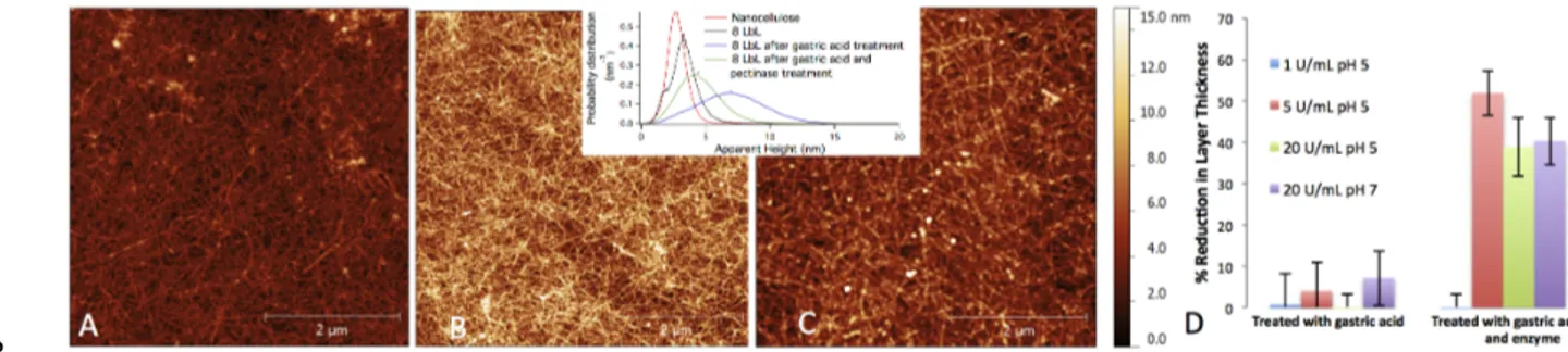
Figure 2. (a) AFM image of the 8 LbL coating. The same surface after exposure to (b) simulated gastric 9
fluid (3h, 2 g/L NaCl, 3.2 g/L pepsin and 0.3 % HCl) and in (c) simulated stomach conditions and pecti-10
nase exposure (3h of enzyme solution). The inset shows the apparent height distributions for the 8 LbL 11
coating before treatment, after treatment with simulated gastric fluid and treatment with gastric flu-12
id/pectinase. (d) The reduction of the total coating thickness after each treatment (gastric fluid or gastric 13
fluid/enzyme). 14
15
3.2 Resistance of the LbL coating to stresses present in the GI tract
16
The stomach fluid consists mostly of pepsin, HCl and NaCl. This environment kills bacteria to 17
prevent infections in the digestive system and moreover the acid gives the right pH for the reac-18
tion of the protease pepsin. We subjected our coated Si wafers to a simulated gastric fluid (2 g/L 19
NaCl, 3.2 g/L pepsin and 0.3% HCl) under stirring for 3 h at 37 °C, and the total ellipsometry 20
thickness and surface topology was characterized with SE and AFM respectively. No major dif-21
ferences were observed in either the thickness or the morphology of the surface – compare the 22
changes in SE thickness in Figure 2d and AFM images in 2b and 2a. Therefore, the coating 23
seems to survive the chemical and physical stresses in the stomach. The pH of this solution re-24
mained constant at a value ~2 during the whole length of the experiments. 25
Once reaching the gut, the food is exposed to a rich flora that includes between 300 and 26
1000 different microorganisms and their levels can reach a total population as high as 1013-1014 27
cells,9 which is essential for human well-being. Some of these microorganisms produce enzymes 28
6 that can degrade pectins including polyGalA.8 To simulate the action of the microbiota enzymat-1
ic action in the gut, a pectinase enzyme was used at various concentrations (1, 5 and 20 U/mL) to 2
determine whether the coating was degraded under any of these conditions. SE shows that the 3
layer thickness decreases by 40-50% once the enzyme concentration was raised to 5-20 U/mL 4
during 3h (Figure 2d). Since the pectinase can only degraded the polyGalA and not cellulose, the 5
SE value suggests the partial collapse of the film structure and/or more porous structure of the 6
cellulose based coating. AFM images, however, showed only a small change in the overall mor-7
phology of the film: The nanofibers seemed more “swollen” and small aggregates or “particles” 8
appeared on the coating, compare Figure 2a and 2c. Also, the roughness (Supporting infor-9
mation, Figure S3) increased and the apparent height profile (inset Figure 2) broadened and 10
shifted to higher values once the samples were exposed to the bacterial enzyme solution. The 11
change in both the roughness and apparent height before and after the sample was exposed to the 12
enzyme solution can be caused by the enzymes creating holes in the coating, although irreversi-13
ble adsorption of enzymes to the coating cannot be disregarded. 14
15
3.3 Oxygen barrier properties
16
The oxygen protective properties of the multilayer coatings were also assessed. In this case, a 17
PLA substrate was coated with 20 LbL on each side (total thickness of coating was estimated to 18
be 249.6 nm based on SE) and measured at 20% relative humidity and 23 °C giving a value of 19
0.033 ml (STP) mm m-2 day-1atm-1 for the coating. The oxygen permeability coefficient (OP) for 20
the coating is comparable to that of EVOH,6 which is typically used in high oxygen barrier pro-21
tection applications. However, from a sustainability point of view, our coating is more attractive 22
since it could potentially be a direct part of the edible system, minimizing the amount of waste. 23
A neat cationic NFC film has an OP value of 0.016 ml (STP) mm m-2day-1atm-1 at 20% RH and 24
23 °C. Pectin films have a reported OP of 17.4 ml (STP) mm m-2 day-1atm-1 at 55% RH and 25
23 °C.11 Note that herein, the measurements were performed at 20% relative humidity, and the 26
OP of a neat pectin film will be lower at this relative humidity, as expected given the known 27
effect of water on barrier properties of pectin films.12 28
The oxygen permeability where also measured after the simulated gastric fluid and bacte-29
rial enzyme treatment. In this case, the oxygen permeability of the coating and substrate was 30
very close to the value of the neat PLA substrate itself, suggesting very little contribution from 31
the coating to the oxygen barrier properties. This could suggest that the coating was partially or 32
7 completely decoupled from the PLA substrate under the exposure to the simulated gastric fluid 1
and the pectinase solution. One explanation to this could be that the adhesion of the LbL-coating 2
to the PLA substrate is probably not as strong as that to the silica wafer. 3
4. Conclusions 4
Spectroscopic ellipsometry, atomic force microscopy and oxygen permeability results showed 5
successful buildup of a multilayer coating based on cationic nanofibrillated cellulose and polyga-6
lacturonic acid. The multilayer coating is dense and thick and presents low permeability to oxy-7
gen (0.033 ml (STP) mm m-2 day-1atm-1), typically in the same order of magnitude as for eth-8
ylene vinyl alcohol films. Moreover, the coating is able to survive in simulated gastric fluid and 9
it is partially degraded (polygalacturonic acid) upon exposure to pectinase enzyme, which simu-10
late the action of the microbial symbionts present in the human gut. After this exposure, the per-11
meability to oxygen has increased, which strengthens the conclusions that the coating has been 12
partially degraded. Such coatings could potentially be used for targeted release of active compo-13
nents (e.g. drugs, probiotics, peptides) in the human colon. 14
15
Acknowledgements Karsten Olsen is acknowledged for supplying the pectinase from Novo-16
zymes A/S. Dr. Roland Seitz from Horiba Scientific is thanked for technical assistance. Funding 17
was provided by the Danish Council for Independent Research (A.J.Svagan). M.C. thanks COST ac-‐ 18
tions CM1101 and MP1106. 19
20
Supporting Information. Experimental section, AFM, ellipsometry data and roughness data. 21
References 22
(1) (a) Svagan, A. J.; Akesson, A.; Cardenas, M.; Bulut, S.; Knudsen, J. C.; Risbo, J.; Plackett, D. 23
Biomacromolecules 2012, 13, 397(b) Priolo, M. A.; Gamboa, D.; Holder, K. M.; Grunlan, J. C.
24
Nano Lett 2010, 10, 4970(c) Dubas, S. T.; Kumlangdudsana, P.; Potiyaraj, P. Colloid Surface A
25
2006, 289, 105(d) Li, Y. C.; Schulz, J.; Mannen, S.; Delhom, C.; Condon, B.; Chang, S.;
26
Zammarano, M.; Grunlan, J. C. Acs Nano 2010, 4, 3325(e) Kim, J. H.; Kim, S. H.; Shiratori, S. 27
Sensors and Actuators B: Chemical 2004, 102, 241(f) Westwood, M.; Noel, T. R.; Parker, R.
28
Carbohyd Polym 2013, 94, 137(g) Westwood, M.; Roberts, D.; Parker, R. Carbohyd Polym
29
2011, 84, 960.
30
(2) Sukhishvili, S. A. Current Opinion in Colloid & Interface Science 2005, 10, 37. 31
(3) Wood, K. C.; Boedicker, J. Q.; Lynn, D. M.; Hammond, P. T. Langmuir 2005, 21, 1603. 32
(4) Saito, T.; Kuramae, R.; Wohlert, J.; Berglund, L. A.; Isogai, A. Biomacromolecules 2013, 14, 33
248. 34
(5) Eichhorn, S. J.; Dufresne, A.; Aranguren, M.; Marcovich, N. E.; Capadona, J. R.; Rowan, S. J.; 35
Weder, C.; Thielemans, W.; Roman, M.; Renneckar, S.; Gindl, W.; Veigel, S.; Keckes, J.; Yano, 36
H.; Abe, K.; Nogi, M.; Nakagaito, A. N.; Mangalam, A.; Simonsen, J.; Benight, A. S.; Bismarck, A.; 37
Berglund, L. A.; Peijs, T. J Mater Sci 2010, 45, 1. 38
(6) Aulin, C.; Gallstedt, M.; Lindstrom, T. Cellulose 2010, 17, 559. 39
8 (7) Buchanan, B. B.; Gruissem, W.; Jones, R. L. Biochemistry & molecular biology of plants; 1
American Society of Plant Physiologists: Rockville, Md. ; [Great Britain], 2000. 2
(8) Martens, E. C.; Lowe, E. C.; Chiang, H.; Pudlo, N. A.; Wu, M.; McNulty, N. P.; Abbott, D. W.; 3
Henrissat, B.; Gilbert, H. J.; Bolam, D. N.; Gordon, J. I. PLoS. Biol. 2011, 9. 4
(9) Zhu, Q. C.; Gao, R. Y.; Wu, W.; Qin, H. L. Tumor Biol 2013, 34, 1285. 5
(10) Bergström, L.; Stemme, S.; Dahlfors, T.; Arwin, H.; Ödberg, L. Cellulose 1999, 6, 1. 6
(11) de Moura, M. R.; Aouada, F. A.; Zucolotto, V.; Mattoso, L. H. C. Polymer-Plastics Technology 7
and Engineering 2011, 50, 1323.
8
(12) Vartiainen, J.; Tammelin, T.; Pere, J.; Tapper, U.; Harlin, A. Carbohyd Polym 2010, 82, 989. 9
10 11