Recent Research on
EMF and Health Risks
Third annual report from SSI's Independent
Expert Group on Electromagnetic Fields 2005
SSI Rapport
2006:02
Rapport från Statens strålskyddsinstitut tillgänglig i sin helhet via www.ssi.seUltraviolet, solar and optical radiation
Ultraviolet radiation from the sun and solariums can result in both long-term and short-term effects. Other types of optical radiation, primarily from lasers, can also be hazardous. SSI provides guidance and information.
Solariums
The risk of tanning in a solarium are probably the same as tanning in natural sunlight. Therefore SSI’s regulations also provide advice for people tanning in solariums. Radon
The largest contribution to the total radiation dose to the Swedish population comes from indoor air. SSI works with risk assessments, measurement techniques and advises other authorities.
Health care
The second largest contribution to the total radiation dose to the Swedish population comes from health care. SSI is working to reduce the radiation dose to employees and patients through its regulations and its inspection activities.
Radiation in industry and research
According to the Radiation Protection Act, a licence is required to conduct activities involving ionising radiation. SSI promulgates regulations and checks compliance with these regulations, conducts inspections and investigations and can stop hazardous activities. Nuclear power
SSI requires that nuclear power plants should have adequate radiation protection for the generalpublic, employees and the environment. SSI also checks compliance with these requirements on a continuous basis.
Waste
SSI works to ensure that all radioactive waste is managed in a manner that is safe from the standpoint of radiation protection.
Mobile telephony
Mobile telephones and base stations emit electromagnetic fields. SSI is monitoring developments and research in mobile telephony and associated health risks. Transport
SSI is involved in work in Sweden and abroad to ensure the safe transportation of radioactive substances used in the health care sector, industrial radiation sources and spent nuclear fuel.
Environment
“A safe radiation environment” is one of the 16 environmental quality objectives that the Swedish parliament has decided must be met in order to achieve an ecologically sustainable development in society. SSI is responsible for ensuring that this objective is reached. Biofuel
Biofuel from trees, which contains, for example from the Chernobyl accident, is an issue where SSI is currently conducting research and formulating regulations.
Cosmic radiation
Airline flight crews can be exposed to high levels of cosmic radiation. SSI participates in joint international projects to identify the occupational exposure within this job category.
Electromagnetic fields
SSI is working on the risks associated with electromagnetic fields and adopts countermea-sures when risks are identified.
Emergency preparedness
SSI maintains a round-the-clock emergency response organisation to protect people and the environment from the consequences of nuclear accidents and other radiation-related accidents.
SSI Education
is charged with providing a wide range of education in the field of radiation protection. Its courses are financed by students' fees.
ISSn 0282-4434
Författarna svarar själva för innehållet i rapporten.
The conclusions and viewpoints presented in the report are those of the authors and do not necessarily coincide with those of the SSI.
tItle / tItel: Recent Research on EMF and Health Risks. Third annual report
from SSI's Independent Expert Group on Electromagnetic Fields 2005.
department / avdelnIng: Department of Emergency Preparedness &
Environ-mental Assessment / Avdelning för Beredskap och miljöövervakning.
Summary: SSI:s international independent expert group for electromagnetic fields was
established in 2002. In December each year a report on the scientific development in the field is delivered to the Director General of SSI. This is the third annual report.
Recent studies from the REFLEX programme suggest genotoxic effects from exposure to relatively weak extremely low frequency and radio frequency (RF) electromagnetic fields. However, results from other recent studies were negative. The recent studies have not estab-lished effects that would change the previous conclusions.
Recent results of studies of the blood-brain-barrier aimed at replicating earlier positive work have been negative. On balance, the evidence that exposure to RF fields alters the blood-brain-barrier is weak.
Two recent Interphone studies on brain tumours suggest that there is no risk increase either for short-term or term use, although term data are sparse. However, for long-term use and acoustic neuroma there is a concern, and more information is required. The few studies that have been published on health risks among populations living near transmitters have had major methodological shortcomings. The exposure to the general population from transmitters is very weak and one would not expect such exposure to produce a health risk. Cancer risk in mobile phone users have been discussed in all reports. Short-term use of mobile phones does not appear to be associated with cancer risks in adults. However, other outcomes have not been studied, no studies on children or adoles-cents have been done, and long-term use has not been fully evaluated.
SammanfattnIng: SSI:s vetenskapliga råd för elektromagnetiska fält bildades
hös-ten 2002 och lämnar varje år en rapport om forskningsläget inom området till SSI:s generaldirektör. Detta är den tredje årliga rapporten
I 2005 års rapport har rådet bl.a. behandlat studier om cancer och DNA-skador vid exponering för lågfrekventa fält, som magnetfält från kraftledningar, däribland någ-ra studier inom EU-projektet REFLEX. REFLEX har också omfattat någ-radiofrekventa fält. Även här har några forskargrupper i projektet rapporterat ökad förekomst av bl. a. DNA-skador. Studierna har begränsningar i metoderna. Sammantaget menar rådet att det inte finns tillräckligt underlag för slutsatsen att lågfrekventa eller radio-frekventa fält skulle orsaka DNA-skador.
En amerikansk forskargrupp har upprepat en studie från en forskargrupp i Lund om läckage i blod-hjärnbarriären och ser inga effekter. Rådet menar att det veten-skapliga stödet är svagt för att låga nivåer av radiofrekvent strålning skulle påverka blod-hjärnbarriären.
Två studier som ingår i det internationella INTERPHONE-projektet rapporterar resultat som antyder en något ökad risk för hörselnervstumör vid långvarig använd-ning av mobiltelefon. För kortare tids användanvänd-ning ses ingen ökad risk för denna tumörform.
De få studier som publicerats om hälsoeffekter hos personer som bor nära radio- och TV-sändare och basstationer för mobiltelefoni har kvalitetsproblem. Expone-ringen är låg och det är osannolikt att strålning från sändarna skulle utgöra någon hälsorisk. Eventuella långtidseffekter, som exempelvis cancer, från användning av mobiltelefon bör studeras ytterligare. Studier på barn och ungdomar saknas.
Summary in Swedish (Sammanfattning) ... 3
Preface... 6
Executive Summary ... 7
Introduction... 10
Extremely Low Frequency Fields (ELF)... 10
Recent biology papers... 10
Genotoxicity... 10
In vivo cancer studies... 12
Recent epidemiology paper... 13
Radiofrequency Fields (RF) ... 14
Recent laboratory studies ... 14
Genotoxicity... 14
Blood-Brain Barrier (BBB) permeability... 16
Human laboratory studies ... 17
Recent epidemiological studies... 19
Mobile phone studies ... 19
Methodological studies ... 23
Current overall conclusion on mobile phone use ... 24
Recent studies on transmitters... 24
WHO International EMF programme... 25
Environmental Health Criteria document on Static fields ... 25
WHO Workshops ... 27
Publication from Istanbul workshop: Childhood Sensitivity to EMFs ... 27
WHO workshop on base station exposures... 28
General concluding discussion ... 28
Update on key issues... 28
Upcoming or newly published reviews... 29
Status of bioelectromagnetics research worldwide ... 30
Current Swedish research... 30
References ... 31
Tabellförteckning
Table 1. Results for epidemiological mobile phone studies of brain tumors:... 37Summary in Swedish (Sammanfattning)
Elektromagnetiska lågfrekventa fält Aktuell biologisk forskning
En tysk forskargrupp har publicerat data som stödjer deras tidigare resultat om att ex-ponering för lågfrekventa magnetfält förstärker utvecklingen av kemiskt initierad bröstcancer hos en speciell sorts råttor. Annan aktuell forskning, bland annat inom REFLEX programmet, antyder att lågfrekventa magnetfält skulle kunna orsaka skador på DNA. Detta strider dock mot resultat från andra aktuella studier som inte kunnat se dessa effekter. Tidigare forskning rörande eventuella DNA-skador på grund av lågfre-kventa magnetfält har i huvudsak varit negativa. Resultaten av nu aktuell forskning le-der inte till någon ändring i den övergripande bedömningen.
Aktuell epidemiologisk forskning
En omfattande engelsk epidemiologisk studie om barnleukemi publicerades under 2005. Då man i studien använt sig av digitaliserad exponeringsinformation har man inte behövt ta kontakt med de personer som ingått i studien. Därmed har man undvikit svårigheter som en del tidigare studier inom området haft på grund av att man saknat information om exponeringen för delar av urvalet. I denna rapport utnyttjas dock av-stånd mellan bostad och kraftledning som markör för exponering. Forskarna medger i rapporten att detta är en mycket grov exponeringsmarkör och vi avvaktar analyser ba-serade på mer sofistikerade magnetfältsskattningar.
Radiofrekventa fält
Aktuell laboratorieforskning Skador på DNA
Flera aktuella djur- och cellkulturstudier har undersökt om skador på DNA kan uppstå vid exponering för radiofrekventa fält. Två forskargrupper har redovisat ökad före-komst av DNA-skador och mikrokärnor vid exponering av radiofrekventa fält. Be-dömningen av dessa resultat påverkas dock av vissa metodologiska invändningar och av att andra forskargrupper inte har sett sådana effekter, vilket också är förenligt med resultat från den övervägande delen av tidigare forskning. Aktuell forskning, inklusive resultat från REFLEX-programmet, ger inte underlag för slutsatsen att radiofrekventa fält orsakar DNA-skador.
Blod-hjärnbarriären
Huvuddelen av den aktuella forskning som syftat till att upprepa tidigare resultat där effekter på blod-hjärnbarriären setts har varit negativa vid låga SAR- värden. Ytterli-gare undersökningar är på gång och förväntas ge mer information. Sammantaget be-döms det vetenskapliga stödet som svagt för att exponering för svaga radiofrekventa fält påverkar blod-hjärnbarriären.
Experimentell forskning på försökspersoner
Aktuell forskning på kognitiv förmåga hos frivilliga försökspersoner (inklusive barn) som exponerats för radiofrekventa fält har varit negativ, men på grund av metodbe-gränsningar kan möjligheten av sådana effekter inte avskrivas helt.
Det har också förekommit rapporter om sömnpåverkan. De resultat som finns om blodflöde i hjärnan tillhör de mest intressanta biologiska fynden även om de inte klart pekar mot en hälsoeffekt.
Efter uppföljning av tidigare rapporter, särskilt de som kommit ut från EU-programmet GUARD, om effekter på hörsel kan man nu konstatera att det inte tycks förekomma några negativa effekter på hörseln på grund av exponering för GSM fre-kvenser för mobiltelefoni.
Aktuell epidemiologisk forskning
Två INTERPHONE-studier om hjärntumörer tyder på att det inte förekommer någon ökad risk, varken vid korttidsanvändning eller långtidsanvändning av mobiltelefon. Det finns dock viss osäkerhet för långtidsanvändning på grund av att antalet långtids-användare fortfarande är begränsat.
En gemensam rapport från ett flertal INTERPHONE-studier rörande hörselnervstumör visade ingen sammantagen effekt vare sig vid kort- eller långtidsanvändning. Däremot visade rapporten en ökad risk vid långtidsanvändning för det öra där telefonen använ-des. Vissa andra svenska data har också presenterats men de har givits mindre tyngd i den samlade bedömningen.
Sammantaget tyder aktuell forskning på att det för hjärntumör inte finns något sam-band med mobiltelefoni för åtminstone upp till tio års användning. Även för längre tids användning tyder forskningen på att det inte finns något samband, men här är osä-kerheten större på grund av att antalet långtidsanvändare ännu är begränsat. För hör-selnervstumör gäller samma slutsats för kortare tids användning. Vid långtidsanvänd-ning finns dock anledlångtidsanvänd-ning till oro och mer forsklångtidsanvänd-ning krävs. Därtill kommer att det ännu inte finns någon forskning på barn eller ungdomar och inte på några andra sjuk-domar än cancer.
Rapporter från WHO
”Environmental health criteria document”; hälsoriskbedömning för statiska fält Statiska magnetfält förekommer överallt där likström förekommer; flödestätheter upp till flera millitesla förekommer till exempel i elektriska tåg. De starkaste fält (0.2- 3 Tesla) allmänheten kan förväntas komma i kontakt med förekommer dock i anslutning till magnetkameror inom sjukvården. Användningen kommer antagligen att begränsas av starka elektriska fält som uppstår kring hjärtat vid mycket hög exponering. Andra akuta effekter som yrsel och illamående har rapporterats från personal som arbetar i närheten av magnetkameror. Dessvärre finns inga informativa epidemiologiska lång-tidsuppföljningar av dem som exponerats. Nationella myndigheter rekommenderas att vidta försiktighetsåtgärder mot besvärande exponering och att initiera ytterligare forskning inom området.
Workshop om barns känslighet
Det finns biologiska och dosimetriska skillnader mellan barn/ungdomar och vuxna, men eftersom bara begränsad forskning har studerat en eventuell extra känslighet hos barn så saknas bra data för att bedöma detta. Vid kraftfrekventa fält har det i upprepa-de studier framkommit att exponeraupprepa-de barn har en ökad risk för leukemi, men inte vuxna.
Workshop om bassationer för mobiltelefoni
Deltagarna i denna arbetsgrupp kom till slutsatsen att risken är mycket liten för att det skulle kunna förekomma hälsoeffekter vid exponering från basstationer. Denna slut-sats baserades på att exponeringen från basstationer är mycket låg, att det inte finns någon känd mekanism som skulle kunna förklara hur en hälsoeffekt skulle kunna upp-stå från denna exponering och på avsaknaden av epidemiologiska data.
Avslutande diskussion Aktuell svensk forskning
Svensk forskning inom elektromagnetiska fält har nyligen utvärderats av en kommitté tillsatt av Vetenskapsrådet. Kommittén konstaterade att Sverige bör bidra till forsk-ningen inom området med högkvalitativa studier i internationellt samarbete och inom områden där Sverige har unika möjligheter, till exempel långsiktig epidemiologi. Kommittén menade också att finansiering bör komma genom gängse forskningsråd och finansieringskällor, snarare än via ett särskilt anordnat nationellt forskningspro-gram som man har i många andra länder, till exempel England, Danmark, Frankrike, Schweiz och Tyskland. Vi konstaterar i expertgruppen att högkvalitativ forskning an-tagligen kan konkurrera framgångsrikt om medel hos svenska forskningsråd och fon-der, men att stora internationella initiativ blir svårare att få till stånd utan ett nationellt forskningsprogram. Vi konstaterar särskilt att forskningsprojekt som samfinansieras av staten och industrin blir särskilt svåra att starta utan ett nationellt program därför att sådana projekt kräver en effektiv brandvägg för att bevara tilltron till forskningens oberoende.
Preface
The Swedish Radiation Protection Authority, SSI (Statens strålskyddsinstitut) has ap-pointed an international independent expert group (IEG) for electromagnetic fields (EMF) and health. The task is to follow and evaluate the scientific development and to give ad-vice to the SSI. With recent major scientific reviews as starting points the IEG in a series of annual reports consecutively discusses and assesses relevant new data and put these in the context of already available information. The result will be a gradually developing health risk assessment of exposure to EMF. The group began its work in the fall of 2002 and presented its first report in December 2003, and the second report the year after. This is the third annual report.
The composition of the group during 2005 has been:
Prof. Anders Ahlbom, Karolinska Institutet and Stockholm Center for Public Health, Stockholm, Sweden (chairman);
Prof. Jukka Juutilainen, University of Kuopio, Kuopio, Finland;
Dr. Bernard Veyret, University of Bordeaux, Pessac, France and University La Sapienza, Rome, Italy;
Dr. Harri Vainio, Occupational Health Institute, Helsinki, Finland (formerly at IARC, Lyon, France);
Prof. Leeka Kheifets, UCLA, Los Angeles, USA (formerly at WHO, Geneva, Switzer-land);
Prof. Anssi Auvinen, University of Tampere, Tampere, Finland;
Dr. Richard Saunders, Health Protection Agency, Centre for Radiation, Chemical and Environmental Hazards, Oxfordshire, UK
Prof. Maria Feychting, Karolinska Institutet, Stockholm, Sweden, has been appointed scientific secretary to the group.
Stockholm in December 2005 Anders Ahlbom
Chairman
Executive Summary
ELF
Recent biology
A German research group has published additional data that support their previous re-sults suggesting enhanced development of chemically-induced mammary tumours in a specific substrain of rats exposed to ELF magnetic fields. Some recent studies, e.g. within the REFLEX programme, suggest genotoxic effects from exposure to relatively weak magnetic fields. However, results from other recent studies were negative. Pre-vious evidence for carcinogenic and genotoxic effects of ELF magnetic fields is in-adequate and mainly negative. The recent studies have not established effects that would change the previous conclusions.
Recent epidemiology
The recent childhood leukaemia study from the UK is very large and uses digitized exposure information, which requires no access to study participants’ homes and thus minimizes potential for selection bias. However, the current publication uses distance between home at birth and power line as proxy for EMF exposure. The authors ac-knowledge that this is a poor marker for exposure and we await further results based on calculated magnetic fields which we hope will be more informative.
RF
Recent laboratory studies
Several recent animal and cell culture studies have evaluated possible genotoxic ef-fects of RF electromagnetic fields. Two research groups reported increased DNA strand breaks and micronuclei. However, these results are weakened by methodologi-cal limitations, and other studies reported no effects, which is consistent with the ma-jority of previous studies. On balance, the recent studies reviewed, including those from the REFLEX programme, do not provide sufficient evidence to conclude that RF fields are genotoxic.
Blood-Brain Barrier
Most of the recent results of studies aimed at replicating earlier positive work have been negative at low SAR levels. Further studies on the permeability of the blood-brain-barrier or on damaged (darkly-staining) neurons are ongoing and should give more information on the issue. On balance, the evidence that exposure to RF fields al-ters the blood-brain-barrier is weak.
Human laboratory studies
Recent work on cognitive functions in volunteers (including children) exposed to RF fields has been negative; however, methodological limitations prevents firm conclu-sions. There have been reports of some positive findings on alterations of sleep. The data obtained on alteration of cerebral blood flow following RF exposure are among the most interesting observations of biological effects even if they do not point clearly to health effects. Following all of the recent publications on hearing and in particular those originating from the European Commission’s GUARD programme, one could now conclude about the absence of effects of RF exposure due to cellular phones at GSM frequencies, on the main parameters of the auditory system.
Recent epidemiology
Two recent Interphone studies on brain tumours suggest that there is no risk increase either for short-term or long-term use, although long-term data are sparse. A joint in-ternational publication of some of the studies taking part in Interphone on acoustic neuroma showed no effect for short-term use and no overall effect for long-term use. However, the laterality analysis did show a risk increase for ten or more years of use. Some other Swedish data have also been presented but they are given less weight in the overall analysis.
On balance the currently available evidence suggests that for adult brain tumours there is no association with mobile phone use for at least up to, say, ten years of use. For longer latency the majority of the evidence also speaks against an association, but the data are still sparse. The same conclusion holds for short-term use and acoustic neu-roma. However, for long-term use and acoustic neuroma there is a concern, and more information is required. Furthermore, studies of children are yet to be done, as well as studies on outcomes other than cancer.
WHO
Environmental Health Criteria document on Static Fields
Static magnetic fields are generated wherever DC currents are used; exposure to flux densities of up to several millitesla (mT) occur for example in electric trains. How-ever, the largest static magnetic fields (0.2 – 3 T) likely to be experienced by members of the public occur during exposure for clinical diagnosis to the fields generated by magnetic resonance imaging (MRI) systems. Effects caused by the large electrical fields generated around the heart are likely to limit patient exposure to MRI at very high field strengths. Other acute effects, such as vertigo and nausea, are reported to occur in workers during movement within and around MRI devices. However, with regard to the possible long-term effects of exposure to such high fields, there are no good epidemiological or long-term experimental studies. National authorities should implement programmes that protect both the public and workers from the untoward ef-fects of such fields, and should conduct further research on the possible health efef-fects of exposure.
Workshop on childhood sensitivity
There are biological and dosimetric differences between children and adults, some of which persist into adolescence, but as little research has specifically addressed poten-tial vulnerability and health effects in children and there is no good evidence one way or another whether children are more susceptible to RF fields than adults. At power frequencies, a consistently increased risk of leukaemia is seen in children but not in adults.
Workshop on base stations
The workshop participants concluded that the likelihood of health effects from base station or other transmitter exposure is low. This was based on considerations of the very weak exposure from base stations, the lack of a known mechanism that could ex-plain any effects of such weak fileds, and the absence of epidemiological data.
General Concluding Discussion Current Swedish Research
Swedish EMF research has been evaluated by a committee commissioned by the Swedish Research Council. The report concluded that Sweden should contribute with high quality studies in international collaboration and in particular in areas where Sweden has unique resources, such as for long-term epidemiology. The report also concluded that funding should come through the usual research councils and funding sources, rather than through national research programs supported by the government, which is in contrast to many other countries, e.g. UK, Denmark, France, Switzerland, and Germany, which have established dedicated national research programs. The IEG acknowledges that competitive research groups might be supported through these channels, but that large initiatives would be difficult. The IEG also concludes that pro-jects jointly funded by government and industry are rather difficult to set up in the ab-sence of a national research programme.
Introduction
In this year’s report the IEG evaluates some recent data on ELF fields, but the major part of the report is about RF fields and in particular about recent epidemiological studies on disease risk in relation to mobile phone use. Intense research has been ongoing for several years and new results are presented regularly. Yet, the IEG is aware of a number of addi-tional studies that will appear in the next couple of years, particularly from the Interphone study. Therefore it would still be premature to make final assessments, except in some restricted areas. The report also comments on some recent laboratory studies, including genotoxicity, e.g. studies within the REFLEX programme, and important attempts to rep-licate some earlier neurological findings that had been given considerable visibility. The WHO International EMF project has now been ongoing for ten years and we report on the conclusions from several workshops and environmental health criteria documents that have been produced by this programme.
Extremely Low Frequency Fields (ELF)
Recent biology papers
Genotoxicity
Ivancsits et al. have reported results from several experiments (performed as a part of the European REFLEX programme) in which cultures of human diploid skin fibroblasts were exposed to 50 Hz magnetic fields (up to 2000 µT) for up to 24 h and DNA damage was assessed using the comet assay. Both the neutral (believed to detect only DNA double strand breaks) and alkaline (believed to detect both single strand breaks and double strand breaks, as well as base damage and alkali-sensitive sites) versions of the comet assay were used. In the first study, continuous EMF exposure for 24 h had no effect on DNA strand breaks while intermittent exposure resulted in a significant increase in DNA strand breaks [Ivancsits, et al. 2002]. A dose-response relationship was reported, with statisti-cally significant effects at flux densities ≥70 µT. Various on/off combinations were tested in case of the intermittent magnetic fields, and the maximum effects were found at 5 min on/10 min off. The 5 min on/5 min off intermittency was used in all subsequent studies. In the second study, the maximum effect was seen after 15-19 hours of exposure (1-24 h tested), and the DNA damage appeared to be repaired at 9 h after the termination of expo-sure [Ivancsits, et al. 2003]. The dose-response was further examined, and significant effects were found at a flux density as low as 35 µT. Cell type specificity was investi-gated in the third study, and effects were seen in fibroblasts, melanocytes and rat granu-loma cells, but not in skeletal muscle cells, lymphocytes or monocytes [Ivancsits, et al. 2005]. The method used the in these studies for quantifying comet tail size (amount of DNA damage) is different from the methods used by other authors. The cells were classi-fied into five categories representing five different degrees of DNA damage, based on visual inspection under microscope. A ‘‘tail-factor’’ was calculated from the classified data for quantitative expression of DNA damage. Because of the fast classification-based method, the authors were able to score 1000 cells per sample (in contrast to the usual 50-100 cells in image analysis). However, only two independent samples were used for each treatment, and the data from these two samples are not reported separately to show the
reproducibility of the data. The statistical method thus seems not to be appropriate for the data. Although only two independent samples were available for each treatment (with a single tail factor value calculated from observations on 1000 cells), the results are re-ported as “mean±standard deviation”, and the authors report they have used Student’s t-test and ANOVA for t-testing the differences between treatments. No positive controls were used in the experiments. The investigators have used continuously growing cultured cells with cell cycle duration of approximately 30 h. As pointed out recently [Vijayalaxmi and Obe 2005], the presence of normal cycling cells and semi-conservative DNA synthe-sis during the prolonged exposures could result in increased tail lengths mimicking dam-aged cells in the comet assay. Thus, the method might not differentiate between true ef-fects on DNA breaks and possible MF efef-fects on cell proliferation, cell cycle and DNA synthesis. The exposure conditions producing maximum strand-break levels (1000 µT, 5 min on/10 min off) were also reported to induce a significant increase of micronuclei and chromosomal aberrations in fibroblasts [Winker, et al. 2005], which can be consid-ered to support the comet assay data. There is no obvious explanation why only the in-termittent, but not continuous MF exposure caused genotoxic effects in this study and the other studies by the same group.
In the confirmation study of Scarfi et al. which was also part of the REFLEX programme [Scarfi, et al. 2005], great care was taken to use the same exposure conditions and assay conditions as in the earlier papers of the Austrian group (see above). Clear responses to positive controls (H2O2 and mitomycin C) were seen, but no MF effects on DNA strand
breaks or micronuclei were observed.
It is not possible at present to conclude whether results from the REFLEX programme indicate the existence of some genotoxic effects from exposure to weak ELF magnetic fields. The failure of the Scarfi group to replicate the Ivancsits results may be due to a flaw in the experimental protocol (in one or both laboratories), or to unknown subtle dif-ferences in the protocols or environmental conditions. Additional experiments are needed to resolve the issue.
Luceri et al. used the comet assay to detect strand breaks and oxidated DNA bases in human lymphocytes exposed to a 50 Hz, 1-100 µT magnetic field for 18 hours [Luceri, et al. 2005]. No significant effect on DNA damage was found, but the study had limited ability to detect any effects because of the high variability of DNA damage level. Gene expression profiles of MF-exposed human and yeast cells were also evaluated with DNA microarrays containing 13,971 and 6,212 oligonucleotides, respectively. No MF-related changes in gene expression were found.
Wolf et al. reported increased cell proliferation, changes in cell cycle and increased DNA damage, assessed by the comet assay, in HL-60 leukaemia cells and two fibroblast cell lines exposed to 50 Hz MFs at 500-1000 µT up to 72 hours [Wolf, et al. 2005]. The in-crease in DNA strand breaks showed two peaks at 24 and 72 h, while no inin-crease was seen at 48 h. A similar time-dependent pattern of DNA damage was observed by measur-ing 8-OHdG adducts. Involvement of MF effects on free radical species was supported by changes seen in intracellular levels of reactive oxygen species measured by a fluorescent probe, and in the expression of proteins that are involved in redox-mediated signals (NFκB p65 and p50). Also, the MF effects were suppressed by pre-treatment of the cells with the antioxidant α-tocopherol. The results of this study are internally consistent, and effects seen on different endpoints support each other. Independent replication of the key findings would be useful to assess their repeatability.
Lai and Singh reported significantly increased DNA damage in rat brain cells after expo-sure of the animals to a 60-Hz, 10-µT magnetic field for 24 or 48 h [Lai and Singh 2004]. The same authors have previously reported similar effects after short (2 h) exposure to much higher magnetic flux densities (100-250 µT). The effect was seen in both the alka-line and neutral versions of the comet assay and, although the size of the effect was rela-tively small, it was seen in several independent experiments. Exposure for 48 h caused a larger increase than exposure for 24 h. The effects were blocked by treatment with a radi-cal scavenger, a nitric oxide synthase inhibitor and an iron chelator, suggesting involve-ment of free radicals and iron in the effects of magnetic fields.
Stronati et al. exposed blood samples obtained from five donors to 50 Hz magnetic fields, and evaluated possible genotoxic effects using several genotoxicity endpoints [Stronati, et al. 2004]. The measurements included DNA breaks (comet assay), sister chromatid ex-change, chromosomal aberrations, and micronuclei, and the effects of a combined expo-sure with X-rays were also evaluated. No genotoxic effects were seen after expoexpo-sure for 2 h at 1000 µT. A small but significant decrease of cell proliferation was reported. Be-cause of the short exposure time, the results are not necessarily inconsistent with the posi-tive findings seen after long-term exposures (described above). The same group con-ducted another study with the same exposures and endpoints, but using 48 h MF exposures [Testa, et al. 2004]. No significant effects were seen on the genotoxicity end-points or proliferation. Because of the small study size (n = 4), the data do not provide strong evidence for a lack of effects.
Current overall conclusion on genotoxicity
Studies on genotoxicity are highly relevant in evaluation of carcinogenicity. Studies on genotoxicity of ELF magnetic fields were reviewed by the International Agency for Re-search on Cancer [IARC 2002], and more recently by Vijaylaxami & Obe [Vijayalaxmi and Obe 2005]. These reviews concluded that there was little evidence of any DNA dam-age from exposure to MFs alone, except for very strong magnetic fields above 50,000 µT. However, it was noted in the IARC review that several groups had reported findings sug-gesting that ELF magnetic fields enhance the effects of known DNA-damaging agents such as ionizing radiation. Results from recent studies have not established effects that would change the conclusions of IARC. However, some of the recent studies, in particu-lar within the REFLEX programme, suggest genotoxic effects at low magnetic field in-tensities (10 µT to 1000 µT) and without combined exposure to known genotoxic agents, but the results are not uniform as another group within the same programme have failed to replicate the data. There is thus a need for a few more investigations on genotoxic ef-fects of ELF to allow for a definitive conclusion.
In vivo cancer studies
Motivated by the epidemiological findings of increased leukaemia risk in children, Som-mer and Lerchl investigated the influence of 50 Hz magnetic fields in a mouse strain netically predisposed to lymphoma [Sommer and Lerchl 2004]. The AKR/J mouse ge-nome carries the AK virus, which leads to spontaneous development of thymic lymphoblastic lymphoma within one year. Groups of 160 female mice were sham-exposed or sham-exposed to 50 Hz magnetic fields at 1 or 100 µT for 24 h per day, 7 days per week, for 38 weeks. There was no effect of magnetic field exposure on body weight gain or survival rate, and the time to lymphoma development did not differ between exposed and sham-exposed animals. The results do not support the hypothesis that chronic
sure to 50 Hz magnetic fields increases the risk of haemopoietic malignancy in this ex-perimental model.However, the relevance of this model to human childhood leukaemia is limited.
Most of the animal studies evaluating possible carcinogenic effects of ELF magnetic fields have been negative. However, a German research group has reported in several separate experiments that 50 Hz magnetic fields at µT flux densities enhance the devel-opment of mammary gland tumours initiated by 7,12-dimethylbenz(a)anthracene (DMBA) in female Sprague-Dawley (SD) rats [Löscher, et al. 1993; Löscher, et al. 1994; Mevissen, et al. 1998; Mevissen, et al. 1996a; Mevissen, et al. 1996b; Mevissen, et al. 1996c; Mevissen, et al. 1993; Thun-Battersby, et al. 1999]. The results were not repli-cated in similar studies by other research groups in the USA [Anderson, et al. 2000; Bo-orman, et al. 1999]. In their most recent study [Fedrowitz, et al. 2004], the German inves-tigators tested the hypothesis that the different results are explained by the use of different substrains of SD rats. Exposure to a 100 µT, 50 Hz magnetic field enhanced mammary tumour development in one substrain of SD rats, but not in another substrain obtained from the same breeder. The tumour data were supported by the finding that exposure to MF increased cell proliferation in the mammary gland of the MF-sensitive strain, but no such effect was seen in the insensitive substrain. This finding is potentially important for explaining the inconsistent results, if the substrain-specific effect of MF exposure is con-firmed in further independent experiments.
Recent epidemiology paper
Draper and colleagues published results of a large case-control study of childhood cancer risk in relation to residential power lines [Draper, et al. 2005]. They identified 33,000 children from birth to 14 years old who had a cancer diagnosis in England and Wales between 1962 and 1995 and selected one control for each case matched on gender, birth date within 6 months, and birth registration district. The final data set included 9,700 matched case-control pairs for leukaemia who had a known birth address that allowed mapping of distance in relation to transmission lines.
While only a small fraction of homes are close to transmission lines (in this study, 3% were within 600 meters of a 275- or 400-kV transmission line and some of the 132-kV lines), in these homes the line is likely to be the main source of a 50 Hz magnetic field. They show an elevated relative risk (RR) of leukaemia, based on 64 cases, 1.69 (95% confidence interval 1.13--2.53), for children whose home address at birth was within 200 meters of a high-voltage line compared to those who lived more than 600 m from the line (mainly 275- and 400-kV). For children born in homes located 200 to 600 meters from such a transmission line, the RR was 1.23 (1.02--1.49). They observed no elevated risk of brain tumours, or other cancers.
Given its large size, the risk estimates in the paper should be stable. Furthermore, because contact with the subjects was not necessary, selection bias due to the differential partici-pation among cases and controls, which plagued some of the previous studies [Ahlbom, et al. 2000], was avoided. Despite this, however, the results seem to be dependent on the chosen control group which was noted by the authors, and further explored in a letter to the editor [Kheifets, et al. 2005a]. Kheifets et al. combined all controls into one group and used it for comparison. This approach was justified based on both theoretical and empiri-cal grounds: exposure at birth among controls chosen for leukaemia, brain tumours and
other cancers should not depend on the cancer subtype; calculated crude odds ratios did not differ (beyond first decimal) from the matched results presented by the authors of the original paper.
Use of the combined control group revealed a pattern different than the one presented in the original paper. As would be expected, results for all cancers combined showed no relation to the distance. For both leukaemia and brain cancer results at two distances are noteworthy: for the 50-100 meters category an excess of leukaemia and a deficit for brain tumours was observed. For the 500-600 meters category the results showed a modest excess for both leukaemia and brain tumours. Of note is that the trend reported in the original paper is not present when the combined control group is used, thus indicating that the trend depended on the leukaemia controls rather than on the leukaemia cases. The authors of the original paper emphasise the uncertainty about whether the association represents a causal relation [Draper, et al. 2005]. The IEG notes, however, that distance is known to be a very poor predictor of magnetic field exposure, and therefore, results of this material based on calculated magnetic fields, when completed, should be much more informative.
Radiofrequency Fields (RF)
Recent laboratory studies
Genotoxicity
DNA strand breaks
Previous studies [Lai and Singh 1995; Lai and Singh 1996; Lai and Singh 1997] have reported increased DNA damage, measured by the alkaline and neutral comet assays, in brain cells of rats exposed for 2 h to 2.45 GHz RF (CW or pulsed) radiation at 0.6 or 1.2 W/kg. However, an attempt to confirm part of these results (alkaline comet assay, CW exposure) found no detectable effects [Malyapa, et al. 1998]. The two research groups used different versions of the comet assay, which might be a possible reason for the dis-crepancy between the results. In a recent study both assay versions were used to investi-gate DNA damage in the brain cells of rats exposed to pulsed 2.45 MHz RF fields at 1.2 W/kg [Lagroye, et al. 2004]. No effects were observed by either of the methods, sug-gesting that low-level exposure does not induce DNA damage, and that the different re-sults between the two earlier research groups were unlikely to be due to using different versions of the comet assay.
Two other groups have also reported lack of effects on DNA damage measured by the comet assay. Hook et al. exposed Molt-4 T lymphoblastoid cells to RF fields using four different signals of the type used by wireless communication devices for periods up to 24 h, at SAR levels from 2.4 mW/kg to 3.2 W/kg [Hook, et al. 2004]. The comet assay was used for quantifying DNA damage and the annexin V affinity assay was used to de-tect apoptosis. No statistically significant differences in the level of DNA damage or apoptosis were observed between sham-treated cells and cells exposed to RF radiation for any frequency, modulation or exposure time. Zeni et al. investigated possible genotoxic effects in human peripheral blood leukocytes following 2 h exposure to 900 MHz radio-frequency radiation [Zeni, et al. 2005]. A GSM signal was used at 0.3 or 1 W/kg. The induction of DNA damage was evaluated using the alkaline comet assay, while structural
chromosome aberrations and sister chromatid exchanges were evaluated in lymphocytes stimulated with phytohemagglutinin. Alterations in kinetics of cell proliferation were determined by calculating the mitotic index. Positive chemical controls were also used. No statistically significant differences were observed between the sham-exposed samples and those exposed to RF fields, while significant responses to the positive controls were seen in all cases.
As a part of the European REFLEX project, the investigators who reported increased DNA strand breaks in fibroblasts exposed to ELF magnetic fields also investigated DNA damage in cells exposed to RF radiation, using the classification-based method to quan-tify DNA damage (tail size) in the comet assay [Diem, et al. 2005]. Cultured human dip-loid fibroblasts and cultured rat granulosa cells were exposed for 4, 16 or 24 h to 1800 MHz fields with GSM type modulations and SAR levels of 1.2 W/kg (talk-modulation, continuous exposure) or 2 W/kg (CW continuous exposure, CW and GSM-modulation intermittent exposure- 5 min on/10 min/off). Again, no positive control was used and only two independent experiments were performed. As in the ELF magnetic field experiments, intermittent exposure (5 min on/ 10 min off) was also tested. Increased strand breaks were seen in both the alkaline and neutral comet assays after 16 and 24 h exposures to all signals tested and in both cell types (maximum of 2-fold increase). In-termittent exposure showed a stronger effect than continuous exposure. No significant differences were seen between the 16 and 24 h exposures, and the effect size was similar at 1.2 and 2 W/kg. The methodological limitations of these experiments have been dis-cussed above (“ELF, Recent biology papers”, page 10-11).
Mutations
Chang et al. investigated the effects of radiofrequency fields (CW 835 MHz, 4 W/kg, 48 h) on bacterial reverse mutation (Ames assay) and DNA stability (in vitro DNA deg-radation) [Chang, et al. 2005]. In the Ames assay, RF fields were tested alone or com-bined with positive chemical mutagens. Some suggestive comcom-bined effects with the posi-tive mutagens were reported in the Ames assay, but the findings were not consistent, and the authors concluded that the RF exposure used neither affected the reverse mutation frequency nor accelerated DNA degradation in vitro.
Prenatal exposure to RF fields (2.45 GHz; 10 sec on/50 sec off; SAR in the dam 4.3 W/kg when the field was on, or 0.71 W/kg time-average; 16 h/d on gestation days 0-15) did not affect the mutation rate or the quality of mutations in spleen, liver, brain or testis of 10-weeks-old MutaTM mice (3 animals per group), although exposure was sufficiently intense to induce a slight increase (0.4 to 0.6°C) in the rectal temperature of the exposed dams [Ono, et al. 2004]. Four pregnant females were exposed, and 3 randomly selected male offspring were selected for the mutation analyses. No concurrent control animals were used in the study; the mutation rates were compared to those observed in unexposed animals in previous studies. Because of the small study size and lack of concurrent con-trols, the study has limited value as evidence for lack of effects.
Micronuclei
Trosic and co-workers [2002-2004] have reported transient changes in both micronucleus (MN) frequency and in counts/proportions of red blood precursor cells in blood and bone marrow of Wistar rats exposed to 2.45 GHz RF fields (CW, SAR 1-2 W/kg: 2 h/day for 2, 8, 15 or 30 days). The same MN data was apparently used in two publications [Busljeta, et al. 2004; Trosic, et al. 2004], so they cannot be considered as independent replications of the finding. Each study consisted of four experiments with ten exposed and six
exposed rats in each experiment. The experiments lasted for 2, 8, 15 or 30 days. In the first study [Trosic, et al. 2002], MNs were observed to be significantly increased in pe-ripheral blood after the 8th day of exposure, but not after other exposure durations. In the second study [Trosic, et al. 2004] bone marrow cells were used for evaluating micronu-clei. The difference in MN level between exposed and sham-exposed animals was statis-tically significant only after the 15th day of exposure. The findings may be explained by random fluctuations of MN levels, resulting in significant differences by chance. In any case, the health significance, if any, of this kind of transient changes is unknown.
Current overall conclusion on genotoxicity
Possible genotoxic effects of RF radiation have been of considerable interest because of their potential importance for cancer risk assessment. It is generally accepted that RF radiation lacks sufficient energy to damage DNA directly, but experimental studies are useful in investigating the possibility of increased DNA damage by indirect mechanisms. The effects of RF fields on many different genotoxicity endpoints have been evaluated both in vitro and in vivo, and most of the studies have reported no effects [Vijayalaxmi and Obe 2004]. On balance, the recent studies reviewed, including those from the RE-FLEX programme, do not provide sufficient evidence to conclude that RF fields are genotoxic.
Blood-Brain Barrier (BBB) permeability
One of the major issues related to the health effects of RF radiation has been the potential increase in permeability of the BBB. The findings of the Salford group in Sweden of an increased permeability of the BBB even at low SAR levels have triggered a lot of interest and several replication studies.
The IEG 2003 report stated that “Overall, results published or communicated on the BBB have drawn a lot of attention but a careful analysis of the available data does not indicate the existence of a health risk. However, further work in this area must be performed.” The situation is such that, two years later, we know more about the existence of the effect since a major replication study was performed at Brooks in Texas in collaboration with the Salford group. The outcome of that study was reported on at the BioEM05 congress in Dublin in June [McQuade, et al. 2005], but is not yet published. Extensive work on hun-dreds of animals exposed in the very same exposure system as in Sweden (TEM cells) yielded negative data. Some years ago the Aubineau group in France performed experi-ments on rats that showed an increase in permeability of the BBB. These data were never published but there is an ongoing replication of this work in the USA.
The recent Japanese work of the Shirai group dealt with young rats (4 and 10 weeks old) which were exposed to the Japanese signal at 1.44 GHz at 2 and 6 W/kg for 90 min/day for 1 or 2 weeks [Kuribayashi, et al. 2005]. Neither BBB related genes (P-glycoprotein, aquaporine-4 and claudin-5) or vascular permeability were affected by exposure and this outcome is most relevant in view of the young age of the animals.
Strong evidence of a lack of effect on the BBB came from the Cassel group in Strasbourg [Cosquer, et al. 2005]. They exposed rats at 2.45 GHz in cylindrical waveguides for 45 minutes and monitored the working memory after whole-body exposure of the animals which had been treated with the scopolamine methylbromide compound which affects memory when it crosses the BBB. However, they found no alteration of the performance
and concluded that, in this very sensitive model, exposure did not induce an increase in permeability of the BBB.
In addition, experiments done in vitro on BBB models have been negative [Franke, et al. 2005a; Franke, et al. 2005b]. Improving the assay used in [Schirmacher, et al. 2000], the authors could not reproduce the increased insulin permeability reported earlier.
Overall, while awaiting the results of a few more studies and the publication of the repli-cation of the data of the Salford group, one can conclude today that on balance the evi-dence of effects of exposure on the BBB at low SAR levels is weak. Recent studies have been better documented in terms of dosimetry and all studies published since the studies of Salford, including confirmation studies, have been negative. This conclusion is in line with that of a recent review by Lin [Lin 2005].
The Salford group had also published on the observation of “dark neurons” or damaged neurons in the brain of rats following acute 2 hour exposures and a delay of 50 days be-fore observation of the damage [Salford, et al. 2003]. There are replications of this study being performed in several laboratories. The outcome of these replication studies will be described in the 2006 report.
Human laboratory studies
Cognitive functions
The most recent pieces of work published on cognitive functions have been the two com-panion papers of the Haarala and the Preece groups in Finland and UK, respectively [Haarala, et al. 2005; Preece, et al. 2005]. Both studies exposed children to 902 MHz GSM signals in a double-blind manner. In Finland, 32 children (10-14 years old) per-formed a battery of cognitive tests twice (sham then exposed or vice versa). The tests were the same as in previous work of the same group on adults [Haarala, et al. 2004]. There were no significant differences between the exposure conditions in reaction times and accuracy over all tests. In the UK, 18 children 10-12 years of age were tested using the cognitive drug research cognitive assessment system. The two exposure levels were 0.025 or 0.25 W. There were no significant alterations in any of the tests and in particular in reaction times which had been found to decrease in adults under exposure to more powerful signals [Preece, et al. 1999]. However, there are some experimental weaknesses in these two studies that limit their interpretation, such as low exposure, limited power, and high variability of the tools measuring cognitive function and their applicability to children), although some improvements had been made compared to the previous studies (e.g. blind design).
Another protocol was used by the de Seze group in France on 55 male and female volun-teers [Besset, et al. 2005]. One group was exposed to a GSM-900 signal while the other was sham exposed. This double-blind study lasted 45 days during which a neuropsy-chological battery of 22 tasks screened information processing, attention, memory, and executive function. No effect was found for any of the tests.
In conclusion, recent work on cognitive functions in volunteers exposed to RF fields has been negative in contrast with the various incoherent findings of the previous years (often performed by these same groups).
Cerebral blood flow
In a study of Huber et al. the effects of 900 MHz mobile-telephony signals on regional cerebral blood flow (rCBF) were investigated in 12 healthy male volunteers [Huber, et al. 2005]. The positron emission tomography (PET) technique was used to monitor rCBF in the brain. Two types of exposure were used: base-station-like and mobile phone-like. The exposure of one side of the head lasted 30 min (1 W/kg averaged over 10 g). Following exposure, an increase in rCBF was observed in the dorsolateral prefrontal cortex on the side of exposure. It is remarkable that only the mobile phone-like exposure elicited the effects on rCBF. The authors interpreted this finding as supporting their previous obser-vations that only pulse-modulated RF (as in the mobile phone-like signal, in contrast with the continuous base-station-like signal) is necessary to induce changes in brain physiol-ogy. However, changes in rCBF are not by themselves an indication of health damage. Based on the studies by Huber et al. [Huber, et al. 2002] and Haarala et al. [Haarala, et al. 2003], one can conclude today that it is possible that there is an influence of RF exposure on rCBF, although these changes are within the range of normal variability. The role of modulation of the RF signals in the elicitation of these effects is thus still open.
Hearing
Following all of the recent publications and in particular those originating from the Euro-pean Commission GUARD programme on hearing, one can now conclude about the ab-sence of effects of RF exposure due to cellular phones at GSM frequencies, on the main parameters of the auditory system. However, there is still one European Commission research project ongoing on cellular phone exposure at UMTS frequency, with reports due by 2007.
Several specific and review papers were given at BioEM05 in Dublin. They all converged towards an absence of effects of RF fields on various parameters of the hearing track. Within the GUARD programme, Galloni et al. had found no alteration of distortion prod-ucts of otoacoustic emissions in rats [Galloni, et al. 2005] and Parazzini et al. made the same observation in humans [Parazzini, et al. 2005].
As part of a collaborative effort of French teams within the French COMOBIO pro-gramme, Maby et al. recorded auditory evoked potentials in healthy and epileptic patients exposed to mobile phone emission [Maby, et al. 2005]. They found no difference among the groups except for a minor alteration of some correlation coefficients which the au-thors could not relate to any health effect.
Sleep
There are continuing investigations of the effects of RF exposure on sleep. In 2004, Mann and Röschke reviewed the published data and concluded that there is some evidence for a slight sleep promoting effect and an increase of the alpha power of the sleep EEG [Mann and Röschke 2004].
More recently, an Australian group has reported minor effects on REM sleep (dream phase) following exposure of volunteers to mobile phone signals at moderate level for 30 min [Loughran, et al. 2005].
It can be concluded today that there may be alterations of sleep caused by exposure, but the data are still inconsistent. Moreover, there is no known effect on health attributable to these low-amplitude alterations of sleep.
Recent epidemiological studies
Mobile phone studies
Acoustic neuroma and brain tumours in the Interphone study
In the international collaborative Interphone study, the first national reports on acoustic neuroma were published 2004 from Sweden and Denmark [Christensen, et al. 2004; Lönn, et al. 2004a], and were reviewed in last year’s report. During 2005, these two coun-tries have published results also on brain tumours. A first international joint analysis has also appeared and more are expected within a year. These new studies are discussed be-low.
A Danish study of glioma and meningioma [Christensen, et al. 2005] was based on cases aged 20-69 years and prospectively identified from five neurosurgery departments during September 2000-August 2002. A total of 464 gliomas were identified, of whom 354 were eligible and 252 (71%) interviewed. Similarly, 291 meningioma cases were identified, 238 were eligible and 175 (74%) interviewed. Controls were identified from the Popula-tion Registry using 1:1 frequency matching. Among controls, a response rate of 64% was achieved. The proportion of exposed subjects (regular mobile phone users, i.e. had used a mobile phone on average at least once per week for 6 months or more) was 42% gliomas, 38% meningiomas and 47% among controls. For low-grade gliomas, a slightly increased risk was associated with use starting at least 10 years earlier, although with wide confi-dence intervals (OR=1.6, 95% CI 0.4-6.1) while for high-grade gliomas, a reduced risk was indicated (OR=0.5, 95% CI 0.2-1.3). In terms of cumulative hours of use, no clear trend was found for either low or high grade tumours. For meningioma, no relationship with time since starting mobile phone use (OR=1.0 for ≥10 years) was found.
A Swedish study of glioma and meningioma [Lönn, et al. 2005] was conducted with Umeå, Stockholm, Göteborg, and Lund regions as the study area. Cases diagnosed be-tween September 2000 and August 2002 were identified from hospitals and cancer regis-tries and had to be aged 20-69 years at diagnosis. Controls were identified from the na-tional Population Registry. Participation among glioma cases was 74% (N=371), meningioma 85% (N=273) and controls 71% (N=674). Proportion of regular mobile phone users was 42% among gliomas, 43% meningiomas and 59% controls. Regular use of mobile phones was not associated with either tumour type (OR=0.8, 95% CI 0.6-1.0 for glioma and OR=0.7, 95% CI 0.5-0.9 for meningioma. No association was found with time since starting mobile phone use overall (OR=0.9, 95% CI 0.5-1.6 for ≥10 years) or for analogue or digital phones. Similarly, time since starting or cumulative hours of use were not associated with meningioma. No association was found for laterality of use (OR=1.1, 95% CI 0.8-1.5 for ipsilateral use in glioma, OR=0.8, 95% CI 0.5-1.1 for men-ingioma). A slightly increased risk of glioma was observed for ipsilateral use with at least 10 years duration, although with wide confidence intervals. A decreased risk on the con-tralateral side for the same duration of use indicates, however, that this result may have been influenced by recall bias. Furthermore, the risk increase was not apparent for the lobes receiving the highest exposure, which further strengthens the hypothesis that recall bias may have affected these results.
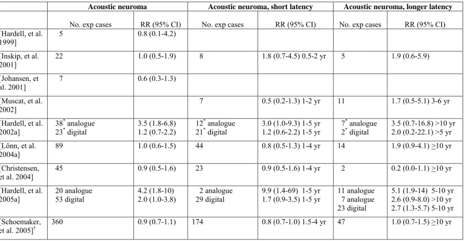
A study using data published previously from Sweden and Denmark as well as cases and controls from Finland, Norway, and the UK [Schoemaker, et al. 2005] included 678 cases of acoustic neuroma and 3553 controls. Of the eligible cases 83% participated and 51% of the controls were successfully interviewed. Regular use of mobile phones was reported
by 53% of the cases and 54% of the controls. No increased risk was found for regular mobile phone use (OR=0.9, 95% CI 0.7-1.1). Acoustic neuroma was not associated with the time since first regular mobile phone use or cumulative years of use. No increased risk was associated with the cumulative number of calls or cumulative hours of use. Risk of acoustic neuroma was not increased on the side were the phone was predominantly held (ipsilateral use/tumour). However, an increased risk of ipsilateral tumours was found for more than 10 years of use (OR=1.8, 95% CI 1.1-3.1). The authors interpreted their findings cautiously, emphasizing the overall lack of association and mentioned both bias and true effect as possible explanations for this finding.
Interphone studies represent a new generation of studies on possible risks related to mo-bile telephony, which provide new information on this rapidly growing and already ex-tremely prevalent exposure. Among the strengths are large and carefully planned interna-tional efforts with emphasis on case definition and ascertainment, exposure assessment and quality control. Nevertheless, some limitations remain. Exposure assessment relies on self-reported mobile phone use and therefore is susceptible to recall bias. Selection bias may be introduced due to low participation rates among controls in some of the centres, since participating controls appear to be more likely to be phone users. There are also still limitations regarding the length of the latency period that can be studied for this new ex-posure. In the studies published so far, results are negative for brain tumours and incon-sistent for acoustic neuroma, for which there is a suggestion for an increased risk among long-term users. Interpretation of the acoustic neuroma results at present is difficult: if the increase is due to recall bias, it is unclear why it would be present for acoustic neuroma only and not operate for brain tumours. Acoustic neuroma is a slowly growing and non-fatal tumour, thus it is possible that it is being diagnosed more often among cell phone users as they experience loss of hearing (detection bias). However, this explanation is not entirely satisfactory as it is unclear as to why such a bias would be present for long-term users only and why it would not have been visible already in nation-wide descriptive data.
Other studies on acoustic neuroma and brain tumours
A Swedish study [Hardell, et al. 2005b] used data from an earlier study [Hardell, et al. 2002a] with 1429 cases diagnosed 1997-2000 and 1470 population-based controls to as-sess differences in risk associated with use of mobile phones in urban and rural settings. The rationale for these analyses is that mobile phones on average use a higher output power level when transmitting in rural areas where base stations are sparse, as compared to urban areas where base stations are more densely situated [Lönn, et al. 2004b]. This would lead to higher levels of RF exposure to the head when mobile phones are used in rural areas. For all brain tumours combined, higher risk estimates were found in rural than urban areas, regardless of latency period used. However, the precision in the risk esti-mates is compromised because of small numbers. The finding was most pronounced for digital phone use, although a similar pattern was observed also for analogue and cordless phones. For the latter type of phone the base station is placed inside the home or office and therefore no difference in the risk estimates between urban and rural areas would be expected.
The same group has also assessed risk of brain tumours by age based on the same mate-rial as described above [Hardell, et al. 2004b]. No consistent trend was found. Instead the highest OR’s tended to be among the youngest age group (20-29 years), followed by the oldest. The numbers of cases and controls in these age groups were, however, too small to
provide meaningful information. No direct analysis of whether or not the relative risks differed across age groups was presented in the paper.
The Hardell group has also published results based on a new material with malignant brain tumours diagnosed in 2000-2003 [Hardell, et al. 2005c]. This is the third material collected by this group. Malignant brain tumours from the Uppsala/Örebro and Linköping areas were identified from the regional cancer registries. As in previous studies by this group, deceased patients were excluded (187 malignant and 18 benign tumours). In addi-tion, 70 patients were excluded for other reasons (not specified whether malignant or benign). Out of 359 contacted malignant cases, 317 participated (88%). Controls were identified from Population Registry with frequency-matching on age and sex. In total, 692 controls participated (84%). Exposure assessment was based on similar methods as in earlier studies, i.e. a mailed questionnaire with supplementation of answers over the tele-phone. In addition, all cases and controls were also interviewed over the phone to “verify exposures and get additional detailed information”. In the data analysis with uncondi-tional logistic regression, adjustment for age, sex, SES, and year of diagnosis was used. Of the cases 66% had used a mobile phone (21% had used analogue and 62% digital phones). The corresponding figure for controls was 51% (11% analogue and 50% digital). The report does not mention any requirement in terms of amount of use needed to be regarded as a mobile phone user (i.e. could be as little as one phone call). For all malig-nant tumours, use of both analogue and digital phones (with >1 yr latency) was associated with approximately two-fold risk (OR for analogue phones 2.6, 95% CI 1.5-4.3 and for digital phones 1.9, 95% CI 1.3-2.7). Slightly higher risk estimates were found for astrocy-toma than other malignant tumours (OR 2.9 for analogue and 1.9 for digital phones rela-tive to 1.6 and 1.9 for other cancers). Increased risks were found already after 1-5 years of using a digital phone (OR=1.6, 95% CI 1.1-2.4) and a cordless phone (OR=1.8, 95% CI 1.2-2.8), although risk estimates were highest after more than 10 years latency (OR for analogue phones 3.5, 95% CI 2.0-6.4), for digital phones 3.6, 95% CI 1.7-7.5, and cord-less phones 2.9, 95% CI 1.6-5.2). These findings differ somewhat from earlier results on malignant brain tumours reported by this research group. The two previous studies found risk estimates close to unity when using a one year latency period [Hardell, et al. 2002b; Hardell, et al. 1999], or 1-6 years latency period [Hardell, et al. 2002b], whereas the newly reported study found increased risks also after a very short period of exposure. In previous studies risk increases for shorter latency periods were confined to subgroups of users. As in earlier reports, highest risks were found for analogue phones and an increas-ing trend with latency was indicated. However, risk estimates were substantially higher than in earlier publications.
Data on meningioma and acoustic neuroma were also included in the most recent study by Hardell and colleagues, published in a separate paper [Hardell, et al. 2005a]. The study included 305 cases with meningioma, 84 with acoustic neuroma, and 692 controls (the same controls were used as in the study described above). The response rates are reported as 89% for cases and 84% for controls. Cases were only included when histopathological diagnosis became available and if they were alive at that time. As in the previously de-scribed work, exposure information was collected by a questionnaire and supplemented over telephone. Case/control status was not disclosed to the interviewer by the investiga-tors. For acoustic neuroma the strongest effect estimates are reported for analogue phone use, with an overall OR=4.2 (95% CI 1.8-10). For >15 years of use the OR=8.4 (95% CI 1.6-45). There are also substantial excess risks for 1-5 years (OR=9.9; 95% CI 1.4-69), 5-10 years (OR=5.1; 95% CI 1.9-14), and for >5-10 years latency (OR=2.6; 95% CI 0.9-8.0).
The laterality analyses are based on >1 years of use. For use of phone at same side as the tumour the OR=5.1 (1.9-14) and for use on opposite side the OR=4.9 (1.2-21). For men-ingioma, the authors found elevated risks that increased with duration of use, particularly for analogue phones. The association, however, is weaker than for acoustic neuroma and the internal consistency is less obvious.
While the Hardell study taken at face value appears to support the recent Swedish Inter-phone study results on acoustic neuroma [Lönn, et al. 2004a], several considerations call for a cautious interpretation. First, looking at table 2 [Hardell, et al. 2005a] it appears that current users of digital phones (> 1 year) in the controls are 50%. At the same time the nation-wide number of subscriptions per person is increasing from 71 to 95% during the study period. One person can have multiple subscriptions, but this is most likely offset by the fact that the denominator includes also the very young and the very old. In the Swed-ish Interphone study over 90% of the controls answered yes to the question if they had ever used a mobile phone (Lönn, personal communication), and 59% reported that they had used a mobile phone regularly, i.e. on average at least once per week during six months or more [Lönn, et al. 2005]. For the corresponding calendar period and age group the proportion of mobile phone users among controls in the latest Hardell et al study was 55% (Hardell, personal communication), but with no requirements regarding the amount of mobile phone use needed to be classified as a mobile phone user. Second, Hardell et al. obtain considerable OR elevations even for the shortest latency periods of 1-5 and 5-10 years, which is remarkable for this type of slow growing tumour. The concern is am-plified by the fact that cases are only included after the histopathological diagnosis has become available. The problem is that this may happen several years after the clinical diagnosis has been made. As a consequence a substantial proportion of the exposure may have taken place after symptoms have occurred. Third, the laterality analyses give no, or weak, support for the notion that the risk is higher on the exposed side of the head. For the malignant tumours a large proportion of the cases have died prior to being identified for inclusion in the study, and were therefore excluded.
The Hardell group’s results are potentially important because they differ from those of other research groups (see Table 1 and 2). It is noteworthy that they differ also for shorter latency periods where most available studies have sufficient precision for adequate analy-sis.
Mobile phone studies on other tumours
The Hardell group has also assessed the risk of salivary gland cancer in relation to mobile phone use [Hardell, et al. 2004a]. The cases were identified from regional cancer regis-tries in entire Sweden 1994-1999/2000 and included malignant tumours only, with de-ceased cases excluded (N=96). Of the eligible cases 66% participated. Controls were from the brain tumour study, with participation of 89%. Exposure assessment was as in the brain tumour studies of the same study group. Of the cases, 12% had used analogue phones and 17% digital phones. For controls, the proportions were 11% and 16%. No increased risks were found for either analogue (OR=0.9, 95% CI 0.6-1.5) or digital phone use (OR=1.0, 95% CI 0.7-1.5). In the analyses by anatomic location, a non-significantly increased risk was found for the submaxillary gland (OR=1.4, 95% CI 0.6-3.5 for any phone use), but not for the parotid (OR=1.0, 95% CI 0.7-1.4) or other locations (OR=1.1, 95% CI 0.5-2.7). Participation among cases was much lower than in other reports by this group.