Interfacial Behavior and Activity of Laccase and Bilirubin Oxidase on
Bare Gold Surfaces
Dmitry Pankratov,
†,‡Javier Sotres,
†Alejandro Barrantes,
†Thomas Arnebrant,
†and Sergey Shleev*
,†,§ †Biomedical Sciences, Health & Society, Malmö University, 205 06 Malmö, Sweden‡Kurchatov NBIC Centre, National Research Centre“Kurchatov Institute″, 123182 Moscow, Russia §A. N. Bach Institute of Biochemistry, 119 071 Moscow, Russia
*
S Supporting InformationABSTRACT: Two blue multicopper oxidases (MCOs) (viz. Trametes hirsuta laccase (ThLc) and Myrothecium verrucaria bilirubin oxidase (MvBOx)) were immobilized on bare polycrystalline gold (Au) surfaces by direct adsorption from both dilute and concentrated enzyme solutions. The adsorption was studied in situ by means of null ellipsometry. Moreover, both enzyme-modified and bare Au electrodes were investigated in detail by atomic force microscopy (AFM) as well as electrochemically. When adsorbed from dilute solutions (0.125 and 0.25 mg mL−1in the cases of ThLc and MvBOx, respectively), the amounts of enzyme per unit area were determined to be ca. 1.7 and 4.8 pmol cm−2, whereas the proteinfilm thicknesses were determined to be 29 and 30 Å for ThLc and MvBOx, respectively. A well-pronounced bioelectrocatalytic reduction of molecular oxygen (O2)
was observed on MvBOx/Au biocathodes, whereas this was not the case for ThLc-modified Au electrodes (i.e., adsorbed ThLc was catalytically inactive). The initially observed apparent kcatapp values for adsorbed MvBOx and the enzyme in solution were found to be very close to each other (viz. 54 and 58 s−1, respectively (pH 7.4, 25 °C)). However, after 3 h of operation of MvBOx/Au biocathodes, kcatappdropped to 23 s−1. On the basis of the experimental results, conformational changes of the enzymes (in all likelihood, their flattening on the Au surface) were suggested to explain the deactivation of MCOs on the bare Au electrodes.
■
INTRODUCTION“Blue” multicopper oxidases (MCOs) (e.g., ascorbate oxidase (AOx), bilirubin oxidase (BOx), ceruloplasmin (Cp), and laccase (Lc)) are copper-containing enzymes efficiently catalyzing oxygen (O2) reduction directly to water (H2O) without the formation of highly reactive oxygen species.1,2 Lc was thefirst redox enzyme for which direct electron transfer (DET)-based bioelectrocatalysis was demonstrated as early as in 1978.3,4 Nowadays, the mechanism of the MCO-catalyzed bioelectrocatalytic reduction of O2 is quite well understood. (See graphical and textual details about the mechanism in Supporting Information (SI), Supporting Figure S2.1,5−7) In spite of this, a scientific dispute concerning the electrochemical behavior of high-redox-potential MCOs (i.e., redox enzymes with redox potentials of the Cu-T1 (ET1) varying from 0.6 up to 0.8 V vs NHE7) adsorbed on gold (Au) electrodes has been taking place since the end of the 1990s.
The debate was initiated by several pioneering studies concerning Lc/Au electrochemistry in which well-pronounced low-redox-potential Faraday processes (with midpoint poten-tials of about 0.4 V vs NHE) related to the adsorbed enzyme were observed without the efficient bioelectrocatalytic reduc-tion of O2on enzyme-modified Au electrodes.
8,9
Later, a similar situation was observed for many different MCOs (e.g., Lc, BOx,
and Cp) adsorbed on bare and modified Au surfaces.6,7,10−12 However, the reason for the absence of bioelectrocatalytic activity was not clearly identified, and in order to explain the experimental results, several suggestions were made. These included both simple, long-standing explanations in thefield of redox protein/metal interface electrochemistry,13 associated with enzyme denaturation on bare Au surfaces or/and the absence of a heterogeneous ET between Au and active centers of MCOs,14,15 as well as a sophisticated hypothesis involving the formation of inactive forms of MCOs as a result of an unnatural ET pathway resulting from enzyme orientation specifically by the trinuclear Cu cluster (Cu-T23), which was assumed to be in DET contact with the Au surfaces.6,7,11,12,16
Recently, the well-pronounced DET-based bioelectrocatalytic reduction of O2 on Au electrodes modified with Lc and BOx was demonstrated. It should be emphasized, however, that bioelectrocatalysis was observed only in the case of atomically planar Au(111) electrodes,17,18 specifically modified polycrys-talline Au surfaces,19,20and 3D (porous) Au electrodes.21−25It was suggested that clear yet short-lived bioelectrocatalytic
Received: June 28, 2013 Revised: January 20, 2014 Published: February 24, 2014
responses on Au(111), which is a highly ordered and smooth surface, were registered because the protein was either held more tightly or there were fewer“side routes” for ET as a result of decreased surface roughness17(i.e., better wiring of BOx on Au(111) was obtained compared to that on the polycrystalline surfaces). Bioelectrocatalysis on modified polycrystalline Au was attributed to proper enzyme orientation20 along with enzyme stabilization.19Similar reasoning could also be given to explain the observed O2 bioelectroreduction process, when porous Au electrodes were used, because the immobilization of MCOs in 3D Au electrodes could favor the exposure of the active site of the immobilized enzymes22 and/or stabilize enzyme molecules incorporated into nanocavities,21 as theoretically predicted in 2001.26
Two questions, however, are still not answered: (i) could MCOs directly adsorbed on bare polycrystalline planar Au electrodes, which are not nanostructured or chemically modified, efficiently catalyze O2electroreduction? If yes, then (ii) why was O2 bioelectroreduction not registered in many previous attempts concerning MCO/Au electrochemistry?
To answer these questions, electrochemical, ellipsometric, and AFM studies of planar polycrystalline Au surfaces modified with Trametes hirsuta Lc (ThLc) and Myrothecium verrucaria BOx (MvBOx) were performed as described below.
■
EXPERIMENTAL SECTIONMaterials. Na2HPO4, NaOH, KH2PO4, K3[Fe(CN)6], NaF, and
H2SO4were obtained from Merck (Darmstadt, Germany). Citric acid,
2,2′-azino-bis(3-ethylbenzothiazoline-6-sulfonic acid) (ABTS), and bovine serum albumin were from Sigma-Aldrich (St. Louis, MO, USA). All chemicals were of analytical grade. Buffers were prepared with water (18 MΩ cm at 25 °C) purified with a PURELAB UHQ II system from ELGA Labwater (High Wycombe, U.K.). Different O2
concentrations were established by bubbling air and oxygen from AGA Gas AB (Sundbyberg, Sweden) through the solution.
Enzymes. Basidiomycete Trametes hirsuta, strain T. hirsuta 56, was obtained from the laboratory collection of the State Research Institute of Protein Biosynthesis (Moscow). Extracellular Trametes hirsuta laccase (ThLc) was isolated from the culture media and purified to homogeneity following well-known procedures.6 The enzyme homogeneity was confirmed by SDS-PAGE (Supporting Figure S2). BOx from Myrothecium verrucaria fungus (MvBOx) was obtained from Amano Enzyme Inc. (Nagoya, Japan) and was additionally purified by Dr. Olga V. Morozova (A. N. Bach Institute of Biochemistry, Moscow, Russia) using gel-filtration and ion-exchange HPLC. Both preparations of the enzymes were stored in 10 mM phosphate buffer at pH 6.5 and −18 °C because the storage stability of both enzymes is close to maximal at this pH value. The preparations were thawed and air-saturated for 2 h before use in order to avoid the possible presence of resting forms, viz., partially reduced enzymes.27The concentration of
Lc and BOx was measured spectrophotometrically at 228.5 and 234.5 nm using BSA as a standard.28
Enzyme Assay and Kinetic Studies. The specific activity of MvBOx and ThLc in homogeneous solution was determined by estimating the initial rates of O2 consumption using an Oxygraph
Clark O2electrode from Hansatech Ltd. (Norfolk, England) at 25°C
with constant stirring. An appropriate concentration of ABTS dissolved in 100 mM buffer (citrate/phosphate, pH 4.0 for ThLc and phosphate, pH 7.4 for MvBOx) was used in order to ensure a measurable linear rate for thefirst 30 s after the addition of prepared enzyme. The concentrations of O2were assumed to be 0.25 and 1.2
mM in air- and O2-saturated buffers, respectively.29
Electrode Preparation. As working electrodes, we used Au substrates manufactured in a Balzers UMS 500 P system by the electron-beam deposition of 2000 Å of Au on silicon(100) wafers (planar polycrystalline Au electrodes) that had been precoated with a 25-Å-thick titanium adhesion layer (Laboratory of Applied Physics,
Linköping University, Sweden) with a geometrical area of about 0.32 cm2. Prior to all measurements (AFM, ellipsometrical, and
electro-chemical), Au electrodes were cleaned with a series of CV scans at a 200 mV s−1scan rate between 0 and−1200 mV versus NHE in 0.5 M NaOH, and they were then rinsed thoroughly with H2O andfinally
cleaned by a series of CV scans at a 200 mV s−1scan rate between 0 and +1900 mV versus NHE in 0.5 M H2SO4.
Clean Au working electrodes were modified by a simple adsorption of ThLc and MvBOx on the electrode surface using dilute and concentrated solutions of MCOs at room temperature (∼25 °C). Specifically, for physisorption, ThLc and MvBOx in 50 mM phosphate buffers with pH values varying from 6.0 up to 7.5 were used, and the enzyme concentration in the bulk solution varied from 0.125 to 4.0 mg mL−1. For electrochemical and atomic force microscopy studies, a drop of solution containing different concentrations of enzymes was evenly deposited on top of the electrodes, adsorption was allowed to occur, and after 15 min the electrode was carefully rinsed with H2O. A
cuvette with a total volume of 4 mL was used (vide infra) in the ellipsometric measurements. It should be emphasized that electrodes did not dry out at any time during modification and investigations.
Au electrodes were connected to a potentiostate using Au-plated alligator clips (model 3289-2) from Pomona Electronics (Everett, WA, USA). The working areas of Au electrodes were determined by direct precise geometric measurements, and the area was also controlled electrochemically.
Electrochemistry. Cyclic voltammetry and chronoamperometry were performed in an electrochemical cell with a volume of 30 mL containing a saturated calomel reference electrode (242 mV vs. NHE) and a platinum mesh counter electrode using a PGSTAT12 potentiostat/galvanostat from Metrohm Autolab B.V. (Utrecht, The Netherlands). As supporting electrolytes, a 100 mM citrate/phosphate buffer solution at pH 4.0 and a 100 mM phosphate buffer solution at pH 7.4 were used for measurements with ThLc- and MvBOx-modified Au electrodes (ThLc/Au and MvBOx/Au), respectively. Electro-chemical experiments were performed at 25°C. All potentials in the present work are given versus NHE.
Ellipsometry Measurements. The adsorption of ThLc and MvBOx onto bare Au was studied in situ by means of null ellipsometry, which measures changes in the polarization of light reflected by a surface (the sample). A thin-film automated ellipsometer (type 43 603-200E, Rudolph Research, Fairfield, NJ, USA) equipped with a xenon arc lamp with afixed angle of incidence (67.96°) was used. Light was detected at a wavelength of 442.9 nm employing an interferencefilter with ultraviolet and infrared blocking (MellesGriot, Netherlands). The Au surface was vertically mounted in a glass trapezoid cuvette (Hellma, Germany) containing 4 mL of solution, which was thermostated at 25°C and stirred using a magnetic stirrer with a rotational speed of 325 rpm. The changes in ellipsometric angles were recorded in situ every 15 s. To determine the refractive index of the Au surface, a four-zone surface calibration in buffer solution was carried out prior to each measurement. When enzymes were to be adsorbed on the electrochemically cleaned Au surface (vide supra),first a stable baseline acquisition was done and then enzyme from the stock solution was added to the cuvette to afinal volume of 4 mL. The formation of protein films was monitored for 60 min, followed by rinsing with enzyme-free buffer solution for 5 min. From ellipsometric data, the protein layer thickness (d, in Å) and the adsorbed amount per unit area (Γ, in mg m−2) were calculated by
assuming a three-layer model (Au−enzyme layer−buffer solution) and using a value of 0.18 mL g−1 as the refractive index increment with respect to the change in protein concentration (dn/dc).30 As an evaluation of the ellipsometric data, the refractive index can be either calculated31or assumed. Because the determination of the thickness of an adsorbed layer is not straightforward at low coverage,32,33 ellipsometry results obtained in this work were evaluated using both procedures. In doing so, the index wasfitted according to the method described by McCrackin and co-workers.31 When assuming the refractive index, two values were taken into account: one based on our own work (n = 1.65)12 and another based on previously reported results n = 1.45.34 Because of the complexity in evaluating the
thicknesses of thin protein layers on Au, as discussed further in the Results and Discussion section, we evaluated the trends (differences between conditions and enzymes) in thickness rather than the absolute values.
Atomic Force Microscopy. AFM images of Au surfaces were obtained using a commercial setup equipped with a liquid cell (MultiMode 8 SPM with a NanoScope V control unit, Bruker AXS). The samples were visualized in water at room temperature by operating the AFM in both tapping and in peak force tapping (PFT) modes. In tapping mode, the cantilever is oscillated at its free resonance frequency, and the tip−sample distance is adjusted during scanning so that the amplitude of the oscillation is kept constant. In this case, Au-coated cantilevers with a nominal spring constant of 0.03 N m−1, a resonance frequency in air of 37 kHz, and a nominal tip radius in the 30−40 nm range (Biolever A, Olympus, Tokyo, Japan) were employed. In PFT mode, force curves are constantly recorded at a constant rate equal to 2 kHz. In this mode, the maximum load exerted on the sample is constantly and automatically minimized. When operating in PFT mode, triangular silicon nitride cantilevers with a nominal spring constant of 0.7 N m−1, a resonance frequency in air of 150 kHz, and a nominal tip radius of 20 nm were employed (ScanAsyst-Fluid, Bruker AXS). Analysis and processing of AFM images was performed with WSxM software.35 Standard image processing consisted of plane subtraction and/or equalization.
The AFM size of adsorbed molecules is given as mean and standard deviation values calculated by averaging the height measured in AFM topography images for at least 20 molecules. It is also important to note that all samples (electrodes) were placed in the AFM liquid cell immediately after electrochemical cleaning and biomodification without letting them dry at any moment.
■
RESULTS AND DISCUSSIONFirst, the MCOs were physisorbed on bare Au surfaces. Because both enzyme preparations were stored in 10 mM phosphate buffer at pH 6.5 (vide supra), phosphate buffer was also used for their immobilization on Au surfaces in order to avoid additional manipulations (e.g., dialysis or chromatography steps). However, the phosphate concentration was increased from 10 to 50 mM in order to adjust the pH during the dilution of the stock solutions properly. Different pH values of phosphate buffers used for the proteins’ physisorption (6.0− 7.5) reflect different pH dependences of the activity and stability of the different MCOs. Specifically, ThLc is very active but simultaneously quite unstable in acidic solutions, whereas it is very stable but inactive in neutral solutions.36 Contrary to ThLc, MvBOx is both active and stable in neutral solutions,37 whereas well-pronounced irreversible deactivation of the enzyme is known to occur in acidic media.38 Thus, for MvBOx physisorption 50 mM phosphate buffer at pH 7.4 was used, whereas the pH of the buffer used for ThLc adsorption was decreased to 6.0. Under these conditions, significant activity of the enzyme could still be registered without fast enzyme deactivation. For comparison, the physisorption of both MCOs was also performed from an identical solution, viz., 50 mM phosphate buffer at pH 7.5 (vide infra)). Both unmodified and modified electrodes, viz., bare Au, ThLc/Au, and MvBOx/Au, were investigated by means of AFM as described below.
Atomic Force Microscopy Studies. As control measure-ments, evaporated Au surfaces that were electrochemically cleaned were imaged in water by operating the AFM in both the PFT and tapping modes (Figure 1a,b, respectively). Clean Au exhibited a granular surface, in accordance with previous reports.39The grains exhibited lateral dimensions in the range of 50−200 nm, with typical heights of about 10−15 nm resulting in a roughness factor of the Au surface ( f) of about
1.3. Thus, from AFM studies, the microscopic (real) Au electrode area (Areal) was 1.3 times higher than the geometric (2D projected) one (Ageom; see additional details concerning the electrode areas in SI). However, it is important to note that this value may be an underestimation because AFM would not Figure 1.(a, b) AFM images of clean evaporated Au surfaces obtained in a water environment by operating the AFM in PFT mode and in tapping mode, respectively (color height scale 25 nm). (c, d) Topography images of Au surfaces obtained with a high surface coverage of ThLc (tapping mode) and MvBOx (PFT mode), respectively (color height scale 12 nm). In these samples, enzyme solutions with concentrations equal to 4.0 mg mL−1 in 50 mM phosphate buffers, at pH 6.0 and 7.4, in the case of ThLc and MvBOx, respectively, were used for protein immobilization on Au surfaces. (e, f) Topography images obtained by operating the AFM in PFT mode and highlighted height profiles for the low surface coverage of ThLc and MvBOx, respectively (color height scale 14 nm). In these samples, enzyme solutions with concentrations equal to 0.125 and 0.25 mg mL−1 in 50 mM phosphate buffer at pH 6.0 and 7.4, in the case of ThLc and MvBOx, respectively, were used for protein immobilization on Au surfaces.
be sensitive to roughness lower than the curvature of the tip. It should also be mentioned that AFM operation in tapping mode provided better lateral resolution than did the PFT technique. MCOs were directly adsorbed on bare polycrystalline Au electrodes using highly concentrated enzyme preparations. A bulk enzyme concentration of 4 mg mL−1was chosen, a value similar to those used in previous studies.9,11,12 The MCO-modified Au surfaces were imaged by operating the AFM in both tapping and PFT modes (Figure 1c,d). A granular structure similar to that of clean Au was found for these surfaces. However, the Au grains were not smooth any longer; they seemed to be almost completely covered by globular features with average lateral dimensions of ca. 20 nm. Although this width is higher than that expected for single ThLc and MvBOx molecules (4−6 nm; see additional details concerning enzyme structures and sizes in SI), it is well known that the visualization of features smaller than the AFM tip results in width values similar to those of the tip (ca. 20 nm) resulting from tip dilation effects,40 with this being the most probable case in our experiments. Thus, it is likely that the topography images show the convolution of the AFM tip and the molecules protruding from the surface. Nevertheless, it is reasonable to assume (i) from the homogeneity of the samples and (ii) from the fact that the images do not show substrate zones without molecules that in both samples full coverage was obtained.
ThLc and MvBOx were also adsorbed on Au electrodes using dilute enzyme solutions (i.e., 0.125 and 0.25 mg mL−1, respectively) in order to obtain AFM images of the submonolayer coverage of the Au surface, from where the dimensions of single immobilized enzymes could be determined. AFM images of these samples obtained by operating in the PFT mode are shown in Figure 1e,f. Both samples showed isolated globular features on top of the wider Au grains.
These features exhibited similar lateral (ca. 20 nm) and vertical dimensions, 2.9± 0.6 and 3.0 ± 0.8 nm for ThLc and MvBOx samples, respectively. As in the case of fully covered samples, the molecules exhibiting larger lateral dimensions than those expected for single molecules can be attributed to tip dilation effects.40
Moreover, both types of molecules exhibited a similar height, which was lower than those expected for both ThLc and MvBOx molecules (4−6 nm, SI). However, this is also expected for single adsorbed proteins because the AFM-determined height of soft biological molecules is usually smaller than its nominal value as a result of tip compression effects.41
Thus, it is reasonable to associate the AFM-determined height of the globular features visualized on top of the Au grains with that of individual protein molecules.
It is important to emphasize that no desorption of either protein from the electrode surface was registered by AFM when modified electrodes were kept in buffer solutions with different pH values (pH 4 and 7.4, i.e., both very close and very far from the pI values of ThLc and MvBOx; SI) for 1 h, indicating that the adsorption of both MvBOx and ThLc on Au has an irreversible character.
The quantitative AFM analysis of MCO/Au surfaces is summarized in Table 1 and Supporting Information Table S1, where height values for both MCOs (ThLc and MvBOx) are presented.
To complement the AFM results, additional ellipsometry studies of Au electrodes were carried out, as described below.
Ellipsometry Studies. Ellipsometric measurements can be used for the determination of the refractive index and thickness of thinfilms by using, for example, the Cuypers32or de Feiter30 formula to determine the adsorbed mass. The accuracy of mass determination has been shown to be more accurate that the determination of refractive indices or thicknesses, particularly at low adsorbed mass.33 Furthermore, refractive indices can be calculated31or assumed. As shown in some previous work,32,33 assuming a plane parallel model for thefilm, the refractive index and thickness co-vary, so an overestimation of one results in an underestimation of the other. In the calculation of mass, this will be compensated for, thus resulting in values with much higher reliability. Therefore, in the present work ellipsometry is mainly used as a mass sensing tool, whereas AFM is used as an enzyme-height-determining technique (vide supra).
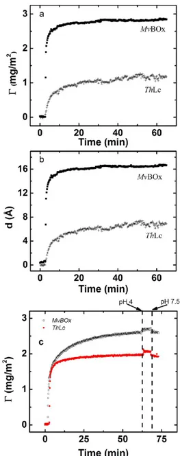
The adsorption of both enzymes onto the electrochemically cleaned Au surfaces was followed by means of ellipsometry, as shown in Figure 2, where the time evolution of the adsorbed amount of protein (Γ) at the electrode with Ageom= 0.32 cm2 and the thickness of the enzymatic layers (d) are presented. Taking into account that the pI of both redox enzymes is close to 4 (see additional details concerning basic biochemical properties of the MCOs in SI), one can conclude that both proteins were negatively charged during all immobilization processes. It is important to emphasize that no desorption of proteins from the electrode surface was registered by ellipsometry measurements when rinsing with enzyme-free buffer solutions with different pH values (pH 4 and 7.5, i.e., both very close and very far from the pI values of both ThLc and MvBOx) after modification (Figure 2), confirming that the adsorption of both MvBOx and ThLc on Au has irreversible character in the experimentally relevant time frame.
Much higherΓ values were obtained for MvBOx, Γ = 2.9 ± 0.1 mg m−2 (∼4.8 pmol cm−2; see calculation details in SI), than for ThLc, Γ = 1.6 ± 0.1 mg m−2 (∼1.7 pmol cm−2) (Figure 2a). Because two different concentrations of MCOs were used during protein immobilization (in analogy with the AFM studies, solutions of ThLc and MvBOx with concen-trations of 0.125 and 0.25 mg mL−1, respectively, were used), the possible dependence of Γ values on the bulk enzyme concentration used for the adsorption procedure was evaluated (SI). As can be seen from the results presented in SI, changes in ThLc bulk concentrations from 0.125 to 4.0 mg mL−1resulted in increasedΓ from 1.2 to 2.2 mg m−2(Supporting Figure S4). However, even when the bulk concentration of ThLc used for the absorption was 16 times higher than that of MvBOx (4.0 vs 0.25 mg mL−1), a lowerΓ value for ThLc was still obtained (cf. 2.2 mg m−2 for ThLc and 2.9 mg m−2 for MvBOx). The determination of thicknesses of thin (submonolayer)films of proteins on solid supports and specifically on Au (see below) is Table 1. Au Surface Coverage with MCOs (Γ) and
Thickness of Enzymatic Layers (Enzyme Height,d) on Aua,b
MCO Γ and d theoretical AFM ellipsometry
ThLc Γ, pmol cm−2 3.95−8.23 n.d. ∼1.7
d, Å 45−65 29 7
MvBOx Γ, pmol cm−2 4.63−10.42 n.d. ∼4.8
d, Å 40−60 30 17
aEllipsometry data were calculated using afixed value for n = 1.65. The
enzyme solutions with concentrations equal to 0.125 and 0.25 mg mL−1 in the case of ThLc and MvBOx, respectively, were used for protein immobilization on Au surfaces.bn.d.− not determined.
not straightforward for a number of reasons. First,fitting to the model (plane parallel homogeneous film) becomes less accurate at low adsorbed mass.33Second, at low coverage the modeling of the layer as a plane parallel homogeneousfilm will result in an average thickness that will be lower than the real dimensions of the protein. Finally, Au has been shown to interact strongly with proteins; therefore, sophisticated modeling (e.g., assuming a mixed protein/Au layer with intermediate optical properties between the surface and the protein) has been proposed in order to obtain realistic values.42 Therefore, our evaluations were carried out using two different approaches: (1) by calculating the refractive index31and (2) by the assumption of two different refractive indices (vide supra).
It is important to point out that because of these uncertainties absolute values of thickness should not be overinterpreted; rather, the focus should be on the differences observed between conditions and enzymes.
The thicknesses of the enzymatic layers (d) calculated from the ellipsometric measurements had very low values (Figure 2b), viz., 1.7± 0.1 and 0.7 ± 0.1 nm for MvBOx and ThLc, respectively. In the case of a very high ThLc concentration during enzyme adsorption (4 mg mL−1), d = 1.3± 0.1 nm was obtained (i.e., it is 6−10 times lower than the height of the native enzyme (Table 1)). Indeed, when an n value for the refractive index equal to 1.45 instead of 1.65 was used for calculations, significantly higher d and slightly lower Γ values were obtained (Supporting Information Figure S5), and d values from AFM and ellipsometry studies almost coincided (Supporting InformationTable S1).
In summary, the thickness of the enzyme layer decreases in the order dMvBOx(0.25 mg mL−1) > dThLc(4 mg mL−1) > dThLc (0.125 mg mL−1) irrespective of the method used for evaluation. This also implies that the obtained results are not a modeling-dependent artifact.
The quantitative analysis of ellipsometrical results is compiled in Table 1 and Supporting Information Table S1, whereΓ and d values for both ThLc and MvBOx molecules are presented. To conclude, AFM and ellipsometry studies confirmed a submonolayer coverage of both ThLc and MvBOx on the Au surface when dilute preparations of the enzymes were used for modification. Both bare Au (control studies) and MCO/Au electrodes, investigated by AFM and ellipsometry, were also electrochemically characterized, as described below.
Electrochemical Investigations. First, an average f value for bare Au electrodes was calculated during electrochemical cleaning. Additional details concerning the calculations as well as some experimental results are described in the SI (Supporting Figure S6). The obtained result ( f = 1.6) correlates well with calculations of real surface areas based on AFM studies of bare Au electrodes (vide supra).
Second, the electrodes were modified with ThLc or MvBOx and placed in air-saturated buffers, and cyclic voltammograms (CVs) of modified Au electrodes were recorded. The catalytic current related to the bioelectroreduction of O2 was clearly visible only for MvBOx/Au electrodes (Figure 3a, Supporting Information Figures S7−S8), whereas adsorbed ThLc was bioelectrocatalytically inactive (Figure 3a, Supporting Informa-tion Figures S11−S12). When electrochemical measurements of MvBOx/Au were performed in the air-saturated buffer, a steady-state bioelectrocatalytic response was obtained with an onset potential of O2bioreduction at about 0.7 V (Figure 3a, curve 2), coinciding with the proposed ET1 value of the enzyme.43
Taking into account motionless electrodes and unstirred solutions used in our studies, the shape of the CV (an almost steady-state potential current curve at high overpotentials) and the observed slight dependence of the biocatalytic current on solution stirring show evidence of O2mass-transfer limitations (additional details in SI). When increasing the O2 concen-tration from 0.25 to 1.2 mM, the maximal current density (jmax) corresponding to bioelectrocatalytic O2 reduction increased only by a factor of 3 (i.e., from 19 μA cm−2 in air-saturated buffer to 53 μA cm−2 in O2-saturated buffer (Supporting Information Figure S7), also suggesting O2diffusion limitations (SI).
Figure 2.Ellipsometric data of MCO adsorption on bare Au surfaces. Adsorption onto the bare polycrystalline Au surface ofMvBOx and ThLc followed in situ by means of null ellipsometry. Enzyme solutions with concentrations equal to 0.125 and 0.25 mg mL−1 in 50 mM phosphate buffers at pH 6.0 and 7.4 in the case of ThLc and MvBOx, respectively (a, b), or 0.125 mg mL−1in 50 mM phosphate buffers at pH 7.5 for both MCOs (c) were used for protein immobilization on Au surfaces. The thickness was calculated by assuming a constant refractive index of 1.65.
The apparent biocatalytic constant (kcatapp) for adsorbed MvBOx was calculated to be 54 s−1 (calculation details can be found in the SI), taking into account that theΓ value for MvBOx on Au equals 4.8 pmol cm−2(Table 1 and Supporting Information Table S1) from ellipsometry studies. It is worthwhile to mention that voltammetric data with a low enzyme concentration (0.25 mg mL−1) used for electrode modification were taken into account during calculations in order to avoid situations in which the formation of multilayers could be suggested (vide supra).
When F−, a known efficient inhibitor of active MCO, was added to the air-saturated buffer solution at a 100 mM concentration, the electrocatalytic current from MvBOx/Au electrodes vanished (Supporting Information Figure S7),
confirming the bioelectrocatalytic origin of the obtained currents.
The operational stability of MvBOx/Au electrodes was investigated in both acidic and neutral solutions (Figure 3b). The half-inactivation time for MvBOx/Au electrodes was calculated to be 0.25 and 2.5 h at pH 4.0 and 7.4, respectively. In other words, after 3 h of biocathode operation kcatappfor the adsorbed MvBOx decreased to 23 s−1 in solution at pH 7.4, whereas under acidic conditions the enzyme was completely inactivated. Fast deactivation of the adsorbed enzyme under acidic conditions is a widely held notion. Indeed, MCO preparations (both BOx and Lc) are usually stored in weak buffers with neutral pH values (10 mM phosphate buffer at pH 6.5 in our studies, vide supra). Because both AFM and ellipsometry studies clearly indicated that the adsorption of MvBOx and ThLc on Au has irreversible character, one can conclude that the time decay of the kcatappis due to the gradual deactivation of the enzyme but not to its desorption from the electrode surface.
As already mentioned, contrary to MvBOx/Au, Au electrodes modified with ThLc were bioelectrocatalytically inactive. Significant increases in enzyme concentration used for modification (Supporting Information Figure S11) and an increase in O2 content in the buffer (Supporting Information Figure S12) did not result in the appearance of even slight bioelectrocatalytic currents from ThLc/Au electrodes.
Enzymatic Assay. The observed kcat values of ThLc and MvBOx toward ABTS in homogeneous reactions were calculated to be 205 and 58 s−1at pH 4.0 and 7.4, respectively (vide supra). To determine the activity of both enzymes in heterogeneous systems (MCO/Au) using ABTS as an electrode donor (qualitative enzymatic assay), a few hundred microliters of air-saturated buffers (100 mM citrate-phosphate at pH 4.0 for ThLc/Au, 100 mM phosphate at pH 7.4 for MvBOx/Au, and both buffers for bare Au electrodes) containing the substrate of the enzymes (i.e., 5 mM ABTS) was dropped on top of the Au electrodes, both unmodified as well as modified with ThLc and MvBOx, as illustrated in Figure 4.
A clear blue color was developed in just a few minutes, when MvBOx/Au wafers were used, whereas no biocatalytic reaction of ABTS oxidation was registered for ThLc/Au or bare Au electrodes, even when incubated with the enzyme substrate for 1 h (Figure 4). Because no desorption of the enzyme was observed in our studies as described above, these measurements confirmed that adsorbed MvBOx was catalytically active whereas ThLc was completely deactivated after immobilization on bare Au surfaces.
General Discussion. The difference in coverage between the two MCOs is a puzzle, which obviously complicates the interpretation of the results. To the best of our knowledge, the hydrodynamic radii of ThLc and MvBOx are unknown. However, there is an investigation showing that the crystallo-graphic size of an MCO (T. versicolor Lc) and its hydrodynamic size are comparable.44Thus, because of ThLc being larger than MvBOx (SI), higher Γ values are expected (in moles) when BOx is used for surface modification. However, the measured difference is too high to take into account only this simple explanation. Nevertheless, the experimental results described above give a positive answer to thefirst question specified in the Introduction concerning the bioelectrocatalytic reduction of O2by MCOs directly adsorbed on polycrystalline bare planar Au electrodes. Specifically, well-pronounced bio(electro)-Figure 3. Electrochemical investigations of MCO-modified Au
electrodes. (a) CVs of MCO-modified Au electrodes: ThLc-Au (curve 1) and MvBOx-Au (curve 2). Enzyme solutions with concentrations equal to 0.125 and 0.25 mg mL−1in 50 mM phosphate buffers, pH 6.0 and 7.4, in the case of ThLc and MvBOx, respectively, were used during the biomodification of Au surfaces. Measurement conditions: 100 mM buffers (citrate/phosphate pH 4.0 for ThLc and phosphate; pH 7.4 forMvBOx); scan rate, 20 mV s−1; second cycle. (b) Chronoamperometric responses from MvBOx-modified Au electrodes recorded at a +400 mV applied potential. Initial jcatvalues
were 18.3 and 7.6μA cm−2at pH 4.0 and 7.4, respectively. Conditions: 100 mM citrate/phosphate buffer at pH 4.0 and 100 mM phosphate buffer at pH 7.4.
catalytic reactions of O2reduction were registered on MvBOx/ polycrystalline Au electrodes when they were electrochemically polarized (Figure 3) as well as when an electron donor was added to the solution (Figure 4).
The second question, viz., why in many previous attempts concerning MCO/Au electrochemistry O2bioelectroreduction was not registered, is also quite easy to answer. Because the well-pronounced bio(electro)catalytic reduction of O2 was never registered for ThLc/Au electrodes in the present studies, neither when the electrodes were electrochemically polarized (Figure 3a, Supporting Figures S11−S12) nor when the electron donor was added directly to the solution (Figure 4), one can conclude that ThLc is completely deactivated on the bare polycrystalline planar Au surface. It is worthwhile to mention that bare carbon electrodes with adsorbed ThLc7 or nanostructured Au electrodes modified with the same enzyme24,25 showed well-pronounced bioelectrocatalytic re-sponses under similar conditions. Moreover, very high kcatapp values are usually calculated for ThLc adsorbed on modified (e.g., thiol-functionalized) Au surfaces (≫100 s−1in refs 19 and 24). Contrary to ThLc/Au, MvBOx immobilized on bare Au is still catalytically active for quite a long time, even if some deactivation of the enzyme definitely occurs (Figure 3b), and the rate of this process depends on the experimental conditions. Actually, the initially calculated kcatappvalue for adsorbed MvBOx (54 s−1) is very close to the kcat value measured in a homogeneous assay (58 s−1). However, after 3 h of operation of MvBOx/Au biocathodes, the kcatapp value decreased to 23 s−1. However, it is difficult to compare kcat values obtained in homogeneous assays and DET-based bioelectrocatalysis because the limiting steps in homogeneous and heterogeneous systems could be different. For instance, while the oxidation of electron donors is known to be the limiting step during homogeneous assays (step 1 in Supporting Information Figure S2), intramolecular ET and the reduction of O2 by the trinuclear copper cluster are the limiting steps during heterogeneous BOx-based bioelectrocatalysis (steps 2 and 3 in Supporting Information Figure S2).37However, the results from stability studies of MvBOx/Au electrodes (Figure 3b), which showed very fast deactivation of the adsorbed enzyme especially in acidic buffers, also strongly support the idea that
ThLc was completely deactivated on the bare Au surface and that ThLc deactivation occurred at much higher rates than did MvBOx. This is actually quite surprising considering the fact that ThLc is heavily glycosylated as compared to MvBOx (SI). On the one hand, there is a widely held notion that carbohydrates are responsible for the stabilization of proteins and deglycosylated enzymes have much lower operational stability than do glycosylated oxidoreductases. On the other hand, it has also recently been shown that the immobilization of deglycosylated redox enzymes results in their stabilization on the electrode surface whereas, as expected, they are unstable in solution.45,46 Thus, in all likelihood, glycosylation is not responsible for enzyme stabilization in the immobilized state.
As described in the Introduction, three main reasons for the absence of O2 bioelectroreduction on MCO/Au exist in the literature: (1) enzyme deactivation, (2) the absence of heterogeneous ET, and (3) the formation of inactive forms of MCOs. Taking into account the experimental results presented herein, it can be concluded that the main reason for the absence of O2 bioelectroreduction in many previous attempts concerning MCO/Au electrochemistry is the fast deactivation of the enzymes on bare Au surfaces. Even if the second and third reasons could be taken into account as possible explanations for the absence of DET-based bioelec-trocatalysis (Figure 3, Supporting Information Figures S11− S12), enzymatic assays in homogeneous and heterogeneous systems (Figure 4) clearly indicate that neither the absence of heterogeneous ET nor the formation of inactive (resting) forms of MCOs are responsible for the inertness of ThLc/Au. Specifically, in the presence of both enzyme substrates, viz., electron donor (ABTS) and electron acceptor (O2), the biocatalytic oxidation of ABTS should occur even if DET between ThLc and Au is not achieved. The biocatalytic oxidation of ABTS should also occur even if resting forms of ThLc are formed on bare Au surfaces. It is well known that resting forms (both the resting oxidized form2and the resting partially reduced states27) in the presence of both substrates will be transformed into the native intermediate (i.e., the catalytically relevant form of MCOs2,47). Even though the rate of this transformation is very low (only 1 s−1),47 3600 s experiments (as demonstrated in Figure 4) should be long Figure 4.Photographs of polycrystalline Au electrodes with drops of 5 mM ABTS in buffers after 1 h of incubation. (Left) MvBOx/Au electrode. (Middle) ThLc/Au electrode. (Right) Bare Au electrode (control). Enzyme solutions with concentration equal to 0.125 mg mL−11in 50 mM
enough to transfer all resting ThLc molecules into the active enzyme.
It is also worthwhile to mention the very fast deactivation of MvBOx on Au(111)17as compared to that when adsorbed on polycrystalline Au (present study). It can be hypothesized that the protein is held much more tightly on Au(111), a highly ordered and smooth surface, compared to the situation where polycrystalline Au is used, resulting in a faster deactivation. The most difficult question to answer on a molecular level without serious speculation is why the fast deactivation of MCOs on a bare Au surface occurs. Although very low d values compared to the height of the native enzymes were obtained for both ThLc and MvBOx, this difference was still much higher for ThLc (Table 1). This indicates seriousflattening of the enzyme on a bare Au surface. Specifically, the very low d values (7−29 Å, i.e., 1.5−9.3 times lower than the height of the native enzyme) registered in AFM experiments are also very strong evidence for ThLc deactivation on bare Au electrodes, in all likelihood being due to the enzymeflattening on the metal surface. By taking into account much higherΓ values for MvBOx than for ThLc (Table 1 and Supporting Table S1), one can speculate that steric effects prevent MvBOx from spreading on the surface, thereby preventing their inactivation due to conformational changes.
■
CONCLUSIONSFor the first time, we provide mechanistic insight into the interfacial behavior and activity of MCOs on bare Au surfaces. The obtained experimental results show without doubt the efficient bioelectrocatalytic reduction of O2 at least by some MCOs (e.g., MvBOx) directly adsorbed on a bare polycrystal-line planar Au surface. Conformational changes of MCOs on the electrode are suggested to explain the observed experimental data (i.e., absence of bioelectrocatalysis for ThLc/Au electrodes) as well as the fast degradation of bioelectrocatalytic signals in the case of MvBOx-based biocathodes. Comparison of the kcatapp values in homogeneous and heterogeneous systems also confirms our suggestion concerning the inactivation of MCOs on bare polycrystalline planar Au surfaces. Taking into account AFM concerning the heights of adsorbed enzyme molecules as well as experimental results in the literature and the present studies concerning the very fast deactivation of MvBOx on highly ordered and smooth Au(111) compared to that on polycrystalline Au, we suggest that the inactivation of MCOs on bare Au is, in all likelihood, due to the enzymeflattening on the metal surface.
■
ASSOCIATED CONTENT*
S Supporting InformationAdditional information about redox enzymes; AFM, ellipsom-etry, and electrochemical data; and the theoretical basis of electrochemical measurements and enzymatic assay results. This material is available free of charge via the Internet at http://pubs.acs.org.
■
AUTHOR INFORMATIONCorresponding Author
*E-mail: sergey.shleev@mah.se.
Author Contributions
S.S. and T.A. conceived and initiated the project. D.P., J.S., and A.B. performed electrochemical, AFM, and ellipsometry studies, respectively, and analyzed the data. All authors discussed the
results and implications and commented on the work. The manuscript was written through the contributions of all authors, and all authors have given approval to thefinal version of the manuscript.
Notes
The authors declare no competingfinancial interest.
■
ACKNOWLEDGMENTSWe thank Dr. Olga V. Morozova (Institute of Biochemistry, Moscow, Russia) and Dr. Karolina Haberska (Malmö University, Malmö, Sweden) for the purification of MvBOx and some ellipsometry measurements with ThLc, respectively. This work has been financially supported by the Russian Foundation for Basic Research (research projects 12-04-33102 and 13-04-12083), the Gustaf Th. Ohlsson Foundation, and the Faculty of Health and Society, Malmö University, Sweden.
■
ABBREVIATIONSAgeom, geometric electrode area; AOx, ascorbate oxidase; Areal, microscopic (real) electrode area; AFM, atomic force microscopy; Au, gold; BOx, bilirubin oxidase; Cp, ceruloplas-min; CVs, cyclic voltammograms; d, thicknesses of the enzymatic layer; DET, direct electron transfer; ET1, redox potential of the Cu-T1 site; f , roughness factor; jmax, maximal bioelectrocatalytic current density; Lc, laccase; MCO, multi-copper oxidase; MvBOx, Myrothecium verrucaria bilirubin oxidase; NHE, normal hydrogen electrode; PFT, peak force tapping mode; qreal, charge associated with the gold oxide reduction process; ThLc, Trametes hirsuta laccase; SI, supporting information; Γ, protein concentration on the electrode surface
■
REFERENCES(1) Sakurai, T.; Kataoka, K. Basic and applied features of multicopper oxidases, CueO, bilirubin oxidase, and laccase. Chem. Rec. 2007, 7, 220−229.
(2) Solomon, E. I.; Sundaram, U. M.; Machonkin, T. E. Multicopper oxidases and oxygenases. Chem. Rev. 1996, 96, 2563−2605.
(3) Berezin, I. V.; Bogdanovskaya, V. A.; Varfolomeev, S. D.; Tarasevich, M. R.; Yaropolov, A. I. Bioelectrocatalysis. Equilibrium oxygen potential in the presence of laccase. Dokl. Akad. Nauk SSSR 1978,240, 615−618.
(4) Tarasevich, M. R.; Yaropolov, A. I.; Bogdanovskaya, V. A.; Varfolomeev, S. D. Electrocatalysis of a cathodic oxygen reduction by laccase. Bioelectrochem. Bioenerg. 1979, 6, 393−403.
(5) Tsujimura, S.; Nakagawa, T.; Kano, K.; Ikeda, T. Kinetic study of direct bioelectrocatalysis of dioxygen reduction with bilirubin oxidase at carbon electrodes. Electrochemistry 2004, 72, 437−439.
(6) Shleev, S.; Christenson, A.; Serezhenkov, V.; Burbaev, D.; Yaropolov, A.; Gorton, L.; Ruzgas, T. Electrochemical redox transformations of T1 and T2 copper sites in native Trametes hirsuta laccase at gold electrode.Biochem. J. 2005, 385, 745−754.
(7) Shleev, S.; Tkac, J.; Christenson, A.; Ruzgas, T.; Yaropolov, A. I.; Whittaker, J. W.; Gorton, L. Direct electron transfer between copper-containing proteins and electrodes. Biosens. Bioelectron. 2005, 20, 2517−2554.
(8) Gelo-Pujic, M.; Kim, H. H.; Butlin, N. G.; Palmore, G. T. Electrochemical studies of a truncated laccase produced in Pichia pastoris. Appl. Environ. Microbiol. 1999, 65, 5515−5521.
(9) Hyung, K. H.; Jun, K. Y.; Hong, H.-G.; Kim, H. S.; Shin, W. Immobilization of laccase onto the gold electrode using b-mercaptopropionate. Bull. Korean Chem. Soc. 1997, 18, 564−566.
(10) Yoon, C.-J.; Kim, H.-H. Electrochemical studies of immobilized laccases on the modified-gold electrodes. J. Korean Electrochem. Soc. 2004, 7, 26−31.
(11) Ramirez, P.; Mano, N.; Andreu, R.; Ruzgas, T.; Heller, A.; Gorton, L.; Shleev, S. Direct electron transfer from graphite and functionalized gold electrodes to T1 and T2/T3 copper centers of bilirubin oxidase. Biochim. Biophys. Acta 2008, 1777, 1364−1369.
(12) Haberska, K.; Vaz-Dominguez, C.; De Lacey, A. L.; Dagys, M.; Reimann, C. T.; Shleev, S. Direct electron transfer reactions between human ceruloplasmin and electrodes. Bioelectrochemistry 2009,76, 34− 41.
(13) Guo, L. H.; Hill, H. A. O. Direct electrochemistry of proteins and enzymes. Adv. Inorg. Chem. 1991, 36, 341−375.
(14) Christenson, A.; Shleev, S.; Mano, N.; Heller, A.; Gorton, L. Redox potentials of the blue copper sites of bilirubin oxidases. Biochim. Biophys. Acta 2006, 1757, 1634−1641.
(15) Shleev, S.; Pita, M.; Yaropolov, A. I.; Ruzgas, T.; Gorton, L. Direct heterogeneous electron transfer reactions of Trametes hirsuta laccase at bare and thiol-modified gold electrodes.Electroanalysis 2006, 18, 1901−1908.
(16) Pita, M.; Shleev, S.; Ruzgas, T.; Fernandez, V. M.; Yaropolov, A. I.; Gorton, L. Direct heterogeneous electron transfer reactions of fungal laccases at bare and thiol-modified gold electrodes. Electrochem. Commun. 2006, 8, 747−753.
(17) dos Santos, L.; Climent, V.; Blanford, C. F.; Armstrong, F. A. Mechanistic studies of the blue Cu enzyme, bilirubin oxidase, as a highly efficient electrocatalyst for the oxygen reduction reaction. Phys. Chem. Chem. Phys. 2010, 12, 13962−13974.
(18) Climent, V.; Zhang, J.; Friis, E. P.; Oestergaard, L. H.; Ulstrup, J. Voltammetry and single-molecule in situ scanning tunneling microscopy of laccases and bilirubin oxidase in electrocatalytic dioxygen reduction on Au(111) single-crystal electrodes. J. Phys. Chem. C 2012, 116, 1232−1243.
(19) Pita, M.; Gutierrez-Sanchez, C.; Olea, D.; Velez, M.; Garcia-Diego, C.; Shleev, S.; Fernandez, V. M.; De Lacey, A. L. High redox potential cathode based on laccase covalently attached to gold electrode. J. Phys. Chem. C 2011, 115, 13420−13428.
(20) Olejnik, P.; Palys, B.; Kowalczyk, A.; Nowicka, A. M. Orientation of laccase on charged surfaces. Mediatorless oxygen reduction on amino- and carboxyl-ended ethylphenyl groups.J. Phys. Chem. C 2012, 116, 25911−25918.
(21) Murata, K.; Kajiya, K.; Nakamura, N.; Ohno, H. Direct electrochemistry of bilirubin oxidase on three-dimensional gold nanoparticle electrodes and its application in a biofuel cell. Energy Environ. Sci. 2009, 2, 1280−1285.
(22) Qiu, H.; Xu, C.; Huang, X.; Ding, Y.; Qu, Y.; Gao, P. Immobilization of laccase on nanoporous gold: comparative studies on the immobilization strategies and the particle size effects. J. Phys. Chem. C 2009, 113, 2521−2525.
(23) Dagys, M.; Haberska, K.; Shleev, S.; Arnebrant, T.; Kulys, J.; Ruzgas, T. Laccase-gold nanoparticle assisted bioelectrocatalytic reduction of oxygen. Electrochem. Commun. 2010, 12, 933−935.
(24) Gutierrez-Sanchez, C.; Pita, M.; Vaz-Dominguez, C.; Shleev, S.; De Lacey, A. L. Gold nanoparticles as electronic bridges for laccase-based biocathodes. J. Am. Chem. Soc. 2012, 134, 17212−17220.
(25) Salaj-Kosla, U.; Poller, S.; Schuhmann, W.; Shleev, S.; Magner, E. Direct electron transfer of Trametes hirsuta laccase adsorbed at unmodified nanoporous gold electrodes. Bioelectrochemistry 2013, 91, 15−20.
(26) Zhou, H.-X.; Dill, K. A. Stabilization of proteins in confined spaces. Biochemistry 2001, 40, 11289−11293.
(27) Shleev, S.; Reimann, C. T.; Serezhenkov, V.; Burbaev, D.; Yaropolov, A. I.; Gorton, L.; Ruzgas, T. Autoreduction and aggregation of fungal laccase in solution phase: possible correlation with a resting form of laccase. Biochimie 2006, 88, 1275−1285.
(28) Ehresmann, B.; Imbault, P.; Weil, J. H. Spectrophotometric determination of protein concentration in cell extracts containing tRNA and rRNA. Anal. Biochem. 1973, 54, 454−463.
(29) Truesdale, G. A.; Downing, A. L. Solubility of oxygen in water. Nature 1954, 173, 1236.
(30) De Feijter, J. A.; Benjamins, J.; Veer, F. A. Ellipsometry as a tool to study the adsorption behavior of synthetic and biopolymers at the air-water interface. Biopolymers 1978, 17, 1759−1772.
(31) McCrackin, F. L.; Passaglia, E.; Stromberg, R. R.; Steinberg, H. L. Measurement of the thickness and refractive index of very thin films and the optical properties of surfaces by ellipsometry. J. Res. Natl. Bur. Stand., Sect. A 1963, 67, 363−77.
(32) Cuypers, P. A.; Corsel, J. W.; Janssen, M. P.; Kop, J. M. M.; Hermens, W. T.; Hemker, H. C. The adsorption of prothrombin to phosphatidylserine multilayers quantitated by ellipsometry. J. Biol. Chem. 1983, 258, 2426−2431.
(33) Tiberg, F. Physical characterization of non-ionic surfactant layers adsorbed at hydrophilic and hydrophobic solid surfaces by time-resolved ellipsometry. J. Chem. Soc., Faraday Trans. 1996, 92, 531− 538.
(34) Voeroes, J. The density and refractive index of adsorbing protein layers. Biophys. J. 2004,87, 553−561.
(35) Horcas, I.; Fernandez, R.; Gomez-Rodriguez, J. M.; Colchero, J.; Gomez-Herrero, J.; Baro, A. M. WSXM: a software for scanning probe microscopy and a tool for nanotechnology. Rev. Sci. Instrum. 2007,78, 013705/1−013705/8.
(36) Pankratov, D. V.; Zeifman, Y. S.; Morozova, O. V.; Shumakovich, G. P.; Vasil’eva, I. S.; Shleev, S.; Popov, V. O.; Yaropolov, A. I. A comparative study of biocathodes based on multiwall carbon nanotube buckypapers modified with three different multicopper oxidases. Electroanalysis 2013, 25, 1143−1149.
(37) Shleev, S.; Andoralov, V.; Falk, M.; Reimann, C. T.; Ruzgas, T.; Srnec, M.; Ryde, U.; Rulisek, L. On the possibility of uphill intramolecular electron transfer in multicopper oxidases: electro-chemical and quantum electro-chemical study of bilirubin oxidase. Electro-analysis 2012, 24, 1524−1540.
(38) Andoralov, V.; Falk, M.; Suyatin Dmitry, B.; Granmo, M.; Sotres, J.; Ludwig, R.; Popov Vladimir, O.; Schouenborg, J.; Blum, Z.; Shleev, S. Biofuel cell based on microscale nanostructured electrodes with inductive coupling to rat brain neurons. Sci. Rep. 2013,3, 3270. (39) Caruso, F.; Furlong, D. N.; Kingshott, P. Characterization of ferritin adsorption onto gold.J. Colloid Interface Sci. 1997, 186, 129− 140.
(40) Margeat, E.; Le Grimellec, C.; Royer, C. A. Visualization of trp repressor and its complexes with DNA by atomic force microscopy. Biophys. J. 1998, 75, 2712−2720.
(41) Moreno-Herrero, F.; Colchero, J.; Baro, A. M. DNA height in scanning force microscopy. Ultramicroscopy 2003, 96, 167−174.
(42) Maartensson, J.; Arwin, H. Interpretation of spectroscopic ellipsometry data on protein layers on gold including substrate-layer interactions. Langmuir 1995, 11, 963−968.
(43) Tsujimura, S.; Kuriyama, A.; Fujieda, N.; Kano, K.; Ikeda, T. Mediated spectroelectrochemical titration of proteins for redox potential measurements by a separator-less one-compartment bulk electrolysis method. Anal. Biochem. 2005, 337, 325−331.
(44) Christensen, N. J.; Kepp, K. P. Stability mechanisms of a thermophilic laccase probed by molecular dynamics. PLoS One 2013, 8, e61985.
(45) Ortiz, R.; Matsumura, H.; Tasca, F.; Zahma, K.; Samejima, M.; Igarashi, K.; Ludwig, R.; Gorton, L. Effect of deglycosylation of cellobiose dehydrogenases on the enhancement of direct electron transfer with electrodes. Anal. Chem. 2012, 84, 10315−10323.
(46) Yakovleva, M. E.; Killyeni, A.; Seubert, O.; O Conghaile, P.; MacAodha, D.; Leech, D.; Gonaus, C.; Popescu, I. C.; Peterbauer, C. K.; Kjellstroem, S.; Gorton, L. Further insights into the catalytical properties of deglycosylated pyranose dehydrogenase from Agaricus meleagris recombinantly expressed in Pichia pastoris. Anal. Chem. 2013, 85, 9852−9858.
(47) Lee, S.-K.; DeBeer George, S.; Antholine, W. E.; Hedman, B.; Hodgson, K. O.; Solomon, E. I. Nature of the intermediate formed in the reduction of O2to H2O at the trinuclear copper cluster active site