Linköping University Medical Dissertations No. 968
Expanding Role of Caveolae in
Control of Adipocyte Metabolism
Proteomics of Caveolae
Nabila Aboulaich
During the course of the research underlying this thesis, Nabila Aboulaich was enrolled in Forum Scientium, a multidisciplinary doctoral programme at Linköping University, Sweden.
© Nabila Aboulaich
Published articles have been reprinted with permission from copyright holder:
Paper II © 2004 & Paper IV © 2006, Elsevier, Inc. Paper I © 2004, The Biochemical Society, London
Paper III © 2006, The American Society for Biochemistry and Molecular Biology, Inc.
Printed in Sweden by Linköpings Tryckeri AB, Sweden, 2006 ISBN 91-85643-58-0
I
Abstract
The primary function of adipose tissue is to store energy in the form of triacylglycerol, which is hydrolyzed to fatty acids to supply other tissues with energy. While insulin promotes the storage of triacylglycerol, catecholamines stimulate its hydrolysis. The development of type II diabetes is strongly associated with obesity, indicating a role of triacylglycerol metabolism in the pathogenesis of diabetes. Caveolae are plasma membrane invaginations found in most cells but are highly abundant in adipocytes. Insulin receptors are localized in caveolae and their function depends on intact caveolae structures. In the present thesis work, mass spectrometry-based methodology allowed identification of a number of new proteins and their posttranslational modifications in caveolae of human adipocytes. Variable N-terminal acetylation and phosphorylation of caveolin-1α and caveolin-1β were identified, which might regulate the function of caveolae. The transcription regulator protein PTRF was identified as the major caveolae associated protein. Specific proteolytic modifications of PTRF at the cytosolic surface of caveolae and phosphorylation on nine serine and one threonine residues were identified. Moreover, insulin induced translocation of PTRF from the plasma membrane to the nucleus. PTRF was previously shown to regulate the activity of both RNA polymerase I and polymerase II, thus a role of PTRF in mediating the anabolic action of insulin on protein synthesis and gene transcription is proposed.
PTRF was also involved in an extranuclear function in the hormonal regulation of triacylglycerol metabolism in caveolae. PTRF was colocalized with the triacylglycerol regulator proteins perilipin and hormone-sensitive lipase (HSL) in the triacylglycerol-synthesizing caveolae subclass. We showed that, while perilipin was translocated to the plasma membrane, both PTRF and HSL were translocated from the plasma membrane to the cytosol as a complex in response to insulin. The perilipin recruited to the plasma membrane was highly threonine phosphorylated. By mass spectrometry, three phosphorylated threonine residues were identified and were located in an acidic domain in the lipid droplet targeting domain of perilipin. The insulin-induced recruitment of perilipin to the plasma membrane might, therefore be phosphorylation-dependent. Isoproterenol, which stimulates hydrolysis of triacylglycerol, induced a complete depletion of perilipin B from the plasma membrane, suggesting a function of perilipin B to protect newly synthesized triacylglycerol in caveolae from being hydrolyzed by HSL. The location of PTRF and HSL was not affected by isoproterenol, indicating that insulin is acting against a default presence of PTRF and HSL in caveolae.
Taken together, this thesis expands our knowledge about caveolae and provided valuable information about their involvement in novel roles, particularly in the hormonal regulation of triacylglycerol metabolism.
III
Original publications
This thesis is based on the following original scientific papers, which will be referred to in the text by their Roman numerals:
I. Nabila Aboulaich, Julia P. Vainonen, Peter Strålfors, and Alexander V. Vener (2004). Vectorial proteomics reveal targeting, phosphorylation and specific fragmentation of polymerase I and transcript release factor (PTRF) at the surface of caveolae in human adipocytes. Biochem J 383: 237-248.
II. Julia P. Vainonen, Nabila Aboulaich, Maria V. Turkina, Peter Strålfors, and Alexander V. Vener (2004). N-terminal processing and modifications of caveolin-1 in caveolae from human adipocytes. Biochem Biophys Res Commun 320: 480-486.
III. Nabila Aboulaich, Alexander V. Vener, and Peter Strålfors (2006). Hormonal control of reversible translocation of perilipin B to the plasma membrane in primary human adipocytes. J Biol Chem 281: 11446-11449.
IV. Nabila Aboulaich, Unn Örtegren, Alexander V. Vener, and Peter Strålfors (2006). Association and insulin regulated translocation of hormone-sensitive lipase with PTRF. Biochem Biophys Res Commun 350, in press.
V
Table of contents
Abstract ___________________________________________________ I Original publications _______________________________________ III Abbreviations ____________________________________________ VII Introduction________________________________________________ 1
Obesity and the adipose tissue _______________________________ 2
Insulin action in adipose tissue ______________________________ 3
The adipocyte and caveolae ___________________________________ 7 Caveolin _________________________________________________ 8 Functions of caveolin-1 ____________________________________ 11 Characterization of caveolae _________________________________ 13
Isolation of caveolae ______________________________________ 13 Protein composition of caveolae_____________________________ 15
Proteomics _____________________________________________ 15 Post-translational modifications ___________________________ 19 Vectorial proteomics of caveolae____________________________ 22
Roles of caveolae in adipocytes _______________________________ 27 Vesicular transport _______________________________________ 27 Insulin signaling at caveolae________________________________ 28 Regulated proteolysis at caveolae ___________________________ 29 Caveolae in insulin-control of gene transcription_______________ 30 Caveolae as centres of lipid metabolism ______________________ 32
Regulation of lipid metabolism _____________________________ 33
Conclusion ________________________________________________ 39 Acknowledgements _________________________________________ 41 References ________________________________________________ 45
VII
Abbreviations
AMP adenosine monophosphate
CREBP cyclic AMP response element binding protein EHD Eps 15 homology domain-containing protein
ESI electrospray ionization
FOXO forkhead transcription factor GLUT glucose transporter
HSL hormone-sensitive lipase
IMAC immobilized metal affinity chromatography MALDI matrix-assisted laser-desorption ionization MAP mitogen activated protein
MS mass spectrometry
PKA protein kinase A
PTRF polymerase I and transcript release factor SDPR serum deprivation response gene product
SRBC serum deprivation response-related gene product SREBP sterol regulatory element-binding protein
Introduction 1
Introduction
Obesity is one of the major environmental factors contributing to the development of various diseases, including type II diabetes, hypertension and cardiovascular disease. The prevalence of obesity among patients with insulin resistance and type II diabetes is very high, reflecting the association of increased body fat mass and impaired insulin action. Conversely, lack of adipose tissue, as in lipodystrophy, is also associated with insulin resistance, which reflects that the adipose tissue per se, as a site for fatty acid storage, is critical for whole body insulin action and development of disease.This thesis will focus on the role of caveolae microdomains in fat cell metabolism. Caveolae are highly abundant structures at the plasma membrane of the adipocytes. The protein caveolin, which is the only marker protein of caveolae, plays a significant role in caveolae. Caveolae have been implicated in fundamental cellular activities such as endocytosis and signal transduction. Different approaches and methods are used to identify and study proteins in caveolae in order to further explore the role of these multifunctional domains. Mass spectrometry-based approaches have allowed identification of new caveolar proteins, which implicated caveolae in novel roles, such as the insulin-controlled gene transcription. Recent findings have also identified a further new role of caveolae as metabolic platforms: the uptake of fatty acids and conversion to
2 Introduction
triacylglycerol appear to take place in caveolae where the metabolism of the newly synthesized triacylglycerol is, moreover, under hormonal control.
Obesity and the adipose tissue
Obesity, which is defined by body mass index ≥ 30 kg/m2, is a consequence of the modern society lifestyle with excess food intake and low physical activity. This imbalance between energy intake and energy expenditure is a major risk factor for the development of several chronic diseases, including hypertension, cardiovascular disease, and type II diabetes. It is known that obesity is associated with insulin resistance, which is the initial defect in patients who will develop type II diabetes. More than 80 % of type II diabetic patients are overweight (Bloomgarden 2000). Furthermore, animal models of genetic and diet-induced of obesity develop insulin resistance and diabetes (Coleman 1978; Liu et al. 2002).
The adipose tissue stores excess energy or fatty acids as triacylglycerols. When energy is required, stored triacylglycerols are hydrolyzed and fatty acids are released from adipocytes to supply different tissues with energy from oxidation of the fatty acids. Abnormalities in the storage and release of fatty acids in adipose tissue lead to elevated plasma fatty acid concentrations. Obese individuals and type II diabetic patients have high plasma fatty acid levels, indicating a role of lipid metabolism in the pathogenesis of insulin resistance and type II diabetes. The inability of the adipose tissue to trap fatty acids leads to their accumulation in other tissues such as muscle, heart, liver, and pancreas. This gives rise to hyperglycemia and hyperlipidemia, which are associated with the development of insulin resistance. The insulin producing β-cells in pancreas increase their insulin secretion to compensate for the insulin
Introduction 3 resistance, which eventually leads to β-cell dysfunction and the development of overt type II diabetes.
It has been proposed that in obesity the adipose tissue is overloaded and resists further lipid storage, which explains the high plasma fatty acid levels and their diversion to accumulation in non-adipose tissues seen in insulin resistant states. Conversely, the lack of adipose tissue, as in the condition of lipodystrophy, is also associated with insulin resistance (Ganda 2000). In both cases a limited storage capacity, due to obesity or lack of the adipose tissue, reflects the association of adipose tissue with the development of insulin resistance and type II diabetes (Frayn 2001).
Moreover, it is well established that the adipose tissue, in addition to being an energy store and provider, is also an endocrine organ. It secretes a number of proteins that have effects on other tissues as well as the adipose tissue itself. The production of these proteins, including leptin, resistin, adiponectin, tumor necrosis factor-α and interleukin-6, extends the function of adipose tissue to control whole body metabolism and energy homeostasis. In obesity, it has been reported that the plasma levels of these proteins are altered, which in turn interferes with the action of insulin and its tissue sensitivity (Jazet et al. 2003).
Insulin action in adipose tissue
Insulin is an anabolic hormone that stimulates cell growth and promotes synthesis and storage of substrates in adipocytes, liver, and muscles. It stimulates the synthesis of lipids, glycogen, and proteins, and inhibits their breakdown. As part of these processes insulin increases glucose uptake in adipocytes and muscles and inhibits hepatic glucose output, thereby regulating the glucose concentration in the blood (Saltiel and Kahn 2001).
4 Introduction
In adipose tissue, the main function of insulin is to clear lipids from the circulation by promoting their storage as triacylglycerol in the adipocyte oil droplet. The source of fatty acids is the plasma triacylglycerol-rich lipoproteins (chylomicrons and very low-density lipoproteins (VLDL)). Extraction of fatty acids from the lipoproteins is catalyzed by lipoprotein lipase, which is activated by insulin. The released fatty acids can enter the adipocyte through caveolae membrane domains, where they are re-esterified with glycerol 3-phosphate to triacylglycerol (Öst et al. 2005). Glycerol 3-phosphate is derived from insulin-stimulated glucose uptake, which is an important function of insulin in adipocyte’s glycolysis. Another function of insulin is to inhibit the hydrolysis of triacylglycerol and the release of fatty acids (lipolysis) from adipocytes. Lipolysis is stimulated by catecholamines, particularly noradrenaline released from nerve endings in the adipose tissue, which activate hormone-sensitive lipase (HSL) and the lipid droplet-associated protein perilipin through phosphorylation by a cyclic AMP-dependent pathway (Strålfors and Belfrage 1983; Honnor et al. 1985; Greenberg et al. 1991). Insulin inhibits lipolysis by decreasing cyclic AMP levels and by dephosphorylation of both HSL and perilipin (Strålfors and Honnor 1989; Mooney and Bordwell 1991).
In addition to fatty acid uptake, insulin stimulates the uptake of glucose in adipose tissue. The adipose tissue account for a very small proportion (~ 10%) of insulin-stimulated whole body glucose uptake compared with the muscles in which 60-70% of the glucose is utilized (DeFronzo et al. 1992). However, animal models with adipose-specific ablation of GLUT4 (insulin-regulated glucose transporter) gene developed whole body insulin resistance (Abel et al. 2001). Conversely, overexpression of GLUT4 in adipose tissue enhanced whole body glucose utilization (Shepherd et al. 1993). Although the mechanisms are not known,
Introduction 5 this reflects the important role of adipose tissue in glucose disposal in the whole body.
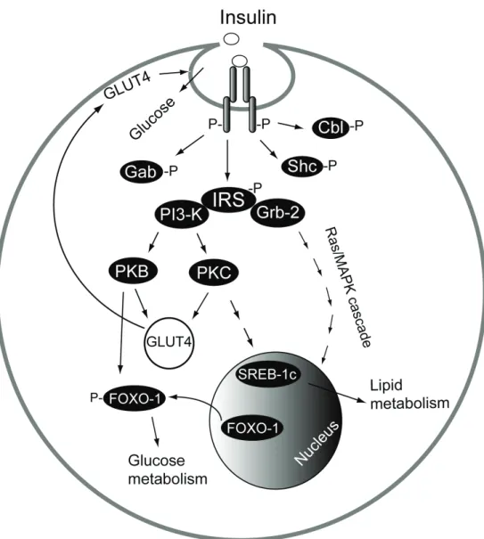
In adipocytes, the insulin receptor is localized in the plasma membrane in caveolae invaginations (Gustavsson et al. 1999). Upon binding of insulin to the two extracellular α-subunits of the insulin receptor, the two cytoplasm-exposed β-subunits that have an intrinsic tyrosine kinase activity, transphosphorylate one another thereby increasing their kinase activity (Figure 1).
Figure 1. Insulin signaling pathways. Binding of insulin to its receptor in
caveolae initiates protein phosphorylation (-P) cascades, which affect glucose and lipid metabolism.
6 Introduction
This phosphorylation allows the insulin receptor to interact with and activate several docking proteins such as the insulin receptor substrate proteins (IRSs), Shc, Cbl and Gab. Once phosphorylated, these substrates act as docking molecules for proteins that contain Src homology 2 (SH2) domains such as phosphatidylinositol 3-kinase (PI3-kinase) and Grb-2. Binding of Grb-2 to IRS stimulates the mitogenic signaling pathway of insulin through a Ras/MAP kinase cascade (Laviola et al. 2006). PI3-kinase mediates the activation of protein kinase B (PKB) and protein kinase C (PKC), which are important in the insulin-stimulated glucose uptake. They mediate the recruitment of GLUT4 to the plasma membrane and to caveolae to increase glucose uptake (Cushman et al. 1984; Gustavsson et al. 1996). PKB mediates also phosphorylation of the transcription factor FOXO-1. Phosphorylation of FOXO-1 leads to its exclusion from the nucleus, preventing it from activating the transcription of different genes in the glucose metabolic pathway (Biggs et al. 1999; Nakae et al. 2001). Insulin increases also the level of the transcription factor sterol regulatory element-binding protein (SREBP-1c) through a PI3-kinase/PKC signal cascade (Matsumoto et al. 2003). SREBP-1c in turn activates the transcription of genes involved in lipid synthesis (Shimano 2001).
The adipocyte and caveolae 7
The adipocyte and caveolae
Adipocytes are very large cells with a diameter of 20-200 μm. As one of the primary functions of these cells is to store energy, the adipocyte is able to store triacylglycerol in a single lipid droplet, which occupies more than 95% of its volume. The nucleus is pushed by the big lipid droplet against the plasma membrane and the cellular organelles are squeezed between the lipid droplet and the plasma membrane in a thin film of cytosol of less than 0.5 μm thickness.The fatty acids that are to be stored in the adipocyte can enter the cell through cave-like caveolae invaginations in the plasma membrane, where the process of converting them to triacylglycerol takes place (Öst et al. 2005). The insulin-stimulated glucose uptake also takes place in caveolae (Gustavsson et al. 1996). By electron microscopy, caveolae were morphologically identified as 50-100 nm omega-shaped invaginations in the plasma membrane of endothelial cells (Palade 1953; Yamada 1955). They are found in the plasma membrane of most cells but are particularly abundant in adipocytes, endothelial cells, fibroblasts, and smooth muscle cells (Razani et al. 2002). In rat adipocytes, about one third of the plasma membrane constitutes caveolae (Thorn et al. 2003), reflecting their importance in adipocytes.
Being gateways of entrance for fatty acids, which act as detergents, caveolae have specific lipid compositions making them detergent resistant. Caveolae are enriched in sphingolipids (glycosphingolipids and
8 The adipocyte and caveolae
sphingomyelin) and cholesterol. It was shown that caveolae have a threefold higher concentration of cholesterol and twofold higher concentration of sphingomyelin compared to the surrounding plasma membrane in rat adipocytes (Örtegren et al. 2004). Interestingly, extraction of cholesterol from caveolae by cholesterol binding agents, such as β-cyclodextrin, led to complete ablation of caveolae invaginations, reflecting the crucial role of cholesterol in maintaining the vesicular morphology of caveolae (Hailstones et al. 1998; Gustavsson et al. 1999; Parpal et al. 2001).
Caveolin
The formation and the morphology of caveolae are dependent on the protein caveolin, which in fact binds both cholesterol and sphingomyelin (Fra et al. 1995; Murata et al. 1995). Expression experiments of caveolin in cells lacking caveolae resulted in de novo formation of membrane invaginations (Fra et al. 1995). Conversely, down-regulation of endogenous caveolin led to loss of pre-existing caveolae (Galbiati et al. 1998). In addition, caveolin-1 deficient mice lack caveolae in all tissues (Drab et al. 2001).
Caveolin is a 21-24 kDa integral membrane protein and is the first and only bona fide caveolae marker protein (Rothberg et al. 1992). The caveolin gene family comprises of three gene products encoding caveolin-1 to 3. While caveolin-1 and 2 are expressed in most tissues, caveolin-3 is muscle specific. Caveolin-1 and 2 have two shorter isoforms derived from alternative translational starting sites. Caveolin-1α corresponds to the full-length protein whereas caveolin-1β starts from the internal methionine at position 32. The starting methionine in both isoforms is excised in the
The adipocyte and caveolae 9 mature proteins and the N-terminus of caveolin-1α is modified by acetylation (Paper II). The N-terminus of caveolin-1β, on the other hand, is found in both acetylated and non-acetylated forms (Paper II), indicating a regulatory role for this modification. The function of N-terminal acetylation varies with the particular protein. In line with the function of caveolin-1 described below, the N-terminal acetylation of caveolin-1 may serve as a recognition site for protein-protein interaction and regulation (Polevoda and Sherman 2000). Caveolin-2 isoforms have not been analyzed in detail.
The membrane topology of caveolin-1, with a hairpin loop configuration where both the C- and N-termini are exposed to the cytosolic face of caveolae (Figure 2), has been confirmed in various studies.
10 The adipocyte and caveolae
It was shown that antibodies recognizing the C- and the N-termini of caveolin-1 were bound only when the cells were permeabilized by detergent (Dupree et al. 1993). Furthermore, when isolated caveolae vesicles with outward-oriented cytosolic surface were treated with the protease trypsin, immunoblotting with polyclonal caveolin-1 antibodies showed disappearance of caveolin-1 immunoresponse (Paper II). Moreover, subjecting the tryptic peptides, released from the cytosolic surface of caveolae, to collision-induced fragmentation and sequencing using mass spectrometry resulted in identification of the N-terminal peptides of caveolin-1α and caveolin-1β (Paper II). Palmitoylation sites on three cysteine residues of caveolin-1 were identified at its C-terminus (Figure 2) (Dietzen et al. 1995). All these data clearly demonstrate cytosolic orientation of the C and N termini of caveolin-1 in caveolae.
The N-terminal domain of caveolin-1 is suggested to be required for its particular membrane-spanning topology (Figure 2) (Monier et al. 1995). As a modulator of signal transduction, the N-terminal domain plays also an important role in regulating the function of caveolin-1. The N-terminal domain is exposed to various posttranslational modifications, such as phosphorylation. Phosphorylation on serine-80 is required for the regulation of the distribution of caveolin-1 between caveolae and the endoplasmic reticulum in epithelial cells (Schlegel and Lisanti 2001). The insulin receptor was suggested to phosphorylate caveolin-1 on tyrosine-14 but the biological significance of this phosphorylation was not shown. It might, however, serve to recruit SH2-domain containing proteins to caveolae (Kimura et al. 2002) . In vivo phosphorylation on serine-37 in caveolin-1α and the corresponding serine-6 of caveolin-1β in caveolae of human adipocytes were recently identified by mass spectrometry (Paper II).
The adipocyte and caveolae 11 The functional significance of these phosphorylations also remains to be examined.
Taken together, caveolin-1 seems to be a tightly regulated protein, especially the N-terminal domain where truncation, variable acetylations, fatty acylations, and phosphorylations have been reported. These modifications are in line with a critical involvement of the N-terminal domain of caveolin-1 in the regulation of caveolae structure and function.
Functions of caveolin-1
The large variety of functions associated with caveolin-1 implicate caveolae in different cellular processes. In addition to its importance as a structural protein for the formation and stability of caveolae morphology, it is clear that caveolin-1 serves as a signal transduction modulator. The tightly regulated N-terminal domain of caveolin-1 contains a scaffolding domain that allows caveolin-1 to bind and concentrate certain signal transduction proteins in caveolae (Figure 2) (Williams and Lisanti 2004). Binding of caveolin-1 to different signaling proteins modulates their activities. While binding of the insulin receptor to caveolin-1 through its scaffolding domain increases the kinase activity of the insulin receptor (Yamamoto et al. 1998), it was shown that caveolin-1 interaction with signaling proteins such as, G-proteins, Src-like kinases, and protein kinase C, inhibits their activity in caveolae (Li et al. 1995; Lisanti et al. 1995; Li et al. 1996; Couet et al. 1997; Oka et al. 1997; Smart et al. 1999). Caveolin-1 also interacts with and inactivates many signaling proteins involved in survival and proliferation, such as platelet-derived growth factor receptor and phosphatidylinositol kinase (Yamamoto et al. 1999; Zundel et al. 2000).
12 The adipocyte and caveolae
As already noted caveolin also binds to cholesterol (Murata et al. 1995) and may thus mediate the transport of newly synthesized cholesterol from endoplasmic reticulum to the plasma membrane via caveolae (Smart et al. 1996). In line with the ability of caveolin-1 to bind cholesterol, it has recently been shown that a specific subclass of caveolae is enriched with the cholesterol-rich lipoprotein receptor class B scavenger receptor-1, which mediates cholesteryl ester uptake from high-density lipoprotein, suggesting caveolae to serve a role in cholesterol metabolism (Örtegren et al. 2006). It has also been suggested that caveolin-1 binds fatty acids with high affinity and therefore might be involved in lipid trafficking between membrane compartments (Trigatti et al. 1999). Binding of fatty acids by caveolin-1 is in agreement with the recent finding implicating caveolae in adipocyte fatty acid uptake (Öst et al. 2005; Örtegren et al. 2006), although the exact function of caveolin-1 in this process has not been examined.
Characterization of caveolae 13
Characterization of caveolae
Isolation of caveolae
Identification of caveolin as a marker for caveolae (Rothberg et al. 1992) allowed the study of caveolae’s function biochemically as well as morphologically. Different methods to isolate and purify caveolae from the bulk of the plasma membrane, or more commonly from whole cell homogenates, utilizing the detergent resistance and/or the low density properties of caveolae were developed to study the protein and lipid content in these membrane compartments. Clearly, the identification of caveolar components is important to understand the function of caveolae.
The commonly used method to isolate caveolae is by extraction of cell homogenates with the detergent Triton X-100 and fractionation by sucrose density gradient ultracentrifugation, utilizing the buoyancy of these low density microdomains (Lisanti et al. 1994). Purification of caveolae from whole cell homogenates may, however, include intracellular membranes like ER and Golgi. These membranes compartments contain caveolin (Ostermeyer et al. 2001) and may therefore wholly or partially end up in an isolated caveolae fraction. To avoid contamination, caveolae were isolated by detergent extraction from purified plasma membranes followed by sucrose gradient ultracentrifugation (Darby et al. 2000). The use of detergents can, however, alter the membrane composition of caveolae, in terms of extraction of proteins and lipids from caveolae or inclusion of
14 Characterization of caveolae
plasma membrane proteins not residing in caveolae (Anderson 1998; Gustavsson et al. 1999; Örtegren et al. 2004).
Detergent free isolation methods were therefore developed using sonication, in 0.5 M sodium carbonate buffer pH 11, to detach caveolae from the plasma membrane (Song et al. 1996). Using the detergent free method (Song et al. 1996), three subclasses of caveolae were isolated by sonication of purified plasma membranes and linear sucrose gradient ultracentrifugation (Öst et al. 2005; Örtegren et al. 2006) (Paper IV). The three caveolae fractions of different densities contain caveolin-enriched vesicles and harbour different sets of proteins, implicating each caveolae subclass in specific functions (Öst et al. 2005; Örtegren et al. 2006). This is in agreement with an electron microscopy study of caveolae structure, in which two classes of caveolae were described. One class had a diameter larger than 50 nm, representing half of the total caveolae. These caveolae have access to the external milieu. The other half of caveolae, which are smaller than 50 nm, are not open to the cell exterior (Thorn et al. 2003).
However, treatment of cell membranes with high concentration of sodium carbonate causes dissociation of peripherally bound proteins, while integral proteins are not solubilized in this buffer (Fujiki et al. 1982). Thus for identification of caveolae associated proteins, the sonication method was modified (Paper I). Sonication of the plasma membranes was performed in 50 mM ammonium bicarbonate buffer, pH 8, to retain the associated proteins bound to caveolae during the isolation procedure (Paper I). Purification of caveolae obtained by different isolation methods was examined by immunoblotting with antibodies against the protein caveolin. Enrichment of caveolin in the caveolae fraction in comparison with the purified plasma membrane fraction was a measure of caveolae purification.
Characterization of caveolae 15
Protein composition of caveolae
The development of different techniques for isolation of caveolae was a necessary initial step in the characterization of the functions of caveolae in different cell types. Specific antibodies have frequently been used for localization of different proteins to caveolae by immunoblotting, immunohistochemistry or immunogold electron microscopy. Among the proteins that have been localized to caveolae by these methods are: G-proteins (Lisanti et al. 1994), membrane G-proteins such as the insulin receptor, platelet-derived growth factor receptor, GLUT4 (Scherer et al. 1994; Liu et al. 1996; Gustavsson et al. 1999), and structural proteins such as annexin II and actin (Sargiacomo et al. 1993; Chang 1994). Measurement of enzymatic activity was also used to confirm the localization in caveolae of enzymes, such as the endothelial derived nitric oxide synthase (Shaul et al. 1996). By micro-sequencing and immunoblotting, a number of caveolae proteins were identified from endothelial cells including scavenger receptors, cytoskeletal elements, an anion transporter and cytoplasmic signaling proteins (Lisanti et al. 1994).
Proteomics
The technical developments and advances made in the application of mass spectrometry for identification and sequencing of proteins, in addition to the availability of the genome sequence databases of different organisms, have provided powerful tools of proteomics. The developments of methods in gel electrophoresis in combination with the capability of the mass spectrometry instruments to perform rapid sequencing of peptides recovered from in-gel digested proteins (Shevchenko et al. 2002), allowed
16 Characterization of caveolae
a rapid and effective identification of a large number of proteins in different cellular compartments of different cell types.
There are two commonly used mass spectrometry-based methods by which protein identification can be performed (Figure 3). One is based on the separation of a protein sample in one- or two-dimensional gels followed by in-gel digestion of the stained protein bands or spots by the protease trypsin. The use of matrix-assisted laser-desorption ionization time-of-flight mass spectrometry (MALDI-TOF-MS) permits accurate mass determination of the extracted peptides from each protein band or spot. Each protein generates a specific peptide mass pattern (fingerprint), which can be matched with theoretically generated peptide mass fingerprints of different proteins in the genome databases. This peptide mass fingerprinting method for protein identification can be combined with peptide sequencing for confirmation of protein identification. For peptide sequencing, which is the other protein identification method, electrospray ionization mass spectrometry ESI-MS and collision-induced fragmentation are used. Peptides generated from gel digested proteins or from in-solution protein digests are separated according to their mass to charge ratio and individual peptides are selected for collision-induced fragmentation. The actual peptide sequence obtained from interpretation of the fragmentation spectrum is then matched against peptide sequences in the databases. Different mass spectrometry instruments allow rapid sequencing of peptides of different sizes. However, sequencing of large peptides containing more than twenty amino acids gives fragmentation spectra that are not always interpretable due to poor peptide fragmentation patterns. On the other hand, sequencing of a quadruply charged peptide
Characterization of caveolae 17
Characterization of caveolae 19
Figure 4. Fragmentation spectrum of a 44-amino acids long peptide obtained by mass spectrometry. Collision-induced fragmentation of the
quadruply charged peptide with m/z 1204.5 was performed using ESI quadrupole-TOF MS. The amino acid sequence is identified from interpretation of the fragment ions from the C-terminus (y) and the N-terminus (b). The amino acid sequence above the spectrum is numbered according to the fragment ions that were identified from the spectrum. Fragment ions with higher charge than 1+ are marked with the corresponding superscript numbers. Indicated is also the corresponding triply charged peptide ion (M-H3+).
containing 44 amino acids was successfully performed using ESI quadrupole-TOF-MS. The fragmentation spectrum obtained from subjecting a quadruply charged peptide ion with m/z 1204.5 was of very high quality that allowed de novo sequencing of the peptide (Figure 4), which originates from the protein polymerase I and transcript release factor (PTRF) (Paper I). Notably, the collision-induced fragmentation of the quadruply charged parent peptide ion occurred in parallel with fragmentation of its corresponding triply charged peptide ion (indicated in figure 4 by M-H3+). The triply charged ion was apparently generated by loss of one proton from the parent ion. This gave rise to more comprehensive fragmentation pattern of the peptide. It can be noted that this peptide is the second biggest peptide sequenced by mass spectrometry. The biggest peptide was 45 amino acids long, which also originated from PTRF protein and was sequenced in the same way (Paper I).
Post-translational modifications
In humans, there are about 30,000 genes coding for more than one million proteins, due to alternative splicing, translation, and different post-translational modifications. Post-post-translational modifications include among others phosphorylation, glycosylation, N-terminal acetylation, acylation,
20 Characterization of caveolae
and proteolytic modifications. Phosphorylation is the most common post-translational modification, which modulates the function of proteins and affects various cellular processes such as metabolism, cell growth, and cell differentiation. Thus studying protein phosphorylation is crucial for understanding the regulation of these biological processes.
Different methods are used to detect protein phosphorylation. Radioactive labeling by incorporation of 32P or 33P into cell cultures is a common technique for detection of phosphoproteins. Such an approach is restricted to a limited range of biological materials like tissue culture samples. Moreover, radioactive 32P-labeling is inefficient due to the high amount of non-radioactive endogenous phosphates with unknown turn-over times. Different phosphorylation sites in caveolin-1 and caveolin-2 have been identified by mutation of individual serine sites to glutamate (mimic permanent phosphorylation) or to alanine (mimic permanent dephosphorylation) followed by comparison of the amount of 32P incorporated by the mutant and wild-type of caveolin (Schlegel et al. 2001; Sowa et al. 2003). Immunoblotting with phosphospecific antibodies is frequently used to detect protein phosphorylations. Insulin-induced tyrosine and threonine phosphorylation of protein in caveolae and at the plasma membrane were studied by phorphospecific antibodies (Corley Mastick et al. 1995; Smith et al. 1998) (Paper III). However, the sensitivity of the antibodies towards the phosphorylated amino acid residue is often dependent on the sequence context and site-specific antibodies are often not available. In addition, the use of phosphospecific antibodies provides information only about the relative level of protein phosphorylation under different conditions. For a thorough understanding of the role of protein phosphorylation, identification of the phosphorylation sites in the corresponding protein and the extent of their phosphorylation state need to be addressed.
Characterization of caveolae 21 Proteomics and mass spectrometry allow identification of phosphorylated proteins without the use of radioactive labeling or antibodies. Phosphorylation increases the mass of the intact protein by 80 Da, which can be measured by MALDI-TOF or ESI mass spectrometry. Sequencing of phosphopeptides, which are obtained by proteolysis of the proteins, using tandem mass spectrometry allows identification of the exact phosphorylation sites in the phosphopeptides. However, due to the low natural abundance of phosphopeptides in complex peptide mixtures, the identification of phosphorylation sites remains challenging. Especially identification of phosphorylation on tyrosine residue, whose occurrence is very low (~0.05%) compared to phosphorylation on serine (~90%) and threonine (~10%) residues. Different approaches are thus required for purification and enrichment of phosphopeptides. Phosphospecific antibodies are used to purify phosphoproteins or phosphopepides prior to mass spectrometry analysis (Pandey et al. 2000; Rush et al. 2005). Immobilized metal affinity chromatography (IMAC), which utilizes the high affinity of the phosphate group to cations such as Zn2+, Fe3+ and Ga3+, has been used to enrich phosphopeptides from proteolytic mixtures (Andersson and Porath 1986; Posewitz and Tempst 1999). A disadvantage of this method is that peptides containing acidic amino acids also bind and co-elute with the phosphopeptides. This nonspecific binding is eliminated by methyl esterification of the carboxyl groups of the side chains of these acidic amino acids (Ficarro et al. 2002). Application of mass spectrometry and IMAC thus resulted in identification of in vivo phosphorylation of twenty-six novel phosphorylation sites in plasma membrane caveolae of human adipocytes (Paper I; Paper II; Paper III).
22 Characterization of caveolae
Vectorial proteomics of caveolae
Several mass spectrometry-based studies of caveolae proteins in different cell types have been reported. The protein composition of caveolae isolated from rat adipocytes by immunoisolation using caveolin antibodies was attempted by peptide mass fingerprinting of in-gel-digested proteins separated on one-dimensional gel. In addition to caveolins, two major proteins were identified in this caveolae fraction; the scavenger lipoprotein receptor CD36 and the semicarbazide-sensitive amine oxidase (Souto et al. 2003). Proteomic studies of caveolae using one- or two-dimensional gel electrophoresis and mass spectrometry have made contributions to the growing list of caveolae proteins (von Haller et al. 2001; Blonder et al. 2004; Sprenger et al. 2004; Vinten et al. 2005; McMahon et al. 2006). Proteomics of membrane lipid rafts, which are detergent resistant and have similar composition of lipids as caveolae, revealed their enrichment in signaling proteins (Foster et al. 2003). In this study two cell populations were grown with normal leucine or with deuterium-substituted leucine. One of the populations was then treated with a cholesterol extracting agent and equal protein amounts from treated and untreated cells were combined prior to isolation of rafts using detergent and sucrose gradient ultracentrifugation. The isolated raft-fraction contained no rafts from the cells treated with cholesterol extracting agent because their raft structures were disrupted. The peptides generated from trypsin treatment of the isolated rafts were then analyzed by liquid chromatography/mass spectrometry (LC/MS). Sequencing of peptides, which have quantitative changes in deuterium labeling, identified cholesterol-dependent and thus raft-localized proteins (Foster et al. 2003).
A new mass spectrometry-based approach, termed vectorial proteomics, allowed differential identification and characterization of
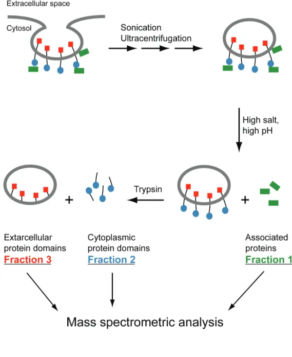
Characterization of caveolae 23 proteins and their domains exposed to the opposite sides of the caveolae membrane, providing information about the orientation and topology of identified proteins. This approach utilizes the intrinsic morphology of caveolae that produces vesicles with an outward-oriented cytosolic surface. Integral membrane proteins, as well as peripheral membrane proteins associated to caveolae, were thus identified (Vener and Strålfors 2005) (Paper I, Paper II, Paper III). In this approach caveolae vesicles were isolated from purified plasma membranes of primary human adipocytes by ultracentrifugation after sonication in mild ionic strength and pH (50 mM ammonium bicarbonate buffer at pH 8) to retain the non-integral membrane associated caveolae proteins (Paper I). The associated proteins were then stripped from caveolae vesicles by 0.5 M carbonate (pH 11) and separated from the membranes by centrifugation (Fraction 1, Figure 5). Treatment of the remaining membrane proteins by trypsin allowed digestion of the cytoplasmic surface exposed domains of integral caveolae membrane proteins (Fraction 2, Figure 5). The generated peptides were separated from the remaining membranes (Fraction 3, Figure 5) by centrifugation. Mass spectrometry analysis of the proteins in these three fractions allowed identification of proteins from the outside (Fraction 1 and 2, Figure 5) and the inside (Fraction 3, Figure 5) of caveolae membranes by applying gel electrophoresis and in-gel digestion.
The two most abundant caveolae membrane proteins resisted trypsin digestion (Fraction 3, Figure 5). In-gel digestion and MS of peptides generated from these two proteins resulted in their identification as CD36 and copper-containing amine oxidase (Paper I). The membrane topology of these transmembrane proteins, possessing short cytosol-exposed N-terminal sequence and the rest of the proteins exposed to the extracellular space (Tandon et al. 1989; Morris et al. 1997), explains their resistance to trypsin
24 Characterization of caveolae
Figure 5. Vectorial proteomics of caveolae.
as they are protected inside the sealed caveolae vesicles. Both proteins had also been identified in caveolae from rat adipocytes (Souto et al. 2003).
The caveolae marker protein caveolin was identified by sequencing of the peptides released by trypsin from the cytoplasmic face of caveolae (Fraction 2, Figure 5). Mass spectrometry analysis of caveolin-1 peptides
Characterization of caveolae 25 revealed that in both isoforms the starting methionine was processed by excision and, moreover, the N-terminus of the mature caveolin-1α was acetylated, while caveolin-1β was partially acetylated (Paper II). It should be noted that, previous analysis of the N-terminus of caveolin-1 isoforms by N-terminal chemical sequencing had failed due to the natural blocking of the N-termini of the proteins (Scherer et al. 1995). Enrichment of the phosphorylated peptides from the cytosol-exposed peptides by immobilized metal ion affinity chromatography (Paper II) and sequencing by mass spectrometry revealed phosphorylation of caveolin-1α on serine-37 and on the equivalent serine-6 of caveolin-1β (Paper II). The phosphorylation of caveolin-1β was found in both the acetylated and non-acetylated forms. These two in vivo phosphorylations of caveolin-1 are at the consensus site for phosphorylation by protein kinase C, which has been found associated to caveolae (Tang et al. 1994; Oka et al. 1997; Mineo et al. 1998).
Proteins released by high salt and pH treatment of caveolae membranes were identified by mass spectrometry. This allowed identification of the peripheral proteins associated with the cytoplasmic face of caveolae (Fraction 1, Figure 5) (Paper I). These proteins have implicated caveolae of human adipocytes in novel functions, as discussed below. In addition to proteins involved in membrane-cytoskeleton interactions such as β-actin, α-parvin, myosin-IC and annexin II, V and VI, a number of signaling proteins were identified, including Eps15-homology domain-containing protein, EHD2; the protein kinase Cα-binding protein, SDPR; the protein kinase Cδ-binding protein, SRBC; R-Ras, and polymerase I and transcript release factor, PTRF. Different posttranslational modifications of the transcription regulator protein PTRF at the cytoplasmic face of caveolae were identified by mass spectrometry. This major caveolae-associated protein was acetylated on its N-terminus,
26 Characterization of caveolae
phosphorylated on one threonine and nine serine residues, and in vivo fragmented into five identified fragments (Paper I; Paper III).
The vectorial proteomics methodology enabled us, in addition to identification of intrinsic and extrinsic proteins in caveolae and characterization of their topology and localization, also to analyze in vivo posttranslational modifications of the proteins.
Roles of caveolae 27
Roles of caveolae in adipocytes
Vesicular transport
Based on their vesicular morphology and analogy with clathrin-coated pits, caveolae have been considered to function as organelles facilitating vesicular trafficking of different molecules taken up from the external milieu in various cell types. Caveolae were also hypothesized to affect vesicular transport of proteins through endothelial cells in a process referred to as transcytosis (Palade 1953). Several membrane bound proteins may be taken up through internalization of caveolae (endocytosis), including the folate receptor and alkaline phosphatase (Anderson 1992; Parton et al. 1994). Transport of molecules to the cytosol by receptor-mediated internalization (potocytosis) was also suggested (Anderson et al. 1992). Indeed, identification and localization in caveolae of proteins known to be involved in vesicular transport, such as SNF, SNAP, and VAMP2, supported caveolae’s participation in this process (Schnitzer et al. 1995). Moreover, the endocytosis-involved protein EHD2 was associated with human adipocyte caveolae (Paper I). However, the involvement of caveolae in vesicular transport has not been studied in adipocytes.
28 Roles of caveolae
Insulin signaling at caveolae
In adipocytes, the caveolar localization of the insulin receptor put caveolae at the centre of insulin signaling (Gustavsson et al. 1999). Being largely confined to caveolae, the insulin receptor is dependent on caveolae for mediating the insulin signals. It was shown that when caveolae invaginations were disrupted by cholesterol extraction, insulin-receptor signaling to enhanced glucose uptake was lost (Parpal et al. 2001; Karlsson et al. 2004). The insulin receptor in the disrupted caveolae bound insulin and was activated, but downstream signaling for the mobilization of glucose transporters was disrupted (Parpal et al. 2001). Previous studies had shown that insulin induced translocation of the glucose transporter protein GLUT4 from an intracellular pool to the plasma membrane where glucose uptake took place (Cushman et al. 1984). It was shown that the insulin-regulated glucose transporter protein GLUT4 translocated from intracellular stores to the plasma membrane and to caveolae in response to insulin (Gustavsson et al. 1996). The enhanced glucose uptake by the cell occurred in parallel with the accumulation of GLUT4 in caveolae but not in the whole plasma membrane (Gustavsson et al. 1996). About 85 % of GLUT4 in the plasma membrane are residing in caveolae of adipocytes (Karlsson et al. 2001).
Recently, the caveolar localization of the cyclic AMP degrading enzyme phosphodiesterase 3B (PDE3B), which is activated by insulin, implicated caveolae in the insulin signaling to inhibit lipolysis in adipocytes (Nilsson et al. 2006).
Roles of caveolae 29
Regulated proteolysis at caveolae
The identification of many different caveolar proteins has suggested the involvement of caveolae in a number of novel functions. Mass spectrometry analysis of caveolae-associated proteins in human adipocytes revealed the presence of a quite large proportion of PEST-domain-containing proteins (Paper I). The PEST-domain is an amino acid sequence enriched in proline, glutamic acid, serine and threonine. This domain can function as a signal for rapid proteolytic degradation in order to regulate the level of signaling proteins in the cell (Rechsteiner and Rogers 1996). Among the identified proteins in this study five proteins contained PEST domains: nuclear transcription regulator protein PTRF, protein kinase Cδ-binding protein SRBC, protein kinase Cα-Cδ-binding protein SDPR, EHD2, and R-Ras (Paper I).
The nuclear transcriptional control factor PTRF is the major caveolae-associated protein in adipocytes (Vinten et al. 2005) (Paper I). It was shown that PTRF resides at the cytoplasmic face of caveolae in intact as well as in five different truncated forms identified by in-gel digestion and mass spectrometry (Paper I). Moreover, two endogenous cleavage sites were identified by the sequencing of several PTRF peptides, one of which was within a PEST-domain. Both cleavage sites were closely flanked by multiply phosphorylated sequences, indicating a possible phosphorylation-dependent cleavage of PTRF on caveolae (Paper I; Paper III). Multiply phosphorylated amino acid sequences were also identified in the PEST- domain-containing proteins SDPR and SRBC. The identified phosphorylation sites in SRBC were located in its PEST-domain (Paper III). However, SRBC fragments were not identified, perhaps due to the low amount of this protein in caveolae and its staining sensitivity.
30 Roles of caveolae
Thus, caveolae of human adipocytes might be involved in the control of PTRF and other PEST-domain-containing proteins and their cellular concentration by phosphorylation and proteolysis.
Caveolae in insulin-control of gene transcription
Examination of the protein sequence of EHD2 revealed the presence of a nuclear localization signal, which might be involved in relocation of the protein from caveolae to the nucleus (Paper I). Similarly, PTRF contained two nuclear localization signals (Paper I). Indeed, incubation of human adipocytes with insulin caused translocation of PTRF from caveolae to the nucleus (Paper IV). Notably, immunological analysis demonstrated that, under basal conditions, a major part of PTRF protein is confined to caveolae, while a surprisingly low amount of PTRF is residing in the nucleus of primary human adipocytes (Paper I). In addition, it should be noted that the perinuclear region of adipocytes is more caveolae dense (Thorn et al. 2003), which could facilitate a rapid translocation of different proteins from the plasma membrane to the nucleus in these big cells.
It was shown that PTRF regulates transcription in vitro by inducing dissociation of the transcription complex and by facilitating the reinitiation of RNA polymerase I (Jansa et al. 1998; Jansa and Grummt 1999; Jansa et al. 2001). It was also suggested that PTRF might be involved in the regulation of RNA polymerase II reaction (Hasegawa et al. 2000).
As an anabolic hormone, insulin effects transcription of a great number of genes and proteins, and a number of insulin transcription mediators have been identified, such as SRBP1-c, the CREBP-binding protein CBP and the forkhead transcription factor FOXO (Desvergne et al. 2006). The insulin-induced translocation of the transcription regulator
Roles of caveolae 31 PTRF to the nucleus in fat cells indicates possible involvement of PTRF in mediating insulin’s metabolic effects by acting as a regulator of gene transcription. However, the genes that are affected by PTRF and the mechanism by which it exerts control has not yet been examined. Moreover, PTRF augments ribosomal RNA synthesis by increasing the activity of RNA polymerase I (Jansa et al. 2001), which may be a novel way by which insulin exerts its effect on stimulation of protein synthesis in general.
It has been shown by autoradiographic detection of incorporated 32 P-orthophosphate in NIH 3T3 cells that PTRF is a phosphorylated protein. Moreover, analysis of cellular PTRF by two-dimensional gel electrophoresis revealed the presence of two PTRF populations with pronounced charge heterogeneity, thereby suggesting phosphorylation of PTRF at multiple sites (Jansa et al. 2001). In addition, cellular PTRF was separated in two fractions, which differed in the capability of PTRF to dissociate the transcription complex and to augment transcription. No correlation of PTRF activity with its phosphorylation pattern was found, but it was suggested that specific phosphorylation may inhibit PTRF activity based on the fact that both recombinant and cellular PTRF are transcriptionally active (Jansa et al. 1998; Jansa et al. 2001). In line with these findings, ten phosphorylation sites were identified in the caveolae-associated PTRF from human adipocytes by mass spectrometry (Paper I; Paper III). It remains to examine to what extent insulin affects the phosphorylation level and phosphorylation pattern of PTRF.
Taken together, the identification and localization of PTRF in caveolae of human adipocytes, the regulation of its amount at the surface of caveolae by proteolysis and the insulin-induced translocation of PTRF to the nucleus indicate a novel role of caveolae in the regulation of gene transcription and protein synthesis.
32 Roles of caveolae
Caveolae as centres of lipid metabolism
Functioning as an energy reserve organ, the adipose tissue plays a central role to provide other tissues with energy. When energy is needed the stored triacylglycerol is broken down and fatty acids are released from the adipose tissue to provide different tissues with energy. The storage and the breakdown of triacylglycerol in adipocytes are two processes that are controlled by different hormones and other factors such as exercise and nutritional status (Large and Arner 1998). Catecholamines activate the breakdown of triacylglycerol (lipolysis), while insulin promotes its storage. Dysfunction in fatty acid processing is associated with insulin resistance and type II diabetes.
Uptake and release of fatty acids by adipocytes have been hypothesized to occur through caveolae. The ability of caveolin-1 to bind fatty acids with high affinity supports this theory (Trigatti et al. 1999). It was shown that the number of caveolae was increased during differentiation of fibroblasts to adipocytes, which accumulate huge amount of lipids compared to fibroblasts, indicating a role of caveolae in fatty acid uptake and metabolism (Fan et al. 1983). In addition, caveolin-1 knockout mice lacking caveolae have high plasma triacylglycerol concentration and are not able to store triacylglycerol in adipocytes (Razani et al. 2002). Furthermore, the disruption of caveolae in 3T3-L1 adipocytes by cholesterol extraction reduces their uptake of long-chain fatty acids (Pohl et al. 2004). The caveolar localization of the fatty acid translocase CD36, moreover, implicates caveolae in fatty acid uptake (Lisanti et al. 1994; Pohl et al. 2005) (Paper I). The high abundance of caveolae particularly in adipocytes, in addition to their detergent resistance properties, is likely providing a high number of ports for influx and efflux of the detergent-like fatty acids at plasma membrane of adipocytes. Indeed, a recent study
Roles of caveolae 33 showed that in insulin-stimulated primary rat adipocytes fatty acids were not only taken up but also converted to triacylglycerol in a specific subclass of caveolae (Öst et al. 2005). This caveolae subclass, which is one of three subclasses obtained after density gradient ultracentrifugation, specifically contained proteins involved in fatty acid metabolism such as fatty acid transport protein-1, fatty acid transport protein-4, fatty acyl-CoA synthetase, which catalyzesthe first step in the synthesis of triacylglycerol, and the lipolysis regulators perilipin and hormone-sensitive lipase (Öst et al. 2005; Örtegren et al. 2006). Moreover, it was found that the newly synthesized triacylglycerol may expand the caveolae membrane to become big enough to make physical contact with the large central lipid droplet (Öst et al. 2005). Thus, caveolae in adipocytes serve not only as ports for influx and efflux of fatty acids but also as sites of triacylglycerol synthesis.
Regulation of lipid metabolism
The major actors in triacylglycerol metabolism are hormone-sensitive lipase (HSL), perilipin, and desnutrin. Desnutrin was recently identified as an adipocyte triacylglycerol lipase that catalyzes the breakdown of triacylglycerol to diacylglycerol (Villena et al. 2004; Zimmermann et al. 2004), while HSL catalyzes the hydrolysis of diacylglycerol to monoacylglycerol. Much is known about the regulation of HSL and perilipin, but very little is known regarding the regulation of desnutrin.
Perilipin coats and protects the central lipid droplet from hydrolysis by the action of HSL. Two forms of perilipin are expressed in adipocytes, perilipin A and perilipin B, derived from differentially spliced mRNA of a single gene. They have identical N-terminal regions but the C-terminus is unique for each form (Figure 6) (Greenberg et al. 1993). Stimulation of lipolysis by catecholamines, such as noradrenaline, activates adenylate
34 Roles of caveolae
cyclase, which increases the production of cyclic AMP. The increased levels of cyclic AMP in turn activates the cyclic AMP-dependent protein kinase, PKA (Honnor et al. 1985), which phosphorylates both HSL and perilipin on serine residues (Strålfors and Belfrage 1983; Egan et al. 1990; Greenberg et al. 1991). When phosphorylated, HSL is activated and translocated from the cytosol to the surface of the central lipid droplet at which the phosphorylated perilipin A, due to conformational changes, no longer blocks access of HSL (Egan et al. 1992; Clifford et al. 1997; Clifford et al. 2000). Insulin, on the other hand, promotes lipid storage by acting as an anti-lipolytic agent inhibiting lipolysis by dephophorylation of both HSL and perilipin A (Strålfors and Honnor 1989; Mooney and Bordwell 1991). This is, at least partly a result of insulin activation of phosphodiesterase, PDE3B, which hydrolyses cyclic AMP, which in turn leads to decreased PKA activity (Eriksson H, 1995).
Perilipin A functions not only as barrier against lipolysis under basal conditions but also as activator of lipolysis. It was shown that upon stimulation of lipolysis in perilipin knockout mice, HSL did not translocate to the central lipid droplet unless perilipin A was introduced (Sztalryd et al. 2003). Furthermore, phosphorylation of different PKA sites in perilipin was required to enhance the activity of HSL (Zhang et al. 2003). The phosphorylation of perilipin A by PKA in turn can be mediated by caveolin-1, which may recruit the catalytic subunit of PKA to perilipin by binding to both perilipin and PKA (Cohen et al. 2003).
Interestingly, the dual function of perilipin in inhibiting and activating lipolysis of triacylglycerol in the central lipid droplet has been suggested to be isoform-segregated in caveolae at the plasma membrane where triacylglycerol is synthesized. It was shown that in primary human adipocytes only perilipin B was completely depleted from the plasma membrane after stimulation of lipolysis, indicating its role in protecting the
Roles of caveolae 35
Figure 6. Domain structure of perilipin A and perilipin B. The acidic domain
(gray boxes), comprising the amino acids 292-319, is a part of the lipid targeting domain, 233-364 (Garcia et al. 2003). Identified phosphorylation on three threonine residues within the acidic domain is indicated by PPP in perilipin B. Black boxes correspond to unique amino acid sequences in the two isoforms of perilipin.
newly synthesized triacylglycerol droplets at the plasma membrane (Paper III). The serine-phosphorylated perilipin A, on the other hand, remained at the plasma membrane after lipolysis activation (Paper III), likely demonstrating the function of this isoform as an activator of lipolysis through HSL. HSL was recently found to localize to the triacylglycerol-synthesizing caveolae (Örtegren et al. 2006) (Paper IV). Insulin, on the other hand, recruited both perilipin A and B to the plasma membrane to protect the newly formed triacylglycerol droplets (Paper III). However, it should be noted that perilipin B isoform is highly enriched at the plasma membrane compared to perilipin A, which comprises more than 90 % of total cellular perilipin in human adipocytes (Paper III). The perilipin translocated to the plasma membrane in response to insulin was
36 Roles of caveolae
phosphorylated on a cluster of threonine residues located within an acidic domain in the lipid-targeting domain of perilipin (Figure 6). Introduction of negative charges through phosphorylation on threonine residues increases the acidic character of this domain. It was therefore suggested that the recruitment of perilipin to the triacylglycerol droplets at the plasma membrane is threonine phosphorylation-dependent (Paper III).
The presence of HSL at the plasma membrane in the triacylglycerol-synthesizing caveolae is under insulin control. While perilipin was recruited to the plasma membrane (Paper III), HSL was rapidly translocated from the plasma membrane to the cytosol in response to insulin (Paper IV). Thus, while the insulin-promoted synthesis of triacylglycerol in caveolae is ongoing the amount of HSL is decreased at the plasma membrane to reduce the hydrolysis process. The recruitment of perilipin to the plasma membrane in response to insulin occurs probably to assure complete protection of the newly synthesized triacylglycerol droplets by acting as a barrier between the remaining HSL at the plasma membrane and the triacylglycerol droplets, as has been described for the central lipid droplet.
In parallel with the insulin-induced translocation of HSL the transcription regulator PTRF was also translocated from the plasma membrane to the cytosol in response to insulin. Moreover, a major part of HSL was bound to PTRF in the cytosol of primary human adipocytes, whether incubated with or without insulin (Paper IV). On the other hand, stimulation of lipolysis by isoproterenol did not change the amount of HSL or PTRF associated with the plasma membrane, which indicated that insulin acts against a default presence of HSL and PTRF at the plasma membrane (Paper IV). Furthermore, at the plasma membrane, PTRF was associated specifically to the triacylglycerol-synthesizing caveolae subclass containing HSL. This, in addition to the association of a very large amount
Roles of caveolae 37 of cellular PTRF with caveolae (Paper I), demonstrates that PTRF serves an extranuclear role in primary human adipocytes. PTRF is thus suggested to be a novel actor in the hormonal control of triacylglycerol metabolism in caveolae.
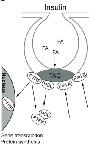
Figure 7. Hormonal control of triacylglycerol metabolism. A). Isoproterenol
stimulates lipolysis of triacylglycerol (TAG) to fatty acids (FA) in caveolae and perilipin B (Peri B) is translocated from caveolae. B) The synthesis of TAG from FA occurs in caveolae. Insulin recruits of perilipin A (Peri A) and perilipin B (Peri B) to caveolae to protect the newly synthesized TAG from hydrolysis by HSL, which is translocated from caveolae together with PTRF, in response to insulin, and form a complex in the cytosol. Indicated is also the translocation of PTRF to the nucleus.
38 Roles of caveolae
It remains to determine whether PTRF, as perilipin, has regulatory effects on the activity of HSL or simply acts as a hormonally controlled docking protein for HSL and other proteins in the primary adipocyte. Another protein found to interact with HSL in 3T3-L1 adipocytes is lipotransin. Lipotransin was found in the fat cake and other membrane fractions, but the extent of its expression in primary adipocytes is not clear (Syu and Saltiel 1999). Neither insulin nor isoproterenol influenced its cellular location, clearly distinguishing any possible role of lipotransin from that of PTRF in lipolytic control (Syu and Saltiel 1999). Figure 7 summarizes the hormonal controlled translocation of perilipin, HSL, and PTRF.
Conclusion 39
Conclusion
Right after their discovery, caveolae were seen as means of transport for different molecules into the cell by endocytosis. Now caveolae are known as signaling platforms due to the identification of many signaling proteins and different receptors in these plasma membrane compartments. Proteomic research, especially a new mass spectrometry-based methodology introduced in this thesis work allowed identification of a number of new proteins and their posttranslational modifications in caveolae. Of particular interest is the transcription regulator PTRF, which is the major caveolae-associated protein. Multiple phosphorylations and specific proteolytic modifications of PTRF at caveolae demonstrate complex and tight hormonal regulation of this protein at this location.Insulin induces translocation of PTRF from caveolae at the plasma membrane to the nucleus. As the function of PTRF was previously shown to regulate the activities of both RNA polymerase I and polymerase II, this has implicated caveolae in mediating insulin effects on protein synthesis and gene transcription. Which genes are affected by PTRF remain to be examined.
PTRF also has an extranuclear function in the hormonal regulation of triacylglycerol metabolism in caveolae. Together with perilipin and HSL, PTRF is localized in the triacylglycerol-synthesizing caveolae subclass. While perilipin is recruited to caveolae in the plasma membrane in response to insulin, both HSL and PTRF are translocated away from
40 Conclusion
caveolae to the cytosol as a complex. The role of PTRF in triacylglycerol metabolism needs to be investigated in more detail. Stimulation of lipolysis, on the other hand, induces complete depletion of perilipin B from the plasma membrane, suggesting that perilipin B’s function is to protect newly synthesized triacylglycerol form being hydrolyzed by HSL. Taken together, this suggests a novel function of caveolae as metabolic platforms.
Acknowledgements 41
Acknowledgements
The research underlying this thesis was performed with financial support from Lions Foundation, the Östergötland County Council, the Novo Nordisk Foundation, the Swedish Diabetes Association, the Graduate Research School in Genomics and Bioinformatics (Forskarskolan för Genomik och Bioinformatik), and theSwedish Research Council.I would like to thank everyone, inside and outside Linköping University who has supported and encouraged me during my thesis work, especially:
Peter Strålfors and Alexander Vener, my supervisors: for your invaluable guidance through the world of caveolae and mass spectrometry, and for encouragement, and support over the years. Thank you for always having the time to discuss results, for your never-ending enthusiasm, and for teaching me all about science.
Fredrik Nyström, Johanna Gustavsson, and Mats Söderström for stimulating discussions during our group meetings.
Unn Örtegren: “the most fabulous scientist” for always having solutions for all problems and for our interesting discussions about politic and religions, despite your somewhat weak geography knowledge ;)
Elisabeth Hallin: “tuffa bruden” who has enriched my Swedish. Thank you for teaching me all about DNA-.... and skruffset-innan. I am going to miss you, hu va ligen…
Karin Stenkula: for spreading enthusiasm and positive energy in the lab and for encouraging me even after moving to Örebro over the phone.
Anita Öst: for stimulating scientific discussions and for serious talks about life in general. Unfortunately, we didn’t get the chance to collaborate.
42 Acknowledgements
Lilian Sauma: “duktiga tjejen” for always being so sweet and for making me feel close to home whenever we spoke Arabic.
Anna Danielsson: the IRS-1 queen, I’m glad that we get the chance to do our PhD studies in the same group after being classmates during the Master studies.
Siri Fagerholm: for patiently listening and comforting when I complained about bad blots. Keep up the good work. I wish you all the best.
Niclas Franck: the “cocky” guy, for fun time in the other lab. Good luck with microarrays and “R”.
Julia Vainonen: for collaboration and clever ideas and for nice time in the lab during your two years in Sweden and of course for all unforgettable Russian parties. I really miss your company.
Margareta Karlsson: “my big sister” for encouragement and enjoyable time, especially during our extreme exercising period after your dissertation. I’m happy that you didn’t move from Linköping.
Hans Thorn: for caveolae discussions, lots of car talks and for being an excellent skiing teacher with the ability to cope with “attack hugs”!
Gennady Bronnikov: for sharing your knowledge about adipocytes and lipolysis. I learned a lot from you during my last year.
Bijar Ghafouri: my dearest friend and ”the future prime minister” (you got my vote), for all invaluable proteomics discussions, for cocking the tastiest rice I have ever eaten. Thank you for making me feel as a member of your family.
Johan Paulsson: my best friend, for all fun and gossiping over the years and for causing “grisbrist” in the country. I have to admit that you won; you were one week faster than me... It’s all about appearance!
Jana Sponarova: for good songs, wonderful music, and tasty dumplings. Friend in need is a friend indeed.
Sebastian Schultz: for always being friendly and for making the best Tiramisu ever.
Acknowledgements 43 Maria Turkina: for friendship and for sharing my shopping interest. I will never forget our crazy shopping days in London. You are the best of the best of the best…
Marina Koulikovska: for nice dinner parties and unforgettable days in Riga during Midsummer. I am very happy that I got the chance to know you. Maria Hansson: for always being so helpful. Thank you for all the days we spent together in the noisy and cold mass spec room during our first year as PhD students.
Helen Karlsson: for being very friendly and a good listener. Thank you for taking care of me during my first downhill skiing and uphill…
Gunilla Westermark, Veronika Patcha, Martin Tinnerfeldt, Jesper Svartz, Pia Druid, Kristina Loinder, Sofia Nyström, and Tobias Strid for creating a nice atmosphere at the work.
Pia Westöö Olsson, Alexandra Rutz, Helena Petterson, and Ramona Wlihelmsson, for great time in Gotland. I wish you all the best in achieving your goals.
My family: there are no words in any language that can express my love and appreciation to you; my father, my mother, Ayman, Amira, Haisam, and hayati Hani ……for everything