Linköping University Medical Dissertations No 1253
Wnt signaling and
metaphyseal bone healing
Fredrik Agholme
Department of Clinical and Experimental Medicine
Linköping University, Linköping,
© Fredrik Agholme 2011 Cover picture by Per Aspenberg
All previously published papers were reproduced with permission from the publishers. Printed by LiU-tryck, Linköping 2011
During the course of the research underlying this thesis, Fredrik Agholme was enrolled in Forum Scientum, a multidisciplinary graduate school at Linköping University Sweden.
ISBN: 978-91-7393-103-8 ISSN: 0345-0082
”Science does not know its debt to imagination.”
Supervisor
Professor Per Aspenberg
Division of Orthopedics, Department of Clinical and Experimental Medicine, Linköping University
Co-supervisors
Professor Pentti Tengvall
Department of Biomaterials, Institute of Surgical Sciences, University of Gothenburg Dr Anna Fahlgren
Division of Orthopedics, Department of Clinical and Experimental Medicine, Linköping University
ABSTRACT
This thesis relates to some new aspects on the regulation of bone healing. In the last few years, Wnt signaling has been shown to play a central role in bone biology. As well as being involved in bone maintenance and repair, Wnt signaling has been presented as one of the ke y pathways through which bone responds to mechanical load. Two secreted extracellular inhibitors of Wnt signaling, sclerostin and dickkopf-1 are potent negative regulators of bone formation.
Using a rat f racture model we investi gated how m etaphyseal bone healing is influenced by changes in Wnt signaling.
Antibodies were used to suppress levels of sclerostin and dickkopf-1, and thereby increase Wnt signaling. Primarily, we investigated if those antibody treatments lead to improved bone healing. Also, we investigated if the response was coupled to the loading conditions of the bone.
Our findings suggest that suppression of either sclerostin or dickkopf-1 leads to increased bone formation and improved bone healing. Apart from just having an effect on healing, the treatment also improved bone formation in other parts of the skeleton. Depending on the loading conditions, the effects were different. Dickkopf-1 appeared to have a stronger ef fect on bone volu me density in unloaded bone, implying a role mainly in mechano-transduction, while sclerostin had similar effect in both loaded and unloaded bone. To confirm these findings, we studied how the expression of several Wnt-related genes changed due to trauma and unloading in metaphyseal bone. We found that trauma led to upregulation of most of the genes with the largest ef fect seen in the u nloaded bone. In untraumatized bone, there was mainly an ef fect on the sclerostin gene.
In conclusion, antibodies against sclerostin and dickkopf-1 appear to be able to improve metaphyseal bone healing. There appear to be s ome differences in how the effect of the two antib odies manifests itself, especially if the loading conditions of the bone are altered. These findings suggest a potential for clinical use to shorten fracture healing time.
POPULÄRVETENSKAPLIG
SAMMANFATTNING
I denna avhandling har jag studerat hur frakturläkning kan förbättras genom läkemedelsbehandling. Benbrott kan drabba de flesta människor någon gång i livet. Frakturer kan upps tå på alla ben i kroppen, men är vanliga st i höf t, handled eller ryggkotor. Ofta drabbas äldre, de flesta är kvinnor, vilka har nedsatt bentäthet på grund av benskörhet.
Benvävnad förekommer i två former, spongiöst ben och kortikalt ben. Det spongiösa benet är uppbyggt av trabekler och återfinns nära lederna och i ryggkotor. Kortikalt ben är tätar e och bildar det rörformade skaftet av benet . De vanliga h öft-, rygg- och handledsfrakturerna inträffar i spongiöst ben.
När ben läker aktiveras en signalväg i benceller som kallas Wnt-signalering. Jag har studerat hur benlä kning påverkas av två olika antikroppsbaserade läkemedel mot proteinerna sclerostin och dickkopf-1 (dkk1), två hämmare av Wnt-signalering.
I tre delarbet en har v i studerat hur benläkning i spongiöst ben påverkas av behandling med dessa antikroppar. I det sista delarbetet har jag ti ttat närmare på vad som händer med Wnt-signaleringen vid läkning och belastning. I samtliga delarbeten har vi använt oss av en modell för frakturläkning i råtta.
I den f örsta studien såg vi att sclerostin-antikroppar leder till en betydande ökning av benbildningen och ger en förbättrad benläkning. I den andra studien behandlade vi djur med dkk1-antikroppar. Även i detta f all såg vi en ökning av benbildningen och en positiv effekt på benläkning. I denna andra studie undersökte vi också hur f rakturläkning påverkas av kombinationen dkk1-antikropp och ändrad mekanisk belastning. Anledningen till detta är att Wnt-signalering också är inblandad i benets svar på mekanisk belastning. Vi kunde då se att antikropp en verkade ha en större positiv ef fekt om belastningen minskades. Detta tyder på a tt dkk1-antikropps behandlingen kan vara fördelaktig i situationer med låg belastning
För att und ersöka om samma effekt kunde ses med sclerostin-antikroppen utförde vi sedan en tredje studie. I denna tredje studie såg vi ingen belastning beroende effekt. Sammantaget visar dessa försök att trots att sclerostin och dkk1 verkar i samma system så verkar de ha olika funktioner i benet vid läkning.
För att bekräfta våra tidigare resultat gjorde vi en fjärde studie där vi undersökte hur de gener som påverkar Wnt-signalering uttrycks vid läkning och då belastningen av benet ändras. Denna studie visade att på gen-nivå ändras både mängden av sclerostin och dkk1 under läkning och belastning.
Under tiden ett benbrott läker har patienten smärta och svårt att röra sig. Ett läkemedel som förkortar läkningstiden skulle därför bidra till öka d livskvalitet för patienten. De studier vi har gjort visar på att benläkning kan förbättras med läkemedel som påverkar Wnt-signalering. Dessa studier ligger nu till grund för framtida studier på människa.
LIST OF PAPERS
The thesis is based upon the following papers which are referred to by their
roman numerals (I-IV).
I
Agholme F, Li X, Isaksson H, Ke HZ, Aspenberg P.
Sclerostin antibody treatment enhances metaphyseal bone healing in rats.
J
Bone Miner Res. 2010;25(11):2412-8.
II
Agholme F, Isaksson H, Kuhstoss S, Aspenberg P.
The effects of dickkopf-1 antibody on metaphyseal bone and implant fixation
under different loading conditions.
Bone. 2011;48(5):988-96.
III
Agholme F, Isaksson H, Li X, Ke HZ, Aspenberg P.
Sclerostin antibody and mechanical loading appear to independently influence
metaphyseal bone in rats.
Acta Orthop. 2011;In Press.
IV
Agholme F, Sandberg O, Aspenberg, P.
Wnt gene expression during metaphyseal bone healing under different load
conditions.
Manuscript.
Other work by the author related to bone healing in rat models.
Aspenberg P, Agholme F, Magnusson P, Fahlgren A. Targeting RANKL for
reduction of bone loss around unstable implants: OPG-Fc compared to
alendronate in a model for mechanically induced loosening.
Bone. 2011
Feb;48(2):225-30
Andersson T, Agholme F, Aspenberg P, Tengvall P. Surface immobilized
zoledronate improves screw fixation in rat bone: a new method for the coating
of metal implants.
J Mater Sci Mater Med. 2010;21(11):3029-37.
Agholme F, Aspenberg P. Experimental results of combining bisphosphonates
with allograft in a rat model.
J Bone Joint Surg Br. 2009;91(5):670-5.
ACKNOWLEDGEMENTS
Arbetet med denna avhandling kunde inte ha utförts utan hjälp. Tack till alla er so m gett råd, uppmuntrat, kritiserat, granskat och hjälpt till under årens lopp.
Min handledare Per Aspenberg, vars kreativa sinne, entusiasm och bildning inspirerat mig från första dagen. Budskapet att forskning ska vara ro lig såväl som intressant tar jag med mig i livet.
Pentti Tengvall, som var en eldsjäl under min grundutbildning och fick in mig på forskningsspåret.
Anna Fahlgren, för din hjälp, stöd och råd.
Mina arbetskamrater på ortopedi so m gör at t det roligt att gå till jobbet varje dag : Therese Andersson, Pernilla Eliasson, Bibbi Mårdh och Olof Sandberg.
Mats Christenson, som tillverkat alla implantat. Min medförfattare Hanna Isaksson.
I want to thank my co-authors at Amgen: Xiaodong Li and David Ke. I want to thank my co-author at Lilly: Stuart Kuhstoss.
Cissi, Nettan, Anders, Linda, Andreas, Jonas, Lena, Jan och Danne på djuravdelningen. Er kunskap och arbete har varit mycket värdefull.
Mina företrädare Björn Skoglund och Karin Wermelin som lade grunden ti ll de modeller som jag arbetat med.
Stefan Klintström och alla i Forum Scientum. Tack för trevligt sällskap på konferenser och resor.
Min kära familj för ert stöd och uppmuntran då ni från håll bevittnat arbetet med denna avhandling.
Min Lotta, för att du förstår, jag hade inte klarat detta utan dig ”…det här är slutet på en resa och början på en ny… med dig”.
TABLE OF CONTENTS
Introduction ... 1
Wnt signaling ... 2 Overview of Wnt-pathway ... 2 Role in bone ... 4 Sclerostin ... 4 Dickkopf-1 ... 5Role in bone healing ... 5
Endochondral bone formation ... 5
Intramembranous bone formation ... 6
Mediation of mechanical information ... 6
Interaction with other signaling systems ... 7
Role in development ... 7
Altered Wnt signaling as a therapeutic tool ... 7
Antibodies ... 7
Sclerostin ... 8
Dickkopf-1 ... 8
Blocking of secreted frizzled-related protein 1 ... 8
Targeting GSK3β ... 8
Wnt-ligand ... 9
Aims and Hypotheses ... 11
Paper I ... 11
Paper II ... 11
Paper III ... 11
Paper IV ... 11
Methods and methodological considerations ... 13
Animals ... 13
Limb unloading ... 13
Animal models of bone healing ... 14
Pull-out testing ... 14
Bone chambers ... 15
Micro computed tomography (µCT) ... 16
Gene expression ... 17
Statistics ... 18
Results and Discussion ... 19
Effects of suppressing sclerostin or dkk1 ... 19
Screw fixation in uninjured bone ... 19
Bone formation around screws ... 19
Bone formation in cancellous bone and its architecture ... 20
Effects of suppressing sclerostin or dkk1 in unloaded bone ... 20
Screw fixation in healing bone ... 20
Bone formation around screws ... 21
Bone formation in cancellous bone and its architecture ... 21
Effect of suppressing dkk1 on formation of new cancellous bone ... 21
Wnt related mRNA expression during healing and unloading ... 22
Effect of trauma ... 22
Wnt ligands... 22
Wnt inhibitors ... 22
Effect of unloading ... 23
Differences between sclerostin and dkk1 suppression ... 23
Effect in untraumatized bone ... 23
Effect on cancellous bone architecture ... 23
Effect during unloading ... 24
Summary ... 24
General discussion ... 25
Changes in sclerostin expression ... 25
Changes in dickkopf-1 expression ... 26
Conclusion ... 26
INTRODUCTION
1
INTRODUCTION
Most people will experience a f racture during their lif etime. One hallmark of fracture repair is the ability of bone tissue to heal without formation of scar tissue. In the callus, damaged bone is removed and replaced with new bone.
Morphologically, bone is classified as cortical or cancellous. These types of bone tissue are confined to specific regions. T he ends of the bone s, the metaphyses, are made up of cancellous bone consisting of trabeculae. These trabeculae form a porous lattice work with the space in between filled with marrow. In contrast, cortical bone is denser and makes up the shaft, the diaphyses, and outer shell of the bones with a marrow cavity in the center (figure 1).
Fracture healing occurs in four different overlapping stages: inflammation, soft callus formation, hard callus formation, and remodeling. However, depending on if the fracture occurs in can cellous or corti cal bone there appear s to be a diff erence in healing mechanisms [1]. This is because during healing new bone is formed by either endochondral or intramembranous bone formation [2]. In most cases a combination of these two pro cesses occur simultaneously, but in so me cases one or the other is primarily used.
In intramembranous bone formation, mesenchymal stem cells are recruited and start making new osteoid on site [2]. In contrast, during endochondral bone formation, a soft cartilaginous callus is first formed. The cartilage is then mineralized and replaced by bone. Endochondral bone formation has similarities to bone formation at the growth plates and during fetal development. It primarily occurs in mechanically unstable regions while intramembranous bone formation occurs in stable regions [2-5]. Hence, fractures of the diaphysis tend t o heal using e ndochondral bone formation while fractures of the metaphysis tend to heal with intramembranous bone formation.
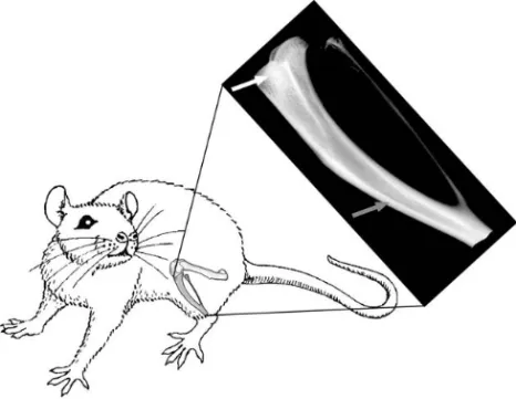
Figure 1: Tibia and femur of the rat hind limb with a tibial x-ray image. Cancellous bone in the
metaphysis (white arrow) has distinctly different morphology compared to the cortical bone of the diaphysis (grey arrow).
INTRODUCTION
2
Most fractures in humans occur in osteoporotic cancellous bone in metaphyseal regions. Screws and other fixation devices such as intra medullary nails may be inserted to stabilize the fractures. The response to the t rauma of inserting a screw in cancellous bone appears similar to metaphyseal fracture repair, even thought the extent and geometry of the injury is different. Similarly, a fracture healing response is involved also in the early fixation of total joint replacements.
For the elde rly patient, the long h ealing time of fractures, combined with the immobility that often accompanies it, lead to high morbidity. Therefore, treatments that increase bone formation and shorten the healing time would benefit the patient. Also, increased bone formation at ear ly stages of the incorporation of joint replacements might provide a better long-term outcome [6].
Two drugs that were initially developed to counter osteoporosis, bisphosphonates and parathyroid hormone, have been evaluated for their potential to improve fracture healing. Bisphosphonates successfully improve implant fixation and bone hea ling in many experiments in animals, and also in patients [7]. However, bisphosphonates do not exert a true anabolic effect, but act mainly on osteoclasts to limit resorption. Parathyroid hormone (PTH) has been shown to have a positive, although weak, effect on fracture healing [8-9]. However, one drawback of using PTH is that it can only be given at low doses, with a limited effect.
To find an anabolic drug for bone one has to examine the systems that regulate osteoblasts. One of the crucial path ways involves bone morphogenetic proteins (BMPs). Over 17 BMPs have been characterized and many of these have been shown to be intimately involved in both bone formation and fracture healing. BMPs bind to and activate serine /threonine kinase cell s urface receptors, leading to activation of intracellular signaling via smads, which leads to change s in tran scription. The exact function of all BMPs is not yet fully understood, but BMP2 appear to be crucial for fracture healing [10]. BMPs 2 and 7 have been used clinically in orthopedic surgery [11].
However, BMPs have to be given locally and find most use in spinal fusion and other situations as a replacement for bone autografts. Complications with the use of BMPs in other parts o f the skeleton have m ostly been related to increased resorption and heterotopic ossification. These complications, combined with the high costs of recombinant BMP proteins, have restricted the use of BMPs in fracture healing.
Owing to these limitations of PTH and BMPs, other pathways have been investigated. Recently, due to naturally occurring mutations in humans, Wnt signaling was discovered to play an important role in bone tissue.
Wnt signaling
Overview of Wnt-pathway
Wnt-ligands (Wnts) are a group of secreted proteins i mportant for embryonic development, cell proliferation and differentiation [12]. Currently, 19 Wnt homologs have been described in humans, with a wide range of functions and expression patterns. Wnt signaling is ubiquitous in the body, and the complete signaling pathway
INTRODUCTION
3
relevant for bone and bone healing. This is because Wnt signaling is crucial for osteoblast differentiation and proliferation [15].
Wnts interact wit h receptors activating several sets of intracellular signaling pathways. These pathways can be subdivided into canonical Wnt signaling and non-canonical Wnt signaling. Canonical Wnt signaling is the most studied and appears to be the most important in bone. The defining feature of canonical Wnt signaling is the stabilization of β-catenin in the cytosol, which enables it to translocate to the nucleus and regulate gene expression. In contrast, the non-canonical pathways function without β-catenin.
The protein interactions during canonical Wnt signaling are shown in figure 2. Initially, Wnts bind to a specific receptor belonging to the frizzled group (there are at least 10 of them). A r eceptor complex is t hen formed with low-density lipoprotein receptor–related proteins (LRP) 4, 5 and 6 . This event pr events an intra -cellular protein complex, consisting of axin, glycogen synthase kinase 3β (GSK3β) and adenomatous polyposis coli (APC), to tag catenin for degradation. As a result , β-catenin accumulates in the cytosol and can translocate into the nucleus, where it interacts with members of the T-cell factor/lymphoid enhancer factor (Tcf/Lef) class of DNA bindi ng proteins and transcriptions factors. This β-catenin accumulation is needed for early osteoblast proliferation and differentiation [15]. Specifically, β-catenin accumulation favors mesenchymal stem cell c ommitment for an osteogenic fate, away from the adipogenic or chondrogenic lineage [16]. However, there are some indications that osteoclast dif ferentiation is af fected since W nt signaling stimulates osteoprotegerin (OPG) release from osteoblasts [15].
In the adult body it appears that part of the system is mostly active in bone tissue. Various Wnt genes, such as Wnt1, Wnt4, Wnt5a, Wnt9a/14 and Wnt7b, are expressed in either osteoblast precursors or adjacent tissues during embryonic development, and Wnt3a and Wnt10b are expressed in bone marrow [17-19]. However, Wnt signaling increases cell proliferation and it is also implicated in cancer [13].
To regulate the ef fect of these ligands, there are several f eedback loops, consisting of both secreted and internal inhibitors (figure 2). These secreted inhibitors include sclerostin, the dickkopfs (dkks), secreted frizzled-related proteins ( sFrps), frizzled related protein (Frzb), Wnt-1-induced secreted protein (WISE), Wnt-inhibitory factor 1 and 2 (Wif1,2) and Chibby [15]. Of these inhibitors, sclerostin and dickkopf-1 have been most investigated.
INTRODUCTION
4
Figure 2: Overview of Canonical Wnt signaling [13-14, 20].. In the active state, Wnt-ligands (Wnt) form a complex
with receptors LRP5 or LRP6 and Frizzleds (Fz). Disheveled (Dsh) is then able to bind to Fz and inhibits the formation of a complex consisting of Axin, APC and GSK3β. This protects β-catenin from proteasomal degradation, so that β-catenin accumulates in the cytosol and can translocate to the nucleus. In the nucleus it interacts with TCF/LEF family of transcriptions factors leading to gene transcription. In the inactive state, inhibitors prevent the formation of the Wnt-Fz-LRP5/6 complex. This leads to β-catenin degradation.
Role in bone
Wnt signaling is needed in normal bone maintenance [21]. It appears that active Wnt signaling is needed to commit mesenchymal stem cells to an osteogenic lineage and for further proliferation and differentiation into osteoblast [15]. Mutations in parts of this system lead to either excessive bone growth or bone resorption. Loss of function mutations in the receptor LRP5 causes the Osteoporosis Pseudoglioma Syndrome [22]. This syndrome is characterized by a very low bone mass, with bone that easily fractures. However, gain of function mutations in LRP5 instead lead to high bone mass [23-25]. Some common polymorphisms of the Wnt-receptor LRP5 gene are also associated with osteoporotic fractures, and polymorphisms of LRP6 are associated with a low bo ne mineral density, explaining some of the heredit ary influence on osteoporosis [26].
Sclerostin
Sclerostin is a secreted glycoprotein and product of the sost gene. Sclerostin binds to the LRP5/6 receptor, thus an tagonizing Wnt signaling and increasing β-catenin degradation [27]. Sclerostin is primarily expressed in bone, exclusively by osteocytes, and appears to be a potent negative regulator of bone formation [28-29]. However, sclerostin is also expressed in hypertrophic chondrocytes in the growth plate and by cementocytes in teeth [29-30]. Hence, sclerostin seem to be expres sed in
INTRODUCTION
5
terminally differentiated cells in mineralized tissues [30]. For some reason, the expression of sclerostin appear higher in the cranium than elsewhere in the s keleton [31-33].
Sclerosteosis and Van Buchems disease, two diseases that causes very high bone mass, due to loss of function mutations in sclerostin [28, 34-36]. This high bone mass is caused by excessive bone f ormation leading to a thickening of the cortice s and denser cancellous bone [28, 32-33, 37]. Characteristic signs of sclerosteosis are syndactyly, enlargement of the jaw and f acial bones, increased intracranial pressure (sometimes leading to sudden death at young age), and entrapment of cranial nerves leading to facial palsy and loss of hearing and smell [32, 38]. It seems these negative effects of sclerostin depletion are largest dur ing youth and then bec ome reduced with age [39]. Van Buchems disease is not as severe and skeletal malformations like syndactyly are absent [28].
This high bone mass phenotype can be replicated in sost knockout mice [37]. However, in mice none of the problem s with nerve opening s or s yndactyly occur. Furthermore, patients heterozygous for mutations in sost have lower levels of circulating sclerostin [39] and a high bone mass [40] without having the problems associated with sclerosteosis. Sclerostin might also be involved in osteoporosis, systemic levels increase in post menopausal women and correlate with decreases in bone density [41-42].
Dickkopf-1
Dickkopf-1 (dkk1) is a secreted glycoprotein, belonging to a family of cysteine-rich proteins with four different forms (dkk1, dkk2, dkk3 and dkk4). Of these, dkk1 is most studied. While the exact mode of action is unclear, it appears that dkk1 competes with Wnt-ligands in binding to LRP6 [43] thus antagonizing Wnt signaling [44-45]. However, dkk1 can also bind to LRP4 and LRP5 as well as LRP6 [44, 46-48]. Dkk1 is expressed at low levels outside bone tissue [49-50]. In bone it is mostly expressed in osteocytes [51] although not as exclusively as sclerostin. Active Wnt signaling has a generally proliferative effect on cells and dkk1 is often downregulated in cancers [49-50].
A decrease in dkk1 gene expression leads to an increase in both bone mass and strength, and deficiency of one or both of the genes f or the inhibitor dkk1 leads to increased bone mass and stronger bone [52-55].
Dkk1 is suspected to mediate many pathological conditions in bone in which excessive bone resorption or f ormation occurs [56]. Serum levels of dkk1 correlate with the extent of lesions in multiple myeloma [57]. Studies of rheumatoid arthritis demonstrated that dkk1 plays an important role in re modeling of the joint [58]. It appears that dkk1 is also involved in the f usion of sacroiliac joints in ank ylosing spondylitis [59-60].
Role in bone healing
Endochondral bone formation
Precise regulation of β-catenin via Wnt sig naling is needed for a proper bone healing response [61]. During bone healing, a large amount of genes, both outside and
INTRODUCTION
6
within the Wnt signaling system, change in expression [62-63]. Wnt-ligands, receptors and inhibitors express a tem poral pattern, and it is not clear ho w this s ystem is regulated. For instance, sclerostin expression is low during the first week of fracture repair, and then gradually increases over time [64].
During healing with endochondral bone f ormation, β-catenin appears to have different effects at di fferent stages of bone repair. Early in the process, when differentiation occurs from pluripotent mesenchymal cells, it controls the ratio between osteoblasts and chondrocytes. At this stage, either too much or too little β-catenin can be detri mental to bone healing [61]. Later on, β-β-catenin promotes the differentiation of osteoblasts and enhances their production of bone matrix. At thi s stage, low levels of β-catenin impair healing, whereas high levels of β-catenin improve healing [61]. A micro array study on closed transverse fractures in rats showed an increased β-catenin expression at day 3 after fracture, peaking at 10 days and leveling out at 21 days, but r emaining up-regulated thereafter [63]. These data wer e later confirmed using RT-PCR, which showed that β-catenin was upregulated during bone regeneration [65].
Another micro array study in mice showed that the ligands Wnt4, 5a, 5b, 10b, as well as dkk1 and sclerostin were all upregulated in a si milar pattern, with a peak around day 10, but there was quite low expression during the first days [66]. In the same study, PTH treatment increased the expression of Wnt-ligands indicating considerable interaction between PTH and Wnt signaling [66]. This is important, since intermittent PTH injections are the only systemic treatment that has shown an ability to improve fracture healing clinically [8-9].
Intramembranous bone formation
Wnt signaling is important also for intramembranous bone formation [67]. The healing of drill holes in the mouse proximal tibia is dependent on Wnt mediated β-catenin signaling [67]. Gene expression during intramembranous bone f ormation caused by marrow ablation has been s tudied in rats at several ti me points af ter the injury. Genes involved in Wnt signaling were upregulated with a peak af ter 10 days and then leveling out [68]. Ligands were upregulated (Wnt2, 5a and 5b ) as well as receptors (Lrp4 and 6 and several of the fz receptors) and inhibitors (sFrps, Wise and frzb) [68]. Apart from the mentioned, few studies have been conducted on intramembranous bone healing. One reason could be the difficulty to study gene expression in cancellous bone in rodents due to the s mall size. Usually whole femurs are analyzed, either by cutting out the entir e calluses [62-64] or the diaph ysis [68]. With this approach it inevitable to include cortical bone in the measurement.
Mediation of mechanical information
Osteocyte function is required for the bone´s ability to respond to mechanical load [69]. Wnt signaling appears to be involved in how the osteo cytes regulate bone formation [15]. Especially sclerostin appears vital for the bone to be able to respond to mechanical loading [70]. In response to loading, sclerostin levels decrease, and conversely, they increase in response to unloading [71]. These changes in sclerostin levels then i nfluence Wnt signaling, leading to either in creased bone formation or increased bone resorption. There are also some indications that dkk1 is involved,
INTRODUCTION
7
although to a lesser extent than sclerostin, in how bone responds to mechanical loading [70].
Interaction with other signaling systems
Far from being an is olated system, canonical Wnt signaling is prone to interact with other anabolic pathways in bone, like PTH [72] and BMP [73-74]. Findings indicate that the f ull actions of PTH require intact Wnt signaling [72] and that PTH can activate the Wnt pathway despite over-expression of dkk1. Additionally, sost is a target of PTH [31] suggesting that some of the effect of PTH might be mediated via Wnt signaling.
Other studies suggest that β-catenin is likely to be involved in BMP signaling. BMP2 enhances Wnt signaling via upregulation of several Wnt-ligands and receptors [61]. Furthermore, active Wnt signaling reduces GSK3-mediated degradation of Smad1 [75]. If Wnt signaling is inactivated, the abilit y of BMP2 to induce ectopic bone formation is reduced [61].
Role in development
In all s tudied animals, Wnt signaling has been s hown to be crucial f or embryogenesis and development [76]. It is required for embryonic developmental processes that regulate the establis hment of head-to-tail axis, limb polarity, neural crest differentiation, kidney morphogenesis and sex determination. Correct formation and function of the nervous s ystem, brain, heart and kidneys also depend on precise regulation of this system [14].
Dkk1 is vital for head and li mb development [45, 77]. In contrast, when the inhibitors sclerostin or sFrp1 are absent in vivo, the only tissue that appears affected is the skeleton [37, 78]. The receptors LRP5 and 6 have received much attention, since they seem to be partially redundant but still vital to development. In vivo experiments show LRP6-deficency to be fatal, while animals with a def iciency in LRP4 and 5 are viable [13]. These developmental studies have been helpful to elucidate the important role of Wnt signaling in cell proliferation and differentiation, not just in bone but also in other tissues.
Altered Wnt signaling as a therapeutic tool
Human mutations point out so me of the pote ntial advantages of targeting Wnt signaling for therapy, as f ew organs except bone see m to be affected. Patients homozygous for these mutations have severe skeletal problems besides having a several-fold increase in bone mass. However, patients that are heter ozygote for these mutations have higher bone mass without having similar health problems [39-40]. This suggests that limited short term use of drugs modulating the pathway could be safe and suitable for clinical practice.
Antibodies
Interfering with Wnt signaling could present a therapeutic potential to increase bone mass. The secret ed inhibitors sclerostin and dkk1 are particul arly interesting, as they can be targeted by therapeutic antibodies.
INTRODUCTION
8
Sclerostin
By using an antib ody to suppress sclerostin levels, it was poss ible to increase bone mass in animal models of postmenopausal osteoporosis [79] and disuse-induced bone loss [80]. The same treatment also improved bone mass in aged male rats [81] and in non-human primates [82]. Clinical trials with focus on osteoporosis are currently being conducted. Results from these trials are n ot yet available, but preliminary results indicate a good tolerance to the treatment [83]. As will be reported later in this thes is, metaphyseal bone healing can be improved by sclerostin-antibody treatment. Other data also support an improvement of fracture healing in rodents and nonhuman primates [84]. Clinical trials are being conducted using sclerostin antibodies to improve fracture healing b oth for metaphyseal (hip) f ractures (NCT01081678) or diaphyseal (tibia) fractures (NCT00907296). However results from these studies are not yet available.
Dickkopf-1
Similar to sclerostin, dkk1 can be blocked using an antibody. This suppression of dkk1 levels inf luences fracture repair in m ice [85] and rats [51], and also leads to improved bone mass in mice [51]. As will be reported later in this thesis, metaphyseal bone healing can also be improved with dkk1-antibody treatment. Clinical trials are underway for a dkk1-antibody to be used to counter o steoporosis (NCT01144377). However, data from this trial is not yet available.
Blocking of secreted frizzled-related protein 1
Knock-out mice lacking sFrp1 have higher bone mass and heal diaphyseal fractures faster. The faster healing, with no loss of bone qualit y, is due to increased intramembranous bone formation [78]. An increased osteoclast activity was also noted, but this could be attributed to the need for more woven bone to be remodeled.
Targeting GSK3β
The intracellular pathway of canonical Wnt signaling appears to open up targets for drug treat ment with small molecules, like conventional drugs. Lithium is an inhibitor of GSK3β, which is a protein in the intracellular cascade that participates in β-catenin degradation. Inhibition of GSK3β using Lithium has a positive effect on bone formation if administered after the tr auma is i nitiated [61]. Lithium promotes bone formation and increases bone mass in mice [86]. There are also some indications that it may have these effects also in the cli nic [87-88]. However, unpublished data from our lab does not show any significant effects of Lithium treatment on bone healing. Therefore, if there is an effect it is not of the magnitude experienced with sclerostin or dkk1 antibodies.
Other drugs can also modify GSK3β function and promote the differentiation of osteogenic progenitors [89]. However, GSK3β appears to have many important functions apart from regulation of bone, and is also regulated b y other systems than Wnt signaling. Therefore, drugs that target β-catenin directly may have too many adverse effects.
INTRODUCTION
9
Wnt-ligand
Another way of influencing Wnt signaling is to supply more Wnt-ligand. Locally delivered Wnt3a accelerated bone healing in mice [90]. Furthermore, administration of Wnt3a increased peri-implant bone formation around stainless-steel implants in mice [91]. However, Wnt-ligands have a general proliferative effect and cannot be given systemically. Taken together, increased Wnt signaling seems to enhance bone formation, and this can be achieved either by administration of more Wnt-ligand or by inhibition of an inhibitor.
AIMS & HYPOTHESES
11
AIMS AND HYPOTHESES
Paper I
The overall aim of the study was to investigate if sclerostin suppression leads to improved fracture healing in cancellous bone.
Specifically, the following hypotheses were tested:
• Sclerostin-antibody treatment will improve the healing response in
cancellous bone, leading to increased bo ne formation and i mproved screw fixation.
• Sclerostin-antibody will promote bone formation in general, also when
no trauma is present.
Paper II
The overall aim of the study was to investigate if dkk1 suppression leads to improved fracture healing in cancellous bone.
Specifically, this study tested three hypotheses:
• Dkk1-antibody treatment will improve the healing response to tra uma,
leading to increased bone formation and improved screw fixation.
• Dkk1-antibody will promote bone formation in regions not subjected to
trauma. This will lead to increased bone density in untraumatized bone and increased bone formation in a titanium chamber.
• The response to dkk1-antibody treatment depends on mechanical
loading.
Paper III
The overall aim of the study was to investigate how the combination of sclerostin antibody treatment and unloading influenced fracture healing in cancellous bone.
Specifically, the following hypotheses were tested:
• The response to sclerostin-antibody treatment depends on mechanical
loading.
• In unloaded bone, the effect of antibody treatment is larger than in loaded
bone.
Paper IV
The overall aim of the study was to describe how the expression of several genes involved in Wnt signaling is changed due to trauma and unloading.
This study tested the following hypotheses:
• The mRNA expression of Wnt rela ted genes changes when bone is
AIMS AND HYPOTHESIS
12
• An upregulation Wnt-inhibitors will be seen during unloading while
METHODOLOGICAL CONSIDERATIONS
13
METHODS AND METHODOLOGICAL
CONSIDERATIONS
Animals
In our effort to have a ho mogenous population, we used male Sprague-Dawley rats of approximate 10 weeks of age weighing 340-370 g for all studies. We used male rats since they are bigger than female rats, making surgical procedures easier. The cellular mechanism behind bone formation and fracture repair is similar between rodents and humans, making it an appropriate model for evaluating the effects of different treatments on bone healing [92]. However, healing models in rat have se veral limitations. The major ones are bone size and animal growth. Rat bone lacks the osteonal organization present in human bone and the collagen composition is different. Also, the bone itself is much smaller. In cortical bone this would facilitate healing and shorten healing times since less tissue has to be formed. Also, the extent of the injury is smaller (compared to cellular size) than w hat usually occur in h umans. However, this might not be an issue in cancellous bone since the siz e of individual trabeculae could be similar in humans and rodents.
Rats grow throughout life and an implant inserted into the cancellous bone of the metaphysis will end up in the cortical bone of the diaphysis. This makes it difficult to study metaphyseal healing for long periods of time. Additionally, rats have f aster metabolism enabling healing to occur at shorter time spans than in humans but also making it more difficult to transf er dosing regimens f rom rodents to hu mans. However, considering these limitations, rats and mice are still much used in musculoskeletal research. A benefit of using rats instead of mice is the larger size of the bone and the pre valence of trabecular bone. We mostly chose to use the proximal tibia as surgery site. This region holds ample trabecular bone, is easily accessible and causes the animal minimal discomfort during the trials.
Limb unloading
In study II-IV, injections of Botullinum toxin A (Botox) into the calf and thigh muscles were used unload the tib ia. This enables us to measure the effects of load protection on th e healing response as we ll as on cancellous bo ne in general. Being minimally invasive, Botox causes clearly visible muscle atrophy and significant bone loss [93-94]. In our studies a halving of the muscle weight in the lower l imb was detected after 17 days. There are several other ways to unload bone in rodents, such as tail suspension, casting, and nerve severing. One of the drawbacks of Botox treatment is that it cann ot be re versed. As lo ng as Botox has an effect, the bone is unlo aded. Also, even though much of the muscles in the lower limb are paralyzed, the animals do not drag their leg around. Instead they keep it close to the stomach. Even though the rats (in theory) should slowly regain function of the limb, we have not seen this in our experiments that have lasted up to 4 weeks.
METHODOLOGICAL CONSIDERATIONS
14
Animal models of bone healing
There are several rodent models for studying different aspects of bone healing. A common model is causing a closed transverse fracture in the diaphysis of the tibia or femur [95]. These types of fractures can then be stabilized using either internal [96] or external fixation [97]. Compared to an osteotomy model, there is minimal periosteal damage or loss of tissue. However, these f ractures lead t o a bone healing resp onse containing both e ndochondral and intramembranous bone formation [2]. To specifically study intramembranous bone formation other models are needed. Cortical defects, in which a hol e is drilled in t he tibia, or femur, heals with a fracture healing response equivalent to a stabilized fracture [67]. This model holds several benefits: It reduces the extent of the skeletal injury and soft tissue damage. This leads to a smaller and better defined callus area, which is beneficial for histological evaluation and also causes less animal morbidity [67].
In our model, we exte nd a cortical def ect in the m etaphyseal tibia to the cancellous bone underneath. This injury and the subsequent insertion of an orthopedic i mplant initiates bone repair in a similar fashion as a stable fracture. For this reason, newly formed bone reaches t he surface of the im plant instead of filling an em pty defect. Hence, the bone repair process is translated into implant fixation. However, there are some differences between the models. In a closed fracture model the primary source of osteogenic cells is the periosteum [2] while in our model these cells have to come from elsewhere, possibly the endosteum and the marrow. Also, in our model we do not get any interference from tissue surrounding the bone; this might be important since stem cells from muscles are suggested to participate in fracture repair [98-99]. We chose to look at healing after 14 to 28 days in our screw experiments. This time period is after the early inflammatory phase and new bo ne have started to f orm. In our model we have earlier seen that inmature bone appear around the screw 7 days after insertion and an increase in healing can be measured up to 8 weeks af ter insertion [100]. However, we are convinced that a positive effect in earl y stages of fracture repair is also beneficial in the long-term.
Pull-out testing
Insertion of an orthopedic implant into bone constitutes an injury, initiating bone repair. Pull-out strength and other measures of fixation correlate with the amount or structure of bone tha t surrounds the fixation device [101]. Peak force in this model may be considered to reflect the end result of cancellous bone healing. This force is proportional to the a mount of bone f ormed. We use stainless steel screws f or these measurements.
The screws are in serted into the cancellous bone in the proximal tibial metaphysis (figure 3). Stainless steel does not osseointegrate as well as titanium, but that is not a co ncern in these comparative studies when we investigate the effect of a systemic agent. However, our choice of screw site in the tibial metaphysis, distal to the growth plate, means that the screw will migrate as the animal grows. Hence, it is hard to conduct long time experiments in this model.
Pull-out testing can also be used t o test th e mechanical strength of uninjured bone. Screws are inserted into the bone po st mortem at the sa me location as screws that are allowed to osseointegrate. The screws are then direct ly pulled-out and reveal
METHODOLOGICAL CONSIDERATIONS
15
information on how strong the bone is. However, due to the inhomogeneous structure of bone, these measurements are sensitive to differences in implant placement.
We generally used 10 animals in each treatment group and considered pull-out force to be t he primary variable. This group size allows for detection of a 40% difference in gr oup means with a statistical power of 80%, assuming a standard deviation of 30% of the mean. However, in paper II and III 12 a nimals were sometimes used to improve precision.
Figure 3: Rat tibia with inserted pull-out screw and loose screw for comparison. Lower (x-ray) image visualizes
the cancellous bone into which the screw is inserted. (Scale bar 3 mm)
Bone chambers
To study new bone formation, we used the bone chamber [102]. The chamber is empty at insertion, and its bone content after a certain time will reflect the capability for new bone f ormation by membranous ossification. Using histology and micro computed tomography (µCT) the bone ing rowth distance and b one volume fraction can be measured. In paper II we saw a discrepancy between the methods for measurement for ingrowth distance. There was a lower ingrowth distance measured by histology compared to µCT. However, we consider the p recision of µCT measurements to be l arger since measurements are performed on an intact specimen. The chamber never fills completely with bone indicating that signals from the originating bone are needed for bone formation. Since the bone in the chamber is well vascularized, a drug administered systemically will also st imulate bone f ormation inside the chamber. However, since there are no other tissues to compete with inside the chamber, the proliferative response might be proportionally larger.
METHODOLOGICAL CONSIDERATIONS
16
Micro computed tomography (µCT)
The density and architecture of bone is related to its m echanical strength. As a complement to pull-out data, we used µCT to measure the a mount of bone f ormed around the screw due to healing and assess the impact on trabecular architecture. We considered the bo ne volume fraction to be our pri mary variable with th e other measurements having mostly a descriptive function. To avoid metal artifacts, we used polymethylmethacrylate (PMMA) screws. These screws have th e same dimensions of the internal portion as the steel screws and were inserted at the sa me site (figure 4). Although the PMMA screws offer a different surface for the bone to adhere to, b one formation appears histologically similar to that seen around stainless steel [103]. Furthermore, differences in surf ace properties should only affect the bone i n direct contact with the i mplant. In our µCT measurements a region consis ting of bone up to 500 µm away from the screws were evaluated.
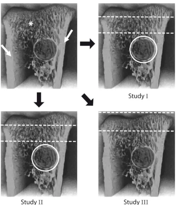
For study I and II w e chose two regions of interest (figure 5). One was a cylindrical portion surrounding the screw. In this region we evaluated the bone volume fraction and bone mineral density. Trabecular parameters were not evaluated since the bone did not h ave a s uitable morphology. The other region consisted of trabecular bone in the metaphysis. This region was chosen not to include the primary spongiosa or the newly formed bone around the screw. In study III, µCT was performed on bone subjected to pull-out testing. The pull-out test caused some damage to the bone around the screw and required the region of interest to be moved more proximally and also to comprise the primary spongiosa (figure 5).
Figure 4: Rat tibia with inserted PMMA screw and loose PMMA screw. The internal portion of the screw has the
METHODOLOGICAL CONSIDERATIONS
17
Figure 5: Image of the proximal tibia. Parts of the cortex (white arrows) have been removed to reveal
the trabecular bone underneath (*). The screw site is visible as a circular opening. In study II and III similar regions of interest were used. White circles show the cylindrical region of interest around the screw consisting of healing bone. The other region of interest is found between the dashed lines and consists of trabecular bone not undergoing healing. In study III a different region was defined consisting of both the primary and secondary spongiosa.
Gene expression
To specifically study the gene expression in metaphyseal bone, mRNA was obtained from trabecular bone in the proximal tibia. The cortex of was removed before taking a bore biopsy from the cancellous bone underneath. We chosed this method to be able to get a biopsy that only contained cancellous bone in contrast to earlier studies in which whole femurs or tibias, or parts of the diaphysis were used. Samples were rinsed in an eff ort to reduce influence of marrow cell Wnt signali ng. However, for technical reasons, we were not able to re move all marrow contained in the bio psy. Hence, it is pos sible that our measurements are inf luenced by Wnt signaling originating from marrow cells. However, there was a large relati ve expression of sost, which is considered exclusive to osteocytes [28]. This suggests that the biopsy contained substantial amounts of bone, bu t could also b e explained by a lo w
METHODOLOGICAL CONSIDERATIONS
18
expression in the embryo standard that we used for comparison, since sclerostin appears not to be involved in fetal bone development [104].
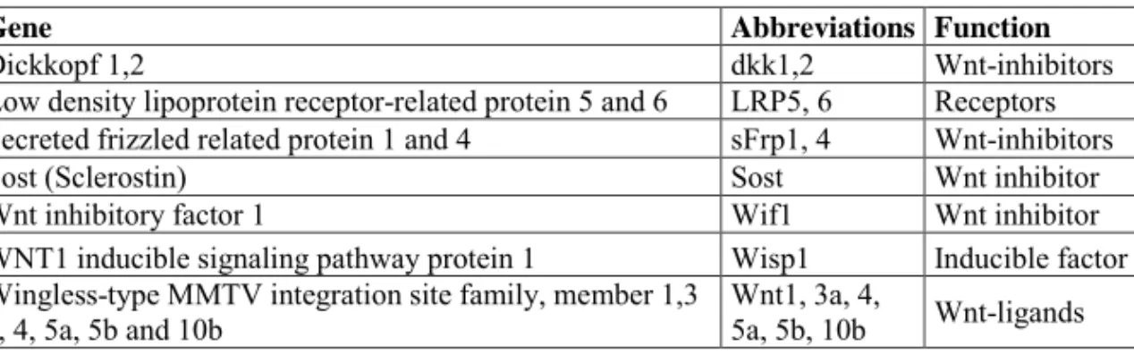
Sample mRNA was analyzed using real-time quantative RT-PCR: a method with high sensitivity and g ood reproducibility. Results were normalized against multiple endogenous control genes; in our case w e choose 18S rRNA, Ubiquitin-C RNA and Cytochrome-A RNA. This enables comparison of mRNA concentrations between different samples. We evaluated mRNA for 15 genes (table 1), chosen to represent the extracellular part of bone Wnt signaling. Since the quality of the primers is essential to a successful result, we obtained all primers from Applied Biosystems, all with m1 quality. PCR reactions were c onducted using a sta ndard curve methodology to quantify the specific gene targets of interest. The stand ard curve was made with embryonic rat RNA. There was concern that the expression of some Wnt components, like sclerostin [104], would be limited in the embryo. However, for all evaluated genes sufficient amount was detected in the st andard. Expression with CT values ove r 33 was considered too low to be anal yzed and such sa mples were e xcluded from the study. In this gene-expression study we chose to look at healing at 7 days after trauma. We chose this time point to see what happens in early stages of bone formation after the inflammatory phase but before remodeling.
Table 1: Genes studied for the mRNA expression.
Gene Abbreviations Function
Dickkopf 1,2 dkk1,2 Wnt-inhibitors
Low density lipoprotein receptor-related protein 5 and 6 LRP5, 6 Receptors Secreted frizzled related protein 1 and 4 sFrp1, 4 Wnt-inhibitors
Sost (Sclerostin) Sost Wnt inhibitor
Wnt inhibitory factor 1 Wif1 Wnt inhibitor
WNT1 inducible signaling pathway protein 1 Wisp1 Inducible factor Wingless-type MMTV integration site family, member 1,3
a, 4, 5a, 5b and 10b Wnt1, 3a, 4, 5a, 5b, 10b Wnt-ligands
Statistics
In all experiments a pre-determined primary outcome variable was defined. Because the primary variable was used for testing the main hypothesis, p-values for other, secondary variables were not corrected for multiple testing. Shapiro-Wilke’s test and Levene’s test was used to determine if data were normally distributed and had homogenous variances. Data were analyzed using 1 - and 2-way ANOVA and Student’s t-test. When comparing loaded and unloaded sides i n the same animal, paired t-test was used. In cases of significant deviation from normal distribution, non-parametric methods were used. Ln-transformation was sometimes used t o obtain similarity in vari ance. In order to describe the size of the treatm ent effect, we calculated the confidence intervals for differences between gr oup means. The confidence limits were then expressed as a percentage of the mean of the corresponding control group. In pap er IV w e assessed the i nfluence of loading b y investigating the gene expression difference between the loaded a nd unloaded li mb after ln-transformation. A 95% confidence interval (CI) was constructed for this ratio, and the confidence limit values were transformed back and expressed as fold change.
RESULTS AND DISCUSSION
19
RESULTS AND DISCUSSION
In this section, I have attempted to summarize the most important findings from each of the included papers (I-IV).
Effects of suppressing sclerostin or dkk1
Screw fixation in healing bone
Pull-out data from paper I and II sh ow that treatment with sclerostin or dkk1-antibodies improved the fixation of a st eel screw in cancell ous bone. This improvement was visible as an almost 50 % increase in peak force after 14 days. For sclerostin antibody treatment this increase was even larger after 28 days. This could be due to an increase in sclerostin production during the course of healing [64]. However, a similar increase with time was not observed in animals treated with dkk1-antibody. In earlier experiments, using a si milar model, an increase in peak force over time has been observed [100]. We suspect that the discrepancy between the sclerostin and the dkk1 experiments, with regard to how peak force developed over time, could be due to a change in rat breeder between study I and II. Nonetheless, the effect on bone healing was in line with other studies showing the positive effects of sclerostin- and dkk1-antibodies [51, 84-85] on bone f ormation. These increases in f ixation are si milar to those reported for bisphosphonates [105] and PTH [106] even though the mechanism of action is dif ferent. This illustrates one of the li mitations of mechanical testing; it measures the end result and we do not obtain information on the healing path that lead to it. Also, we cannot separate the different components that contribute to the increase in pull-out force, we do not know if it is due to an increase in cortical thickness or increase in cancellous bone density.
Screw fixation in uninjured bone
Screws inserted into the proximal tibia post mortem require much less force to be pulled-out than those that had been inserted 14 or 28 days earlier. The difference in pull-out force between the injured and uninjured sides i llustrates the contribution of healing to screw fixation. Although the ef fect of both antibodies was positive in healing bone, there was a difference between the antibodies in their effect on uninjured bone. With sclerostin-antibody treatment, the peak force was significantly increased by almost 50 %, while there were no significant changes with dkk1-antibody treatment.
Bone formation around screws
µCT data from paper I and II show that both sclerostin- and dkk1-antibody treatment increased the amount of bone surrounding the total intraosseous part of the plastic screw. However, the increase was only significant for sclerostin antibody treatment. For the cortical portion of the screw, a significant increase of bone volume fraction was detected for both antibo dies. For the marrow porti on, no significant increases in bone volume fraction occurred.
These data indicate th at the observed improvement in screw f ixation could be due mainly to an increase in bone formation at the cortex. This is probably because of immature bone, initially formed at the screw surface in the marrow cavity, was
RESULTS AND DISCUSSION
20
removed over time. However, it could also be due to specific bone formation at the cortex. In paper II, we saw a thickening of the cortical bone due to dkk1-antibody treatment. These measurements were not performed in paper I, but similar results have been reported both for sclerostin deficient mice [81] and with dkk1 suppression [51].
One potential problem with the se measurements is th at we cannot distinguish between newly formed bone and bone formed before implant insertion. Increased Wnt signaling has an antiresorptive effect due to t he secretion of OPG by osteoblasts [15] and osteocytes [21]. This could lead to a portion of non resorbed necrotic cortical and cancellous bone surrounding the screw. He nce, there is the risk that the increase in bone density around the screws comes at the expense of bone quality.
Bone formation in cancellous bone and its architecture
µCT data from paper I and II show that both sclerostin- and dkk1-antibody treatment changed the cancellous bone density and architecture. Sclerostin-antibody treatment did not significantly increase trabecular bone volume but clearly increased trabecular thickness.
Other studies have found an i ncrease in trabecular bone volume in cancellous bone due to sclerostin suppression [79, 81] and it is possible that w e were not able to detect this ef fect due to specif ic conditions or lack of measurement precision. Nonetheless, dkk1-antibody treatment did increase trabecular bone volume in the proximal tibia but did not significantly affect trabecular thickness. Instead, there was an increase in trabecul ar number. Furthermore, we could see that the eff ect of dkk1-antibody treatment was less pronounced in the vertebrae. This is in line with data from sclerosteosis patients in which vertebrae are not affected to the sa me degree as other bones [32]. Possibly, dkk1 is more important in the tibial metaphysis, close to the growth plate, than in the vertebrae. This is supported by the apparent larger effect of dkk1-antibody treatment in young rats compared to old [51].
Effects of suppressing sclerostin or dkk1 in unloaded
bone
Screw fixation in healing bone
In both s tudy II and I II, Botox administration had a profound effect on scre w fixation, causing the peak force to drop by half. In this unloaded bone, dkk1-antibody treatment caused no significant increase in peak force like the one detected in loaded bone. Sclerostin-antibody treatment had a positive effect on screw fixation in unloaded bone, causing a signi ficant increase in pea k force. However, sc lerostin-antibody treatment did no t restore the peak force to values correspo nding to those f or loaded bone. This contrasts to results with hind-limb suspension, where sclerostin antibody treatment restored bone density and structure [80].
In study III, the response to the sclerostin-antibody seemed proportionally similar in loaded and unloaded bone. This is different from previous data in which a larger effect was seen in lo aded bone c ompared to unloaded [80]. The absence of an interaction between healing and unl oading for the response to sclerostin-antibody treatment suggests that they may be independent of each other. This could infer a
RESULTS AND DISCUSSION
21
double role for sclerostin, one as a regulator of response to mechanical load and one as a participant in the healing response.
Bone formation around screws
µCT data from study II sh ow that dkk1-antibody treatment gave different increases in bone formation in loaded a nd unloaded bone. The effect seems larger in unloaded bone. Antibody treatment restored bone den sity around the screw s in unloaded limbs to values similar for loaded controls. It appears that the effect is larger in the cortical bone than in the marrow cavity, which is similar to the effects in loaded bone discussed earlier.
Bone formation in cancellous bone and its architecture
µCT measurements from study II and III show that Botox treatment caused a large decrease in bone density and deterioration in trabecular architecture with reduced thickness and number.
In study II, dkk1-antibody treatment could partly counter this effect and the bone density was in creased by over 200% in unloaded bone . This increase was proportionally larger than the one in loaded bone, which indicates that dkk1 signaling is important for loss of bone after disuse, but less involved in regulation under normal conditions. However, the antibody could not restore the bone den sity to va lues for loaded bone. As in lo aded bone, the increase in bone densit y could be attributed to a large increase in the number of trabeculae. The reason these effects were not detected as a significant effect on the pull-out experiments could be due t o higher precision of the µCT measurements.
In study III, sclerostin-antibody treatment increased bone density and caused an increase in trabecular thickness in unloaded bone. Similar to the pull-out experiments, there was no significant interaction between unloading and antibody treatment.
Effect of suppressing dkk1 on formation of new
cancellous bone
In paper II we also show, by using implanted bone cha mbers, that the regenerative capacity of bone is increased by dkk1-antibody treatment. Both histological and µ CT measurements show a n almost 80% incr ease in bone volu me fraction inside the chambers. There was also an increase in ingrowth distance, albeit to a lesser degree. The effects appear similar to those obtained with intermittent PTH injections [107] which clearly have an a nabolic effect on bone healing [8-9]. This contrasts to results with bisphosphonates [108] where a lack of resorption leads to increased bone density in the cha mber but le aves the ingr owth distance unchanged. The results suggest that dkk1 suppression increases the strength of the bone healing response and not just by limiting resorption.
RESULTS AND DISCUSSION
22
Wnt related mRNA expression during healing and
unloading
Effect of trauma
In study IV we descri be the changes in mRNA expression of 15 Wnt related genes due to a combination of trauma and unloading. By trauma to unloaded bone, 8 genes were significantly upregulated compared to the intact c ontrols. By trauma to loaded bone, 3 genes were significantly upregulated. Among the upregulated genes we find Wnts, inhibitors and receptors, suggesting a general increase in Wnt activity due to trauma.
Wnt ligands
Compared to intact b one Wnt5b was upregulated by trauma in both unloaded and loaded bone. Wnt4 was u pregulated only in unloaded bone. The role of these ligands in bone is und etermined but, compared to the W nt inhibitors, Wnts appear to not be specific for bone. However, there are reports that Wnts are loosely associated with osteoblast progenitors [109]. Wnt5a is suggested to be inv olved in f racture healing and Wnt10b has been identified as a regulator of bone mass [110]. We saw no significant change in expression of these genes due to trau ma. In previous studie s it has been shown that the addition of Wnt3a leads to improved healing [90] and implant fixation [91]. This illustrates that Wnt-ligands don´t need to be bone specific to have a positive effect on healing.
Wnt inhibitors
Several Wnt-inhibitor genes were significantly upregulated due to trauma, among them were dkk 1, sFrp1, Wif1 and sost. The increase in dk k1 expression in unloaded traumatized bone might explain the proportionally larger effect on bone formation in unloaded bone reported in paper II. With more dkk1 production in unloaded bone, a larger effect of the antibod y could be expected. SFrp1 was upregulated in in tact and traumatized unloaded bone and has been shown to be involved in fracture healing [78] but its role in mechano regulation is unclear. Wif1 was strongly upregulated by trauma in both loaded and unloaded bone. Wif1 has only minor effects in the skeleton [111] but is expressed in cartilage [90]. This presence of Wif1 may indicate that endochondral bone f ormation occurs in this model. However, we have never seen it histologically.
Sost was upregulated in loaded healing bone compared to loaded intact bone. An explanation for this might be that sost is upregulated as a response to the larger Wnt activity experienced during healing. Another explanation might be that th e biopsy samples obtained from traumatized bone contained proportionally more bone cells. Due to the osteocyte specificity of sost, an increase in the prop ortion of bone tissue would lead to an increase in apparent expression. This is a general limitation of our model. Even though care was taken to normalize data t owards housekeeping genes, these are not specific for bone cells so there is a possibility that the ratio between bone cells and marrow cells was distorted.
One further limitation is that we studied gene expression at just one time point, 7 days after trauma and 10 da ys after unloading. Hence, important changes in gene
RESULTS AND DISCUSSION
23
expression could be missed. It is known that Wnt gene expression changes during the course of bone healing [68]. For instance Wnt5a is more expressed during the early inflammatory phase than later [ 63, 68] and sclerostin expression generally increases with time [64].
Effect of unloading
In intact bone we saw few effects of unloading. As expected, sost expression was significantly upregulated, due to its k nown role in mechanotransduction [70-71]. We saw no significant effects on dkk1 expression, contrary to earlier reports that shows dkk1 to be upregulated due to unloading [70]. No other genes were significantly affected and we could exclude large changes in e xpression. This lack of significant changes in Wnt signaling could be attributed to temporal effects. It is obvious that the bone is degraded after 10 days of unloading and it is possible that most of the tissue response to unloading occurs during the first days. For instance, both sost and dkk1 expression is altered 24 h after changes in loading, but this effect is not seen after 3 or 7 days [70].
Differences between sclerostin and dkk1 suppression
Even though both sclerostin and dkk1 antibodies have a positive effect during healing there are some differences in between them.
Effect in untraumatized bone
The differences in peak force between sclerostin and dkk1 antibody treatment in untraumatized bone suggests that in cancellous bone, not involved in healing, sclerostin is more important than dkk1. However, these pull-out data are countered by µCT data showi ng an increase in cancellou s bone f ormation in u ntraumatized bone
with dkk1-antibodies. This could be du e the unspecif ic nature of pull-out
measurements and it is possible that sclerostin suppression improves some aspects of the trabecular structure that makes it more suitable to mechanical testing, for instance by increasing cortical thickness.
A general increa se in cancellous bon e mass by sclerostin-antibodies has been described earlier [79] and is obvious in pat ients with scleros tin deficiency [34-35]. Also for dkk1 it has been described that a reduction leads to increased cancellous bone mass [52, 55]. However, a recent stu dy shows that in untraumatized bone dkk1 antibodies only has a limited effect on bone formation in the adult skeleton and that the effect on bone healing is much larger [51].
Effect on cancellous bone architecture
Sclerostin-antibody treatment increased trabecular thickness but dk k1-antibody treatment increased trabecular number. This finding is potentially interesting, since it may hint at a difference in mechanism between the two wnt-inhibitors. An explanation for this may be that sclerostin and dkk1 does not bind to the same sites on the LRP-receptors [112-113]. However, the seemingly different effects of sclerostin and dkk1 on trabecular architecture might also be due to the change in rat br eeder between the experiments. On the other hand, data from a recent study support the notion that dkk1
![Figure 2: Overview of Canonical Wnt signaling [13-14, 20].. In the active state, Wnt-ligands (Wnt) form a complex with receptors LRP5 or LRP6 and Frizzleds (Fz)](https://thumb-eu.123doks.com/thumbv2/5dokorg/4655770.121143/18.892.128.772.99.568/figure-overview-canonical-signaling-ligands-complex-receptors-frizzleds.webp)