Bisphosphonates and implants in the jaw bone
Jahan Abtahi DDS, MDDepartment of Clinical and Experimental Medicine, Linköping University, Sweden.
Department of Oral & Maxillofacial Surgery, University Hospital, Linköping, Sweden.
©Jahan Abtahi, 2013
Cover/picture/Illustration/Design: Jahan Abtahi.
Published article has been reprinted with the permission of the copyright holder.
Printed in Sweden by LiU-Tryck, Linköping, Sweden, 2013
ISBN 978-91-7519-724-1 ISSN 0345-0082
Supervisor Per Aspenberg,
Department of Clinical and Experimental Medicine, Division of Orthopedics,
Linköping University. Co-supervisor Agneta Marcusson
Department of Oral & Maxillofacial Surgery, University Hospital, Linköping.
Faculty opponent Prof. Lars Rasmusson,
Department of Oral and Maxillofacial Surgery, Institute of Odontology, the Sahlgrenska Academy at University of Gothenburg.
Committee board Prof. Christer Tagesson,
Division of Occupational and Environmental Medicine,
Department of Clinical and Experimental Medicine, Faculty of Health Sciences,
Linköping University. Prof. Thomas Albrektsson,
Institute of Clinical Sciences, Department of Biomaterials, Gothenburg University.
Johan Thorfinn,
Department of Plastic Surgery, Hand Surgery and Burns, University Hospital, Linkoping.
POPULÄRVETENSKAPLIG SAMMANFATTNING ... 11 ABSTRACT ... 13 LIST OF PAPERS ... 15 ABBREVIATIONS ... 17 INTRODUCTION ... 19 BONE METABOLISM ... 21
Bone tissue structure ... 21
Bone cells ... 22
Osteoclast-osteoblast interplay ... 26
INTEGRATION OF TITANIUM IMPLANTS IN BONE TISSUE ... 29
The initial events ... 29
Bone-implant interface ... 30
The role of micromovement ... 31
The role of surface topography ... 33
Surface Roughness ... 33
Surface Chemistry ... 34
Surface Orientation ... 35
IMPLANT STABILITY MEASUREMENTS ... 36
Evaluation before and during implantation ... 37
Post-implantation evaluation ... 39
Experimental studies ... 39
Clinical studies ... 40
The technique ... 42
Clinical and experimental studies ... 43
BISPHOSPHONATES... 46
Structure and bioactivity of bisphosphonates ... 46
Local and systemic delivery, experimental studies ... 50
Local and systemic delivery, clinical studies ... 51
OSTEONECROSIS OF THE JAW (ONJ)... 53
Definition ... 53
Pathogenesis ... 54
Treatment ... 55
Prevention... 57
THOUGHTS BEHIND THE START OF THE PROJECT ... 59
HYPOTHESES ... 61
MATERIAL AND METHODS ... 63
STUDY DESIGNS (I AND II), CLINICAL STUDIES ... 63
Study I ... 63
Study II ... 63
RESONANCE-FREQUENCY MEASUREMENT (STUDIES I AND II) ... 64
STUDY DESIGN (III-V), EXPERIMENTAL STUDIES ... 65
Study III ... 65
Study IV ... 66
Study V ... 67
COATING TECHNIQUE ... 68
SHORT SUMMARY OF RESULTS OF CLINICAL STUDIES (I AND II) ... 71
Marginal bone height (I and II)... 71
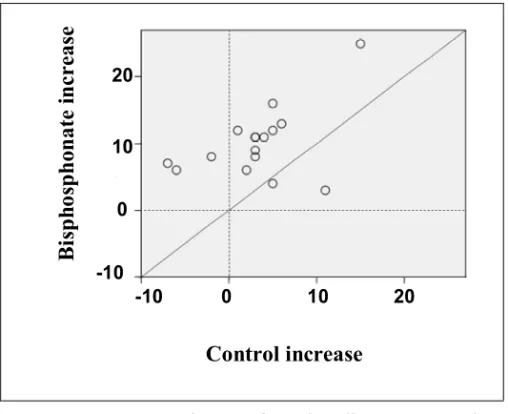
Resonance-frequency analysis (I and II) ... 72
Histology (I). ... 74
SHORT SUMMARY OF RESULTS OF EXPERIMENTAL STUDIES (III) ... 75
SHORT SUMMARY OF RESULTS OF EXPERIMENTAL STUDIES (IV) ... 76
SHORT SUMMARY OF RESULTS OF EXPERIMENTAL STUDIES (V) ... 77
DISCUSSION ... 79
IMPLANTS AND LOCAL DELIVERY OF BISPHOSPHONATE ... 79
RESONANCE-FREQUENCY ANALYSIS... 83
RAT MODEL OF ONJ ... 84
IMPLANTS AND COATING TECHNIQUE ... 86
LACTATE DEHYDROGENASE ANALYSIS ... 88
PATHOPHYSIOLOGY OF ONJ ... 89
CONCLUSIONS ... 93
WHAT NEXT? ... 95
ACKNOWLEDGEMENTS ... 97
11
Populärvetenskaplig sammanfattning
Insättning av metall-implantat för ersättning av förlorade kroppsdelar är ett vanligt förekommande behandlingsmetod inom odontologi och ortopedi. Lyckandefrekvensen för dessa behandlingar är direkt kopplade till implantatens stabilitet, som i sin tur beror på kringliggande benvävnad. När man sätter in en skruv av titan i käkbenet utlöses ett frakturläkningssvar som skapar nytt ben runt skruven. Frakturläkningssvaret innehåller både uppbyggnad och nedbrytning av ben. Genom att selektivt minska bennedbrytningen med ett läkemedel (bisfosfonat) kan man med bibehållen uppbyggnadskomponent få mer och starkare ben. Bisfosfonater används kliniskt bland annat för att hämma bennedbrytning hos patienter med benskörhet eller skelettmetastaser. Under de senaste åren har bisfosfonatbehandling för att förbättra implantatfixation testats i både djurförsök och kliniska studier, men inte i käkar. Detta kan bero på att det finns ett samband mellan användningen av bisfosfonat (speciellt intravenöst) och förekomst av ett tillstånd som kallas för ”osteonekros i käken”. Patofysiologin och behandlingen av detta tillstånd är kontroversiell.
Syftet med denna avhandling är att öka förståelsen om hur bisfosfonater förstärker benvävnaden runt ett implantat. Kan bisfosfonatbeläggning på metallytan förbättra fixeringen av implantat i käken? Kan man reproducera eller förhindra uppkomsten av osteonekros i käken i en djurmodell?
Totalt opererades 96 implantat i överkäken på 21 patienter, som alla fick ett implantat med bisfosfonat.
12
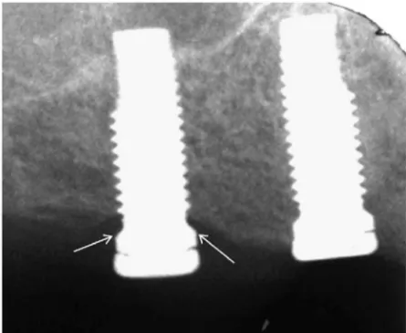
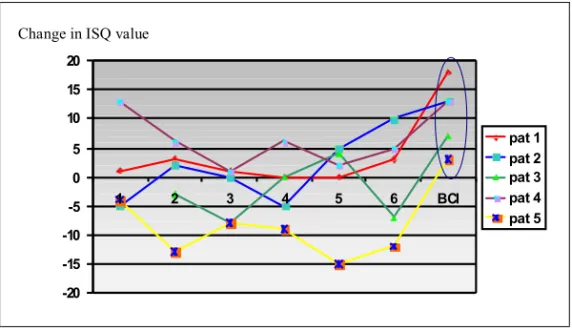
Resonansfrekvensmätning visade att de bisfosfonat beklädda tandimplantaten hade bättre stabilitet jämfört med kontrollimplantaten efter 6 månaders läkning.
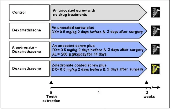
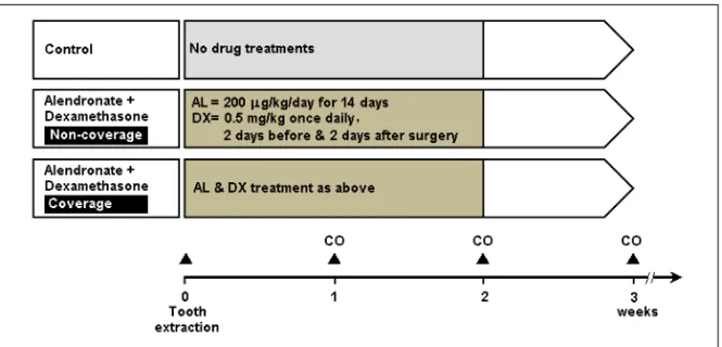
Röntgenundersökning visade mindre benförlust kring bisfosfonatbeklädda implantat. Vi utvecklade tre djurmodeller för att studera osteonekros i käken. I ett experiment studerades effekten av lokal och systemisk bisfosfonatbehandling på käkbenet. Skruvar beklädda med ett potent bisfosfonat (zoledronat) orsakade bättre implantatinläkning, även under betingelser där systemisk bisfosfonat framkallar osteonekros i käken. Vi har också visat att osteonekros i käken inte uppkommer förrän benet exponerats, t ex genom tandborttagning. Slutligen kunde vi förebygga uppkomsten av detta tillstånd genom omedelbar täckning med slemhinna efter tandborttagning.
Slutsatsen är att lokalbehandling med bisfosfonat ger bättre fixering av implantat i käkarna. Detta kan leda till nya möjligheter för ortopedisk och dental implantatkirurgi. Patofysiologin av osteonekros i käken är relaterad till exponering av benvävnad och till läkemedel som förhindrar nedbrytning av benvävnad.
13
Abstract
Insertion of metal implants in bone is one of the commonest of all surgical procedures. The success of these operations is dependent on the fixation of the implants, which, in turn, depends on the strength of the bone that holds them. If the quality of the bone holding the implant could be improved locally, surgical procedures would become simpler and rehabilitation would become faster. Bisphosphonates are anti-resorptive drugs that act specifically on osteoclasts, thereby maintaining bone density and strength. Once released from the surface of a coated implant, bisphosphonates reduce osteoclast activity, thereby changing the balance of bone turnover in favor of bone formation, leading to a net gain in local bone density. During the last decades, the effects of bisphosphonate treatment on the stability of implants have been tested in several clinical and animal studies, but not in human jaws. This may be because it has been suggested that there is a link between the use of bisphosphonates (especially those given intravenously) and a condition called osteonecrosis of the jaw (ONJ). The pathophysiology and treatment of ONJ is controversial. The difficulty in treating ONJ has highlighted the importance of prevention.
The overall aim of the present thesis was to evaluate the effect of local and systemic use of bisphosphonates on bone tissue. Could a thin, bisphosphonate-eluting fibrinogen coating improve the fixation of metal implants in the human jaw? Would it be possible to reproduce ONJ and prevent the development of this condition in an animal model?
In two clinical studies, a total number of 96 implants were inserted in 21 patients. In a randomized trial with a paired design, one implant in each pair
14
was coated with a thin fibrinogen layer containing two bisphosphonates (pamidronate and ibandronate). The bisphosphonate-coated implants showed better stability as measured by resonance-frequency analysis. Radiographic intraoral films also showed less bone loss. Three animal models were developed. In a study comparing local and systemic effects of bisphosphonates, zoledronate-coated screws inserted in rats showed better fixation in spite of a drug treatment that is known to induce ONJ-like lesions when given systemically. In another rat model, ONJ-like lesions were reproducibly induced at sites of tooth extraction whereas there were no signs of bone cell death in uninjured sites. Finally, rat experiments showed that the development of ONJ-like lesions after tooth extraction could be prevented by early mucoperiosteal coverage.
In conclusion, a thin, bisphosphonate-eluting fibrinogen coating can improve the fixation of dental implants in human bone. This principle may lead to new possibilities in orthopaedic surgery and dentistry. The pathophysiology of ONJ is strongly linked to bone exposure in combination with drugs that reduce resorption.
15
List of papers
This thesis is based on the following papers, which will be referred to in the text by their Roman numerals.
I. Bisphosphonate coating might improve fixation of dental implants in the maxilla: A pilot study.
Abtahi J, Tengvall P, Aspenberg P
Int J Oral & Maxillofac Surg 2010 39(7): 673-7.
II. Bisphosphonate-coating improves the fixation of metal implants in human bone. A randomized trial of dental implants.
Abtahi J, Tengvall P, Aspenberg P Bone 2012 50(5): 1148-51.
III. Bisphosphonate-induced osteonecrosis of the jaw in a rat model arises first after the bone has become exposed. No primary necrosis in unexposed bone.
Abtahi J, Agholme F, Sandberg O, Aspenberg P J Oral Pathol Med 2012 41(6): 494-9.
IV. Effect of local versus systemic bisphosphonate on dental implant fixation in a model of ONJ.
Abtahi J, Agholme F, Sandberg O, Aspenberg P Journal of Dental Research 2012 [Epub ahead of print].
V. Prevention of osteonecrosis of the jaw by mucoperiosteal coverage in a rat model.
Jahan Abtahi, Fredrik Agholme, Per Aspenberg
Int J Oral & Maxillofac Surg 2013, accepted, manuscript number: IJOMS-D-12-00927R1.
17
Abbreviations
ATP Adenosine triphosphate BIC Bone-to-implant contact BMP Bone morphogenetic protein BRONJ Bisphosphonate-related
osteonecrosis of the jaw Cbfa 1 Core binding factor 1 CSF Colony-stimulating factor ISQ Implant stability quotient FPPS Farnesyl diphosphate synthase
GTP Guanosine triphosphate
HAC Hydroxyapatite coating
OH Hydroxyl group
ONJ Osteonecrosis of the jaw
OPG Osteoprotegerin
PTH Parathyroid hormone
PTV Periotest value
RANK-L Receptor activator of NF-kappa B ligand
RFA Resonance-frequency analysis TGF-β Transforming growth factor-β TNF- α Tumor necrosis factor-α
19
Introduction
During the last decades there have been problems with the condition called “bisphosphonate-related osteonecrosis of the jaw” (BRONJ). This condition is defined as an area of exposed bone in the maxillofacial region that does not heal within 8 weeks of identification by a healthcare provider, in a patient who currently receives or has been exposed to a bisphosphonate and has not had radiation therapy to the craniofacial region (from here on, I use the shorter acronym ONJ). The pathophysiology of ONJ is poorly understood and as maxillofacial surgeon, I have wondered why these lesions localize specifically in the jaws. It is remarkable that orthopedic surgeons and osteoporosis researchers consider bisphosphonates to be beneficial and useful in many areas, while dental practitioners reject these drugs. By working with an orthopedic research team, I had the opportunity to address the problem of the paradoxical effects of bisphosphonates. Previous animal studies by this team have shown that bisphosphonate coatings can improve the fixation of implants in bone. Clinically, this idea has been tested in orthopedics but not in dentistry. From the literature it can be deduced that high implant survival rates would be expected in jaws. However, dental implant surgery can be risky in bone of low density. We therefore decided to use bisphosphonates in the hope of improving the fixation of implants in the maxilla. Considering that ONJ was apparently non-existent ten years ago, the field has progressed through knowledge gained from case reports, population-based studies and emerging animal models. Still, there are preconceptions that need to be tested and important clues that need to be investigated in order to translate pathophysiology into improved patient care. In the clinic, I have also met cancer patients who have suffered from exposed bone in the jaw associated with intravenous bisphosphonate
20
therapy. Is it possible to treat or prevent the development of ONJ? I hope that my research will improve our knowledge oft this condition.
21
Bone metabolism
Bone tissue structure
The skeleton consists of specialized cells, mineralized and unmineralized connective tissue matrix, and spaces that include bone marrow cavities, vascular canals, canaliculi, and lacunae. At the nano-scale level, bone tissue is a composite material composed of an organic phase, consisting mainly of the protein-based material collagen, and a mineral phase, consisting primarily of hydroxyapatite (1, 2). Hydroxyapatite is present as plate-like crystals, 20-80 nm long and 2-5 nm thick, which are themselves composed of calcium and phosphate. The crystals are found in and around collagen fibers and give bone its compressive strength. The organic matrix determines the structure and mechanical properties of the bone. Of the organic matrix, approximately 90% is type-1 collagen. The remainder consists of non-collagenous matrix proteins, minor collagen types, proteoglycans, and lipids.
At the microscopic level, there are two types of bone tissue, woven bone and lamellar bone. Woven bone is considered to be immature, with collagen arranged randomly. At birth, it makes up all the bone in the body, and in later years it is found at sites of fracture healing or in response to extreme mechanical loading (3). Lamellar bone is the name given to bone that eventually replaces woven bone. By the age of 4 years, most of the skeleton is lamellar bone. Anatomically, both woven bone and lamellar bone can be organized into compartments as either cortical or trabecular bone (cancellous bone). An important difference between cortical bone and trabecular bone is in the way the bone matrix and cellular elements are arranged. Between 80% and 90% of cortical bone volume is mineral, but only 15-25% of trabecular
22
bone volume is mineral (4). The trabecular arrangement allows bone marrow, blood vessels, and connective tissues to be in contact with bone.
The main function of cortical bone is to give structure and protection. In cortical bone, lamellae are arranged concentrically around a central vascular channel (Haversian canal). This arrangement of cortical bone around a vessel is called an osteon. Osteons are usually aligned with the long axis of bone and are connected to each another by Volkmann’s canal, which runs at right angles to the osteon. The outer surface of cortical bone, facing the soft tissue is covered by periosteum, while the inner surface that faces the bone marrow is covered by endosteum. Cells lining the endosteum are metabolically active and very involved in bone formation and resorption.
Bone cells
There are mainly four types of bone cells. These are the osteoprogenitor cells, osteoblasts, osteocytes and osteoclasts. The osteoblasts are mononucleate bone-forming cells that are derived from the local osteoprogenitor cell line (mesenchymal cells) located in the deeper layer of periosteum and the bone marrow (5-7).
These progenitors are capable of
differentiating into other mesenchymal cell lineages such as
chondrocytes, fibroblasts, myoblasts, and bone marrow stromal cells
including adipocytes (
8-
12).
Mature osteoblasts have an average lifespan of 1 month, after which they either undergo apoptosis, to be replaced by newly differentiated osteoblasts, or alternatively about one-third of them may be incorporated into deposited bone matrix as osteocytes (15).23
Several growth factors and hormones regulate osteoblast differentiation. Growth factors are soluble proteins that act as signaling agents for cells and influence critical functions, such as cell division, matrix synthesis, and tissue differentiation, by receptor-ligand binding.
Of these,
bone morphogenetic proteins (BMPs) are the most potent inducers and stimulators of osteoblast differentiation (13, 14).These proteins not only stimulate osteoprogenitors to differentiate into mature osteoblasts but also induce non-osteogenic cells to differentiate into cells of the osteoblast lineage. Proliferation and differentiation of osteoblasts is also regulated by many transcription factors. Expression of the transcription factor core binding factor 1 (Cbfa 1) is an absolute requirementfor osteoblast differentiation and bone formation (15). Similarly, the transcription factor ppar-c 2 can specify adipocyte differentiation (16), and sox-9 expression is required for chondrocyte differentiation (17).Osteoblasts express receptors for various hormones including parathyroid hormone (PTH), glucocorticoids, 1α, 25-dihydroxyvitamin D3, and estrogen, which are involved in the regulation of osteoblast differentiation (18-20). Parathyroid hormone acts directly on the skeleton to promote calcium release from bone and on the kidney to enhance calcium reabsorption by binding to its receptor. The anabolic effect of PTH on bone tissue occurs by upregulation of expression of transcription factor c-fos, which is a key regulator of osteoblast and osteoclast differentiation (18). Inactivation of c-fos causes the bone-remodeling disease osteopetrosis, which is characterized by impaired osteoclastic bone resorption, resulting in a net increase in skeletal mass (21). In contrast, when c-fos is overexpressed in tissues, bone tumors develop that are typically chondroblastic osteosarcomas, containing large amounts of neoplastic bone with foci of cartilage (22, 23).
24
Glucocorticoids also play an important role in the normal regulation of bone remodeling (24). The precise role of glucocorticoids in bone formation is still poorly understood. In vivo studies have shown that continued exposure of bone to pharmacological doses of glucocorticoids excess results in osteoporosis (25, 26). This event is due to the effect of glucocorticoids on bone cells. Glucocorticoids increase the expression of receptor activator of NF-kappa B ligand (RANK-L) and reduce the expression of its decoy receptor, osteoprotegerin (OPG), in stromal and osteoblastic cells (27). They also enhance the expression of colony-stimulating factor (CSF)-1, which in the presence of RANK-L induces osteoclastogenesis (28). Furthermore, several in vitro studies have shown that glucocorticoids reduce the number of cells of the osteoblastic lineage and shift the differentiation of stromal cells towards the adipocytic lineage (29) Glucocorticoids also increase peri-lacunar osteocytic bone resorption to an extent that negatively influences bone material properties (30).
Osteoclasts are large, multinucleate cells derived from hematopoietic stem cells, and they are equipped with phagocytic-like mechanisms similar to those of circulating macrophages (31). Osteoclast literally means “bone eater”.Osteoclast differentiation has various characteristic features, such as multinucleation induced by the cell fusion of mononuclear osteoclasts to cover a larger area, synthesis of the vacuolar proton pump and acid to dissolve the bone mineral, the formation of ruffled borders to secrete protons and acid, and the formation of a sealing zone to prevent proton and acid leakage (31). Their proliferation and differentiation (osteoclastogenesis) depend on the presence of two different osteoblast expressed cytokines, macrophage colony-stimulating factor (M-CSF) and RANK-L (32, 33). The protein osteoprotegerin (OPG), which is secreted by osteoblasts, acts as a
25
decoy receptor that competes with RANK for RANK-L and thereby inhibits osteoclast differentiation (34, 35).
Osteoclasts have developed efficient and unique machinery to dissolve mineral and degrade bone matrix. To maximize bone resorption, osteoclasts expand their surface area by fusion to many mononucleated macrophages (36). After migration of the osteoclast to a resorption site, a specific membrane domain, the sealing zone, forms adjacent to the bone surface (37, 38). Inside the sealed region, extensive infoldings of the cell membrane form, the so-called “ruffled border” which increases the membrane surface (38). A cytoplasmic proton pump (H+-ATPase) produces protons that generate a pH of 4-5 in the extracellular space adjacent to the bone surface (39). This event results degradation of the mineral component of bone, which is composed of hydroxyapatite (31). The organic matrix of the bone (collagen) is removed through enzymatic activity, by cathepsin K. This enzyme reaches the bone surface by exocytosis through the basolateral membrane of the osteoclast (40).
Osteocytes are found within individual lacunae in the mineralized bone matrix. The lifespan of osteocytes is higher than that of osteoblasts, which is an estimated 3 months in human bone (41) and 10–20 days in newly formed murine bone (42). The lifespan of osteocytes is probably largely determined by bone turnover and they may have a half-life of decades if the particular bone they reside in has a slow turnover rate (4).
The morphology of embedded osteocytes is dependent on the bone type. Indeed, osteocytes found in trabecular bone are more rounded than osteocytes from cortical bone (43). Each osteocyte communicates with its
26
neighbors and with the cells lining the surface of bone through long, slender cytoplasmatic processes (canalicular processes) that connect by means of gap junctions (44-46). Osteocytes are capable of detecting mechanical stimuli, which are mediated by loading-induced dynamic fluid flow in the canaliculi (47, 48). Osteocytes differentiation is under the influence of several bone markers such as alkaline phosphatase, bone sialoprotein, osteocalcin, and collagen type I (49). Once the osteoid mineralizes, osteocyte ultrastructure undergoes further changes including a reduction in endoplasmic reticulum and Golgi apparatus corresponding to a decrease in protein synthesis and secretion (50). At this stage, many of the previously expressed bone markers are downregulated in the osteocyte (50).
Osteocytes are cells that not only play a physiological role during their lifetime, but also achieve functions throughtheir apoptosis.Osteocytes have been hypothesized to play a role in this targeted remodeling process (51, 52). Damage to the bone matrix, such as micro-cracks induces apoptotic death of osteocytes, which initiate signals for bone resorption by expression of osteoclast-stimulatory factors, such as RANKL and M-CSF (53, 54).
Osteoclast-osteoblast interplay
Throughout life, bone is constantly renewed through a two-stage process called remodeling (55, 56). This condition is a dynamic process that relies on the correct balance between bone resorption by osteoclasts and bone deposition by osteoblasts. In healthy adults, under normal circumstances, bone resorption is always followed by an equal degree of bone formation, a tightly balanced process referred to as coupling (57).The regulation of bone resorption involves a complicated set of hormonal and/or cytokine
27
interactions that initially stimulate osteoblasts, which then elaborate factors that signal osteoclasts to degrade bone (58, 59). Osteoblast differentiation is promoted by lipid-modified glycoproteins of wingless (Wnt), bone morphogenic proteins (BMPs), and several transcription factors (15, 60). Furthermore, Wnt signaling has been shown to reduce osteoblast and osteocyte apoptosis in vivo and to increase bone formation by stimulating differentiation and replication of osteoblasts (61).
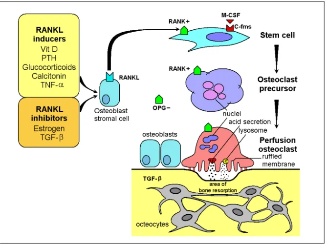
Cells of the Osteoblast lineage produce the osteoclastogenic cytokines RANKL and M-CSF, which recognize their respective receptors RANK and c-fms on macrophages, prompting them to take on the osteoclast phenotype (32, 33). RANKL activity is negatively regulated in the circulation by osteoprotegerin (OPG), which competes with RANK as a soluble decoy receptor (34). Administration of RANKL to mice causes osteoporosis (35), whereas disruption of the RANKL gene in mice leads to severe osteopetrosis, impaired tooth eruption, and the absence of osteoclasts (60). Several hormones including calcitonin, parathyroid hormone, vitamin D, estrogen, interleukins, glucocorticoids and tumor necrosis factor-α (TNF- α), transforming growth factor-β (TGF-β), among others, regulate osteoclast and osteoblast function (18-20, 24, 62, 63) (Figure 1).
28
Figure 1. Differentiation and activation of osteoclasts. M-CSF and RANKL are essential for osteoclastogenesis. OPG can bind to RANKL and thereby inhibit osteoclast differentiation.
The coupling of bone resorption and formation suggests that autocrine and paracrine factors are produced and released within the local bone environment (64, 65). Transforming growth factor β (TGF-β) is a multifunctional cytokine with potent effects on bone metabolism (66, 67). Both osteoblasts and osteoclasts synthesize and secrete latent TGF-β (65). Resorption of bone by osteoclasts releases latent TGF-β from the organic matrix, where it potently stimulates osteoblastogenesis and at the same time inhibits RANKL expression by osteoblasts. (65).
29
Integration of titanium implants in bone tissue
The initial events
Insertion of metal implants in bone is one of the most common of all surgical procedures. Brånemark et al. first defined “osseointegration” in 1969 as a direct structural and functional contact (at the light-microscopic level) between living bone and implant.
The initial host response after implantation is characterized by an inflammatory reaction elicited mainly by the surgical trauma. Inflammatory cells, initially polymorphonuclear granulocytes and later monocytes, emigrate from post-capillary venules into the tissue surrounding the implant (69). At this stage, damage to the pre-existing bone in the implant- bone cavity is often a consequence of heating, located within 100 μm (70). Immediately after the surgical damage, the walls of bone are covered with blood, which initiates a clotting reaction. This is the first tissue to come into contact with the implant surface after insertion of the implant in the bone cavity (71). It has been shown that the implant-blood interface is composed of a fibrin film containing platelets and red blood cells, and they appear to respond differently to different implant surface topographies (71).
A strong correlation has been found in several studies between the adsorption of fibrinogen and surface adhesion of platelets (71-74). The enhanced aggregation may be due to the increased surface area of the micro-roughened surface, suggesting that this topography induces more agglomeration of red blood cells/platelets than machined surfaces (71).
30
A few days after implantation, osteoblasts produce collagen matrix directly on the early- formed lamina limitans layer on the implant surface (69-75). During the first week, mesenchymal cells and multinuclear giant cells are present in the area around the implant (75). Areas of newly formed bone (woven bone) can be seen at the endosteal surfaces towards the implant 1-2 weeks after implantation. Woven bone contains osteocytes, and the trabeculae are lined with osteoblasts. At this time, in areas that are responsible for primary mechanical stability, the bone tissue shows signs of ongoing bone remodeling, resorption, and apposition. After 4 weeks, the dense woven bone often combined with lamellar bone approaches the implant surface, to fill the threads. The remodeling process for rabbits and dogs starts after 4 weeks and is complete after 90 days (69, 75).
Bone-implant interface
The interface zone between bone and implant has been the subject of a vast number of recent publications (76-86). Many investigators have shown an interface zone consisting of connective tissue (76, 77). Initially, it was suggested that the goal for the surgeon should be a periodontal membrane around the implant (76). Several authors believe that direct contact between implant and bone is possible only if the implant is ceramic, and not if it is metal (78, 79). Furthermore, light microscopy of the interface zone has revealed intimate contact between newly formed bone and the oxidized surface of titanium implant (80). Early studies on bone-implant contact at the electron microscopic level showed a close relationship between implants and collagenous filaments from bone (81). Albrektsson and co-workers (82) compared interfacial arrangements around stainless steel implants with those seen around commercially pure titanium. The titanium implants, in contrast
31
to stainless steel ones, became directly anchored in bone without any cellular layer at the interface. Moreover, the authors found a thin (20-40 nm) layer of amorphous material consisting proteoglycans adjacent to the implant surface (83). Similar results have been found by others (84-86). Further ultrastructural studies of machined implants by Sennerby et al. (85, 86) showed a 100-nm- wide electron-dense line (lamina limitans) at the border between the mineralized bone and the non-calcified amorphous layer. This finding indicated that the stability of the implant is mainly mechanical.
The role of micromovement
In the literature, implant movement relative to the surrounding bone has been suggested to be a crucial parameter in the prognosis of implant osseointegration (87, 88). Early movement of the implant during the initial healing phase will lead to a preponderance of interfacial connective tissue (89-92). There appears to be a consensus that excessive micromotion impairs osseintegration (93, 94). However, many of these studies are not comparable due to the influence of other factors such as implant geometry, surface characteristics, and implant site. The threshold of micromotion, as experimentally evaluated in animals, is between 50 and 100 µm (95). In a cadaver study by Burke et al. (96) micromotion of knee and hip prostheses was measured using sensitive displacement transducers. Such movement has been shown to be in the range of 100-600 µm (97).
Søblle et al. (97) studied the influence of micromotion between bone and titanium implant with and without a hydroxyapatite coating (HA) in a dog model. They showed that micromotion of 150 µm inhibits bone ingrowth and results in development of a fibrous membrane. Moreover, 4 weeks after
32
implantation, a fibro-cartilaginous membrane was seen around unstable HA-coated implants, whereas around titanium implants, only fibrous connective tissue was found. Furthermore, continuous loading of initially unstable titanium implants resulted in the development of a permanent fibrous membrane, whereas HA-coating had the capacity to replace the motion-induced fibrous membrane with bone. These findings show that micromotion has a role in tissue differentiation (98).
In an experimental study by Akagawa et al. (99) dental implants were inserted in dog mandible by one-stage or two-stage procedures. The animals were fed with hard pellet food for 3 months. The submerged implants showed direct bone apposition and sparse fibrous, dense connective tissue. The unsubmerged implants also showed direct bone apposition. However, the apical part of the implant was frequently in contact with dense connective tissue. From these experimental studies, it appears that micromovements like those induced by early loading of dental implants should be avoided if the intention is osseointegration.
Trisi et al. (100) evaluated in vitro the correlation between the micromotion of cylindrical- screw implants and the insertion torque in fresh bovine bone of different densities ex vivo. They found that increasing the peak insertion torque reduced the micromotion between the implant and the bone. However, micromotion in soft bone is always high and immediate loading of implants in low-density entails a higher risk of loosening.
33
The role of surface topography
Surface Roughness
Titanium and its alloys are the materials most often used in implant manufacture because of their excellent biocompatibility, favorable mechanical properties, and well-documented beneficial results. When exposed to air, titanium immediately develops a stable oxide layer which forms the basis of its exceptional biocompatibility. In the last decade, most dental implant manufacturers have focused on implant surfaces to improve bone-to-implant contact (101). It is well known that modification of the implant surface (including topography and chemistry) alters the cellular and bone tissue responses (102, 103). Earlier studies (104, 81) have suggested that implant surface topography is the only parameter that significantly affects bone-to-implant contact (BIC). These findings were later confirmed by studies with animals, which indicated that a certain degree of surface roughness favored BIC, as assessed by the removal torque test (105-108). Albrektsson and Wennerberg (109) defined smooth surfaces to have an Sa
value of < 0.5 μm; minimally rough surfaces were identified with an Sa of
0.5-1 μm, moderately rough surfaces with an Sa of 1-2 μm, and rough
surfaces with an Sa of > 2 μm.
Another advantage of a roughened titanium surface is a shorter healing period and the option of using shorter implants, still with a good long-term prognosis because of the better bone anchorage (110). Therefore, many surface modifications of titanium implants have been developed to achieve better osseointegration (electron-polished, anodization, plasma-spraying grit-blasting or acid-etching).
34
One way to produce an increased surface roughness is to blast the surface. Wennerberg et al. (111) showed higher bone-to-implant contact and higher removal torque values for implants blasted with TiO (25 µm) rather than Al2O3 (75 µm) and machined implants. However, no differences were seen
when implants were blasted with different materials (A12O3 and TiO), but
the same degree of roughness (25 µm) was used (112).In recent years, high-strength ceramics have become attractive as new materials for dental implants, due to their biocompatibility and higher fracture resilience (113). At the ultrastructural level, there was no difference between the tissue response of zircornium implants and that of titanium implants (114, 115). The TiOblast ™ surface is grit-blasted with titanium dioxide particles to achieve a moderately rough surface. Several studies (116-118) have shown promising clinical outcome after 5 years of loading of this surface. In vivo and experimental studies (119-121) have shown a high cell biocompatibility and better anchorage in bone when using titanium oxide-blasted implants rather than implants with a machine-prepared surface.
Surface Chemistry
Beyond surface topography, surface chemistry may provide important and possibly synergistic cues for bone formation at titanium implants. Recent studies advocated that surface chemistry is changed by many surface topographic modifications (122, 123).
Ellingsen et al. (124) placed 80 implants with and without a fluoride-modified surface in the tibia of 20 rabbits. Removal torque measurements were performed and biopsies were obtained after 1 and 3 months of healing. While no differences in removal torque were detected between the two types of implants after 1 month, the fluoride-modified (test) implants in the
3-35
month healing group had significantly higher torque values than the control implants. Furthermore, examinations done in ground sections representing both 1 and 3 months of healing revealed that at fluoride-modified implants, there were larger proportions of BIC than at the control implants. Berglundh et al. (125) found that the amount of new bone that formed in the voids within the first 2 weeks of healing was larger at fluoride-modified (test) implants than at TiOblast (control) implants. Similar findings have been reported by Cooper et al. (126). Calcium phosphates, such as hydroxyapatite (HA),have been successfullyused as bone replacement materials in the case of periodontal lesions, or as a pulp-capping agent (127, 128). Synthetic hydroxyapatite that is very similar to the inorganic component of bone has been found to be osteoinductive. It is capable of supporting ingrowth of osteoprogenitor cells into the graft or implant. Hydroxyapatite coatings on titanium alloy are used in dental and orthopedic procedures (129).
Surface Orientation
The orientation of the surface topography has been the subject of a number of studies (130-132). Ivanoff and co-workers (131) investigated microimplants in a human test model. They found that when blasted and turned implants with similar roughness were compared, the blasted implants had significantly more bone-to-implant contact than turned implants. Despite the fact that both implants had the same grade of roughness, the turned implants had a clear orientation of the topography with an orientation perpendicular to the long axis of the implant. This finding indicated that surface orientation may be as important as the degree of surface roughness. Moreover, Wennerberg et al (132) found no differences between the
36
horizontally grooved implants and the vertically grooved implants when measuring bone-to-implant contact.
Burgos et al. (133) showed that titanium implants with a turned or an oxidized surface are integrated in bone in different ways. As observed at the light-microscopic level, bone formation occurs directly on the moderately oxidized surface, while turned titanium surfaces are integrated by growth of bone from the adjacent bone marrow and bone tissue. The mechanisms behind the different integration patterns are not fully known, but surface topography most certainly plays an important role. Moreover, a positive effect of titanium surface roughness on osteoblast proliferation and gene expression associated with collagen biosynthesis has been discussed by several authors (134-136). The surface profile in the nanometer range plays an important role in the adsorption of proteins, adhesion of osteoblast cells, and thus, the rate of osseointegration (137). However, the optimal size of nonometer particles applied on implant surfaces is still unknown.
Implant stability measurements
It is clear that stability both at placement and during function is an important criterion for the success of dental implants. Primary implant stability is a mechanical phenomenon influenced by factors related to the implant (design and dimensions of the fixture), the patient (quality and quantity of bone), and the operator (surgical technique). Primary implant stability is highest just after implant placement, because of mechanical compression of the fixture on bone walls, and it decreases with time. Secondary stability is the progressive increase in stability related to biological events at the bone-implant interface such as new bone formation and remodeling (138). The
37
clinical definition of implant osseointegration considers the level of stable marginal bone and absence of mobility in the bone (139). The diagnosis is therefore based on radiographic and mechanical stability criteria.
Evaluation before and during implantation
Imaging studies
Different radiographic techniques (intraoral radiographs, panoramic radiographs, lateral cephalograms, and tomographic images) are commonly used for evaluation of jaw bone quality and volume when planning for implant treatment (140). The use of computerized tomography (CT) for the evaluation of the bone density of patients requiring implant therapy was introduced by Schwarz et al. (141) and this method has been used in several studies (142-146). The authors suggested that there are strong correlations between bone density as estimated by CT, the insertion torque, and resonance-frequency values at implant placement. These findings support the idea that the preoperative CT examination may be a helpful technique for predicting primary stability. Although CT examinations have advantages over conventional radiography techniques, CT exposes the patient to higher doses of radiation. The recently developed limited cone-beam CT involving a two-dimensional X-ray sensor has been reported to be useful in presurgical evaluation. Limited cone-beam CT provides high-resolution images and does not need the use of high doses of radiation (147, 148).
Drilling/cutting resistance
Currently the most popular method of bone quality assessment is that developed by Lekholm and Zarb, who introduced a scale of 1to 4, based on both the radiographic assessment and the sensation of resistance experienced by the surgeon when preparing the implant site (149). The grading refers to
38
individual experience and provides only a rough mean value for the entire jaw. This classification has therefore been questioned recently due to its poor objectivity and reproducibility (150, 151). A similar index of four different density classes, based on the tactile feeling during drilling and implant insertion was introduced by Mish and Friberg (152, 153). Norton et al (143) concluded that there is a need for an objective quantitative classification of bone quality, which can be applied preoperatively and is not operator- dependent. They proposed a new classification based on the Hounsfield units of the bone on the CT scan, and related it to the existing classification of Lekholm & Zarb (149). A Hounsfield unit represents a normalized index of x-ray attenuation based on a scale where air corresponds to -1000 units water at standard pressure and temperature to 0 (154). Klinge et al. (155) proposed an individualized healing period after implant placement based on an objective score of bone quality. Altogether, 15 bone biopsies from 12 patients were harvested at mandibular sites before insertion of an implant. However, this idea has not been developed for practical reasons. Furthermore, a more objective method has been described by Johansson and Strid (156), who measured the cutting resistance during implant insertion as a function of the electric current drawn by the handpiece. In a series of studies by Friberg et al. (157-159), a positive correlation was found between values of cutting resistance, bone density, and resonance-frequency measurements. However, it was not possible to use this instrument to identify implants that are at risk of failure already at implant placement (159). The majority of failures were seen in bone of medium-to-high density, while implants inserted in bone of poor density gave a better outcome, perhaps due to an adapted surgical protocol and an extended healing period.
39
Post-implantation evaluation
Invasive and non-invasive clinical tools are available for objective assessment of implant stability. Invasive biomechanical tests including removal torque measurements, pullout tests, and histological and histomorphometric evaluation can give valuable information about the fixation of the implant. However, these destructive methods can be used mainly in experimental studies.
Experimental studies
Biomechanical tests
The removal torque test is a simple method that has often been used to evaluate the tissue response to titanium and other materials in experimental studies (160-166). Johansson and co-workers (160) reported a gradually increasing implant removal torque and bone-to-implant contact within a 12-month period. The stability and resistance to shear forces of implants also appear to be dependent on the mechanical properties of the bone at the bone-implant interface. Eulenberger et al. (161) used small cortical screws of stainless steel and titanium to evaluate the stability of implants by measurement of removal torque in rabbit tibia. They found higher removal torque value for titanium implants after 12 weeks. In the rabbit study by Sennerby et al. (162), higher removal torque values were recorded in commercially pure titanium implants than in vitallium implants.
The importance of cortical bone fixation of implants has been discussed in several studies (26, 27). Ivanoff et al. (163) studied the influence of different implant diameters on removal torque after 12 weeks of healing in the rabbit
40
tibia. The authors also showed higher removal torque values inrabbit tibia when implants engaged two cortical layers rather than one (164).
Rasmusson et al. (165) studied the effect of barrier membranes on bone resorption and implant stability in onlay bone grafts. Disc-shaped bone grafts were harvested from the calvarium and placed with titanium implants in the tibia of rabbits. On one side (test), the bone graft/implant was covered with a membrane, while the contralateral side (with no membrane) served as a control. The results showed that postsurgical bone graft resorption was inhibited as long as the membrane was in place. However, after removal of the membrane at 8 weeks, the resorption rate was higher on the test side. No differences were found between the test and control sites after 24 weeks, as measured by removal torque. Furthermore, Rasmussen et al. (166) used a rabbit model to study the healing and stability of titanium implants in free bone grafts, placed simultaneously or after 8 weeks of healing and followed for 24 weeks. Removal torque tests after 24 weeks did not reveal any differences between the two procedures.
Clinical studies
Non-destructive conventional methods, such as clinical evaluation (through manipulation with forceps or judgment of percussion sound) are highly subjective and lack reliability. Other objective methods such as Periotest® (Bensheim, Germany) or the Dental Fine Tester® (Kyocera, Kyoto, Japan) have been used for monitoring of the stability of implants over the healing period. Their lack of resolution, however, and their poor sensitivity and susceptibility to operator variables has been criticized (167).
41
The Periotest system is an electronic instrument that was originally designed to quantify signs of stress resorption by the periodontal ligament surrounding the tooth, as a measure of mobility (168). The Periotest instrument comprises a hand piece containing a metal slug that is accelerated towards a tooth by an electromagnet. The duration of contact of the slug with the tooth is measured by an accelerometer. The software in the instrument is designed to relate contact time as a function of tooth mobility. The result is displayed digitally and audibly as Periotest values (PTVs) on a scale from -8 (low mobility) to 50 (high mobility). Inter-operator and inter-instrument variability has been studied extensively (169-171). Several studies (172, 173) have shown the sensitivity of this method for the variation in the occlusal-gingival position. Furthermore, it has also been found that Periotest values are dependent on the angulations of the hand piece. A change in position of 1 mm in striking height may produce a difference in Periotest values of between 1 and 2 (169, 174).
Osseointegrated implants placed in the mandible have shown systematically lower PTVs than those placed in the maxilla (175). Moreover, Salonen et al. (176) recorded Periotest values of four different implant systems after an average of 22.5 months after installation. 14 of 204 implants lost stability. The lost implants showed significantly higher Periotest values than stable implants, except for ITI implants in the maxilla. In a clinical study by Drago et al. (177), implant stability was evaluated by Periotest at abutment connection, 6 and 12 months after occlusal loading. Periotest values recorded at abutment connection and after 12 months of functional loading failed to predict loss of implant stability. The positive predictive value for these two occasions was 64%. Thus, the prognostic value of Periotest to detect loss of implant stability has been questioned.
42
In recent years, the Ostell™ device for resonance-frequency analysis (RFA) has been advocated to provide an objective measurement of primary implant stability and to monitor implant stability over the healing period.
Resonance-frequency analysis
The technique
Resonance-frequency analysis (RFA) is a non-invasive, objective method for evaluation of implant stability and has been validated through several in vitro and in vivo studies (178-180). Meredith et al. (181) measured the frequency response of the transducer attached to an implant fixture in an aluminium block using abutments of various lengths (0-5 mm). A strong correlation was found between the RFA value and the exposed height of the implant above the block, while the overall implant length was of no significance. Resonance frequency was determined by the stiffness of the bone–implant complex as demonstrated by performing repeated measurements of implants placed in self-curing resin. A significant increase in resonance frequency was found to be correlated with increase in stiffness.
The RFA technique used in this study (Osstell; Integration Diagnostics, Sävedalen, Sweden) is based on magnetic pulses (3,500 to 8,500 kHz) instead of electrical excitement. A transducer called a “SmartPeg” is attached to an implant or abutment and a measurement is made by holding a probe near to the peg. The peg is excited and the resonance frequency is expressed electromagnetically in implant stability quotient (ISQ) units, on a scale from 1 (lowest) to 100 (highest). An increase in ISQ of 1 unit appears to correspond to 50Hz in resonance.
43 Clinical and experimental studies
Early studies by Meredith et al. (182) showed in increase in implant stability from placement to abutment connection in 54 of 56 implants inserted in the maxilla, as measured by RF. Moreover, the authors found a significant correlation between effective implant length (abutment length and bone loss) and RF.
Friberg et al. (183) correlated cutting resistance (bone density) with primary stability for 61 maxillary implants placed in different densities of bone. Repeated measurements indicated that all implants reached similar ISQ values at abutment connection and after 1 year of loading irrespective of initial stability at the time of installation. Similar findings have also been reported by other authors (184-186). These results indicate that the stiffness of the implant-bone contact is low in soft bone and high in dense bone. Furthermore, the remodeling process of soft trabecular bone seems to result in increased stiffness of the peri-implant bone.
Moreover, the influence of implant diameter and location on RFA values has been studied by Östman et al. (187). Measurements of a total of 905 Brånemark dental implants in 267 consecutive patients showed higher ISQ values for wide-platform implants in comparison to regular/narrow-platform implants. Moreover, a lower stability was seen with increased implant length, which may be explained by the fact that long implants may have a reduced diameter in the coronal direction.
The resonance-frequency technique has shown higher implant stability in mandibular bone than in maxillary bone (185, 188). A correlation has been found between bone quality (Lekholm & Zarb) and ISQ values by some
44
authors (187, 188) but not by others (189). In a clinical study by Miyamoto et al. (190), a total of 225 implant stability measurements were made at the time of implant placement using a resonance frequency analyzer. Before surgery, cortical thickness was determined by using CT scans. The authors showed that dental implant stability at the time of surgery was weakly influenced by implant length, but strongly related to cortical bone thickness.
To monitor the outcome of implant installation and determine the prognostic value of RFA in predicting loss of implant stability, Huwiler et al. (191) assessed ISQ values at the time of implant installation and 1, 2, 3, 4, 5, 6, 8, and 12 weeks thereafter. ISQ values of 57–70 represented stable implants. One implant lost stability at 3 weeks. At this time, its ISQ value had dropped from 68 to 45. However, the latter value was determined after the clinical diagnosis of instability. In a longitudinal study by Glauser et al. (192), ISQ values of 72 stable implants were compared with those of nine implants that had lost stability during 1 year according to an immediate/early-loading protocol. The implants that failed during the course of the study showed significantly lower stability already after 1 month. The risk of loss of stability was 18%, if ISQ values were between 49 and 58. In contrast to these findings Bischof et al. (188) performed RFA measurements on immediate and delayed loaded implants during the 3 months of healing.The resonance-frequency analysis method did not reveal any differences between these groups.
It is well known that when performing longitudinal measurements of implant stability, the position of the transducer must be highly reproducible because the measured values are dependent on the orientation of the transducer. The role of direction-dependence of the OsstellTM transducer was evaluated in a
45
parametrical finite element study (193). The data indicated that, when measuring perpendicularly to the long axis of the alveolar crest, the deviation must not exceed 30° from the ideal perpendicular position. In this case, the first resonance frequency is recorded. When measuring in the position parallel to the long axis of the alveolar crest, however, the deviation must not exceed 10°. In order to monitor the stability of an implant over time correctly, it seems important that the same transducer orientation is kept during the different measurements. In a prospective study (194), resonance- frequency measurements at different orientation were performed in a total of 55 implants in the maxillae of nine patients. The results showed that, when measuring the RFA perpendicular (buccopalatal) to the bony crest, the ISQ values may be up to approximately 10 units lower compared to paralel (mesiodistal) orientations. Similar results were found by Pattijn et al. (195). Moreover, Park et al. (196) found no differences when measuring RF from different directions.
The resonance frequency technique has also been used to measure implant stability in grafted bone (197-198). Recently, Rasmusson et al. (199) evaluated implant stability in particulate bone, onlay block bone, interpositional bone, and non-grafted maxillary bone using RFA. A total of 260 TiO2-blasted implants were placed 5-6 months after bone grafting, and
the abutments another 6 months later. The results showed that implants placed in non-grafted and grafted maxillary bone using a two-stage protocol had similar stability. Similar findings have been presented by others (200). Degidi et al. (201) showed higher RFA values for implants in a site previously treated with a sinus augmentation procedure than for implants in non-grafted maxillary bone.
46
Bisphosphonates
Structure and bioactivity of bisphosphonates
Bisphosphonates are anti-resorptive drugs that act specifically on osteoclasts, thereby maintaining bone density and strength (202). Bisphosphonates are used in many clinical settings, including prevention and treatment of primary and secondary osteoporosis, Paget’s disease of bone, hypercalcemia, multiple myeloma and osteolysis associated with bone metastases of malignant tumors (203, 204). They may directly inhibit the bone-resorbing activity of osteoclasts by mechanisms that can lead to osteoclast apoptosis (205). Moreover, a study by Sahni et al. (206) suggested that part of the inhibitory action of bisphosphonates on the osteoclasts is mediated through an action on the osteoblasts. However, it is not yet known whether this plays any important role in vivo.
Bisphosphonates also directly promote the proliferation and differentiation of human osteoblast-like cells in vitro (207). It has been reported that these drugs cause a number of effects on other cells, including inhibition of cell proliferation (208) and causing a decrease in cell adhesion, in fibroblasts (209) and in macrophages (210, 211).
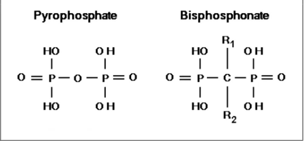
Bisphosphonates are synthetic pyrophosphate analogs with a P-C-P bond instead of the P-O-P bond of inorganic pyrophosphates, which are used as anti-tarter agents in toothpastes and as a bone-specific radionuclide in technetium 99m methylene diphosphonate (Tc 99m MDP) bone scans. Unlike pyrophosphates, bisphosphonates are resistant to breakdown by enzymatic hydrolysis, which explains their accumulation in the bone matrix
47
and their extremely long half-life (212). The P-C-P structure (Figure 2) allows a great number of possible variations, especially by changing the two lateral chains (R1 and R2) in the carbon atom. The two phosphate groups are
essential for binding to bone mineral such as hydroxyapatite and together with the R1 side chain they act as a “bone hook”. A hydroxyl (OH) group or
an amino group at the R1 position increases the affinity for calcium and thus
for bone mineral (213, 214).
Figure 2. The chemical structure of pyrophosphate and bisphosphonate. R1 and R2 signify the side chains of bisphosphonate.
The structure and three-dimensional conformation of the R2 side chain determine the anti-resorptive potency and the enhanced binding to hydroxyapatite (213, 215). It has been shown that bisphosphonates containing a basic primary nitrogen atom in an alkyl chain are 10-100 times more potent at inhibiting bone resorption than earlier-generation bisphosphonates, such as clodronate, which lack this feature. Compounds that contain tertiary nitrogen, such as ibandronate and olpadronate, are even more potent at inhibiting bone resorption. Residronate and zoledronate are among the most potent of bisphosphonates, containing a nitrogen atom
48
within a heterocyclic ring; they are up to 10,000 times more potent than etidronate (216).
The non-nitrogen-containing bisphosphonates (etidronate, clodronate, and tiludronate) inhibit bone resorption by generating the cytotoxic, methylene-containing analogs of ATP that interfere with mitochondrial function and induce apoptosis of osteoclasts (217-219). In contrast, the nitrogen-containing bisphosphonates (alendronate, zoledronate, pamidronate, risedronate, and ibandronate) bind to and inhibit farnesyl pyrophosphate synthase (FPPS), a key enzyme of the mevalonate pathway, thereby preventing the prenylation and activation of small GTPases that are essential for the bone-resorbtion activity and survival of osteoclasts (220-222).
There is no evidence that orally or intravenously administered bisphosphonates are metabolized in animals or humans (223, 224). The gastrointestinal uptake of oral bisphosphonates is low, with a bioavailability of 0.7 % for alendronate (225) and 0.3% for pamidronate (226). The poor absorption of bisphosphonates can probably be attributed to their very poor lipophilicity, which prevents transcellular transport across the epithelial barriers. Consequently bisphosphonates must be absorbed by the paracellular route, which means passage through the pores of the tight junctions between the epithelial cells. Oral absorption of alendronate in rats is fourfold to fivefold higher in the fasted state than in the fed state (227). The same effect of food on the absorption of alendronate has also been observed in healthy volunteers (225).
Intravenous administration of a single dose of alendronate leads to rapid accumulation of this drug in bone tissue: 30% in 5 min and 60% in 1 hour.
49
At 5minutes after dosing, 63% of the dose is present in non-calcified tissues. This drops to about 1% 6-24 hours post-dose (227). The distribution of alendronate in bone is determined by blood flow and favors deposition at sites of the skeleton that is undergoing active resorption (227). Following administration of a single dose of 14C-alendronate in rats, over 70% of the bone resorption surface was densely labeled in comparison with 2% of the bone formation surfaces (228). This preferential localization of alendronate (in areas of high bone turnover) could be due to exposure of hydroxyapatite at sites that are undergoing bone resorption and the accessibility of these bone surfaces to substances in the circulation
Following administration of 14C-alendronate in rats a larger proportion of the dose is taken up by trabecular bone than by cortical bone, and in the latter at the metaphysis rather than the diaphysis (229, 230). Similar results were observed in dogs when etidronate was given intravenously (231). Furthermore, Lin et al. (230) showed that the uptake of alendronate is proportional to the intravenous dose up to 5 mg/kg IV, but at 10 mg/kg or higher, the concentration of drug in bone increases less than linearly
In recent years, it has been hypothesized that another target of bisphosphonates may be osteoblasts, which subsequently influence osteoclasts. The mitogenic effect of bisphosphonates on osteoblasts has been reported by several authors (232, 233). Furthermore, it has been shown that these drugs inhibited the expression of RANKL in a rat osteoblast cell line (234) and increase the expression of OPG in human osteoblastic cells (235), suggesting that the anti-resorptive effect of bisphosphonates is mediated by the influence of osteoblasts on RANKL signaling (234, 235). Moreover, it has been shown that bisphosphonates promote osteoblast
50
differentiation in cultures of osteoblast-like cell lines in a dose-dependent manner and inhibit the osteoblast apoptosis (236). However, the clinical relevance of this is unclear.
Local and systemic delivery, experimental studies
Extraction of teeth necessitated by factors such as developmental problems, trauma, severe periodontal disease, or unsolvable endodontic problems often causes reduction of the residual alveolar ridge height and width in the jaws. These reductions usually cause difficulties in prosthetic restoration, poor aesthetics and insufficient function. Many investigators have shown that tooth extraction stimulates osteoclastic activity with varying amounts of alveolar crest loss (237, 238). Systemic alendronate was found to be significantly effective in reducing bone loss associated with experimental periodontitis in monkeys and beagle dogs (239, 240). Yaffe et al. (241) found that local delivery of alendronate reduced alveolar bone resorption activated by mucoperiosteal surgery. In orthodontics, topical administration of amino bisphosphonate caused significant reduction of tooth movement in
rats, when orthodontic force was applied (242, 243).
Several methods have been reported to increase the bone density around experimental porous implants, but to varying degrees (244-254). In animal models, several investigators have shown that surface-immobilized bisphosphonates improve the mechanical fixation of metal screws in terms of an increased pullout force and bone-to-implant contact (255-259). Yoshinari et al. (256) used plasma-sprayed HA-coated titanium dental implants that were immersed in pamidronate and implanted in mandibular bone of beagles. This study showed a 10% increase in bone contact area. Tengvall et al. (260) showed an increase (by 28%) of the pullout force of