Cell cycle alterations and 11q13 amplification in
breast cancer
-prediction of adjuvant treatment response
Marie Ahnström Waltersson
© 2009 Marie Ahnström Waltersson ISBN 978-91-7393-690-3
ISSN 0345-0082
Published articles have been reprinted with permission of respective copyright holder.
Paper I © Springer
Paper II © Spandidos Publications Paper III © Nature Publishing Group
The growth and development of the breast is to a large extent regulated by oestrogens through the oestrogen receptor (ER). Activation of the ERα triggers transcription of genes that are important for cell proliferation and stimulates entry into the G1 phase of the cell cycle. In breast cancer the ERα is often upregulated and is therefore a suitable target for adjuvant therapies such as tamoxifen. Although tamoxifen is an effective treatment in most cases, tumours sometimes acquire resistance to the drug. The aim of this thesis was to investigate the impact of G1 phase proteins and 11q13 amplification on prognosis and treatment response in breast cancer. The material used was from a clinical trial in which postmenopausal breast cancer patients were randomised to chemotherapy or radiotherapy and tamoxifen or no adjuvant treatment. We studied the expression of cyclin D1, cyclin E and Rb with immunohisochemistry and amplification of CCND1 and PAK1 with real-time PCR. We found that among patients with high tumour expression of cyclin D1, overexpression of ErbB2 was associated with reduced recurrence-free survival. Both cyclin D1 and cyclin E overexpression were associated with reduced tamoxifen response. High expression of cyclin D1 has been found to induce ligand independent activation of ERα in breast cancer cells and might also switch tamoxifen from acting as an antagonist to an agonist. Overexpression of cyclin E has been shown to be associated with expression of low molecular weight isoforms of the protein that possess an increased kinase activity and are insensitive to p21 and p27 inhibition. Furthermore, amplification of 11q13, and in particular the gene PAK1, was a strong predictor of tamoxifen resistance. The pak1 protein is involved in phosphorylation and ligand independent activation of the ERα. We also found that lost expression of either p53 or Rb reduced the patients benefit from radiotherapy compared with patients with normal expression of both proteins. Normally, ionizing radiation upregulates p53 resulting in G1 arrest or apoptosis. If either functional p53 or Rb is missing the cells can proceed from G1 to the S phase despite damaged DNA. The expression of the microRNA, miR-206, was analysed with real-time PCR, and the results showed that high expression of miR-206 correlated to low expression of ERα and 11q13 amplification. In vitro studies have shown that miR-206 negatively regulates the expression of ERα. Taken together the G1 regulators and amplification of 11q13 seem to have an important role in predicting the patient’s response to adjuvant therapy.
Table of contents
Populärvetenskaplig sammanfattning ... 9 Abbreviations... 11 List of publications... 13 Introduction... 15 The breast... 15The oestrogen receptor... 17
Breast cancer... 18 Therapy... 20 Surgery... 20 Radiation ... 20 Chemotherapy... 20 Endocrine treatment ... 21 Herceptin... 21
The cell cycle ... 22
The G1 phase ... 23 Cyclin D1... 24 Cyclin E ... 25 Rb... 26 11q13 amplification ... 26 CCND1... 27 PAK1 ... 27 microRNA... 27 miR-206... 29
Aims of the thesis... 31
Paper I ... 31 Paper II ... 31 Paper III ... 31 Paper IV ... 31 Material ... 33 Patients ... 33 Methods ... 37 Immunohistochemistry ... 37 Real-time PCR ... 38
Real-time PCR detection of micro RNA... 40
Results and discussion... 43
Paper II ... 46 Paper III ... 48 Paper IV... 51 Conclusions ... 53 Acknowledgements ... 55 References ... 57
Populärvetenskaplig sammanfattning
Bröst cancer är den vanligaste cancerformen bland kvinnor i Sverige då omkring 7000 kvinnor får diagnosen varje år. Mammografiscreening och effektivare behandling har förbättrat överlevnaden betydligt under senare år men fortfarande avlider ca 1500 kvinnor i sjukdomen varje år. Tillväxt och utveckling av bröstvävnaden stimuleras av tillväxtfaktorer. Framför allt har hormonet östrogen en framträdande roll. Östrogen aktiverar östrogenreceptorn och stimulerar därigenom bildandet av proteiner som är viktiga för celldelning. Cancer utvecklas genom att fel ackumuleras i de gener som kodar för proteiner som styr celldelning och programmerad självdöd (apoptos). Felen ger cellerna ett evolutionärt övertag genom att inaktivera tillväxtbromsande proteiner och aktivera eller överuttrycka tillväxtfrämjande proteiner. I bröstcancer är ofta östrogenreceptorn överuttryckt vilket leder till ökad stimulering av celldelningen. Genom att behandla bröstcancerpatienter med läkemedel som hämmar östrogenreceptorn kan tumörens tillväxt minskas. Tamoxifen är en hämmande substans som tävlar med östrogen om att binda in till receptorn. Tamoxifen ges som adjuvant (förebyggande) behandling efter operation till kvinnor med tumörer som har höga nivåer av östrogenreceptor för att hindra återfall i sjukdomen. Annan adjuvant behandling som kan ges är strålbehandling och cytostatika. Ofta fungerar den adjuvanta behandlingen mycket bra men vissa patienter utvecklar resistens mot behandlingen och får återfall i sjukdomen.Målet med den här avhandlingen var att undersöka hur förändringar av proteiner och gener inblandade i östrogenreceptorsignalering och celldelning påverkar patienternas nytta av adjuvant behandling. Genom att öka förståelsen för vad som gör att resistens mot behandling uppstår skapas nya möjligheter att ta fram effektivare läkemedel. Det material som användes var fryst tumörvävnad från postmenopausala kvinnor som randomiserats till att få strålbehandling och tamoxifen, enbart strålbehandling, cytostatika och tamoxifen eller enbart cytostatika.
Våra resultat visade att höga nivåer av proteinerna cyclin D1 och cyklin E båda var associerade till sämre nytta av tamoxifenbehandling. Andra studier har visat att när cyklin D1 är överuttryckt kan det hjälpa till att aktivera östrogenreceptorn även när östrogen inte är bundet till receptorn vilket kan leda till tamoxifenresistens. Överuttryck av cyklin E innebär ofta att även kortare versioner av proteinet finns i tumören och dessa har en högre aktivitet
än normalt cyclin E och de är också okänsliga för vissa inhiberande signaler. Vi såg också att onormalt antal kopior av gener i kromosomområdet 11q13 var starkt korrelerat till tamoxifenresistens. I området finns bland annat genen som kodar för cyklin D1 och den som kodar för pak1, ett protein med många funktioner och som är inblandat i aktivering av östrogenreceptorn. Proteinerna p53 och Rb är tillväxthämmande proteiner och p53 stoppar också celldelningen när cellen utsatts för DNA-skada, till exempel efter exponering av strålning. Vi såg att förlust av antingen p53 eller Rb var associerat till sämre svar på strålbehandling jämfört med om båda proteinerna var normala. MicroRNA är små molekyler som hämmar bildandet av proteiner genom att binda till den RNA-mall som används vid syntesen. Vi analyserade microRNA:t miR-206 och såg att höga nivåer av miR-206 var associerat till låga nivåer av östrogenreceptorn. Experimentella studier på celler har tidigare visat att miR-206 är inblandat i regleringen av östrogenreceptorn.
Sammantaget visar våra resultat att de studerade generna och proteinerna sannolikt spelar en stor roll i mekanismerna bakom hur patienter svarar på adjuvant behandling.
Abbreviations
Akt v-akt murine thymoma viral oncogene homolog AF-1 activation function 1
AF-2 activation function 2 CAK cyclin activating kinase
cAMP cyclic adenosine monophosphate
CCND1 the cyclin D1 gene
CDK cyclin dependent kinase
cDNA complementary DNA
CKI cyclin dependent kinase inhibitor
CMF cyclophosphamide, methotrexate, fluorouracil DAB 3,3-diaminobenzidine tetrahydrochloride DGCR8 DiGeorge syndrome critical region gene 8 DNA deoxyribonucleic acid
ER oestrogen receptor
ErbB2 HER2, human epidermal growth factor 2 ERE oestrogen response element
ESR1 ERα gene
FGF fibroblast growth factor
G0 gap 0
G1 gap 1
G2 gap 2
GSK3β glycogen synthase kinase 3 beta
Gy Grey
HRP horse radish peroxidase
IHC immunohistochemistry
INK4 inhibitor of cyclin-dependent kinase 4 MAPK mitogen-activated protein kinase miRISC miRNA-induced silencing complex
miRNA microRNA
M-phase mitosis
mRNA messenger RNA
pak1 protein p21-activated kinase 1
PAK1 the pak1 gene
PCR polymerase chain reaction PI3K phosphatidyl inositol 3 kinase
PgR progesterone receptor
Ras rat sarcoma viral oncogenes homolog
Rb retinoblastoma protein
RNA ribonucleic acid
RT radiotherapy
RT-PCR reverse transcriptase polymerase chain reaction S-phase synthesis phase
SRC-1 steroid receptor coactivator-1
TAM tamoxifen
List of publications
Marie Ahnström, Bo Nordenskjöld, Lars Erik Rutqvist, Lambert Skoog and Olle Stål
Role of cyclin D1 in ErbB2-positive breast cancer and tamoxifen resistance
Breast Cancer Research and Treatment (2005) 91:145-151
Marie Ahnström Waltersson, Marie Stenmark Askmalm, Bo Nordenskjöld, Tommy Fornander, Lambert Skoog and Olle Stål
Altered expression of cyclin E and the retinoblastoma protein influences the effect of adjuvant therapy in breast cancer
International journal of oncology (2009) 34:441-448
Josefine Bostner, Marie Ahnström Waltersson, Tommy Fornander, Lambert Skoog, Bo Nordenskjöld and Olle Stål
Amplification of CCND1 and PAK1 as predictors of recurrence and tamoxifen resistance in postmenopausal breast cancer
Oncogene (2007) 26:6997-7005
Marie Ahnström Waltersson, Elin Karlsson, Bo Nordenskjöld, Tommy Fornander, Lambert Skoog and Olle Stål
miR-206 expression is downregulated in cyclin D1 amplified breast tumours
Manuscript
Introduction
Cancer arises through genetic and epigenetic changes that affect the protein expression pattern of the normal cells. Accumulation of alterations in the systems controlling growth and survival provides the cells with a growth advantage where they no longer respond to repressive signals. This will eventually result in tumour progression. The combination of different alterations within a tumour decides how well the patient will respond to different treatments. The objective of this thesis was to identify deregulated genes and proteins that can predict the efficiency of adjuvant therapy in breast cancer. This kind of knowledge is vital to be able to give each patient the best treatment available. It is not only important to provide the correct treatment, but also to avoid giving unsuitable medication, as that can cause unnecessary side effects and perhaps even promote tumour growth.The breast
The mammary glands develop from the ectodermal ridges at six weeks gestation. During the remainder of the gestation, small primitive ductal structures branch out from the nipple (E. Anderson & Clarke, 2004; Geddes, 2007). From birth up to puberty the mammary glands grow in proportion to the rest of the body. At puberty the mammary glands evolve under the influence of growth hormones, mainly oestrogen and progesterone. The ducts
elongate and branch into an extensive ductal network. The increase in size is mainly due to adipose tissue. During the menstrual cycles the ductal system grows and regresses following the plasma levels of endocrine hormones.
areola ducts
lobules with alveoli
areola ducts
lobules with alveoli
areola ducts
lobules with alveoli
Figure 1. The anatomy of the mammary glands.
The adult breast consists of glandular and adipose tissue supported by Cooper’s ligaments. The glandular tissue consists of around 10 ducts branching close to the areola into tree-like structures ending with the lobules (Figure 1) (Ramsay et al., 2005). The lobules contain 10-100 alveoli in which the milk is produced. The ductal system is lined with a single layer of luminal epithelial cells surrounded by a layer of myoepithelial cells (E. Anderson & Clarke, 2004). The basal membrane is found outside the myoepithelial cells. The luminal cells proliferate in response to endocrine signals, whereas the myoepithelial cells rarely proliferate at all. During pregnancy the ductal branching continues, and lobules and alveoli enlarge (Geddes, 2007). Contrary to previous belief the ducts seem to function as a transport system for the milk during lactation rather than a reservoir (Geddes, 2007; Ramsay et al., 2005).
The glandular tissue of the mammary gland grows in response to oestrogens and progesterones (E. Anderson & Clarke, 2004). Receptors for these hormones are only present in a proportion of the luminal cells and not at all in the myoepithelial cells. Instead growth is induced in a paracrine manner when the hormones induce transcription of other growth factors by binding to their receptors. The exception is oestrogen receptor (ER) β, which is expressed in equal amounts in all the cell types of the breast.
The oestrogen receptor
The growth of the mammary gland is dependent on oestrogen signalling. Oestrogens are steroid hormones and the most common are; 17β-oestradiol (E2), oestrone and oestrol of which 17β-oestradiol is the most abundant and potent (Hall et al., 2001; Speirs & Walker, 2007). Oestrogens mediate their effect through the oestrogen receptors α and β. Before menopause, the ovaries are the main source of oestrogens.
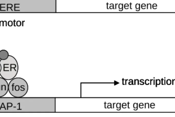
The oestrogen receptors are transcription factors that belong to the steroid receptor super family (Hall et al., 2001; Speirs & Walker, 2007). Both receptors have a DNA (deoxyribonucleic acid) binding domain, a ligand binding domain and two activating domains, viz. the ligand independent activation function 1 (AF-1) and the ligand dependent activation function 2 (AF-2). However, the AF-1 domain in ERβ is non-functional (Hall & McDonnell, 1999). In the absence of a ligand, the ER is sequestered in a large inhibitory complex in the nucleus (Bohen et al., 1995). On ligand binding, the ER is released from the inhibitory complex, and a conformational change enables receptor dimerisation and recruitment of cofactors. The conformational change also displays the AF-2 domain, where coactivators can bind and phosphorylate ER. The receptors can dimerise in homo or hetero complexes. ERβ is thought to have a repressive effect on transcription in breast tissue both in homodimers and in complex with ERα (Hall & McDonnell, 1999). ERα can activate transcription in several ways (Figure 2). The classical pathway involves direct binding to oestrogen responsive elements (ERE) in the promoter region of the genes and thereby activation of the transcriptional machinery (Klein-Hitpass et al., 1988). Around one third of all ER-responsive genes lack the ERE sequence. Instead activation is thought to function through secondary binding to AP-1 responsive elements via fos and jun (Jakacka et al., 2001; Webb et al., 1999). ERα also has non-genomic functions by signalling from the cell membrane.
Overexpression of coactivators and downregulation of corepressors have been seen in breast tumours. Deregulation of these proteins can lead to ligand independent activation of the ER and uncontrolled growth.
target gene AP-1 promotor ER ER E E CO CO target gene ERE promotor transcription a b jun fos ER CO E transcription target gene AP-1 promotor target gene AP-1 promotor ER ER E E CO CO target gene ERE promotor transcription transcription a b jun fos ER CO E transcription transcription
Figure 2. Mechanisms of ER-induced transcription. The classical pathway
where the ER complex binds to an ERE sequence in the target gene promoter (a) and signalling through the jun and fos transcription factors (b).
Breast cancer
The most common cancer form among Swedish women is breast cancer; approximately 7,000 women are diagnosed with the disease every year. Although the incidence is slowly increasing, mortality is decreasing; about 1,500 women die from breast cancer each year (Figure 3). The decrease in mortality is achieved by more effective treatment and early diagnosis with mammography screening. The high survival rates mean that around 82,000 women live with their disease in Sweden.
Most known risk factors for developing breast tumours concern the life exposure of the breast to oestrogen (Hankinson et al., 2004; Trichopoulos et al., 2008). Such factors include time of menarche and menopause, which will directly affect the number of ovulations and thereby also the amount of hormones released. High endogenous oestrogen levels and hormone replacement therapy also increase risk. Pregnancy early in life reduces the total risk of developing a breast tumour after menopause but increases the risk of developing a tumour within the next decade, probably by promoting an
initiated cancer with the temporarily high levels of hormones. During pregnancy, the mammary tissue matures into a state in which it is less susceptible to cancer initiation, hence the decreased long-term risk. Risk factors unrelated to hormone exposure are ionising radiation and family history of breast cancer. 0 50 100 150 200 250 1960 1970 1980 1990 2000 Year /1 00 00 0 Incidence Mortality
Figure 3. Comparison of breast cancer incidence and mortality rates over time
among women in the southeast of Sweden (Onkologiskt centrum, 2009).
After diagnosis it is important to predict the patient’s prognosis in order to choose the most effective treatment (Greene & Sobin, 2008). The tumour stage is determined with the TNM (tumour-node-metastasis) classification. The TNM classification describes the anatomic extent of the cancer and separately classifies primary tumour size (T), the amount of involved lymph nodes (N), and metastatic cancer, spread beyond the lymph nodes (M). The scores of the three classifications are then weighted together in a stage from 0 to IV, of which Stage 0 is non-invasive cancer in situ, Stages I-II indicate increasing tumour sizes, Stage III denotes involved lymph nodes and Stage IV indicates the presence of distant metastasis. Other important factors for choice of treatment are histological grade, fraction of cells in the S phase (synthesis phase) and expression of the ERα, PgR (progesterone receptor) and ErbB2 (HER2, human epidermal growth factor 2) receptors.
Most breast tumours arise from the epithelial cells of the terminal ducts or the alveoli. The aetiology of breast cancer is not clear but the recent availability of
microarray technology has revealed several distinct subtypes based on the gene expression patterns of the tumours, suggesting separate origins (Calza et al., 2006; Perou et al., 2000; Sorlie et al., 2001). Most ER-positive tumours cluster to the groups luminal A, characterised by expression of ER-responsive genes and normal p53, and luminal B, with low expression of ER-responsive genes and frequent p53 mutations. The ER-negative tumours fall into the groups basal-like, with low expression of ErbB2 and PgR, the ErbB2+ group, overexpressing genes in the ERbB2 locus, and normal-like, expressing high levels of genes normally expressed by non-epithelial cells. Patients in the ErbB2+ and basal-like groups show the worst prognosis of these types, whereas patients with luminal A tumours have a good prognosis. Luminal B seems to be associated with reduced response to adjuvant tamoxifen treatment despite the expression of ERα.
Therapy
Surgery
Surgery is the most important treatment for breast cancer (Holmberg, 1995). If the tumour is found in an early stage, breast conserving surgery is used. If the tumour is larger than 4 cm, is inflammatory or has a diffuse growth, mastectomy is chosen. Axillary dissection is mainly a diagnostic procedure and is often done with the sentinel node method (Giuliano et al., 1994). The sentinel node - the first lymph node to drain the tumour - is removed and analysed during the operation. If there are tumour cells in the sentinel node, a complete axillary dissection is done. Surgery is almost always followed by other therapies.
Radiation
Radiotherapy (RT) is used as an adjuvant treatment after surgery to decrease the risk of local recurrence (Rutqvist et al., 2003). If lymph nodes are involved, the axillary is radiated to prevent additional spreading. Radiation is given in 25 fractions during 5 weeks, with a total dose of 50 Grey (Gy). Irradiation induces DNA damage in the cells both by the direct exposure to the radiation and through the secondary effect of free radicals created by the irradiation. The DNA damage is too severe for the cell to repair and induces cell cycle arrest and apoptosis.
Chemotherapy
Adjuvant chemotherapy is given to reduce the risk of remaining cancer cells establishing metastatic growth distant from the primary tumour (Early Breast
Cancer Trialists' Collaborative Group, 2005). A combination of different cytotoxic drugs has proven more efficient than monotherapy. Cytotoxic drugs affect the cell’s ability to divide by interfering with the replication, or mitotic, machinery. The patients in the present thesis were given CMF (cyclophosphamide, methotrexate, fluorouracil) when randomised to chemotherapy. However, it is now established that antracycline-based chemotherapy or antracycline combined with Taxol is more effective.
Endocrine treatment
Approximately 70% of the women diagnosed with breast cancer have tumours that express high levels of ERα. These tumours are likely to depend on oestrogen for their growth. By giving the patients a substance that inhibits the actions of oestrogen, tumour progression can be repressed. Tamoxifen (TAM) is a weak agonist to ERα and binds to ERα with higher affinity than oestrogen (Howell et al., 2000). When tamoxifen binds ERα, the conformational change allows receptor dimerisation and DNA binding. The AF-2 domain, however, remains inactive and unable to recruit cofactors, and transcription is inhibited. Tamoxifen is used as an adjuvant therapy to reduce the risk of tumour relapse, usually with good results and few side effects (Clemons et al., 2002). However, some tumours are resistant or acquire resistance over time to tamoxifen. The mechanisms behind resistance are not clear but are thought to involve loss of ERα dependence or upregulation of coactivators. Overexpression of coactivators has been shown to activate ERα in the absence of ligand and even increase the agonistic effects of tamoxifen in experimental models(Osborne et al., 2003; Zwijsen et al., 1998).
A number of alternatives to tamoxifen also modulate the actions of ERα. Faslodex is a compound which associates with ERα with high affinity, but unlike tamoxifen it does not have agonistic properties. Instead, it marks the receptor for nuclear export and degradation (Howell et al., 2000). For premenopausal women goserelin is often used. Goserelin is an analogue of the gonadotropin releasing hormone and inhibits oestrogen production in the ovaries (Cheer et al., 2005). Aromatase inhibitors interfere with the conversion of androgens to oestrogens (Trunet et al., 1997).
Herceptin
ErbB2 is a transmembrane receptor tyrosine kinase which is amplified and overexpressed in around 20% of all breast cancers (Yeon & Pegram, 2005). ErbB2 is involved in the mitogen-activated protein kinase (MAPK) and phosphatidyl inositol 3 kinase (PI3K) signalling pathways. Overexpression of this receptor is associated with worse prognosis in breast cancer. Herceptin is
an antibody designed to target the ErbB2 receptor and thereby diminish its effects. Herceptin is given to women with tumours overexpressing ErbB2.
The cell cycle
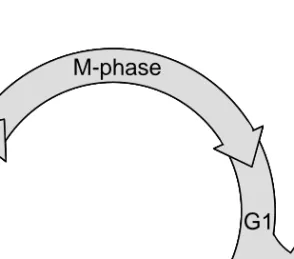
All living organisms are dependent on controlled cell division to develop and reproduce. The control of the cell division is maintained in the cell cycle, in which the goal is to produce two identical daughter cells (Leake, 1996; Vermeulen et al., 2003). To accomplish that, the cell’s DNA is duplicated (replication) preceding the cell division in the cycle. As correct cell division is vital to the organism, the cell cycle is highly regulated. The cell cycle is divided into four phases (Figure 4). The M phase (mitosis), in which cell division takes place, and S phase, in which the DNA is replicated, are separated by two checkpoints, gap 1 (G1) and gap 2 (G2), which regulate entry into the next phase. During G1 and G2, the cells grow and prepare for mitosis. The regulatory system checks for DNA damage and other abnormalities and if there is any damage to the cell the cycle will stop at the restriction point and either repair the damage or commit suicide (apoptosis). The phases in the cell cycle are regulated by cyclins and cyclin dependent kinases (CDK). The cyclins are expressed in a cyclic manner and form complexes with the more stably expressed CDKs. The complexes then phosphorylate key components of the cell cycle machinery to enable progression through the cycle. The G1 phase is triggered by growth factors. In the absence of stimulating factors, the cell will leave the cycle and enter a resting state, gap 0 (G0), instead of proceeding through the G1 phase and into the S phase.
S-phase
G1
G2
M-phase
G0
S-phase
G1
G2
M-phase
S-phase
G1
G2
M-phase
G0
During cancer development, the equilibrium between proliferation and cell death among the cells is disrupted. When mutations arise in important regulators of the cell cycle, the cell acquires a growth advantage and, when enough alterations have accumulated, proliferates uncontrollably (Sandal, 2002).
The G1 phase
The G1 phase regulates the cells entry into the S phase (Figure 5). In response to mitogenic signals - in breast tissue often the growth hormone oestrogen - cyclin D is expressed (Sherr & Roberts, 1999; Steeg & Zhou, 1998). Cyclin D forms a complex with CDK4 or CDK6, and this complex phosphorylates the retinoblastoma protein (Rb). Phosphorylation of Rb releases members of the transcription factor E2F family, which is responsible for transcription of proteins needed in the S phase. E2F also enables transcription of cyclin E, which forms a complex with CDK2 to continue the phosphorylation of Rb throughout the G1 phase. The CDK/cyclin complexes are transported into the nucleus and activated by CDK-activating kinases (CAK) before phosphorylating Rb. transcription
G1
Rb E2F E2F CDK4/6 cyclin D p21 p27 mitogens p21 p27 cyclin E CDK2 P P p21 and p27 titrationS
Rb P P P Rb E2F P transcriptionG1
Rb E2F Rb Rb E2F E2F E2F CDK4/6 cyclin D p21 p27 CDK4/6 cyclin D p21 p27 mitogens p21 p27 cyclin E CDK2 p21 p27 cyclin E CDK2 P P p21 and p27 titrationS
Rb P P P Rb Rb P P P Rb E2F P Rb E2F Rb E2F PFigure 5. The G1 phase of the cell cycle.
The CDK/cyclin complexes are also regulated by CDK inhibitors (CKI) (Sherr & Roberts, 1999). The CKIs can be divided into two groups: the INK4 (inhibitor of cyclin-dependent kinase 4) proteins, including p16 and p19, that
specifically inhibit the activity of CDK4 and CDK6, and the Cip/Kip family acting on cyclin D-, E- and A-dependent kinases. The Cip/Kip family includes p21 and p27. When bound to the CDK4/6-cyclin D complex, p21 and p27 act as stabilisers and enable transport into the nucleus (Cheng et al., 1999; LaBaer et al., 1997). On the other hand, when associated with the CDK2-cyclin E complex, they act as inhibitors. As cyclin D levels rise in response to mitogens, the increased number of CDK4/6-cyclin complexes recruit p21 and p27 from the CDK2-cyclin E complex, leaving it active and able to continue the phosphorylation of Rb (Polyak et al., 1994; Sherr & Roberts, 1999). As the amount of active CDK2-cyclin E complexes rises they start to phosphorylate p27, marking it for ubiquitination, hence further increasing complex activity (Sheaff et al., 1997). When mitogens signalling declines, cyclin D is degraded and the inhibitors move back to cyclin E. Rb is kept phosphorylated throughout the cycle by CDK-cyclin complexes in the other phases.
Cyclin D1
Cyclin D1 is one of three D-type cyclins and, unlike cyclin D2 and D3, it has been found altered to a high extent in breast cancer. Overexpression of cyclin D1 has been found in 40-70% of all breast cancers and is strongly associated with ER-positive tumours (Elsheikh et al., 2008; Gillett et al., 1996; van Diest et al., 1997). Its relation to prognosis is not as clear. Some studies have shown a correlation between high cyclin D1 expression and increased survival (Gillett et al., 1996; Nielsen et al., 1997; Stendahl et al., 2004), whereas others have not (Reed et al., 1999; van Diest et al., 1997). Kenny et al (1997) report worse prognosis among patients with high expression of cyclin D1 mRNA.
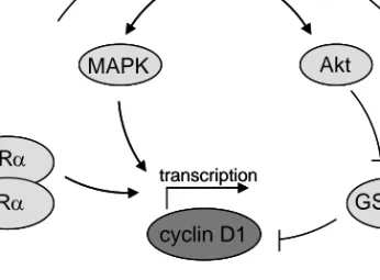
The expression of cyclin D1 in breast tissue is regulated by both ErbB2 and the ERα (Figure 6). Activation of the ERα by oestradiol leads to transcription of the cyclin D1 gene, not through the classical oestrogen response element (ERE) sequence which is absent in the cyclin D1 gene but through a cAMP (cyclic adenosine monophosphate) response element (Doisneau-Sixou et al., 2003; Sabbah et al., 1999). Breast tumours often have high expression of both ERα and oestradiol and therefore also have high levels of cyclin D1. Downregulation of the cyclin D1 protein is facilitated by glycogen synthase kinase 3 beta (GSK3β), which phosphorylates cyclin D1 thereby marking it for ubiquitination (Diehl et al., 1998; Lenferink et al., 2001). GSK3β is regulated negatively by the ErbB2/Akt (v-akt murine thymoma viral oncogene homolog) pathway. Dysregulation of members in this pathway may lead to accumulation of cyclin D1 protein. ErbB2 also regulates cyclin D1 on the transcriptional level by activating the MAPK pathway.
cyclin D1 Akt MAPK GSK3-β ERα ERα transcription ErbB2 cyclin D1 cyclin D1 Akt Akt MAPK MAPK GSK3-β GSK3-β ERα ERα ERα ERα transcription ErbB2
Figure 6. Regulation of cyclin D1 expression.
Apart from its central role in the cell cycle, cyclin D1 also functions as a transcription factor coactivator (Neuman et al., 1997; Zwijsen et al., 1998). In breast tissue, cyclin D1 binds to ERα along with other cofactors and increases its transcriptional activity. When overexpressed, cyclin D1 is believed to be able to activate ERα in the absence of oestradiol, thus creating a positive feedback loop for its own expression.
Cyclin E
Cyclin E has been found overexpressed in 10-50% of all breast cancers (Lindahl et al., 2004; Milde-Langosch et al., 2000; Nielsen et al., 1996). High expression of cyclin E tends to correlate to ER-negative tumours, tumour size and increased proliferative activity (Aaltonen et al., 2009; Lindahl et al., 2004). Several studies have shown that overexpression of cyclin E is associated with worse prognosis (Kuhling et al., 2003; Lindahl et al., 2004; Nielsen et al., 1996). Unlike cyclin D1, expression of cyclin E is not regulated by mitogens. Instead the release of E2F from Rb triggers its transcription. Apart from being overexpressed, cyclin E is also proteolytically processed to produce up to 5 low molecular weight isoforms in tumour cells (Porter et al., 2001; X. D. Wang et al., 2003). These isoforms are not present in normal cells (Harwell et al., 2000). The isoforms bind CDK2 more efficiently than the full-length protein and enhance the kinase activity of the complex (Akli et al., 2004). Additionally, these isoforms are insensitive to p21 and p27 inhibition.
Rb
The Rb family consists of the three members Rb/p105, Rb2/p130 and p107 (Sun et al., 2007). All three members are involved in cell cycle control, but whereas Rb/p105 is expressed continuously through the whole cycle, Rb2/p130 and p107 are predominantly expressed during G1 and G0. The Rb proteins function as cell cycle regulators by binding members of the transcription factor family, E2F. There are activating and repressive E2Fs (Cam & Dynlacht, 2003). Rb/p105 associates with and inactivates the activating E2Fs. Rb2/p130 and p107 binds repressive E2Fs and these complexes mediate histone deacetylation and chromatin condensation, hence repressing the transcription of proteins needed for S-phase entry (Ferreira et al., 1998). On mitogen signalling, the Rb proteins are phosphorylated and the E2Fs released. As a result, repressive E2Fs can no longer promote chromatin condensation, and the activating E2Fs are free to transcribe S phase promoting proteins (Sun et al., 2007).
Although loss of Rb expression is reported in 20-30% of all breast cancers, most studies have failed to see any prognostic importance of the loss (J. J. Anderson et al., 1996; Pietilainen et al., 1995; Sawan et al., 1992). Rb expression does not seem to be connected with ER status, although Ceccarelli et al. (1998) reported that tumours with lost Rb are predominantly ER negative.
11q13 amplification
Gene amplification is a common event and an activator of oncogenes in human cancer (Albertson, 2006; Myllykangas & Knuutila, 2006). Amplifications are defined as an abnormal number of copies of a gene present in the genome, often amplified in combination with other genes in an amplicon 0.5-10Mb in length. Gene amplification is believed to be a non-random event and is more common at fragile sites in the genome. Fragile sites are often present in the outer regions of amplicons. Amplified regions are presumed to contain one or several oncogenes, with the increased number of copies providing the cells with a selective advantage (Ormandy et al., 2003).
The chromosomal region, 11q13, is found amplified at a rate of around 15% in breast cancer (Karlseder et al., 1994; Ormandy et al., 2003). This is a gene-rich area with several oncogene candidates such as CCND1 (the cyclin D1 gene) and
PAK1 (the pak1 gene) (Albertson, 2006). The amplicon consists of 4-8 core
regions that can be amplified together or independently (Albertson, 2006; Ormandy et al., 2003). Although amplifications can result in up to 500 copies of a gene the 11q13 area usually only has a 5-10 fold increase in copy numbers when amplified (Schwab, 1998). Amplification of 11q13 shows strong
associations with ER-positive cancer and has also been reported to be related to worse prognosis (Al-Kuraya et al., 2004; Bieche et al., 2002; Schuuring et al., 1992).
CCND1
CCND1, encoding the cell cycle protein cyclin D1 is the most extensively
studied gene in the 11q13 amplicon and is found amplified in around 10-15% of all breast cancers (Bieche et al., 2002; Cuny et al., 2000). As with the whole 11q13 area CCND1 amplification is strongly associated with ER positivity and reduced recurrence-free survival. CCND1 is often coamplified with other genes in 11q13, especially the fibroblast growth factors (FGF) FGF3, FGF4 and
FGF19 (Haverty et al., 2008; Lammie et al., 1991; Theillet et al., 1990). These
genes are situated very close to and are seldom amplified independently of
CCND1.
PAK1
PAK1 encodes the protein, p21-activated kinase 1 (pak1). This gene has not
been studied as intensely as CCND1, but amplification has been reported in both breast cancer and ovarian cancer in frequencies between 10-20% (Lundgren et al., 2008; Schraml et al., 2003). Coamplification with CCND1 has also been observed.
The pak1 protein is a serine/threonine kinase which is activated by small GTPases in response to growth factors such as ERα (Rayala et al., 2006a; Rayala et al., 2006b). pak1 is involved in cytoskeletal reorganisation, motility and cell survival (Coniglio et al., 2008; Li et al., 2008; Vadlamudi et al., 2000). pak1 can also activate ERα by phosphorylation of ser-305 in the AF-2 domain (Rayala et al., 2006b; R. A. Wang et al., 2002). This interaction promotes ligand independent activation and transcription of ERα responsive genes such as cyclin D1. Overexpression of pak1 has been reported in breast cancer; both in vitro studies and studies on tumour material show that pak1 overexpression is involved in tamoxifen resistance and invasive tumour type (Holm et al., 2006; Rayala et al., 2006b; Vadlamudi et al., 2000).
microRNA
microRNAs (miRNA) are small non-coding RNA (ribonucleic acid) molecules which regulate gene expression on the translational level (Filipowicz et al., 2008). The single stranded miRNA can either suppress translation by imperfect base pairing to the 3’ untranslated region of the target mRNA or by perfect complementary binding to the protein-coding sequence leading to degradation
of the mRNA. Suppression of translation rather than degradation of the mRNA is the most common mechanism in mammals. Many miRNAs are tissue-specific and control the gene expression profile of that tissue by suppression of multiple mRNAs (messenger RNA) (Lim et al., 2005).
Drosha DGCR8 nucleus Exportin Dicer cytoplasm miRISC RNA polymerase II Drosha DGCR8 Drosha DGCR8 nucleus Exportin Exportin Exportin Dicer Dicer cytoplasm miRISC RNA polymerase II
Figure 7. The mechanism of miRNA maturation.
miRNAs are transcribed by RNA polymerase II from either separate miRNA genes or from introns within protein-coding genes (Figure 7)(Y. K. Kim & Kim, 2007; Y. Lee et al., 2004). The transcribed molecule is the miRNA precursor pri-miRNA (Esquela-Kerscher & Slack, 2006). The pri-miRNA consists of a single-stranded RNA molecule with an imperfect hairpin structure. The flanking single-stranded sequences are spliced away by the Drosha-DGCR8 (DiGeorge syndrome critical region gene 8) complex to produce a pre-miRNA consisting of only the approximately 70 nucleotide large hairpin structure (Esquela-Kerscher & Slack, 2006; Y. K. Kim & Kim, 2007). The pre-miRNA is transported to the cytoplasm by exportin-5 and there further processed into the approximately 20 nucleotide single-stranded miRNA by the Dicer enzyme (Bohnsack et al., 2004; Filipowicz et al., 2008; Hutvagner et al., 2001). The mature miRNAs associate with miRNA-induced silencing complexes (miRISCs) and the whole complex is needed for translational repression.
miR-206
The miRNA miR-206 is normally tissue-specific to skeletal muscle (McCarthy, 2008). There it regulates differentiation through repression of DNA synthesis and induction of cell cycle arrest (H. K. Kim et al., 2006; Rao et al., 2006). A direct target for miR-206 in skeletal muscle is DNA polymerase α which is important in DNA synthesis. Expression of miR-206 has also been detected in breast cancer (Iorio et al., 2005; Kondo et al., 2008). Studies on breast cancer cells have shown that miR-206 can negatively regulate the expression of ERα (Adams et al., 2007; Kondo et al., 2008). According to those results, the expression levels of miR-206 in breast tumours are associated with ER-negativity (Iorio et al., 2005; Kondo et al., 2008).
Aims of the thesis
Paper I
To investigate the expression of cyclin D1 and its prognostic and treatment predictive role in postmenopausal breast cancer both alone and when coexpressed with ErbB2.
Paper II
To analyse the expression of cyclin E and loss of Rb in relation to treatment effect and prognosis in postmenopausal breast cancer.
Paper III
To study how amplification of CCND1 and PAK1 affect the postmenopausal breast cancer patient’s prognosis and response to adjuvant treatment and to analyse the importance of pak1 overexpression.
Paper IV
To explore whether tumours with 11q13 amplification, in particular those with
CCND1 amplification, are less prone to express the micro RNA miR-206 than
tumours with normal copy numbers, and whether miR-206 expression is related to oestrogen receptor expression.
Material
Patients
The patient material used in the present thesis was from a randomised trial opened in 1976 and closed in 1990 (Rutqvist & Johansson, 2006). The trial, conducted by the Stockholm Breast Cancer Study Group, included postmenopausal women with operable unilateral breast cancer with either a tumour diameter exceeding 30mm or lymph node metastases. The tumours were removed with modified radical mastectomy.
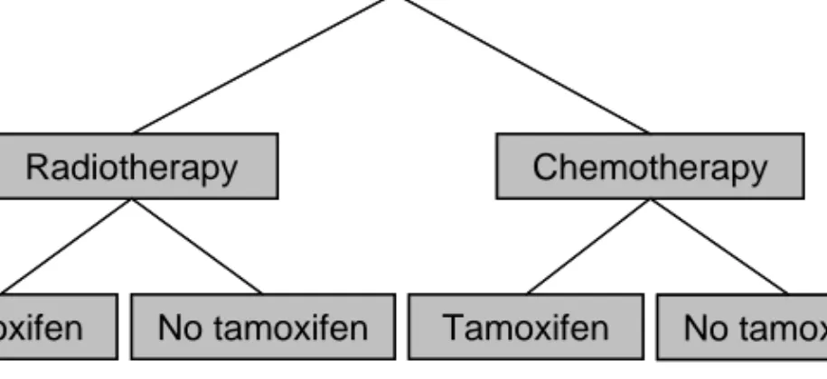
The aim of the trial was to investigate the benefits of postoperative radiation versus adjuvant chemotherapy. When the study opened, the standard treatment for high-risk node-positive patients was primary surgery and postoperative radiation. Adjuvant chemotherapy was still considered experimental. The study also included a tamoxifen trial. In 1976 little was known about the effects and mechanisms of tamoxifen, and only a few small clinical studies had been conducted (Cole et al., 1971; Ward, 1973). Hence, tamoxifen was given to both oestrogen receptor positive and negative patients. The patients were randomised into four groups; radiotherapy alone, radiotherapy and tamoxifen, chemotherapy alone, or chemotherapy and tamoxifen (Figure 8).
The trial was closed in 1990 when convincing evidence of the benefit of chemotherapy and tamoxifen was available and withholding this treatment from the patients became unethical.
Postmenopausal breast cancer patients
Radiotherapy Chemotherapy
Tamoxifen No tamoxifen Tamoxifen No tamoxifen
Postmenopausal breast cancer patients
Radiotherapy Chemotherapy
Tamoxifen No tamoxifen Tamoxifen No tamoxifen
Figure 8. Randomisation of the patients into treatment groups.
A total of patients 679 patients entered the trial. Parts of the tumour tissue were frozen awaiting receptor determination, and the remaining tissue after those analyses was kept frozen for research purposes. The material left consisted of tumours from 284 patients with a distribution of tumour and treatment characteristics corresponding to the original material (Table 1). The tumour tissue was used for frozen tissue sections and preparation of DNA and RNA. In the immunohistochemical studies in the present thesis, all patients from whom sections were still available were included. In the amplification analysis only tumours with at least 50% tumour cells, as determined by light microscopy evaluation, and where DNA was still available were used.
This material presents a unique opportunity to study the impact of biological factors on prognosis and treatment response in breast cancer. The randomised trial includes well-documented patient data and long-term follow-up. Due to the unethical aspect of withholding tamoxifen treatment from patients, it is no longer possible to obtain an untreated patient group in new trials. Therefore this material holds valuable possibilities to compare treated and untreated patients.
The original data set n=679
The subset of patients available for further studies n=284 %(n) Age <60 54(365) 55(154) >60 46(314) 45(126) Lymph node status N0 13(77) 11(31) N1-3 62(381) 58(161) N4+ 25(155) 31(88) Tumour size <20mm 44(293) 43(119) >20mm 56(368) 58(161) ER status Negative 23(138) 30(83) Positive 77(456) 70(196) Postoperative treatment RT 22(148) 21(59) RT/TAM 24(160) 23(64) CMF 27(182) 29(83) CMF/TAM 28(189) 27(78)
Table 1. Comparison of the whole patient material from the clinical trial with
the subset available for research purposes.
Methods
Immunohistochemistry
Immunohistochemistry (IHC) is a common method of detecting proteins in tissue specimens. Antibodies have a fundamental role in this method, but the visualisation can be done in several different ways. Primary antibodies are created by administering the protein or part of the protein of interest into animals, most commonly rabbit or goat. The animal’s immune system will produce antibodies against the foreign protein, and these can be harvested through the animal’s blood and purified by affinity chromatography. Antibodies produced in this way are polyclonal, a mix of antibodies against several different epitopes on the same protein. Monoclonal antibodies, i.e. all antibodies in the sample are immunochemically identical, are produced by an individual clone of plasma cells, most often from mice. The results are analysed through a microscope, which allows the localisation of the protein to be detected in the cells.
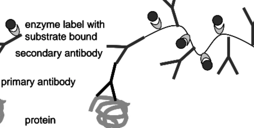
In the present thesis, we have used a two-step indirect immunohistochemical method by which an unlabeled primary antibody first reacts with the tissue antigen. Then a secondary antibody, which is directed against the animal in which the primary antibody is produced, is applied and reacts with the primary antibody (Figure 9). In Papers I and II a biotinylated Multilink secondary antibody was used. Multilink is a larger construct than an ordinary antibody and is used to enhance the signal as it has more binding sites for biotin than an ordinary antibody. Biotin conjugation is used because of its high affinity for
streptavidin, which can be enzyme labelled. Here we have used streptavidin with horseradish peroxidase (HRP) conjugation. When substrate is added, a colour is produced and the intensity of the colour indicates the extent to which the protein is expressed. In Paper III the EnVision technology was used. The EnVision molecule is a polymer of dextran. It binds a large amount of secondary antibodies and even more enzyme molecules, which enhances the signal. The EnVision system is faster than the classical method because the enzyme is bound directly to the polymers and the streptavidin-HRP incubation step is excluded. The HRP substrate used for both methods was 3,3-diaminobenzidine tetrahydrochloride (DAB), which gives a brown colour in the part of the cell in which the protein is located.
protein primary antibody
secondary antibody enzyme label with substrate bound
a b
protein primary antibody
secondary antibody enzyme label with substrate bound
protein primary antibody
secondary antibody enzyme label with substrate bound
protein primary antibody
secondary antibody enzyme label with substrate bound
a b
Figure 9. Comparison of immunohistochemical detection of proteins using
traditional secondary antibody (a) versus the EnVision technology (b).
Real-time PCR
Real-time PCR is based on the polymerase chain reaction (PCR) technique. PCR is a method with which specific parts of the DNA can be amplified to a concentration at which it is possible to analyse further with other methods. In short, PCR is based on the use of primer pairs specific to the area of interest, a thermostable polymerase that can amplify the DNA, and oligonucleotides as building blocks for the synthesised PCR product. Primers are short DNA sequences that are complementary to the end sequences of the DNA fragment. When they anneal to the DNA strands the polymerase is able to copy the existing DNA into new DNA molecules with the primers as starting points.
Forward primer Reverse primer
R
Q
ProbeQ
Q
R
R
Forward primer Reverse primerR
Q
Probe Forward primer Reverse primerR
Q
ProbeQ
Q
R
R
Q
Q
Q
R
R
R
Figure 10. The principle of real-time PCR.
PCR is not a quantitative method; it merely serves as an indication that the product exists but not to what extent. To obtain a quantitative measure of the DNA contents it is possible to use real-time PCR, with which the increase in PCR product is measured over time. An internal control is used to enable reliable comparison between samples.
Real-time PCR is basically an ordinary PCR reaction, but usually with a probe added (Figure 10). The probe is conjugated with a fluorescent reporter in one end and a quencher in the other. As long as the probe is intact, the quencher suppresses the fluorescent signal from the reporter (Heid et al., 1996). When the polymerase that travels along the DNA strand reaches the probe, it digests the probe with its endonuclease activity to be able to continue the synthesis of the DNA. When the probe is cleaved, the quencher is no longer close enough
to suppress the fluorescent signal from the reporter. The reporter signal grows stronger exponentially over time during the PCR reaction, and this signal is recorded every PCR cycle within the real-time PCR machine. A threshold is chosen in the exponential phase of the reaction. The cycle number when a sample reaches the fluorescent intensity of the threshold is inversely proportional to the concentration of start material.
In the present thesis we have used real-time PCR to detect DNA amplification. Genomic DNA was added in the PCR reaction, and the earlier the signal reached the threshold the more copies of the gene were present in the genome. A standard curve and an internal control were used to normalise the runs.
Stem-loop primer miRNA R Q Forward primer Reverse primer Probe Stem-loop primer miRNA R Q Forward primer Reverse primer Probe miRNA R Q Forward primer Reverse primer Probe
Figure 11. Real-time PCR of an miRNA.
Real-time PCR detection of micro RNA
The most common use of real-time PCR is to measure gene expression. The RNA samples must first be translated to cDNA (complementary DNA) in a reverse transcription PCR (RT-PCR). The RT-PCR is similar to PCR except
that the reaction is completed in only one cycle and therefore does not amplify the sample. The enzyme used is a reverse transcriptase, which can copy RNA into cDNA. The primers in the RT-PCR are either random hexamers that enable translation of all RNA molecules in the sample or are specific to the RNA of interest.
In the present thesis we have also used real-time PCR to analyse the expression of an miRNA. miRNAs require a modified RT-PCR because they are too short to harbour both primers and the probe in the real-time PCR reaction (Chen et al., 2005). The primer used in the RT-PCR is a stem-loop primer specific to the miRNA of interest (Figure 11). The primer consists of a hairpin structure with an extended tail with an miRNA-specific sequence. The stem-loop ensures specificity to the mature miRNA by sterical inhibition of binding to larger structures such as miRNA precursors or genomic DNA. In the RT-PCR the stem-loop primer binds to one end of the miRNA and the reverse transcriptase transcribes the miRNA to cDNA. The stem-loop serves as an extension of the short cDNA product of the miRNA. Both the reverse primer and large parts of the probe bind to its sequence in the real-time PCR reaction, in which the hairpin structure unfolds during the denaturation step.
Results and discussion
Paper I
In the present study we analysed the protein expression of cyclin D1 in tumours from postmenopausal women included in the randomised trial previously described (Rutqvist & Johansson, 2006). Cyclin D1 expression was measured with IHC. ErbB2 expression had been measured with flow cytometry in an earlier study (Stal et al., 2003). The aim of this study was to investigate the prognostic and treatment predictive role of cyclin D1 alone or in combination with ErbB2.
We found that 24% of the tumours were strongly positive for cyclin D1, similar to the findings of by Nielsen et al. (1997) and Gillett et al. (1996). Like several others we found an association between cyclin D1 expression and ER-positive cancer but not with nodal status or tumour size (Gillett et al., 1996; Nielsen et al., 1997; van Diest et al., 1997). We and others failed to see any correlation between cyclin D1 expression and prognosis (Reed et al., 1999; van Diest et al., 1997).
Activated ErbB2 can increase cyclin D1 transcription and expression through activation of the Ras (rat sarcoma viral oncogenes homolog)/MAPK and PI3K/Akt pathways (Lenferink et al., 2001). Several studies on breast cancer cells have shown that ectopic expression and activation of these pathways
elevate the expression of cyclin D1 (R. J. Lee et al., 2000; Lenferink et al., 2001). Consequently, abrogation of the same pathways diminish cyclin D1 expression. Similar results were found in studies on mice in which ErbB2-driven transformation of mammary glands was dependent on cyclin D1 to function (Bowe et al., 2002; Yu et al., 2001). In the present study we showed that among patients with cyclin D1 overexpression, ErbB2 overexpression is associated with a high rate of recurrence (RR=4.7; 95% CI, 2.1-10.4). The present findings suggest that cyclin D1 and ErbB2 can cooperate to produce a more malignant tumour type, with worse prognosis as a consequence. However, a recent study showed improved prognosis for patients with overexpression of both cyclin D1 and ErbB2 (A. Lee et al., 2007). It is worth noting that the grading system used in their study differed from ours. We graded cyclin D1 in three steps according to staining intensity. Only the group with high intensity was considered overexpressed. Lee et al. (2007) graded the frequency of cyclin D1 cells and used the median as a cut off between high and low expression. Consequently, the patients we distinguished as high risk were grouped together with lower risk patients, which may have caused the different results. Another recent publication showed that tamoxifen treatment upregulates cyclin D1 through the ErbB2 pathways in tamoxifen resistant cells and that inhibition of these pathways or cyclin D1 directly restores the tamoxifen sensitivity (Kilker & Planas-Silva, 2006). The authors suggest the use of a cyclin D1 inhibitor in combination with tamoxifen to prolong the response period in patients. Nahta et al. (2002) found a synergistic effect between flavopiridol, which inhibits CDK4/6 activity and decreases cyclin D1 levels, and Herceptin, an ErbB2 monoclonal antibody, when treating ErbB2-overexpressing cell lines with a combination of the drugs, and suggests adding of flavopiridol to enhance the effect of Herceptin treatment in patients.
Several in vitro studies have shown that cyclin D1 can bind directly to the ERα and increase its transcriptional activity (McMahon et al., 1999; Neuman et al., 1997; Zwijsen et al., 1998). A number of coactivators have been shown to cooperate with cyclin D1 in the activation of ERα. Cyclin D1 acts as a stabiliser of ERα complexes with SRC-1 (steroid receptor coactivator-1) and P/CAF and thereby enhance the activation of the receptor (McMahon et al., 1999; Zwijsen et al., 1998). Increased levels of coactivators can enhance tamoxifen agonist properties, especially in conjunction with ErbB2 overexpression (Kilker & Planas-Silva, 2006; Osborne et al., 2003). Cyclin D1 overexpression has been shown to induce tamoxifen resistance in cultured cells, at least in the short term (Hui et al., 2002; Pacilio et al., 1998; Wilcken et al., 1997) Our results indicate that only patients in the cyclin D1 moderate group respond to tamoxifen (RR=0.42, 95% CI, 0.21-0.82) whereas the weak (RR=0.82, 95% CI, 0.36-1.86) and strong (RR=0.72, 95% CI, 0.25-2.57) groups do not. Stendahl et al. (2004) and a recent report by Rudas et al. (2008) present similar results suggesting that
cyclin D1 overexpression is connected with tamoxifen resistance. Decreased effect of tamoxifen in the group with strong cyclin D1 staining might be a result of cyclin D1 mediated activation of the ERα, as described above, through which the agonist properties of tamoxifen maybe brought out. A moderate expression of cyclin D1 might indicate that the tumour is still oestrogen dependent and is probably not strong enough to switch tamoxifen from antagonist to agonist. Low cyclin D1 expression has been shown to correlate with dysfunctional Rb (Bartkova et al., 1994). When Rb is dysfunctional, cyclin D1 is no longer essential for cell cycle progression, hence regulation through the ERα is lost (Neuman et al., 1997; Wilcken et al., 1997). As a consequence this may lead to tamoxifen resistance. However, in Paper II we analysed the expression of Rb in this material and could not see any correlation between lost Rb and cyclin D1 expression.
Paper II
In this study we continued to explore cell cycle proteins in the breast cancer material. Protein expression of cyclin E and Rb was analysed with immunohistochemistry, and p53 status was previously detected with a combination of IHC and mutational analysis (Askmalm et al., 2004). We aimed to investigate the involvement of these proteins in treatment resistance and their prognostic value in breast cancer.
We found overexpression of cyclin E in 32.1% of the breast tumours. Overexpression of cyclin E correlated to ER negativity (P=0.007), high S-phase fraction (P=0.015), tumour size (P=0.04) and negative lymph nodes (P=0.009). This is consistent with previous studies (Kuhling et al., 2003; Lindahl et al., 2004; Nielsen et al., 1996). Nielsen et al. (1997) found an inverse correlation between cyclin E and cyclin D1 expression. In contrast, our results showed that high expression of cyclin E was associated with high expression of cyclin D1 (P=0.03). However, the association between cyclin D and cyclin E expression has recently been shown to depend on ER status (Aaltonen et al., 2009). Unlike many other authors (Keyomarsi et al., 2002; H. K. Kim et al., 2001; Kuhling et al., 2003; Nielsen et al., 1996), we did not find that cyclin E expression affected the risk of recurrence (RR=0.74; 95% CI, 0.49-1.13). This was still true when the material was divided by ER status. However, the relationship between cyclin E and prognosis seems to be complex and has been reported to be dependent on the growth pattern (Berglund et al., 2008). Ahlin et al. (2008) suggests that coexpression of cyclin E and cyclin A might be a marker for worse prognosis in high-grade breast cancer, although they did not see this connection in low grade tumours.
We found Rb loss in 25% of the tumours. Our result is in line with Anderson et al. (1996) and Sawan et al. (1992). It appears, as there is no association between Rb loss and ER status or prognosis (J. J. Anderson et al., 1996; Milde-Langosch et al., 2000; Pietilainen et al., 1995; Sawan et al., 1992). Our findings were similar (RFS; RR=1.50; 95% CI, 0.96-2.32). We did find an association between Rb loss and large tumours (P=0.011).
In response to radiation-induced DNA-damage, p53 is expressed, leading to increased p21 levels followed by G1 arrest (Pawlik & Keyomarsi, 2004). In our study patients with high tumour expression of cyclin E showed decreased radiosensitivity (RT vs CMF; RR=0.68; 95% CI, 0.2-2.32). As high expression of cyclin E was associated with high expression of cyclin D1 and abnormal p53 (P=0.003), it is possible that radiation will fail to induce p21 and cyclin E inhibition in this case. At the same time, high expression of the two cyclins will
continue to move the cells forward in the cell cycle. Abnormal p53 has been associated with resistance to radiotherapy (Askmalm et al., 2004), probably caused by diminished p21 induction and failure to induce apoptosis. Diminished Rb function may undermine the role of p53 in G1 arrest upon DNA damage as E2F1 is constantly free. In this respect, Rb positive tumours are expected to respond better to radiation than tumours without Rb. We found that tumours with normal Rb expression did respond to radiotherapy (RT vs CMF; RR=0.40; 95% CI, 0.19-0.84), although the difference from how tumours with lost Rb responded (RT vs CMF; RR=0.27; 95% CI, 0.032-2.33) was not as big as might be expected. However, it is worth noting that the correlation between normal Rb and lost p53 function, may affect the radiosensitivity. Indeed, in our material, tumours with both normal p53 and Rb expression showed increased response to radiotherapy (RT vs CMF; RR=0.17; 95% CI, 0.052-0.58) compared with tumours with altered expression of either p53 or Rb or both (RT vs CMF; RR=0.70; 95% CI, 0.28-1.73).
Our results suggest that patients with high cyclin E tumours benefit less from tamoxifen (TAM vs no TAM; RR=0.97; 95% CI, 0.36-2.60) than do patients whose tumours show low expression (TAM vs no TAM; RR=0.41; 95% CI, 0.24-0.72). Several experimental studies show the importance of cyclin E and the Rb/E2F signalling pathway in tamoxifen response (Christov et al., 2003; Dillon et al., 2004; Hui et al., 2002). Overexpression of cyclin E in breast cancer cells results in anti-oestrogen resistance. Tamoxifen causes cell cycle arrest through up-regulation of p21 and p27 protein levels, and increasing their binding to the cyclin E/CDK2 complex (Cariou et al., 2000; Planas-Silva & Weinberg, 1997). Inhibition of either p21 or p27 will lead to anti-oestrogen resistance. In support of this, Pérez-Tenorio et al. (2006) found that p21 delocalisation to the cytoplasm was associated with tamoxifen resistance, whereas nuclear localisation predicted benefit from the treatment. Akli et al. (2004) analysed the importance of overexpression of low molecular weight forms (LMW) of cyclin E and found higher kinase activity and a more effective binding between them and CDK2 compared with the full length protein. The LMW forms were also resistant to inhibition by p21 and p27 despite normal binding of the inhibitors to the CDK2/cyclin E complex. The inability of p21 to inhibit the complex caused anti-oestrogen resistance. In a clinical material, Akli et al. (2004) could not see any benefit from tamoxifen among patients with tumours overexpressing cyclin E. In Paper I we found that overexpression of cyclin D1 indicated decreased benefit from tamoxifen treatment. Here we found that high expression of cyclin E was associated with cyclin D1 overexpression, hence, overexpression of the two cyclins might have a combined effect on anti-oestrogen response. However, elevated expression of both cyclins did not show higher level of anti-oestrogen resistance, than did overexpression of cyclin E alone in our material.
Paper III
In this paper we studied amplification of the CCND1 and PAK1 genes, both present in the 11q13 chromosomal band. CCND1 encodes the cell cycle protein, cyclin D1, that was analysed in Paper I and PAK1 encodes the protein pak1 which is, among other things, involved in ERα activation and cyclin D1 expression. Amplification of the two genes was measured with real-time PCR. Protein expression of pak1 was also analysed, using IHC. The purpose of the study was to investigate to what extent the two genes were amplified and if they were coamplified in the breast cancer material. We also aimed to study the relation between amplification and the corresponding protein and the impact of amplification on prognosis and treatment effect.
Amplification is a non-random event and a result of genomic instability at fragile sites. Fragile sites have been identified in both ends of the 11q13 region (Bieche et al., 2002). Amplification of 11q13 has been detected in ductal carcinoma in situ, indicating that amplification in this region is an early event in cancer development (Rennstam et al., 2001). Amplification of the CCND1 gene has been extensively studied, and the frequencies reported lie between 10 and 15% (Al-Kuraya et al., 2004; Bieche et al., 2002; Cuny et al., 2000; Jirstrom et al., 2005; Karlseder et al., 1994). PAK1 amplification has not been studied as thoroughly as CCND1 but specific gene copy number gain has been reported in small groups of breast (Bekri et al., 1997) and ovarian cancers (Brown et al., 2006; Mayr et al., 2006; Schraml et al., 2003). Recently microarray and CGH investigations have shown PAK1 amplification of around 20% of the analysed breast tumours (Haverty et al., 2008; Lundgren et al., 2008). In accordance with these results we found amplification of CCND1 and PAK1 in 12.5% and 9.3%, respectively, and at least one of the genes was amplified in 17.5% of the cases. Thirty-seven percent of the tumours with CCND1 amplification also carried coamplified PAK1. In contrast, Lundgren et al. (2008) suggests low level of coamplification of the two genes based on lack of coexpression of the corresponding proteins. Both PAK1 and CCND1 amplification was associated with positive ER status (P=0.038 and P<0.001 respectively), which is in agreement with previous investigations (Bieche et al., 2002; Fantl et al., 1990; Jirstrom et al., 2005). Among the ER-positive patients in our material both 11q13 amplification (defined as amplification of CCND1 and/or PAK1) and CCND1 amplification alone predicted reduced recurrence free survival. This has also been shown by other authors (Bieche et al., 2002; Champeme et al., 1995).
We analysed pak1 protein expression in a subset of the tumours. Overexpression of pak1 was seen in the nucleus (36% of the cases) and the
cytoplasm (32% of the cases). The pak1 staining in the nucleus displayed a spotted pattern, which has been suggested to indicate association with mitotic chromosomes during prophase (Rayala et al., 2006a). In agreement with Holm et al. (2006) we observed no link between pak1 overexpression and prognosis. Amplification in 11q13 is common in the ERα-expressing tumour types breast cancer, ovarian cancer and head and neck squamous cell carcinoma but is rarely seen in most other types of cancer (Schuuring, 1995). The strong correlation between ERα expression and 11q13 amplification suggests that amplification of the genes in this region might result in a selective growth advantage in ER-positive tumours. Supporting this, both cyclin D1 and pak1 have been found involved in the regulation of the ERα (Balasenthil et al., 2004; Neuman et al., 1997; Rayala et al., 2006b; R. A. Wang et al., 2002). We suggest that the 11q13 amplicon drives tumourigenesis partly through activation of the ERα. We did not, however, see any association between amplification of CCND1 and expression of the cyclin D1 protein (P=0.39). This is not in agreement with what is commonly found in breast cancer (Elsheikh et al., 2008; Jirstrom et al., 2005; Lundgren et al., 2008). We were not able to obtain reliable data concerning the association between PAK1 amplification and pak1 protein expression due to the limited number of amplified tumours present in the subset of tumours analysed for protein expression. However, Lundgren et al. (2008) saw significant gene to protein correlation despite few cases in the analysis. Active pak1 has been reported to phosphorylate the ERα at Ser305, leading to oestrogen independent activation of the receptor (R. A. Wang et al., 2002). Balasenthil et al. (2004) showed that activation of the receptor by an amino acid switch (S305E) upregulates the expression of cyclin D1. Despite this, overexpression of cyclin D1 protein was not correlated to pak1 overexpression in our material. Vadlamudi et al. (2000) were unable to find any correlation between the expression level of pak1 protein and its kinase activity. The pak1 antibody used in our study did not select for activated pak1, which might explain why we could not see any link between pak1 and cyclin D1 overexpression.
Experimental studies have shown increasing evidence for the cyclin D1 and pak1 connection to tamoxifen response (Hodges et al., 2003; Holm et al., 2006; Rayala et al., 2006b). In this study we found reduced benefit from tamoxifen among ER-positive patients with tumours carrying amplification of PAK1 (TAM vs no TAM; RR=1.62; 95% CI, 0.47-5.55) compared with patients without amplification (TAM vs no TAM; RR=0.53; 95% CI, 0.32-0.88). Amplification of CCND1 did not affect the response to tamoxifen as evidently as PAK1. However, the statistical power of these analyses is limited due to the small number of patients in the amplified groups. Jirström et al. (2005)
previously found significantly reduced benefit from tamoxifen among patients with CCND1 amplification and they suggest an agonistic effect of tamoxifen in
CCND1 amplified ER-positive breast cancer. Recently those results were
confirmed by two other groups (Elsheikh et al., 2008; Kirkegaard et al., 2008), although their materials lacked the untreated arm that is present in both our study and the study performed by Jirström et al. (2005). The role of PAK1 amplification in tamoxifen resistance has yet to be investigated by other groups. In agreement with Holm et al. (2006) we found an association between reduced tamoxifen response and cytoplasmic overexpression (TAM vs no TAM; RR=0.81; 95% CI, 0.27-2.41) of pak1 protein compared with normal expression (TAM vs no TAM; RR=0.35; 95% CI, 0.16-0.80).
Reports show that CCND1 amplification is a more useful prognostic factor than its overexpression, suggesting that other genes in the 11q13 chromosomal region contribute to tumour cell selection and poor clinical outcome in breast cancer (Bieche et al., 2002; Jirstrom et al., 2005) and oral squamous cell carcinoma (Kyomoto et al., 1997; Miyamoto et al., 2003). We show that amplification of PAK1 is a potential predictor of tamoxifen resistance. In addition, pak1 protein overexpression also seems to be able to predict tamoxifen resistance although not as strongly as PAK1 amplification. Hence, our results do not discard PAK1 as one of the potential candidate genes in the 11q13 amplicon.