MALMÖ Universit y he AL th A nd societ y doct or AL dissert A tion 20 1 2:2 Ann A G U s tA fsson MALMÖ U niversit y MALMÖ University
AnnA GUstAfsson
AsPects on sePsis:
treAtMent And MArKers
isbn/issn 978-91-7104-428-0/ 1653-5383 A s P ect s on se P sis: t re A t M ent A nd MA r K ers
Malmö University
Health and Society, Doctoral Dissertation 2012:2
© Anna Gustafsson, 2012 Fotograf omslag: Emil Larsson
ANNA GUSTAFSSON
ASPECTS ON SEPSIS:
TREATMENT AND MARKERS
Till min familj
Science is organized knowledge. Wisdom is organized life.
Immanuel Kant
CONTENTS
ABSTRACT ... 9
ABBREVIATIONS ... 10
LIST OF PAPERS ... 11
INTRODUCTION ... 13
AIMS OF THE THESIS ... 30
EXPERIMENTAL PROCEDURES ... 31
RESULTS ... 36
ONGOING STUDIES AND DISCUSSION ... 40
CONCLUDING REMARKS ... 49
POPULÄRVETENSKAPLIG SAMMANFATTNING ... 52
TACK TILL ... 55
REFERENCES ... 57
ABSTRACT
Sepsis is one of the greatest challenges in critical care medicine today, while the treatment of sepsis and evaluation of its severity is complicated. The first part of this thesis presents two approaches on the use of antimicrobial pep-tides in sepsis treatment, relying on both soluble and immobilized peppep-tides. All peptides tested, truncated from human and non-human antimicrobial peptides, did neutralize LPS activity in a dose-dependent manner. Immobilization of the peptides did not inhibit their ability to bind LPS, therefore, the peptides can be considered for extracorporeal LPS removal in sepsis therapy. Interestingly, the soluble peptides inhibited LPS induced cytokine production but potentiated LTA induced cytokine production in human blood. Consequently, care should be taken when considering these peptides in treatment of Gram-positive infec-tions. The second part of this thesis evaluates the inflammatory marker soluble urokinase plasminogen activator receptor (suPAR) in sepsis prognosis. Also, an investigation whether suPAR can be detected in human saliva was under-taken. The results indicate that plasma levels of suPAR are increased in sepsis patients compared to controls, but there was no significant difference between survivors and non-survivors. Plasma levels of suPAR did not correlate with other inflammatory markers, suggesting that suPAR reflects general activation of the immune system rather than exerting inflammatory actions. Moreover, suPAR can be detected in saliva and the levels are more than 10 times higher than the corresponding plasma levels in healthy individuals.
ABBREVIATIONS
AMP Antimicrobial peptide BIAcore Biomolecular interaction analysis
BPI Bacterial/permeability-increasing protein CARS Compensatory anti-inflammatory reaction CRP C-reactive protein
DAMPs Damage associated molecular patterns ELISA Enzyme linked immunosorbent assay Hb Hemoglobin
ICU Intensive care unit IL Interleukin
KD Equilibrium dissociation constant
LTA Lipoteichoic acid LPS Lipopolysaccharides
PAMPs Pathogen-associated molecular patterns PCT Procalcitonin
PMB Polymyxin B RU Response units
SIRS Systemic inflammatory response SOFA Sequential organ failure assessment SPR Surface plasmon resonance
suPAR Soluble urokinase plasminogen activator receptor TLR Toll-like receptor
TNF Tumor necrosis factor alpha
uPA Urokinase plasminogen activator uPAR Urokinase plasminogen activator receptor
LIST OF PAPERS
The thesis is based on the following papers, which are referred to in the text by their roman numerals:
I. LPS interactions with immobilized and soluble antimicrobial peptides. Gustafsson A, Olin AI, Ljunggren L. Scand J Clin Lab Invest. 2010;70:194-200
II. The antimicrobial peptide LL37 and its truncated derivatives potentiate proinflammatory cytokine induction by lipoteichoic acid in whole blood. Gustafsson A, Sigel S, Ljunggren L. Scand J Clin Lab Invest. 2010;70:512-8
III. The prognostic value of suPAR compared to other inflammatory mark-ers in patients with severe sepsis. Gustafsson A, Ljunggren L, Bodelsson M, Berkestedt I. Biomarker Insights. 2012;7:39-44
IV. Detection of suPAR in the saliva of healthy young adults: comparison with plasma levels. Gustafsson A, Ajeti V, and Ljunggren L. Biomarker Insights. 2011;6:119-25
The publications are reproduced with permission from the publishers. Contributions by the respondent
In paper I, II and IV, I took part in the planning process, was responsible for the experimental work and analysing the data. I measured suPAR and PCT in paper III. All papers are written by me with support from the co-authors.
INTRODUCTION
The early recognition, diagnosis, and treatment of sepsis remain one of the greatest challenges in the field of critical care medicine today. Current therapy is primarily supportive and includes timely administration of antibiotics, source control of infection, aggressive fluid therapy and organ support. Even so, employing current practices and the most up to date treatment strategies, the mortality rate of the most severe form, septic shock, still exceeds 30%1
. Bacterial mediators including endotoxin are critical to sepsis pathogenesis and can be targeted with antimicrobial peptides (AMPs) by medical devices. AMPs, in general, show significant sequential variation, suggesting that a spe-cific sequence is not crucial for biological activity. Modification of AMPs is a common strategy for improving their antimicrobial activity. The first part of this thesis will cover two approaches on the use of truncated AMPs in sepsis treatment, using both soluble and immobilized peptides. A biomarker that could pre-select sepsis patients in urgent need of treatment would be of great value to ensure the optimal use of health care resources. The second part of this thesis will elucidate the role of a biomarker called soluble urokinase plas-minogen activator receptor (suPAR) in sepsis prognosis. Furthermore, an in-vestigation regarding suPAR detection in human whole saliva of healthy indi-viduals was undertaken.
In the following pages a brief introduction to sepsis and its connection to in-nate immunity will be presented, and also, AMPs and suPAR will be intro-duced. Then, an overview of methods, results, discussion and concluding re-marks will be presented. For details, the reader is referred to the separate pa-pers at the last section of this thesis.
Sepsis
Sepsis is def infection an results in a (figure 1). S that a least o syndrome c patients surv gan failure2 despite trea one of the m Figure 1. C The criteria Chest Physi decade later conference11 fined as a cl nd a systemi cascade of Severe sepsis one organ h haracterized viving the fi . Patients w tment at an major causes Criteria for in a was defined icians and t r (2001) by 1 . linical syndr ic inflamma clinical eve s occurs whe as become d d by severe h irst hours us with this con n intensive c of death in infection, SIR ed in 1991 a the Society o the participa rome charact tory respon ents with incen the septic dysfunctiona
hypotension sually lead to ndition have
care unit (IC the industria IRS, sepsis, s at a conferen of Critical C pants of the I terized by th se (SIRS), th creasing sev c process ha al. Septic sho n and hyperp o tissue inju high morta CU)1,3 . Henc alized countr severe sepsis nce by the A Care Medic Internationa he presence he sepsis sy erity and m as become s ock is a catas perfusion, w ury and mult ality rate (30 ce, sepsis rep
ries4-9 . s, and septic American Co cine10 and re al Sepsis Def of both ndrome mortality o grave strophic which in tiple or-0-60%), presents c shock. ollege of efined a finitions Infection SIRS Sepsis Severe sepsis Septic shock
The presence of at least 2 of the following 4 criteria, 1 of which must be abnormal temperature or leukocyte count:
Core temperature of >38.5°C or <36.0°C
Tachycardia, defined as a mean heart rate at 2 SDs above normal for age
Mean respiratory rate at 2 SDs above normal for age
Leukocyte count elevated or depressed for age
A suspected or proven infection caused by any pathogen; or A clinical syndrome associated with a high probability of infection
SIRS with a presumed or confirmed infection Sepsis with 1 sign of organ failure Cardiovascular Renal Respiratory Hepatic Hematologic CNS Sepsis-induced hypotension that is unresponsive to adequate fluid resuscitation; and Presence of perfusion abnormalities
Innate immune system in sepsis
The innate immunity, also called unspecific immunity, is the first line of de-fence and is activated upon the encounter of a pathogen. The players in innate immunity are neutrophils, macrophages, monocytes, dendritic cells, NK cells, mast cells and the complement system. The cells have pattern recognition re-ceptors (PRRs) that respond immediately to certain pathogen-associated mo-lecular patterns (PAMPs)12. The best-studied examples of PRRs are toll-like receptors (TLRs)13. Additionally, TLRs, together with other PRRs, like CD36 and C-reactive protein (CRP) recognize host derived epitopes, i.e. damage as-sociated molecular patterns (DAMPs)14,15. Injury of host components in sepsis causes cell lysis, and thus many pro-inflammatory DAMPs are released, e.g. high mobility group box 1 (HMGB1) and AMPs16,17. Many PRRs are evolu-tionarily conserved, suggesting that these proteins play essential roles in innate immunity18,19. Signalling through PRRs results in the production of cytokines, type I interferons, AMPs and chemokines, all of which contribute to the host inflammatory response20. Cytokines are soluble proteins that mediate immune and inflammatory reactions. Chemokines are cytokines that induce chemo-taxis, or directed migration, of leucocytes to the sites of inflammation.
The sepsis process begins when PAMPs, e.g. lipopolysaccharide (LPS) from Gram-negative bacteria or lipoteichoic acid (LTA) from Gram-positive bacteria, are sensed by the host immune cells. The host microbial interaction initiates the production of inflammatory cytokines that contribute to the innate immune re-sponse. When a limited number of bacteria invade, the local responses are suffi-cient to clear the pathogens. Sepsis develops when this initial, appropriate in-flammatory response becomes deregulated and rises to such a level that it may injure the host21. The inflammatory cytokines activate lipid mediators and reac-tive oxygen species, as well as upregulating cell adhesion molecules resulting in the initiation of inflammatory cell migration into tissues22. PAMPs and cytokines can also induce excessive production of nitric oxide, resulting in vasodilation, contributing to the hypotension during sepsis23,24. Cytokines are also important in inducing a procoagulant effect in sepsis. The natural balance of coagulation gets disturbed because of the increased coagulant and diminished fibrinolytic ac-tivity. Then, small clots form faster than they can be dissolved, and the clots can inhibit the microcirculation, causing tissue hypoxia and may contribute to the multiorgan failure in septic patients25. The key elements in the sepsis syndrome are summarized in figure 2.
Figure 2. A the innate i molecular p that contrib A representat immune resp patterns (PAM bute to circul tion of the m sponse to ba AMPs) simult latory collap microvascula acteria causi ltaneously ac pse and mult
lature showi ing sepsis. P ctivate sever tiple organ fa
ing key elem Pathogen-ass ral parallel c failure. ments of sociated cascades Immunomodulatory cytokines Oxygen radicals Lipid mediators
Binding of PAMPs to host cells
Upregulation of adhesion molecules
↑Coagulation ↓Fibrinolysis Intravascular coagulation
Endothelial damage, tissue injury, hypotension, microthrombosis Circulatory collapse and multiple organ failure
Death Endothelium
Invading bacteria
Monocytes Neutrophils
Increased capillary permeability Vasodilation
If a pathogen can surpass innate immunity and invade the body, it will be rec-ognized by cells in the adaptive immunity. The adaptive, specific, immunity includes cell mediated and humoral responses and leads to recognition of an extensive diversity in pathogen epitopes continuously throughout life26. Cells of both the innate and adaptive immunity will then cooperate, with the ulti-mate goal of eliminating the invading bacteria.
The cytokine cascade in sepsis
Sepsis is characterized by simultaneous release of both pro-inflammatory and anti-inflammatory mediators and if the cytokine balance cannot be estab-lished, a massive pro-inflammatory reaction (SIRS) or a compensatory anti-inflammatory reaction (CARS) will arise27. Both SIRS and CARS can ulti-mately lead to shock and death either by excessive inflammation, or indirectly through immune dysfunction. Four pro-inflammatory cytokines, tumor necro-sis factor (TNF), interleukin (IL)-1, IL-6, IL-8, have been most strongly associated with sepsis28. It has been suggested that IL-10, transforming growth factor- (TGF-), IL-1 receptor agonist (IL-1Ra), and soluble TNF receptor serve to counteract the pro-inflammatory cytokines29. However, there are fur-ther cytokines released during sepsis, which in addition to those mentioned above, interact in a complex network involving several interaction points and feedback loops. Achieving the correct balance of SIRS and CARS, as well as the intensity of these responses, should greatly influence host survival. How-ever, in contrast to animal data, attempts to modulate the inflammatory re-sponse in sepsis patients have failed to improve survival30-32. Perhaps a better definition of sepsis patients for targeted therapy is necessary, as these popula-tions might respond differently to pro-inflammatory or anti-inflammatory agents.
Pathogen-associated molecular patterns
Considering that PAMPs are a major cause of the myriad of physiological changes seen in the septic patient, targeting bacterial PAMPs, in theory re-mains a very attractive approach in sepsis therapy. Here, only LPS and LTA will be described because they are included in the papers of this thesis, other examples of PAMPs are peptidoglycans, bacterial DNA, and double stranded RNA.
Lipopolysaccharide
The cell envelope of a typical Gram-negative bacterium is composed of a thin layer of peptidoglycan, an outer membrane, and LPS and phospholipids. The LPS molecule consists of three parts: an outer variable O polysaccharide side chain, a relatively conserved core region, and a highly conserved lipid A com-ponent. The LPS molecule is embedded in the outer membrane of Gram-negative bacteria and the lipid A tail of the molecule serves to anchor LPS in the bacterial cell wall21. Upon cell division, death, or antibiotic treatment, LPS is released in the circulation and can trigger an immune response in the pa-tient; LPS is therefore also known as endotoxin. LPS can enter the blood in two ways: a) through a local or systemic infection by Gram-negative bacteria and b) by translocation of Gram-negative bacteria across the intestinal mem-brane during various disease processes33.
In human blood and other body fluids, LPS binds to the LPS-binding protein (LBP), an acute-phase reactant. LBP transfer LPS to CD14, a glycosyl phos-phatidylinositol-linked molecule on the surface of myeloid cells. Although CD14 is membrane bound it is unable to generate a transmembrane signal; this is achieved by the activation of the TLR4 on the cell surface and mediated through the adaptor protein known as myeloid differentiation factor-2 (MD-2)34. A pro-inflammatory response is then set in motion with cytokine produc-tion and activaproduc-tion of the complement and coagulaproduc-tion cascade35.
It is generally believed that LPS triggers early SIRS in Gram-negative sepsis36. Moreover, in patients with severe sepsis, increased concentrations of plasma LPS is correlated to higher mortality compared to patients without measurable LPS37-39.
Lipoteichoic acid
In recent years, the proportion of sepsis induced by Gram-positive bacteria has increased and currently, between one-third and one-half of all cases are caused by Gram-positive organisms40
. As there are no LPS in Gram-positive bacteria, other microbial cell wall components must be responsible for the pro-inflammatory activity. A widely accepted hypothesis is that LTA and pepti-doglycans serve as PAMPs during Gram-positive sepsis.
Gram-positive bacterial cell walls are composed of multiple peptidoglycan lay-ers, wall teichoic acids linked to the peptidoglycan and LTA linked to the cy-toplasmic membrane. The amphiphilic LTA from most Gram-positive bacte-rial strains is generally made up of a hydrophilic backbone with repetitive glycerophosphate units and D-alanine or N-acetylglucosamine substituents, and a lipophilic glycolipid anchor41
. LTA is shed during bacterial replication, and after antibiotic administration42,43.
Publications regarding the biological activity of LTA, such as induction of in-flammatory mediators in various types of cells, are contradictory. In most of the published studies, commercial phenol extracted LTA preparations are used. It has been demonstrated that these preparations have a high degree of compositional heterogeneity and also contain significant amounts of LPS or other immunostimulatory substances44,45. Furthermore, purification of LTA by standard methods using phenol extraction results in loss of D-alanine substitu-ents, which are important to maintain the LTA pro-inflammatory activity46. Taken together, the heterogeneity of the LTA preparations, contamination by endotoxin, and inappropriate methods of purification might explain the con-tradictory results in the literature. In paper II of this thesis LTA were purified using butanol/water extraction to isolate highly pure and biologically active LTA from Staphylococcus aureus (S. aureus)46
. Recent studies using highly pu-rified LTA prepared from S. aureus have shown that staphylococcal LTA can efficiently stimulate cytokine production through TLR247-50.
Antimicrobial peptides
The presence of antimicrobial components in blood and other body fluids, leukocytes and tissues has been known since the end of the 1800s and many were reported in the 1900s51. For example, in the 1950s Hirsch reported the presence of a bactericidal protein in rabbit granulocytes that he called phagocytin52,53 and then, in the 1960s, Zeya and Spitznagel found cationic pro-teins in leukocytes, isolated from rabbit and guinea pig, to be antimicrobial
54-56
. Today, more than 1950 AMPs have been observed in virtually all species, including bacteria, fungi, insects, amphibians, birds, fish, mammals, and hu-mans57.
AMPs are generally defined as having less than 50 amino acid residues, a net positive charge and contain around 50% hydrophobic amino acids58. In
mammals, AMPs are mainly expressed in the epithelia and in the blood cells. Secretion of biologically active peptides is induced by factors such as bacterial products, injury, and/or inflammatory stimuli59
.
Most AMPs are membrane active and lyse the target cell by disrupting the in-tegrity of the membrane. They fold into a variety of secondary structures (of-ten after they insert into membrane bilayers) in which the charged and polar, and hydrophobic residues form patches on the surface of the molecule60
. Due to differences in the microbe and mammalian membranes the peptides prefer-entially attack microorganisms. The cationic peptides are more attracted to the negatively charged membranes of bacteria than to the neutral mammalian. This difference in membrane charge is due to a higher level of acidic phosphol-ipids in the outer leaflet of bacterial membranes, while eukaryotic membranes have more cholesterol and zwitterionic phospholipids facing the extracellular space, and the acidic phospholipids at the cytoplasmic side61
.
Despite the similar general physical properties of AMPs, individual peptides have very limited sequence homologies and a wide range of secondary struc-tures with at least four major themes. The most prominent strucstruc-tures are 1) amphiphilic peptides with two to four -strands, including the defensins 2) amphipathic linear -helical peptides such as the cecropins and melittins 3) peptides that form loop structures with one or more disulfide bridges, like bac-tenecin 4) extended peptides which often have a single amino acid predomi-nating such as histidine, glycine, proline or tryptophan62,63
.
Although many AMPs demonstrate direct antimicrobial activity against bacte-ria, fungi, eukaryotic parasites and/or viruses, recently it has become evident that they also have a key modulatory role in the innate immune response. An overall scheme of separate effects is presented in figure 3.
Figure bacteri induce tosis an rate eff cyte ch 3. Function ia AMPs int e cytokine an nd wound he ffects. LPS, l hemoattracta n of AMPs i teract with h nd chemokin healing. The o lipopolysacc ant protein-1 in immunity host cells to ne release, ne overall schem charide; LTA 1. y61,64-66. Besid o indirectly e eutralize PAM me presented A, lipoteicho
des from dire eliminate pat MPs and, pro ed is a compo oic acid; MC ect killing o athogens, like romote apop osite of sepa CP-1, mono of ke p- a-
o-AMPs
AMPs
AMPs
AMPs
Chemotaxis of leukocytesPromote wound healing Direct killing of bacteria
Promote apoptosis
Induce cytokine and chemokine release
Blood vessel Blood vesselBlood vessel Blood vessel Epidermis EpidermisEpidermis Epidermis LPS/LTA Neutralize endotoxins Monocyte Neutrophil Macrophage IL-8 IL-1 Bacteria IL-8 MCP-1 TNFα
Below the peptide origins of the truncated peptides used in this thesis are de-scribed (see table 1, experimental procedures).
hCAP18/LL-37
Cathelicidins are characterized by a conserved N-terminal (the cathelin do-main) that is proteolytically cleaved to generate the mature, active peptide contained within the C-terminus67. In some mammals, multiple cathelicidins are found. The only human cathelicidin identified to date is called human cationic antimicrobial protein (hCAP18), the name refers to the mass of the full-length polypeptide (approximately 18 kDa) and the cationic character of the C-terminal sequence. hCAP18 is predominantly produced in the specific granules of neutrophils, where it has been detected at extremely high concen-trations (~0.63 µg per 106 cells)68,69. The hCAP18 precursor is cleaved upon neutrophil degranulation by proteinase 3 to yield the 37 amino acid AMP LL-3770. During infection and inflammation high concentrations of LL-37 will be released at the sites of neutrophil accumulation71. As a result of proteolytic cleavage, LL-37 derivatives KR-20, RK-31 and KS-30 are generated. These three peptides exhibit stronger antimicrobial activity compared to full length LL-3772. hCAP18 is also found in various blood cell populations that are in-volved in immune responses, including NK cells, B cells, monocytes and mast cells73,74 and in different tissues such as the squamous epithelia of the skin, airways, mouth, tongue, esophagus, intestine, cervix, vagina, and epididymis
75-78
.
LL-37 is potent against Gram-negative and Gram-positive bacteria61,66, the bactericidal activity involves membrane disruption (pore formation, change of lipid packing and organization) following interaction with negatively charged bacterial molecules and insertion into the membrane. Considering that the an-tibacterial activity of LL-37 is inhibited by apolipoprotein A-1, other factors in human plasma, and physiologically relevant salt concentrations79-81, the pri-mary function of LL-37 might not be to kill bacteria directly but instead modulate immune responses. In this regard, several other functions have been described for LL-37. It acts as a chemotactic factor for monocytes, neutro-phils, eosinoneutro-phils, T cells, and mast cells82-84 and is capable of neutralizing the pro-inflammatory response to the TLR4 ligand LPS85-89. Some published data shows that LL-37 can neutralize the TLR2 ligand lipoteichoic acid86,90,91, how-ever these experiments are made with high concentrations of commercial
available LTA which might indicate that it is rather a neutralization of the contaminated LTA induced cytokine production. Paper II in this thesis demon-strated that LL-37 potentiates highly purified LTA induced cytokine produc-tion in human whole blood. LL-37 has also been shown to stimulate wound healing and angiogenesis92,93, induce apoptosis94,95, and increase the expression of costimulatory molecules on dendritic cells96. Overall, LL-37 plays a critical role in selectively balancing responses to inflammatory stimuli, such as bacte-rial endotoxin, in human immune cells. The amino acid sequence of LL-37 is NH2-LLGDFFRKSKEKIGKEFKRIVQRIKDFLRNLVPRTES-COOH90. In this thesis two derivatives of LL-37 were used, LL33 (peptide 1-32) and IG23 (peptide 13-35), with proven LPS neutralization activity97.
Bacterial/permeability-increasing protein
Bacterial/permeability-increasing protein (BPI) is a cationic protein with a mo-lecular weight of 55 kDa, present in the azurophil granules of human neutro-phils and the specific granules of eosinoneutro-phils98,99
. It can also be detected on the surface of neutrophils and monocytes100,101
. The highly cationic N-terminal half of BPI has high affinity for the conserved lipid A region of LPS102
. Binding of BPI to LPS results in increased permeability of the outer membrane of Gram negative bacteria, inhibition of cell division, and also to potent neutralization of endotoxin activity103,104
. In animal models, BPI is protective against lethal and sub-lethal challenges with Gram negative bacteria and endotoxin105-109
. BPI has a -turn with alternating cationic and hydrophobic residues in its LPS-binding domain. A peptide, -pep-25, is a designed peptide with 9 residues of the LPS-binding domain of BPI flanked by -turn–inducing elements from IL-8110,111
. A series of dodecapeptides (SC1-SC8), which “walk through” the amino acid sequence of -pep-25 has been investigated for the ability to kill bacteria and to neutralize LPS112
. One of these peptides, SC4 (KLFKRHLKWKIIC-NH2), had the best bactericidal activity and displayed
bactericidal activity at nanomolar concentrations against Gram-negative bac-teria and at submicromolar concentrations against Gram-positive bacbac-teria. SC4 also effectively neutralized LPS and showed no haemolytic activity below 100 µM112
. In papers I and II of this thesis cysteine terminated SC4 (KL12) was investigated for LPS binding and neutralization of LPS/LTA induced cyto-kine production in whole blood.
Factor C protein of the Limulus (horseshoe crab)
LPS can activate the coagulation cascade found in Limulus amebocyte lysate (LAL). This response underlies the important defence mechanism of Limulus against invasion of Gram-negative bacteria113. In the presence of LPS, the LPS-sensitive Factor C, serine protease zymogen, is autocatalytically activated. The active Factor C then activates zymogen Factor B to active Factor B, which sub-sequently activates proclotting enzyme to clotting enzyme. The resulting clot-ting enzyme converts soluble coagulogen, an invertebrate fibrinogen-like sub-stance, to an insoluble coagulin gel114. This mechanism results in entrapment of the invading Gram-negative bacteria. Being the initial activator of the clot-ting cascade, Factor C functions as a biosensor that responds to subnanogram levels of LPS or lipid A115. LPS binds with high sensitivity and avidity to at least two domains, Sushi 1 and Sushi 3, within the Factor C molecule116, and four peptides with high affinity for LPS and antimicrobial activity have been designed based on these domains116, of which S3 (NH2-HAEHKVKIKV
KQKYGQFPQGTEVTYTCSGNYFLM-COOH) is one example. HA27 in this thesis is composed of peptide 1-27 of S3.
CEME
CEME is -helical peptide derived from a hybrid of silk moth cecropin and bee melittin117. A series of CEME variants with small amino acid changes have been designed to gain insight into peptide characteristics that are important for activity118
. No significant correlation was found between the length, charge, or hydrophobicity of the peptides and antimicrobial activity. The most active peptides had good antimicrobial and antiendotoxin activities, as well as higher LPS-binding affinity. One of the most active peptides called CP207 (NH2-KWKSFIKKLTSVLKKVVTTAKPLISS-COOH) had good antimicrobial activity against Gram-negative bacteria, high LPS binding affinity and, slightly better ability to neutralize LPS induced cytokine production in RAW macro-phage cells compared to CEME118. Therefore CP207 (KW27) was selected for further investigations in this thesis.
Polymyxin B
Polymyxin B (PMB) is a bacterium-derived cyclic AMP that is bactericidal and able to permeabilize the bacterial outer membrane119. PMB is considered the “golden standard” of AMPs and it was used as a model substance in papers I
and II. Due to the fact that PMB is toxic to eukaryotic cells in therapeutic doses it is unsuitable as a systemic drug120.
Antimicrobial peptides in treatment of sepsis
In principle, AMPs represent almost ideal candidate drugs in treatment of sep-sis; they can be used as single anti-infective agents or in combination with conventional antibiotics as well as immunostimulatory agents that enhance in-nate immunity. AMPs have excellent antimicrobial activity in vitro and, there is some evidence that this good in vitro activity can translate to in vivo activ-ity. For example, in mice models, LL-37 protects against the lethal effects of LPS injection through neutralization of LPS and suppression of the cytokine production86,121-123. Unfortunately, only a few AMPs have entered clinical trials based on promising data from in vitro and animal studies. Two bacterium-derived AMPs, gramicidin S and PMB63 have found use in topical creams and solutions. However, these molecules tend to be toxic and this characteristic limits their potential for systemic use120. Another reason for poor or incom-plete in vivo activity is lack of stability due to the action of host proteases. Understanding the full clinical potential of AMPs in sepsis therapy will require much more research.
Antimicrobial peptides in extracorporeal removal of endotoxin
In general, the host has developed innate mechanisms that orchestrate a rapid response to eliminate pathogenic bacteria, and blockade of these pathways may lead to disastrous consequences. The mediators of response to endotoxins such as cytokines and activated complement products play important role in the pathogenesis of sepsis; instead of removing these mediators a better solu-tion would be to remove the reason of the immune response. Removal of PAMPs would inhibit the activation of cells, which in turn inhibits the release of inflammatory cytokines, controlling the inflammatory response and stabiliz-ing the circulation. The removal of endotoxins from solution is well estab-lished but selective removal from blood is more difficult and requires the de-velopment of adsorbents capable of retaining high endotoxin selectivity under physiological conditions.
In 1983 Toray Industries Inc. developed a blood endotoxin removal cartridge with PMB-immobilized fibers which could be clinically applied by direct hemoperfusion (Toramyxin)124. Studies have shown that Toramyxin clearly
reduces the amount of circulating endotoxin without releasing any PMB to the circulation125,126. Treatment of severe abdominal sepsis with extracorporeal PMB adsorption was recently reported to significantly improve blood pres-sure, vasopressor requirement, and organ dysfunction and reduce 28-day mor-tality in a targeted population with severe sepsis and/or septic shock from in-tra-abdominal Gram-negative infections127
. In Japan, this device has been widely used for the treatment of sepsis since 1994, when Toraymyxin was ac-cepted by the national health insurance scheme. However, despite widespread use in Japan, PMB devices have not been fully adopted elsewhere.
The Alteco LPS adsorber (Alteco Medical, Lund, Sweden) is a newly devel-oped LPS adsorber based on a synthetic AMP bound to porous polyethylene discs. It received its European Approval of Conformity certificate in 2005. There are some limited human data that shows that the Alteco LPS adsorber can be used safely128
but it failed to show a significant reduction in circulating endotoxin concentrations129. An observational study from Finland with nine septic shock patients showed that treatment with the Alteco LPS adsorber was associated with a decrease in endotoxin activity and sequential organ failure assessment (SOFA) scores130. Another study comparing the Alteco LPS Ad-sorber with Toraymyxin in patients with Gram-negative sepsis showed im-provement in clinical and laboratory variables with both devices. However, the 28-day mortality was 69% (9 of 13 patients), with only two survivors in each group131.
Biomarkers in diagnosis and prognosis of sepsis
Biomarkers are molecules that are correlated with disease states or states of altered physiology. Biomarkers may not actually cause the disease, but they do represent a marker of biological process. Considering the complexity of the septic response, there is a great interest in finding biomarkers to accurately di-agnose sepsis and to monitor critically ill patients. A desirable biomarker for identifying patients that need more intense monitoring and treatment would be an accurate marker that is easily and quickly obtained bedside. The sys-temic nature of sepsis and the large numbers of cell types, tissues and organs involved expand the number of potential biomarker candidates, compared with disease processes that involve individual organs or are more localized. Amongst the many biomarkers evaluated in sepsis none has sufficient specific-ity or sensitivspecific-ity to be routinely employed in clinical practice132.
The two biomarkers that have been most widely studied and used in patients with severe sepsis are CRP and procalcitonin (PCT)133-135. Compared to CRP, PCT is more specific for bacterial infections and may help to distinguish bacte-rial infections from viral illnesses136-138. In addition, it has been proposed that PCT is a better prognostic marker in sepsis than CRP139.
Cytokine levels are notoriously variable in the blood and have proven rather difficult to assess from routine blood samples. The most reliable and widely utilized cytokine measure in septic patients is IL-6. However, its measurement has proven to be of limited clinical relevance as a biomarker for severe sep-sis140,141.
soluble urokinase Plasminogen Activator Receptor
The urokinase-type plasminogen activator receptor (uPAR) is present on vari-ous immunologically active cells including monocytes, neutrophils, activated T lymphocytes, and macrophages, and also on endothelial cells, keratinocytes, fibroblasts, smooth muscle cells, megakaryocytes, and certain tumor cells142. uPAR consists of three homologous domains (DI, DII and DIII) and is linked to
the cell surface by a glycosylphosphatidylinositol (GPI) anchor143. Domain DI
carries the main binding site for the urokinase-type plasminogen activator (uPA)143. The binding of uPA to its receptor uPAR mediates a variety of cellu-lar activities like migration, adhesion, differentiation, and proliferation142. A soluble bioactive form of uPAR (suPAR) is shredded or cleaved from the cell surface142. Full-length suPAR (suPARI-III) consists of all three domains but lacks
the GPI anchor and can be cleaved into two soluble forms, suPARII-III and
su-PARI. The soluble receptor has similar extracellular functions as uPAR 144
and has been detected in various body fluids, including blood, plasma, serum, urine, ovarian cystic fluid, and cerebrospinal fluid142,145-147. Paper IV of this the-sis demonstrates that suPAR also can be detected in human saliva.
suPAR is a rather new biomarker. Two research groups headed by K Danø, and F Blasi cloned uPAR in 1990148, and identified suPAR in 1991149. Since then intensive research regarding suPAR as a risk marker in various diseases has been carried out. In healthy individuals, suPAR levels are low and quite stable while the concentration increases in conditions that involve immune ac-tivation150
. Additionally, suPAR levels increase with age and are slightly higher in plasma from females when compared to males151. In 1997 it was shown that
plasma suPAR levels were elevated in breast and colon cancer patients152 and in 2000 it was demonstrated that suPAR had strong prognostic value in HIV infected patients153
. Moreover, it has been suggested that plasma suPAR may be a general marker of low grade inflammatory processes and that elevated suPAR levels correlate with risks of developing cancer, diabetes, and cardio-vascular disease in the general population151,154,155
.
As early as 1995, elevated plasma suPAR levels were reported in a small group of septic ICU patients156
. In 2004 it was shown that suPAR levels are elevated in patients with pneumococcal sepsis and predicts mortality in these pa-tients157. Though, it was primary in 2011 that several studies indicate that an elevated suPAR level in plasma is associated with a negative outcome in condi-tions of SIRS, bacteriemia, sepsis, and septic shock150,158-163. However, suPAR is more a severity marker than a sepsis marker considering that high suPAR lev-els are associated with increased mortality in both septic and non-septic popu-lations.
Saliva biomarkers
Saliva has multiple roles: it is important not only for protection of the oral cavity, but also for general health, digestion and wellbeing. Saliva is secreted by exocrine glands; the two main sources are the major gland and the minor saliva glands. Saliva can be considered as gland-specific saliva and whole sa-liva. Whole saliva is a mixture of glandular secretions and gingival crevicular fluid. Human saliva contains many kinds of proteins and peptides; each of them carries several significant biological functions. Saliva, as a clinical tool, has become a more and more attractive option because of its ability to mirror both oral and systemic health conditions164. A thin layer of epithelial cells separating the saliva ducts from the systemic circulation enables the transfer of substances to the saliva by means of active carriage, diffusion through the cell membrane, or passive diffusion via a concentration gradient. Although some molecules are transported into saliva from blood, others are synthesized by the saliva glands165.
There is a great interest in exploring the utility of biomarkers in saliva, since compared to blood drawing, saliva collection is simple and non-invasive and does not carry any of the inconveniences or risks of drawing blood. However, saliva volume, viscosity, content of mucins, abundance of particulate matter,
and bacterial load vary considerably between individuals. These factors have the potential to influence the reliability and validity of the measurements of saliva biomarkers. Also, the most important limitation of saliva sampling in the ICU is the difficulty in collecting the samples in intubated and dehydrated patients, as shown in a previous study166.
Both CRP and PCT have been detected in saliva167-169 however, neither of these markers has been investigated as saliva biomarkers in sepsis. It has been dem-onstrated that saliva cortisol levels can be used as a surrogate of free serum cortisol level in patients with septic shock with very good correlation170,171.
AIMS OF THE THESIS
The specific aims of each individual paper were: Paper I
To investigate LPS neutralization/binding of soluble and immobilized trun-cated AMPs. Moreover, to examine the ability of these peptides to inhibit LPS-induced cytokine production in human whole blood.
Paper II
To analyse the effect of LL-37 and its truncated derivatives on human whole blood responses to LTA.
Paper III
To analyse plasma levels of suPAR in patients with severe sepsis and correlate it to the level of inflammatory activation, severity and mortality.
Paper IV
To investigate whether suPAR can be detected in saliva and if it correlates to plasma suPAR in healthy young individuals.
EXPERIMENTAL PROCEDURES
Cationic peptides (papers I and II)
The peptides used in this thesis were truncated derivatives of known AMPs (see table 1) with proven capacity to bind LPS. In paper II intact LL-37 was used. The cationic peptides were obtained from Innovagen (Lund, Sweden) and all were terminated with cysteine in the carboxy end in order to specifi-cally immobilize them onto a solid matrix in an equal manner (for sequence see table 2). The molecular weight of the peptides was confirmed by mass spectral analysis. The 95 % purity was determined by HPLC.
Table 1. Peptide origin of the peptides investigated
Peptide Based on amino acid sequence from Source
KL12 SC4112 -pep-25 peptide/BPI
IG23 LL-37 (peptide 13-35)97
hCAP18 KW27 CEME (CP207)172
Hybrid of silk moth cecropin and bee melittin HA27 S3(peptide 1-27)116
Factor C protein of the horseshoe crab LL33 LL-37 (peptide 1-32)97
hCAP18 Table 2. Sequences of the truncated peptides
KL12 NH2-KLFKRHLKWKIIC-COOH
IG23 NH2-IGKEFKRIVQRIKDFLRNLVPRTC-COOH
KW27 NH2-KWKSFIKKLTSVLKKVVTTAKPLISSC-CCOH
HA27 NH2-HAEHKVKIKVKQKYGQFPQGTEVTYTC-COOH
Limulus amoebocyte test (paper I)
Endotoxin neutralization was performed using a kinetic chromogenic Limulus amoebocyte test (LAL) test, an indicator of the presence of free, non-neutralized LPS173. LPS catalyses the activation of a Limulus proenzyme into an enzyme which cleaves a colourless substrate. When the enzyme cleaves the substrate, a coloured compound is produced and the developing colour was measured automatically every minute during 1 h at 37°C in a Bio-Tek Elx808 microplate reader. Data was transferred and analysed using the Endoscan-V software (Charles River Endosafe, Charleston, USA). The peptide concentra-tion required to inhibit half of the maximum LPS-induced Limulus proenzyme activation (IC50) was calculated using a linear interpolation between adjacent
peptide concentrations.
Biomolecular interaction analysis (paper I)
In order to monitor binding kinetics between the cationic peptides and LPS surface plasmon resonance (SPR) were analysed using a biomolecular interac-tion analysis (BIA) core 2000 instrument (BIAcore, Uppsala, Sweden). The SPR technique is an optical method for measuring the refractive index of very thin layers of material adsorbed on a metal. The protocol involves one inter-acting molecule (in our case the peptide) that is immobilized to a gold sensor chip and its counterpart (LPS) injected into a continuous buffer flow. If bind-ing occurs to the immobilized target, the reflected light intensity changes. The change in angle is proportional to the mass of bound material and is recorded in a sensorgram. The interaction is monitored in real time, thereby offering the possibility of calculating the kinetic rate constants for the association and dis-sociation phases of the reaction. The ratio of these values gives the apparent equilibrium constant (affinity).
Whole blood incubations (papers I and II)
Heparinized blood was obtained from volunteers. Stock solutions of the dif-ferent cationic peptides were prepared in endotoxin free water. Each peptide was mixed with LTA or LPS and diluted with 0.9 % physiological saline prior to addition of blood. The mixtures were incubated for 1, 3, 6, 12, 16 or 24, hours respectively at 37 °C and 5% CO2 in 1.5 ml polypropylene reaction
1 000 g) and supernatants were analysed immediately or stored at -80 °C until cytokine measurement.
Isolation of human leukocytes (paper II)
Mononuclear cells (PBMCs) and polymorphonuclear cells (PMNs) were iso-lated from whole human peripheral blood by centrifugation with a density gradient medium (Polymorphprep, Nycomed Pharma AS, Majorstua, Nor-way). After centrifugation, two leukocyte bands (PBMCs in the top band and PMNs in the lower one) were obtained. The yield and purity of both types of leukocytes are approximately 95 % according to the manufacturer. The autologous plasma on top of the PBMCs was also collected. To obtain a mix of leukocytes, both PBMCs and PMNs were resuspended in the autologous plasma.
Alamar blue (paper II)
The impact on cell viability of the cationic peptides and LTA was evaluated using the one step Alamar Blue assay174. The oxidized form of Alamar Blue is a dark blue colour, when taken into cells the dye becomes reduced and turns red. This reduced form of Alamar Blue is highly fluorescent. The extent of this conversion, which is a reflection of cell viability, can be quantified by fluores-cence measurement. The fluoresfluores-cence values were normalized by untreated cells and expressed as per cent viability.
Immunoassays (papers I-IV)
Enzyme linked immunosorbent assay (ELISA)
In order to measure the cytokine concentrations in cell culture medium, plasma, serum or saliva, different ELISAs were used. ELISA methods are im-munoassay techniques used for detection or quantification of a substance based on an immunological reaction. The cytokine analysis kits are con-structed as 96 well plates were each well is coated with specific capture anti-bodies. Plasma, serum, cell culture medium or saliva was added to the wells and cytokines therein attached to the capture antibody. Different kinds of de-tection system were used. ELISA is an equilibrium method, and the results are correlated with the expression levels of the cytokine.
Luminescence immunoassay (LIA)
The PCT measurements were made using a LIA by BRAHMS (Henningsdorf, Germany). The serum was added to tubes that were coated with a monoclonal antibody against the katacalcin sequence. Then, PCT was detected with an anti-calcitonin monoclonal antibody labelled with a luminescent acridine de-rivative. The tubes were placed in a luminiometer where hydrogen peroxide and sodium hydroxide reacted with the acridine derivate, which emits light as it transforms into acridone. The emitted light intensity is directly proportional to the PCT concentration.
Subjects
Paper III
The local research ethics committee approved the project and informed con-sent was obtained from patients or next of kin of the unconscious patients. Sepsis patients admitted to the ICU of Lund University Hospital, Sweden, be-tween 23 March 2004 and 7 December 2007 were included. All patients ful-filled at least 2 out of 4 criteria for SIRS, had a suspected or verified underly-ing infection and respiratory and/or circulatory dysfunctions requirunderly-ing inten-sive care. Therefore, they fulfilled the criteria for severe sepsis or septic shock. Severity of organ dysfunction was defined with the SOFA score on the basis of measurements during the first 24 h of admission. The median age of the septic patients was 65 years (range 28-87 years; n= 27); 10 males and 17 females. The mortality rate within 90 days after admission was registered. Survival of the septic patients in 90 days post submission to the ICU was 56 % (15/27 pa-tients). Controls consisted of patients scheduled for neurosurgery. Informed consent was obtained. Sex distribution and age were similar in sepsis and con-trol patients.
Paper IV
The study included a total of 20 healthy volunteers (10 female and 10 male) with a median age of 28 years (range 21-41 years). All participants gave writ-ten consent and the study was conducted in accordance with the sixth revision of the Declaration of Helsinki. Participants were students recruited at Malmö University.
Saliva collection (paper IV)
Unstimulated whole saliva was collected from the individuals using an oral swab (5001.02, Salimetrics, PA, USA). Participants rinsed their mouth with water and then the swab was placed under the tongue on the floor of the mouth for 1-2 minutes. After collection, the swab was centrifuged at 1500×g for 15 minutes. Saliva aliquots were immediately frozen at -20 C.
Statistical analysis
Papers I and II
The differences between induced/inhibited cytokine productions by LPS/LTA/AMPs were analysed with the paired t-test. Differences were consid-ered significant when P < 0.05 (*, 0.01 < P < 0.05; **, 0.001 < P < 0.01; *** P < 0.001).
Paper III
The differences in the plasma levels of suPAR between patients and controls, men and women, and between survivors and nonsurvivors were assessed using the Wilcoxon rank sum test. The difference between levels at admission com-pared to the four-day samples was assessed with the Wilcoxon signed rank test. Correlations between suPAR and the other inflammatory markers or age were calculated with linear regression on log-transformed data. Differences and correlations were considered as statistically significant when P < 0.05.
Paper IV
The differences in the suPAR levels between plasma and saliva and between males and females were evaluated by using Pearson´s correlation coefficient (r). The significance was tested using the paired t-test. Differences and correla-tions were considered as statistically significant when P < 0.05.
RESULT
This section complete ve
Papers I a
Biological q
Can the trun of the peptid in whole blo
Most impo
Peptides tru and CEME Immobilizat LPS (typical tides inhibit potentiated 5) however; induced cytoTS
n summarize rsion of eachand II
questions a
ncated pepti des change t ood stimulatrtant finding
uncated from neutralized tion of these l sensorgram ted LPS indu LTA induce ; on isolated okine produ es the most h paper is praddressed
ides interact their ability t ted with PAMgs
m the known LPS activity e cationic pe m see figure 4 uced cytokin ed cytokine d monocytes ction. Figure 4. T LPS bindin IG23, imm surface. Inj at the arro stant. RU = important f resent in the with LPS in to bind LPS? MPs like LPS n antimicro y in a dose-eptides did n 4). Interestin ne productio production s the peptide The BIAcor ing (8-125 mobilized a jection of LP rows. KD = = response un findings from last section n solution? D ? Do the pep S and LTA? bial peptide -dependent m not inhibit t ngly, some of on (IL-1, IL in human w es inhibited re plot show nM) to p at the senso PS starts and equilibrium unit. m each pap of this thesi Does immobi ptides have a es LL-37, SC manner in so their ability f the truncat L-6 and TN whole blood both LPS an ws the peptide orchip d stops m con-per. The is. ilization an effect C4, S3 olution. to bind ted pep-F) but d (figure nd LTA 500 1000 1500 2000 2500 0 100 200 300 400 500 600 IG23 KD0.84 nM R es p on se (R U )Impact to the field
Since immobilization of these peptides did not inhibit their ability to bind LPS, these peptides can be considered for extracorporeal LPS removal in sepsis therapy. Considering that these peptides stimulated LTA induced pro-inflammatory cytokine production rather than inhibit it, care should be taken when considering cationic peptides, especially originated from humans, in treatment of Gram-positive infections.
Figure 5. Cytokine potentia-tion/inhibition. Human whole blood was stimulated with LTA (A) or LPS (B) in combi-nation with cationic peptides (A;20 M B;0.02-20 M) for 16 h, 37 C. After centrifuga-tion plasma samples were ana-lysed with ELISA to determine the concentration of cytokines. 20 µM LL33 and IG23, origi-nating from human LL-37, in combination with LTA poten-tiated IL-1 secretion 4-fold and TNFα secretion 30-fold. Pure peptides (20 µM) showed no detectable cytokine produc-tion (not shown). Note the log scale on the x-axis. Results are presented as means ± SD of at least three independent ex-periments. (∗) indicates a sig-nificant difference compared to LTA induced cytokine produc-tion. 10 100 1000 104 KL12 IG23 KW27 LL33 PMB IL-1β IL6 TNFα LTA C y toki ne r el ea se (% of L T A i nduc ed) * * ** * ** * * * A A A A 0 20 40 60 80 100 120 0,01 0,1 1 10 100 KL12 IG23 KW27 LL33 PMB IL -1 β r ele a se (% of L PS i nhi bi ti on) Peptide concentration (µM) B B B B
Papers III and IV
Biological questions addressed
Is suPAR the super biomarker that can be used as a prognostic mortality marker in patients with severe sepsis? Is suPAR correlated with the level of in-flammatory activation in patients with severe sepsis? Furthermore, can suPAR be detected in human whole saliva of healthy individuals?
Most important findings
Plasma levels of suPAR were increased in sepsis patients upon admission to the ICU and remained elevated during the first days of treatment (figure 6). The level of suPAR did however not significantly correlate with mortality, but the mean suPAR values were higher in nonsurvivors compared to survivors. Levels of suPAR did correlate with admission SOFA scores and the neutrophil granule protein myeloperoxidase (MPO) but not with the inflammatory mark-ers CRP, PCT, IL-6 or IL-10 in patients with severe sepsis.
suPAR can be detected in saliva and levels are more than 10 times higher than the corresponding plasma levels in healthy young individuals (figure 7).
Impact to the field
Considering the fact that suPAR correlated with MPO but not inflammatory cytokines might suggest that suPAR reflects activation of the cellular immune system rather than exerting pro-inflammatory or anti-inflammatory actions. Nevertheless, since the suPAR level had almost equal prognostic value as the admission SOFA score perhaps a simple suPAR test can be included in clinical practice in the ICUs as an early assesment for low or high risk for developing organ failure.
The advantages of using saliva as a medium for non-invasive sampling com-pared to blood sampling has increased the interest for the use of saliva as a tool within systemic disease screening and diagnostics. Our detection of su-PAR in saliva is clearly just an observation and future studies have to focus on the use of saliva suPAR as a marker for diagnosis or prognosis of any disease.
Figure 6. Plasma levels of suPAR in patients with severe sepsis and controls. P-values refer to statistically significant differences in mean levels of sepsis pa-tients compared to controls, between deceased and alive sepsis papa-tients at 90 days after admission or of sepsis patients at day 4 compared to the day of ad-mission. The circles represent individual patients and the means is indicated with a horizontal line.
Figure 7. suPAR levels in plasma and saliva, healthy females and males. The cir-cles represent individuals and the solid bars indicate mean values. Day 0 Day 4 n.s. Alive Deceased n.s. 0 5 10 15 20 25 30 35 40 Controls Sepsis suPA R ng /m l P < 0.001 1 10 100
Female Male Female Male
suPA R ( ng /ml ) Plasma Plasma Plasma
Plasma SalivaSalivaSalivaSaliva
P < 0.001
n.s
ONGOING STUDIES AND DISCUSSION
Cationic peptide interactions with lipopolysaccharide
LPS derived from invading bacteria can generate a significant inflammatory host response, which in some cases can cause significant tissue damage to the host itself. Therefore, removal of LPS has therapeutic potential. The use of immobilized AMPs in order to eliminate LPS is one such clinical application. Paper I in this thesis is an in vitro study that examines the degree of LPS bind-ing by five peptide fragments derived from naturally occurrbind-ing and synthetic AMPs.
LPS binding was compared between immobilized peptides and peptides in so-lution. The results indicated that the peptides LL33 and IG23 derived from LL-37 were the best LPS-neutralizers in solution with IC50 values below 1 µM,
just as good as PMB. The SPR-based system BIAcore was used to analyse binding capacity and stability of the interactions between LPS and the peptide fragments. The association (ka) and dissociation (kd) rate constants were
de-termined simultaneously using a local curve fit to the equation for 1:1 Lang-muir binding in the BIA evaluation 4.1 software (BIAcore). From these values, the equilibrium dissociation constants (KD) were calculated. The result showed
high association between LPS and KL12, IG23 and KW27 respectively, while the peptides HA27 and LL33 showed lower association. However, it is not just high association that is necessary for a peptide to be useful in removal of circulating LPS by extracorporeal therapy, it is also important that the peptide show very little tendency to release LPS with time. The interaction between LPS and IG23, KW27, LL33 respectively was quite strong (KD 0.6-1.5 nM).
After the LPS injection stopped and LPS solution was replaced by running buffer, only minor amount of LPS dissociated from these peptides. This was
not the case for the peptides KL12 and HA27 (KD 5 nM). KL12 showed high
association but LPS desorbed readily from the peptide when LPS solution was replaced with running buffer, hence KL12 showed no potential to be a candi-date for extracorporeal LPS removal in comparison to the other peptides tested.
To further examine the potential utility of these peptides as a valuable tool in sepsis therapy, a key issue is whether the peptides can effectively interact with LPS present in authentic settings, and hence whole blood experiments were made. Stimulation with IG23, KW27, and LL33 significantly inhibited LPS in-duced pro-inflammatory cytokines (IL-1β, IL-6 and TNF) in human whole blood. The LPS concentration of 80 pg/ml used for stimulation of leucocytes mirrors a clinically relevant blood concentration in the early phase of Gram-negative sepsis175.
Over all, the peptides IG23, KW27 and LL33 were more efficient in LPS bind-ing/neutralization both in solution, in whole blood, and when immobilized to a solid matrix compared to KL12 and HA27. HA27 could not be evaluated in solution or in whole blood considering that HA27 induced a strong Limulus proenzyme activation and cytokine activation independently of LPS. This phe-nomenon has not been previously described. It is known that a number of protelytic enzymes, peptidoglycans, simple polysaccharides, and dithiols have the ability to activate the Limulus assay115. Further investigations are needed to explain this unknown activation, one could speculate that HA27 possesses an allosteric effect on Factor B.
Cationic peptide interactions with lipoteichoic acid
The interactions of bacterial and host components in activating the innate immune system is an important area of work. The role of LTA in these inter-actions is particularly important to address because it is less well studied in comparison to other factors like LPS. The prevalence of Gram-positive sepsis is nearly similar to that of Gram-negative40 so there is definitely a need for ef-ficient treatment regimens and the use of AMPs could have a great impact on this area. Though, the clinical development of therapeutic agents against LTA has been frustrated, maybe because there are no good assays with which to demonstrate the presence of LTA in the circulation.
There are many studies evaluating the inhibitory effect of LL-37 on LPS in-duced cell stimulation however, to date, there have been relatively few studies on the effect of LL-37 on responses to LTA by human leukocytes. Scott et al 86
showed that LL-37 inhibited TNF production by LTA in the RAW 264.7 macrophage cell line. Furthermore Kandler et al 176
proved that LL-37 inhib-ited the production of IL-6, TNF and IL-12 induced by LTA on dendritic cells. Nell et al 90
demonstrated that LL-37 inhibited IL-8 production in whole blood stimulated with LTA. All these results are based on commercially avail-able LTA. Paper II of this thesis showed the contradictory result that LL-37 and its derivatives potentiated highly purified LTA induced TNF and IL-1 production in human whole blood. However, IG23 and LL33 inhibited LTA induced TNF production on monocytes. These results indicate that a whole blood system, i.e. interactions between the leukocytes, rather than isolated monocytes is essential for the enhanced LTA induced cytokine secretion by LL-37. Accumulated data suggest that platelets play a role in inflammation. A few experiments on platelet depleted whole blood demonstrated that LL37 in-hibited LTA induced TNFα secretion (not shown). In addition, experiments with flow cytometry showed that platelets form complexes with other leuko-cytes in whole blood incubated with LL-37 and LTA (not shown). The ability of platelets to store, produce and release several pro-inflammatory and anti-inflammatory factors, make it an important modulator of the regulation of other immune cells177,178.
Bowdish et al 179
demonstrated that LL-37 promotes the activation of ERK1/2 and p38 in monocytes at concentrations likely to be found at sites of acute in-flammation (20-25 µg/ml) and that in the presence of GM-CSF, activation of these kinases occurred at concentrations of LL-37 as low as 5-10 µg/ml. They hypothesized that low concentrations of LL-37 (< 5 µg/ml), as predicted to be present at the onset of inflammation, are homeostatic and do not activate the effector cells of the innate immune response unless they exist in the presence of inflammatory signals such as inflammatory cytokines. GM-CSF is pro-duced by macrophages and T lymphocytes upon stimulation with TLR ago-nists and pro-inflammatory cytokines180
. In addition, Mookherjee et al 91
dem-onstrated that LL-37 enhanced IL-6, IL-8, and TNFα by PMBC stimulated with IL-1β.
Our results demonstrate the importance of whole blood experiments due to the complex interactions of LL-37 with leukocytes and platelets. The peptide concentrations used in paper II was rather high, but recent results show that LL-37 in low concentrations also potentiates whole blood cytokine production induced by LTA (figure 8). In summary, the results indicate that the cytokines produced by leukocytes and platelets stimulated with LTA may interact with LL-37 and promote increased TNFα and IL-1β production.
Hasty et al 181,182
have observed that butanol extracted LTA forms complexes and synergizes with hemoglobin (Hb) to potently increase the amount of IL-6 and TNFα secreted by macrophages and monocytes in a whole blood prepara-tion. Their hypothesis was that LTA forms a complex with Hb in a way that facilitates the presentation of LTA to the TLRs. In the present study, hemoly-sis is likely to contribute to the effect in whole blood considering the hemolytic effect of LL33 and IG23. However, paper II shows that stimulation of leuko-cytes by LTA is enhanced by IG23 both with and without Hb. Moreover it is well known that the hemolytic effect of cationic peptides is drastically reduced in the presence of plasma79
.
To determine whether the synergistic effect of the LTA-peptide mixtures could be due to physical interactions between the two molecules a non-denaturating gel electrophoresis was performed followed by silver staining. Although LTA migrates on a SDS-PAGE gel at ~ 8 to10 kDa, it migrates much slower and in
Figure 8. Cytokine release in human whole blood stimu-lated with LL-37 (1-20 g/ml) and LTA (5 g/ml) for 16 h, 37 C. Values are pre-sented as means ± SEM of three independent experi-ments. Values marked (*) in-dicate a significant difference compared to LTA-induced cytokine release. 50 150 250 350 450 5 0 1 5 10 20 TNFα IL-1β C y toki ne pr oduc ti on (pg /ml ) LL-37 (µg/ml) LTA (µg/ml) 0 5 5 5 5 5 ** * ** ** * ***
a diffuse manner on a nondenaturating gel. This migration patter may be ex-plained by the ability of LTA to form micelles in aqueous solution183. Paper II shows that when incubated with LL33 or IG23, at a weight ratio of 1:1, LTA disaggregates and migrates faster and more diffusely. It has been suggested that the bioactivity of LPS decreases with the increased size of LPS aggregates and that this can be explained by the fact that the binding sites for mammalian proteins such as LBP, CD14, and others are hidden in multilamellar aggregates rather than in cubic aggregates184. Maybe the LTA activity increases as the size of LTA aggregates decrease, making LTA more attractive for activation of human leukocytes. On the other hand, another possible explanation to the mi-gration in gel electrophoresis might be that the LTA and peptide form com-plexes and that the comcom-plexes may more efficiently bind to TLRs, which may result in an enhanced response.
Considering that the peptides and LTA physically interact with each other, as shown by their interaction by nondenaturing gel electrophoresis it would be very interesting to see how immobilized peptides interact with LTA and there-fore we have tried to analyse the biding capacity and stability of the interac-tions between LTA and the peptide fragments with BIAcore. Surprisingly, the results showed that LTA was not able to bind to immobilized peptides (data not shown).
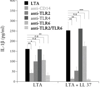
To further investigate the relationship between LL-37 and LTA, the effects of LL-37 on cellular responses to LTA stimulation in combination with different PRRs were analysed. The production of both TNF (not shown) and IL-1 (figure 9) in whole blood stimulated with LTA and LTA/LL-37 was signifi-cantly inhibited by CD14, TLR2, and a combination of anti-TLR2/TLR6. As expected, pre-treatment with antibodies against TLR4 did not affect the LTA induced cytokine production since TLR4 is the main recep-tor for LPS. CD14 is well known to mediate activation of monocytes by LPS. Here, CD14 was also shown to mediate LTA induced activation, which has previously been reported185,186. TLR2 has been shown to form functional het-erodimer complexes with TLR6 to recognize LTA187
. This can be seen in the current experiment since the combination of antibodies against TLR2 and TLR6 were more efficient in blocking the cytokine production induced by LTA compared to anti-TLR2 alone. An antibody against only TLR6 had low blocking effect. According to several lines of evidence we conclude that
activation of cellular responses by highly purified LTA is mediated by TLR2 and CD14, but is clearly independent of TLR4. However, the results did not add any information about the cytokine production induced by LL-37/LTA compared to LTA since there was no difference in cytokine inhibition by PRRs on LL-37 and LTA/LL-37 challenge. It is not entirely clear whether LL-37 ini-tiates its immunomodulatory activities in a specific (i.e. receptor-mediated) or nonspecific manner. Studies have indicated that there are receptors on differ-ent cell types that interact with LL-37, e.g. formyl peptide receptor-like 184 for chemoattraction and the transactivator P2X7
188
for IL-1β processing, respec-tively. It would be informative and interesting to block these receptors and find out if the cytokine production by LL-37/LTA is changed.
Figure 9. Effect of anti-CD14, anti-TLR2, anti-TLR4 and anti-TLR6 antibod-ies on LTA/LL-37 induced cytokine production. Whole blood was incubated with antibodies 20 minutes before LTA (5 µg/ml)/LL-37 (20 µg/ml) challenge. All antibodies were used at 5 µg/ml. Release of IL-1 was quantified after 16 hours of incubation. Results are presented as cytokine concentrations (pg/ml), means ± SD of three independent experiments. Pure LL-37 (20 µg/ml) did not induce any detectable cytokine production. The differences between cytokine release were analysed with paired t-test, values marked (*) indicate a signifi-cant difference to LTA or LTA/LL-37 induced IL-1β (*, 0.01 < P < 0.05; **, 0.001 < P < 0.01; *** P < 0.001). 0 50 100 150 200 250 300 350 400 LTA LTA + LL 37 LTA LTA LTA LTA anti-CD14 anti-CD14 anti-CD14 anti-CD14 anti-TLR2 anti-TLR2 anti-TLR2 anti-TLR2 anti-TLR4 anti-TLR4 anti-TLR4 anti-TLR4 anti-TLR6 anti-TLR6 anti-TLR6 anti-TLR6 anti-TLR2/TLR6 anti-TLR2/TLR6 anti-TLR2/TLR6 anti-TLR2/TLR6 IL -1 β ( p g /m l) ** * ** n.sn.s *** ** ** n.s*
Prognostic value of suPAR in severe sepsis
More than two decades of research has shown that suPAR participates in a range of immunological effector functions including cell adhesion, migration, chemotaxis, immune activation, and the activity of these events may all add to the systemic suPAR concentration142. When a pathogen invades the blood stream, all of these effector functions are activated and the suPAR level may reflect the severity of the infection. To date, the main part of the published re-sults regarding the role of suPAR in sepsis indicates that suPAR is more a prognostic marker than a diagnostic marker189.
suPAR is still a rather new risk marker and when we analysed plasma suPAR in sepsis patients in 2010 there were only two articles published regarding this topic157,190. In the first one, a study of 141 adult patients with Streptococcus pneumoniae bacteremia, Wittenhagen et al 157
found that suPAR levels were higher in the 17% of patients who died from the infection than in those who survived and that suPAR levels above 10 ng/ml independently predicted mor-tality. In the other study, Kofoed et al 190
compared the prognostic value of su-PAR in 151 sepsis patients to that of other inflammatory markers and of the Simplified Acute Physiology Score (SAPS) II and the SOFA score. Their results showed that suPAR levels had a better prognostic value than PCT and CRP, equal to that of the admission SOFA score and almost as good as the SAPS II score.
Then, in 2011 four large cohort (55-200 sepsis patients) studies were pub-lished on the prognostic value of suPAR in bacteremia and sepsis158-160,191. All four studies found that plasma suPAR concentrations were increased in in-fected patients compared to non-inin-fected patients and that high levels of su-PAR were associated with increased mortality. Despite the relatively low number of patients included in our study, the results are concordant with the larger cohort studies. We found significantly higher levels of plasma suPAR in patients with severe sepsis upon admission to the ICU compared to non-sepsis patients. However, the plasma level of suPAR did not significantly correlate with mortality, only with severity, although the mean suPAR values were higher in non-survivors compared to survivors (figure 6). A probable explana-tion to the lack of correlaexplana-tion between suPAR and mortality might be the low number of patients included in our study (n=27, ICU mortality = 44 %). Still, on the basis of the results in paper III and other studies, increased suPAR