Linköping Studies in Science and Technology Dissertation No. 1961
Investigation of nanoparticle-cell
interactions for
development of next generation
of biocompatible MRI
contrast agents
Natalia Abrikossova
Division of Molecular Surface Physics and Nanoscience Department of Physics, Chemistry and Biology
Linköping University, Sweden
© [Natalia Abrikossova, 2018]
Cover: activated neutrophil granulocytes visualized with fluorescence microscopy Abrikossova, Natalia
Investigation of nanoparticle-cell interactions of next generation of biocompatible MRI contrast agents
ISBN 978-91-7685-190-6 ISSN 0345-7524
Linköping studies in science and technology. Dissertations, No. 1961
“There's Plenty of Room at the Bottom” Richard Phillips Feynman
ABSTRACT
Progress in synthesis technologies and advances in fundamental understanding of materials with low dimensionality has led to the birth of a new scientific field, nano-science, and to strong expectations of multiple applications of nanomaterials. The physical properties of small particles are unique, bridging the gap between atoms and molecules, on one side, and bulk materials on the other side. The work presented in this thesis investigates the potential of using magnetic nanoparticles as the next gener-ation of contrast agents for biomedical imaging. The focus is on gadolinium-based nanoparticles and cellular activity including the uptake, morphology and production of reactive oxygen species.
Gd ion complexes, like Gd chelates, are used today in the clinic, world-wide. However, there is a need for novel agents, with improved contrast capabilities and increased biocompatibility. One avenue in their design is based on crystalline nanopar-ticles. It allows to reduce the total number of Gd ions needed for an examination. This can be done by nanotechnology, which allows one to improve and fine tune the physi-co-chemical properties on the nanomaterial in use, and to increase the number of Gd atoms at a specific site that interact with protons and thereby locally increase the sig-nal. In the present work, synthesis, purification and surface modification of crystalline Gd2O3-based nanoparticles have been performed. The nanoparticles are selected on the
basis of their physical properties, that is they show enhanced magnetic properties and therefore may be of high potential interest for applications as contrast agents.
The main synthesis method of Gd2O3 nanoparticles in this work was the modified
“polyol” route, followed by purification of as-synthesized DEG-Gd2O3 nanoparticles
suspensions. In most cases the purification step involved dialysis of the nanoparticle samples. In this thesis, organosilane were chosen as an exchange agent for further functionalization. Moreover, several paths have been explored for modification of the nanoparticles, including Tb3+ doping and capping with sorbitol.
Biocompatibility of the newly designed nanoparticles is a prerequisite for their use in medical applications. Its evaluation is a complex process involving a wide range of biological phenomena. A promising path adopted in this work is to study of nanoparti-cle interactions with isolated blood cells. In this way one could screen nanomaterial prior to animal studies.
The primary cell type considered in the thesis are polymorphonuclear neutrophils (PMN) which represent a type of the cells of human blood belonging to the granulo-cyte family of leukogranulo-cytes. PMNs act as the first defense of the immune system against invading pathogens, which makes them valuable for studies of biocompatibility of newly synthesized nanoparticles. In addition, an immortalized murine alveolar macro-phage cell line (MH-S), THP-1 cell line, and Ba/F3 murine bone marrow-derived cell line were considered to investigate the optimization of the cell uptake and to examine the potential of new intracellular contrast agent for magnetic resonance imaging.
In paper I, the nanoparticles were investigated in a cellular system, as potential
probes for visualization and targeting intended for bioimaging applications. The pro-duction of reactive oxygen species (ROS) by means of luminol-dependent chemilumi-nescence from human neutrophils was studied in presence of Gd2O3 nanoparticles. In
paper II, a new design of functionalized ultra-small rare earth-based nanoparticles
was reported. The synthesis was done using polyol method followed by PEGylation, and dialysis. Supersmall gadolinium oxide (DEG-Gd2O3) nanoparticles, in the range of 3-5 nm were obtained and carefully characterized. Neutrophil activation after expo-sure to this nanomaterial was studied by means of fluorescence microscopy. In paper
III, cell labeling with Gd2O3 nanoparticles in hematopoietic cells was monitored by
magnetic resonance imaging (MRI). In paper IV, ultra-small gadolinium oxide nano-particles doped with terbium ions were synthesized as a potentially bifunctional mate-rial with both fluorescent and magnetic contrast agent properties. Paramagnetic behav-ior was studied. MRI contrast enhancement was received, and the lumines-cent/fluorescent property of the particles was attributable to the Tb3+ ion located on the crystal lattice of the Gd2O3 host. Fluorescent labeling of living cells was obtained.
In manuscript V, neutrophil granulocytes were investigated with rapid cell signaling
communicative processes in time frame of minutes, and their response to cerium-oxide based nanoparticles were monitored using capacitive sensors based on Lab-on-a-chip technology. This showed the potential of label free method used to measure oxidative stress of neutrophil granulocytes. In manuscript VI, investigations of cell-(DEG-Gd2O3) nanoparticle interactions were carried out. Plain (DEG-Gd2O3) nanoparticles,
(DEG-Gd2O3) nanoparticles in presence of sorbitol and (DEG-Gd2O3) nanoparticles
capped with sorbitol were studied. Relaxation studies and measurements of the reac-tive oxygen species production by neutrophils were based on chemiluminescence. Cell morphology was evaluated as a parameter of the nanoparticle induced inflammatory response by means of the fluorescence microscopy.
The thesis demonstrates high potential of novel Gd2O3-based nanoparticles for
development of the next generation contrast agents, that is to find biocompatible com-pounds with high relaxivity that can be detected at lower doses, and in the future ena-ble targeting to provide great local contrast.
Populärvetenskaplig sammanfattning
De små nanopartiklarnas fysikaliska egenskaper är unika, och befinner sig i gränslan-det mellan atomer och molekyler, å ena sidan och bulkmaterial å andra sidan. Arbetet som presenteras i denna avhandling visar på möjligheten att använda flera olika typer av nanopartiklar för biomedicinsk bildbehandling, med särskild inriktning på kon-trastmedel för signalförstärkning vid magnetisk resonansbildning (MRI). Syntes, upp-rening, ytmodifiering och karakterisering av både kristallina Gd2O3-nanopartiklar har gjorts. Dessa nanopartiklar har valts utifrån deras fysikaliska och kemiska egenskaper, det vill säga de uppvisar förbättrade magnetiska egenskaper och kan därför vara högin-tressanta för tillämpningar såsom kontrastmedel inom biomedicinsk avbildning. En stor utmaning vid utformning av nya kontrastmedel baserade på nanopartiklar (jämfört med Gd-komplex som idag används inom sjukvården på klinik världen över) är att minska det totala antalet Gd som behövs för en undersökning. Detta kan göras genom nanoteknik, dvs att förbättra och finjustera de fysikalisk-kemiska egenskaperna hos det nanomaterial som används. Vidare öppnar sig möjligheter för målsökande nanoprober, då kan man ytterligare öka signalen, genom att endast lokalt öka koncentrationen av Gd-joner på en specifik plats eller sätt leder detta till en förstärkt signal. En nanopar-tikel består av ca 1000 Gd joner vilket möjliggör en 1000-falt förstärkt kontrast lokalt jämfört med de existerande kontrastmedlen som endast bär en Gd-jon per komplex. Det är också av högsta relevans att utvärdera effekter av nanopartiklar i biologiska system. Cellstudier har potential att ersätta och / eller minska användandet av djurför-sök. Detta är i linje med nya strategier för riskbedömning av kemikalier där man vill ersätta/reducera antalet djurförsök med mer prediktiva test. Syftet med denna avhand-ling är att undersöka biokompatibiliteten hos lovande nanopartiklar som syntetiserats av mig och andra medlemmar av vårt forskarteam. Långsiktigt vill man åstadkomma effektivare, billigare och mer relevant riskbedömning av kemikalier och kombination-er av kemikalikombination-er. Detta kommkombination-er att kunna uppnås genom ökad kunskap om effektkombination-erna på människor och med hjälp av nya celltester. Dessa studier kräver nära samarbete mellan olika vetenskapliga discipliner, dvs medicin, biologi, kemi och fysik. En slå-ende fördel med detta tillvägagångssätt är att resultatet av flera strategier gällande nanopartikel-funktionalisering kan utvärderas med hjälp av fysisk karaktärisering, probing-kapacitet och biologisk aktivitet redan i ett tidigt skede, under design och optimering av en ny typ av nanopartikel, vilket stämmer väl överens med den internat-ionella rekommendationen ”safe by design” för framtidens hållbara nanomaterial. Biokompatibiliteten förbättras med hjälp av olika tekniker, t.ex. olika syntesvägar, via capping, ytmodifiering och funktionalisering, samt genom att introducera en liten mängd av något annat element/grundämne. För att studera cell-nanopartikel interakt-ion används isolerade blodceller såsom neutrofila granulocyter, men även cell-linjer THP-1 och makrofager från mus-lungvävnad. Våra in vitro-studier inkluderar under-sökning av effekterna av Gd2O3 nanopartiklar på funktionen av neutrofila granulocyter
List of publications
Paper I
Natalia Abrikossova, Caroline Skoglund, Maria Ahren, Torbjörn Bengtsson and Kajsa Uvdal
Effects of gadolinium oxide nanoparticles on the oxidative burst from human neutrophil granulocytes.
Nanotechnology 2012;23(27); 275101.
Paper II
Maria Ahren, Linnea Selegård, Anna Klasson, Fredrik Söderlind, Natalia Abrikossova, Caroline Skoglund, Torbjörn Bengtsson, Maria Engström, Per-Olov Käll and Kajsa Uvdal
Synthesis and Characterization of PEGylated Gd2O3 Nanoparticles for MRI Contrast
Enhancement.
Langmuir 2010;26(8):5753-62.
Paper III
Anna Hedlund, Maria Ahrén, Håkan Gustafsson, Natalia Abrikossova, Marcel Warntjes, Jan-Ingvar Jönsson, Kajsa Uvdal, Maria Engström.
Gd2O3 nanoparticles in hematopoietic cells for MRI contrast enhancement.
International Journal of Nanomedicine. 2011;6:3233-40.
Paper IV
Rodrigo M. Petoral, Jr., Fredrik Söderlind, Anna Klasson, Anke Suska, Marc A. Fortin, Natalia Abrikossova, Linnea Selegård, Per-Olov Käll, Maria Engström and Kajsa Uvdal
Synthesis and Characterization of Tb3+-Doped Gd
2O3 Nanocrystals: A Bifunctional
Material with Combined Fluorescent Labeling and MRI Contrast Agent Properties. Journal of Physical Chemistry C. 2009;113(17):6913-20.
Paper V
Kalle Bunnfors, Natalia Abrikossova, Joni Kilpijärvi, Peter Eriksson, Jari Juuti, Niina Halonen, Caroline Brommesson Anita Lloyd Spets and Kajsa Uvdal
Lab-on-a chip for capacitive measurements on human neutrophil granulocytes and their activation triggered by Ce/Gd oxide nanoparticles. In manuscript
Paper VI
Natalia Abrikossova, Caroline Brommesson, Emanuel Larsson, Peter Eriksson, Zhangjun Hu and Kajsa Uvdal
Sorbitol capping of gadolinium oxide nanoparticles for magnetic resonance imaging contrast enhancement. In manuscript
Related papers not included in thesis:
Emanuel Larsson, Christian Dullin, Natalia Abrikossova, Caroline Brommesson, Urša Mikas, Chiara Garrovo, Agostino Accardo, GiulianaTromba, Igor Serša, Kajsa Uvdal. Optimization of the loading efficacy for dual-modal CT/MRI macrophage tracking in lungs of an asthma mouse model. Manuscript.
Emanuel Larsson, Christian Dullin, Natalia Abrikossova, Urša Mikas, Caroline Brommesson, Chiara Garrovo, Agostino Accardo, GiulianaTromba, Kajsa Uvdal, Igor Serša.
Dual-modal CT and MRI functional and anatomical imaging using barium sulphate and gadolinium nanoparticle loaded macrophages in a preclinical asthma mouse mode. Manuscript.
Linnea Selegård, Alexei Zakharov, Andreas Skallberg, Natalia Abrikossova, Kajsa Uvdal.
PEEM, LEED and PES temperature study of Eu doped Gd2O3 nanoparticles and their
Author contributions
Paper I
I was responsible for the study design and implementation, including synthesis of nanoparticles, isolation of neutrophils, measurements of ROS, and examination of the cellular behavior and morphological changes by fluorescence staining. I was responsi-ble for summarizing the results and writing the major part of the manuscript.
Paper II
I carried out synthesis of gadolinium oxide (DEG- Gd2O3) nanoparticles followed by
their PEGylation, and dialysis. I was responsible for studies of neutrophil activation after exposure to this nanomaterial by means of fluorescence microscopy and contrib-uted to the manuscript writing.
Paper III
I carried out synthesis of gadolinium oxide (DEG- Gd2O3) nanoparticles. I participated
in culturing and labeling of a monocytic cell line, THP-1, MRI measurements and contributed to the manuscript writing.
Paper IV
I acquired a knowledge on synthesis of gadolinium oxide nanoparticles doped with rare-earth ions. I was involved in sample preparation for the microscopy analysis of THP-1 cell line incubated with the particles and contributed to the manuscript writing.
Paper V
I participated in the project planning and design of the study. I carried out isolation of neutrophils and initiated testing of the Lab-on-a-chip principle based on the CMOS/LTCC sensor device to monitor the neutrophils activity. I participated in anal-ysis of the results and contributed to the manuscript writing.
Paper VI
I was responsible for the study design and implementation. I carried out gadolinium oxide nanoparticles synthesis and their surface modification, relaxivity measurements
using Bruker minispec mq60 NMR analyzer, macrophage cell line culturing and label-ing, isolation of human neutrophil granulocytes, measurements of ROS, and examina-tion of the cellular behavior and morphological changes by fluorescence staining. I was responsible for summarizing the results and writing the major part of the manu-script.
Acknowledgement
First of all, I would like to thank to my principal supervisor, Kajsa Uvdal, for giving me a chance to do research, for sharing your knowledge with me, for your patience and valuable discussion. You opened to me an exciting field of nanomaterials. Your visionary advice to focus on studies of cellular activity determined the main direction of my work. Your energy and optimism helped me all the way, from my first experi-ment to summarizing the results in this thesis. Your leadership was stimulating. Fan-tastic atmosphere of friendship, collaboration and engagement in your group is some-thing that I would nether forget. Last, but not least, I truly appreciate your understand-ing of my wish to work part time to combine my research and my family in a way that suited me best.
I am deeply grateful to my co-supervisor, Caroline Brommesson for continuous and friendly support. Carro, you are great teacher! You thought me to deal with cells, to make advanced measurements on uptake, morphology and production of reactive oxy-gen species, to collect and analyze results. We spent many hours together, and work-ing with you was a real pleasure.
I would like to thank Maria Engström for introducing me into field of MRI science and biomedical imaging.
Maria Sunnerhagen was my mentor during all these years. Thank you, Maria for keeping me focused!
I would like to express my sincere gratitude to all persons around me who support-ed me in different ways. Many thanks to all the current members and alumni of Mo-lecular Surface Physics and Nanoscience Division: Maria, Linnea S, Cecilia, Linnea A, Andreas, Peter, Kalle, Zhangjun, Xuanjun, Frank and Jiwen for interesting discussions and nice time together.
I would like to acknowledge all my co-authors: Torbjörn Bengtsson, Maria Ahren, Linnea Selegård, Anna Hedlund, Emanuel Larsson, Peter Eriksson, Zhangjun Hu, Per-Olov Käll, Fredrik Söderlund, Håkan Gustafsson, Anita Lloyd Spets, Joni Kilpijärvi, Anke Suska. Interactions with you were extremely useful for my thesis work.
All my friends, colleagues and collaborates at IFM, thank you for your assistance and for building the department where excellent science is combined with continuous consideration and improvement of work environment.
Finally, I would like to take the opportunity to thank my family and my friends for encouragement and support.
Tack alla!
Linköping, November 2018 Natalia Abrikossova
Abbreviations
DEG DIC DLS DTPA fMPL IR MRI MPTES NADPH NEXAFS NMR PBS PEG PMA PMN RES RF ROS TEM XPD XPS Diethylene glycolDifferential interference contrast Dynamic light scattering
Diethyltriamine pentaacetic acid
N-formyl-methionyl-leucyl-phenylalanine Infrared spectroscopy
Magnetic resonance imaging
3-mercaptopropyl trimethoxy silane
Nicotinamide adenine dinucleotide phosphate
Near-edge X-ray absorption fine structure spectroscopy Nuclear magnetic resonance
Phosphate buffered saline Polyethylene glycol
Phorbol 12-myristate 13-acetate Polymorphonuclear granulocytes Reticuloendothelial system Radio frequency
Reactive oxygen species
Transmission electron microscopy X-ray diffraction
Table of Contents
1. Nanoparticles: general definitions...1
1.1. Nanomaterials in modern science in technology……….1
1.2. Nanoparticles properties enabling biomedical application ………...5
2. Synthesis of nanoparticles …………...9
2.1. General overview of synthesis methods ……….…9
2.2. Wet chemistry synthesis ...……….….…9
2.3. Modified “polyol” synthesis method ...11
3. Methods of nanoparticles characterization ... 13
3.1. Transmission Electron Microscopy ...14
3.2. Dynamic light scattering system ………..……….17
3.3. Infrared spectroscopy …...……….…….…18
4. Hematopoietic cells for studies of nanoparticle-cell interaction………...……….21
4.1.Polymorphonuclear neutrophils ...……….22
4.2. Isolation of polymorphonuclear neutrophils from human blood...…23
4.3. ROS production …….………...25
4.4 Macrophages ………...27
4.5.THP-1 ……….………...28
4.6.BA/F3cells ………..………29
5. Techniques for studies of cell-nanoparticle interactions...31
5.1. Magnetic resonance imaging ………...31
5.2. Morphological studies of cell-nanoparticle interactions using fluorescence microscopy…….……….….………...36
5.3. Capacitive sensors based on Lab-on-a-chip technology...39
6. Nanoparticles as MRI contrast agent ..………...…..……….... 43
6.1. General considerations ………..43
6.2. Fe-oxide based contrast agents ………..……44
6.3. Gd chelates based contrast agents ………...………..……45
6.4.Gd2O3 nanoparticles………48
7. Modification of Gd2O3 nanoparticles ………...………... 53
8. Summary of papers………..………..…... 59
8.1. Paper I: Effects of gadolinium oxide nanoparticles on the oxidative burst from human neutrophil granulocyte ………...59
8.2. Paper II: Synthesis and Characterization of PEGylated Gd2O3 Nanoparticles for MRI Contrast Enhancement………..……….…..60
8.3. Paper III: Gd2O3 nanoparticles in hematopoietic cells for MRI contrast enhancement………..………....…..62
8.4. Paper IV: Synthesis and characterization of Tb3+-doped Gd2O3 nanocrystals: a bifunctional material with combined fluorescent labeling and MRI contrast agent properties…...………..………..…… ...…..63
8.5. Paper V: Lab-on-a chip for capacitive measurements on human neutrophil granulocytes and their activation triggered by Ce/Gd oxide nanoparticles….………...65
8.6. Paper VI: Sorbitol capping of gadolinium oxide nanoparticles for magnetic resonance imaging contrast
enhancement………..….…66 9. Conclusions and outlook………..69 10. References...71
Chapter 1
Nanoparticles: general definitions
1.1. Nanomaterials in modern science in technology
Most formal definitions of engineered nanomaterials revolve around the manipulation of materials roughly 1 to 100 nm in size1. Recent progress
in synthesis technologies, as well as advances in fundamental under-standing of materials with low dimensionalities, coupled to strong ex-pectation in terms of applications boosted huge interest in the field, and led to the birth of a new scientific discipline, the nanoscience. It is im-portant to underline, that the size is not the sole reason for the interest, and it cannot be used as the only motivation for studies and use of nano-materials. The point is that physical properties of small particles are unique, bridging the gap between atoms and molecules, on one side, and bulk materials on the other side.
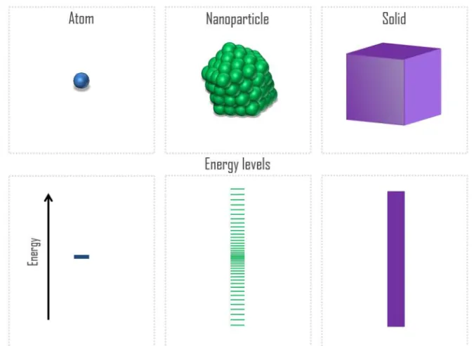
In particular, the main distinction of a nanomaterial from its bulk counterpart is considered to be the presence of quantum effects in the former. Indeed, at the atomic scale, electronic states are typically char-acterized by discrete energy levels that are often separated by electron volts; the spatial distribution of these states is highly localized. In con-trast, electronic states in crystalline matter are typically characterized by energy bands: the energy states form a quasicontinuous spectrum with delocalized spatial distributions. A nano-particles in this scheme may possess hundreds or thousands of atoms. Therefore, at the nanoscale the distribution of energy states resides between these limits: there are many states within energy interval of the order of 1 eV, but the states are still separated from each other2.
This evolution of an energy level from an atom towards bulk systems through the nano-particle is illustrated in Figure 1.1. The confinement effects in a nanomaterial make quantum effects stronger in comparison to the bulk materials, leading to qualitatively new physical properties of the former. Moreover, the electronic, optical and magnetic properties of small particles, has a non-linear behavior between the two general limits given by the atomic and the bulk-like behavior3-4. In particular, small
clusters show substantial dependence of magnetic properties, e.g. orbital and spin moments, as well as interactions between magnetic moments in the system on its dimensionality and size. Improved fundamental
knowledge of the behavior of nanomaterials is of crucial importance for their applications.
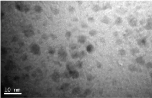
The small size of the nanoparticles is essential for their practical use. Indeed, a 10 nm particle (Figure 1.2) is too tiny for us to observe with a naked eye, and consequently, to operate nanoparticles in a stand-ard way, as we deal with bulk materials and conventional engineering tools. Nanoparticles, for instance, can be suspended in fluids, the so-called nanofluids5. Due to the small size of the nanoparticles, their
Brownian motion caused by collision with surrounding fluid molecules is sufficient to overcome gravitational force. Properly chosen nanoparti-cles will enhance the thermal, electrical, magnetic, and/or chemical properties of the original fluid. Moreover, if they are designed in a way that prevents their agglomeration, they will remain suspended and can be used in applications, which are discussed in this thesis. Another pos-sibility to use the nanoparticles is to collect them on an appropriate sub-strate.
Figure 1.1 Evolution of electronic states from atom via nanoparticle towards bulk solid.
Figure 1.2 Transmission electron microscope (TEM) image of as-synthesized DEG-capped 5Tb:Gd (5Tb:Gd-DEG) nanoparticle. Reprint-ed with permission from Paper IV. Copyright 2009 American Chemical Society.
Quantum phenomena are enhanced in the nanoparticles due to their small size (Figure 1.1) and determine majority of their applications in modern science in technology, existing, as well as potential. Let us con-sider, for example, a metallic nanoparticle interacting with a quantum of light, with a photon. The oscillating electromagnetic field leads to a collective motion of the nanoparticles valence electrons, or to the so-called plasmon oscillations. The plasmons may be localized at the sur-face of the nanoparticles, in which case they are called sursur-face plasmons. The plasmonic response becomes heightened at a resonance optical fre-quency that depends on the size and shape of the nanoparticles, as well as on the density of its valence, mostly free-like, electrons5. Varying
these parameters, one can ensure that plasmons resonate in specific, well-defined bands ranging from ultraviolet to infrared parts of the spec-trum, depending on the need for a specific application. Interestingly, this is perhaps the first known application of the nanoparticles, dating back to ancient Rome technology of making glass with varying color. Nowadays, nanofluids which exploit a physical phenomenon of plas-mon resonance have been deployed in biological imaging and tumor-destroying hypothermia treatments, and they constantly find new
appli-cations, e.g. in photovoltaic thermal collectors that generate electricity and usable heat simultaneously5.
A strong motivation to study nanoparticles is their use in heteroge-neous catalysis. A catalyst increases the rate of a chemical reaction, and therefore it is very important and often used in the chemical and phar-maceutical industries6. The catalytic activity depends critically on the
number of active sites, which makes nanoparticles superior in the appli-cations. Catalysts are often designed by dispersion of sub-nanometer and nano-sized metal particles on oxide supports7.
A rapidly growing field for applications of nano-systems is their use for sensing. Their functionality is related to the quantum confinement effect. For example, the size-dependent band gap of a nanomaterial may result in a variable photoluminescence emission. The adsorption of cer-tain ions or molecules could modify the gap, quenching or enhancing photoluminescence. Optical detection of the change can be utilized for sensing of the ions or molecules8. In fact, biosafety regulations requiring
rapid and cost-effective evaluations give strong motivation for devel-opment of novel sensors, often involving nano-systems. For example, traditional methods for in vitro cytotoxicity evaluation are often expen-sive and time-consuming, because they include cell cultivation and la-bel-based assay kits. This has led to a growing interest in noninvasive, label-free, real-time, data-rich biosensing techniques, like surface plas-mon resonance spectroscopy, electrochemical quartz crystal microbal-ance measurements, optical sensing, impedimetric sensing, and capaci-tive sensing9. In particular, cell-impedance measurement using the
lab-on-a-chip (LoC) concept has been used in this work, Paper V. Moreover, nanoprobes combining different modalities, for instance magnetic reso-nance imaging and fluorescence attract increasing interest, e.g. as a use-ful tool for biomedical imaging10.
Magnetism is another quantum phenomenon, which is important for applications of nanoparticles in general, and in particular for imaging applications in medicine11. The source of magnetism in condensed
mat-ter is identified with the spin of the electrons. The spontaneous ordering of the magnetic moments on individual atoms in a material, known as ferromagnetism (FM) or antiferromagnetism (AFM). It is a manifesta-tion of electrostatic Coulomb interacmanifesta-tions constrained by Pauli’s exclu-sion principle, which forbid the occupancy of a quantum state by two electrons with the same spin12. Note that in contrast to the free-electron
often contribute to magnetic phenomena are localized electrons belong-ing to d or f shells of the atoms. With temperature increasbelong-ing above spe-cific limit, known as Curie temperature TC of a ferromagnet or as a Neel temperature TN of an antiferromagnet, the net magnetization of the ma-terial vanishes, and the mama-terial becomes paramagnetic (PM). Note, however, that the local magnetic moments still exist in most magnetic materials in their PM phase above TC or TN, and net magnetization van-ishes due to the disorder of the local moments.
Even below TC or TN bulk FM (AFM) materials may be demagnet-ized on the macroscopic scale in the absence of magnetic field because the existence of magnetic domains: microscopic, but still large enough parts of the material with FM(AFM) order inside each domain, but with disordered orientation of magnetization between the domains due to magnetic dipolar interactions. However, magnetic behavior can be in-fluenced by scaling down the system size11,13. In particular, magnetic
nanoparticles can be made small enough to remain in the monodomain magnetic state, which gives qualitatively new opportunities for their applications. For example, consider FM nanoparticles in the monodomain state. Their individual magnetization directions will be disoriented with respect to each other in the absence of magnetic field, and the temperature will flip the directions randomly, making them sim-ilar to the bulk PM material. However, their magnetization directions will order in the magnetic field. Because their moments, which are pro-portional to number of atoms in the nanoparticles, are larger than mo-ments of individual atoms, the magnetic susceptibility of the nanoparti-cles will be much larger as well. This gives rise to the phenomenon of superparamagnetism. In fact, interest in magnetic nanoparticles has in-creased in the past few years because of the high potential of their ap-plications in several fields, like ultrahigh-density recording and medi-cine. The latter is of primary interest for this work.
1.2. Nanoparticles properties enabling biomedical application
In the past year one of the most promising and rapidly developing areas of the use of magnetic nanoparticles is their applications in biomedicine. When the size of a particle decreases from the micro- to nano-meters, their magnetic state is often strongly affected. In particular, the reduc-tion of the size makes the particles effectively one-dimensional, leading to the appearance of the superparamagnetic effects 14-15. In this respect,
metal oxide nanoparticles attract substantial attention, because of their general availability, well-developed synthesis methodologies, relatively straight-forward manipulation to obtain controlled magnetic behavior and acceptable levels of magnetic signal. The possibility to control their behavior with an external the magnetic field makes magnetic nanoparti-cles into highly attractive material. Indeed, they possess appropriate physicochemistry and their surface properties may be tailored via sur-face modification, Superparamagnetic nanoparticles of iron oxide, have earlier been shown to produce negative contrast in Magnetic Resonance Imaging (MRI). The Uvdal group at Linköping University has shown that when scaling down in size this material, with a size of 2-6 nm, capped with a passivizing non-ionic material, is very promising as posi-tive contrast agent11,13. This shows the potential in nanomaterial design
for biomedical imaging.
The magnetic nanoparticles have been extensively investigated for various applications, such as drug delivery, tissue engineering and re-pair, biosensing, biochemical separations, and bioanalysis. In the field of disease therapy, the development of “theragnostics”, which facilitates simultaneous drug delivery and imaging, represents an important break-through16.
In addition, ferromagnetic magnetite nanoparticles are characterized by very high values of magnetization and coercive force, which ignites a considerable interest, for example, for their use for therapeutic purpos-es17. For instance, an application of ferrofluids for the treatment of
on-cological diseases by local hyperthermia was proposed in the beginning of 1990s. Jordan et al.18 has proved high efficiency of the conversion of
the energy absorbed by suspension of superparamagnetic nanoparticles in an oscillating magnetic field into heat. The effect can be used in vivo to destroy the pathological cells by heating certain tumor tissues, the so-called “leaky cells”. Preclinical and clinical data show that hyperthermia is in fact feasible and effective in combination with radiation therapy19.
Of course, lot of research must still be undertaken to address multi-ple challenges faced in nanomedicine and to ensure safe and efficient biomedical applications of magnetic nanoparticles. Considering metal toxicity and pharmacology important issues must be addressed, such as dose response, kinetics etc. Indeed, the biochemical and physiological effects of the nanoparticles depend upon the chemical form of the metal, e.g. oxidation state, salt or complex. Moreover, toxicity is dependent upon the route of exposure20. Accurate analytical tools need to be
de-veloped further for studies of interactions of nanoparticles with the im-mune system. A wise strategy is to screen nanoparticle on the cellular level, in parallel with nanomaterial design, synthesis and development. This is also the strategy we used in Paper I, where Gd2O3 nanoparticles
of our own design were tested in presence of blood cells, more specifi-cally, neutrophils. The cellular response on the presence of three sets of Gd2O3 nanoparticles was investigated, i.e. as synthesized, dialyzed and
functionalized and dialyzed, and an increase of their biocompatibility by the surface modification of these nanoparticles with polyethylene glycol (PEG) was demonstrated. In such a way one obtains an early sign on the nanomaterial with high potential, as well as at early stage sort out mate-rial with negative response. This is also in line with the strive, national-ly and internationalnational-ly (EU, USA) that present and future material devel-opment should involve sustainability from step one, in this case, safe by
design.
Finally, multiple preclinical and clinical studies in relevant animal models and disease states will be required to substantiate proof of con-cept using different controls especially in MRI molecular imaging. Safety and biocompatibility studies, in particular long-term toxicity studies, will be required, as well in addition to proof-of-concept studies19. Thus, the development of methodologies for efficient
evalua-tion and improvement of biomedical performance of novel nanoparticles at the initial stages of their design is of primary importance. This is the subject of the present thesis.
Chapter 2
Synthesis of nanoparticles
2.1. General overview of synthesis methods.
Rapid development of techniques for synthesis of nanoclusters has re-sulted in remarkable advances in our knowledge of these systems and pathed the way to their advanced applications in different fields, from physics and chemistry to biology and medicine. Already in the 1960s cluster sources were developed to produce metallic clusters21. However,
they were composed of only a few atoms in the gas phase. In the first half of the 1980s clusters of alkali metals with up to about 100 atoms were produced and detected22. This has modified the perception of these
systems originally viewed as a kind of small molecules and stimulated the development of numerous synthesis techniques. For instance, seeded supersonic nozzle sources are used to produce intense cluster beams of low-boiling-point metals. Gas-aggregation sources are particularly effi-cient in the production of large clusters with more than 10000 atoms of low-to-medium-boiling-point materials. Laser vaporization sources pro-duce clusters in the size range from the atom to typically several hun-dreds of atoms per cluster. In pulsed-arc cluster-ion sources an intense electrical discharge rather than a laser is used to produce the clusters. Ion sputtering sources are primarily used to produce intense continuous beams of small singly ionized clusters of most metals. Liquid-metal ion sources produce singly and multiply ionized clusters of low-melting-point metals23.
2.2. Wet chemistry synthesis
Efficient methods for nanoparticle synthesis employ chemical reactions in the so-called “wet-chemistry” synthesis16. For example, in the sol−gel
method the hydroxylation and condensation of precursors in a solution phase yields a colloidal solution (sol) of nanosized particles. Oxidation method can, for instance, be used for the synthesis of small-sized ferrite colloids, like Fe3O4 by crystallization starting from the ferrous
hydrox-ide gels. It involves partial oxidation of the Fe(II) hydroxhydrox-ide suspen-sions with different agents (e.g., nitrate ions). The hydrothermal synthe-sis method is used to synthesize nanoparticles in aqueous media in
reac-tors or autoclaves at high temperature, above 200 °C and high pressure. This allows one to achieve the formation of small-sized NPs due to rap-id nucleation and faster growth.
Further discussion of numerous of “wet-chemistry” synthesis tech-niques can be found in Ref.16, where the authors also point out
consider-able efforts that have been spent in the development of magnetic nano-particles (MNPs), because of their applicability in many different areas, for example for bioimaging10,15. Precise control over the synthesis
con-ditions and surface functionalization of MNPs is pointed out as the cen-tral issue. Indeed, it governs the physical, as well as chemical properties of the nanoparticles, their stability, and their biocompatibility. The fol-lowing conditions are of primary importance for pharmaceutical and biomedical purposes:
- the nanoparticles should possess very small size and narrow size distribution;
- magnetization values must be high;
- high magnetic susceptibility for an optimum magnetic enrich-ment must be combined with loss of magnetization after removal of the magnetic field;
- optimal surface coating must ensure tolerance and biocompati-bility, as well as specific localization at the biological target site. Concluding this section, it is worth to note that the synthesis of na-noparticles, especially those that exhibit superparamagnetic properties of interest for this thesis, is a complex process because of their colloidal nature19. There are several reasons for this. Most importantly, there is a
need to defining experimental conditions, leading to a monodisperse population of magnetic grains of suitable size. Next, the process must be reproducible, and in addition it should be possible to industrialize it in such a way that complex purification procedures (like ultracentrifuga-tion, size-exclusion chromatography, magnetic filtraultracentrifuga-tion, or flow field gradient) are not needed. Thus, it is challenging to use wet-chemistry-based process for the synthesis of sophisticated nanomaterials with ho-mogeneous composition and narrow size distribution. At the same time, in order to design nanoprobes with unique capabilities to deliver en-hanced local signal contrast in MRI very high control of the nanoprobe shape and geometry becomes very important for selective cell uptake. Moreover, the requirements for the future nanoprobes will increase re-garding both hard core (core/shell) and soft outer bio-shell. Therefore, novel synthesis techniques could be of interest for future research24.
2.3. Modified “polyol” synthesis method
Precipitation in a high boiling alcohol (polyol method) has been used with success for the synthesis of sub-micrometer transition metal parti-cles and selected oxides. In 2003 Bazzi et al.25 employed this route to
prepare colloidal suspension of lanthanide nano-oxides with the aim to incorporate lanthanide impurities in ultra-small, sub-5-nm metal oxide hosts. The technique is now known as modified “polyol” protocol. Studying the direct precipitation of rare earth doped oxides from metal-lic salts in polyalcohol, Bazzi et al. mixed suitable rare earth precursors, LnCl3 ∙ 6H20 with Ln=Eu, Tb, Nd, Gd or Y with a defined amount of
water in a high boiling diethylene glycol (DEG). To reduce the possibil-ity of lanthanide hydroxide formation, the precipitation was carried out in a non-aqueous environment. Indeed, Bazzi et al. observed the for-mation of oxide, rather than hydroxide particles, and explained this by the dehydrating properties of the alcohol associated with the high tem-perature of the solution during the synthesis.
One year later, Bazzi et al. further refined the modified “polyol” pro-tocol and made it highly suitable for the synthesis of the magnetic nano-particles. In this work, we primarily use this protocol, so we describe it in details below following closely the original description of Bazzi et al.26.
All reactions should occur at temperatures lower than 200◦C.
Rare-earth chloride RECl3·6H2O with RE = Eu, Gd, or Y (99.9% Aldrich)
should be dispersed in 20 ml of DEG with a global metal concentration of 0.1–0.5 mol l−1. After strong stirring for 30 min, 1 ml of aqueous
NaOH solution (3mol/l) should be added and the mixture should be heated in a silicon oil bath at 140◦C for 1 h. After complete dissolution
of the compounds, the solution should be heated at 180◦C for 4 h under
vigorous stirring in refluxing DEG.
In the experiment of Bazzi et al., the procedure described above re-sulted in transparent suspensions of particles dispersed in organic sol-vent. Monitoring the oxide particle suspension, the authors concluded that it was colloidally stable for weeks, independently of the particles content (pure Eu, Eu/Gd or Eu/Y solid solution oxides). Moreover, the nanoparticles consisted of a single phase. At the same time, the initial metal ion concentration influenced the size of the synthesized particles. Bazzi et al. suggested that varying the concentration one could adjust the size to some degree, though the main parameter for controlling the
grain size in their experiment was water/DEG ratio, because it was found to directly determine the nucleation rate. The duration of heating at 180◦C was also important, and it was set to 4 h in the experiment,
because longer treatment did not lead to further increase of the grain size.
Bazzi et al. recommended to ensure that OH− concentration of the
starting solution should be small. This prevented the rapid precipitation of hydroxides in his experiment. The addition of one equivalent of NaOH was thus suggested to be a good compromise. Lower values were found to give too small reaction yields while higher values led to irre-versible precipitation of hydroxides.
An important suggestion by Bazzi et al. was to dialyze the obtained colloidal suspension against pure DEG for 24 h to achieve purification. In addition, the authors verified that a partial low-pressure distillation of solvent could be performed to achieve concentrated suspension. In summary, the protocol described above allowed the authors of Ref.26 to
obtain solution containing up to 10 wt% rare earth oxide without parti-cle agglomeration.
As motioned above, DEG process the main contribution to stabilize the nanoparticles in DEG solution. Other capping molecules also can be added during the synthesis. For instance, Söderlind and coworkers stud-ied that the addition of oleic acids can give oleic acid-stabilized Gd2O3
nanoparticles, which can be well-dispersed in non-polar solvents. How-ever, it is not suitable for MRI applications due to its poor water-solubility27.
Meanwhile, our group has made efforts to apply the nanoparticle synthesized from this method for biological studies. We used Bazzi’s method to obtain the DEG-suspended Gd2O3 nanoparticles, but the
dial-ysis of the as-synthesized Gd2O3 nanoparticles was processed against
milli-Q water instead of DEG. In Paper I, we showed that the presence of DEG, even in a low amount, can induce negative effects on neutro-phil granulocytes. Actually, the small amount of DEG on the surface of Gd2O3 after dialysis could be easily exchanged by customized
PEGylat-ed-molecules. Paper II demonstrated that the delivered pegylated Gd2O3
exhibits much better biocompatibility and is very promising for MRI application.
Chapter 3
Methods of nanoparticles characterization
The properties of nanoparticles and their performance in applications depend crucially on the chemical composition, the size and the shape of the particles, as well as on their microstructure. In addition, for biomed-ical applications the polydispersity, charge, and nature of the coating play essential role19. Because of the small size of the nanoparticles,
most advanced experimental techniques must be employed for the char-acterization of the above-mentioned properties.
In fact, it is not straightforward to define unambiguously what is the size of the nanoparticle. Is the size determined by the crystalline part alone? How should we characterize the size for nanoparticles consisting of the core, the shell, and the cover layers? Moreover, even state-of-the-art synthesis protocols most often give polydisperse nanopstate-of-the-articles with certain heterogeneity of sizes. In order to study the size distribution, the shape and crystallinity of the particles transmission electron microscopy (TEM) is often employed15,28-30. Moreover, high-resolution transmission
electron microscopy (HRTEM) allows one to investigate the structure with atomic-scale resolution, as well as to get access to local micro-structures and defects, like lattice vacancies, as well as to characterize structural and chemical parameters of particles surfaces31.
X-ray diffraction (XRD) experiments can be performed to determine the crystalline structure of the particles. In addition, intensity of a dif-fraction pattern can be used to quantify the proportion of different phas-es formed in a mixture15,32. Extended X-ray absorption fine structure
(EXAFS) also gives information on the particle size. Moreover, it has recently been demonstrated that near edge X-ray absorption fine struc-ture (NEXAFS) studies can be used to characterize oxidation states of chemical elements composing nanoparticles30. The advantage of the
energy dispersive X-ray diffraction (EDXD) is that it can be carried out on the suspension leading to an improved knowledge of fine structural details.
Photon correlation spectroscopy (PCS), also called dynamic light scattering (DLS) or quasi-elastic light scattering (QELS), gives access to the so-called hydrodynamic radius of a particle and to the polydisper-sity of the colloidal solution33. In addition, the DLS can be utilized to
study the stability of the nanoparticles. X-ray photoelectron spectrosco-py (XPS) and infrared spectroscospectrosco-py (IR) are powerful experimental techniques for molecular composition analysis15,29-30.
To investigate the surface properties of coated nanoparticles, to char-acterize the bonding between the particle surface and the coating as well as to understand the influence of the coating on the magnetic properties of the nanoparticles, several experimental techniques can be used, in-cluding atomic and chemical force microscopy (AFM and CFM), Fouri-er transform infrared spectroscopy (FTIR), as well as othFouri-er techniques reviewed in Ref. 19. Below we present more detailed description of the
experimental techniques, which are most relevant for this thesis.
3.1. Transmission Electron Microscopy
XRD experiments that are traditionally used for highly precise structural characterization of solids, give access to the average structure of the studied materials. However, they are not suitable for studies of local regions of a specific material, and its applicability for studies of indi-vidual nanostructures is limited. At the same time, there is an interest and a great need in a detailed characterization of nanomaterials with atomic-scale resolution. This makes the transmission electron microsco-py highly attractive for applications in the field. Indeed, it is capable to form images of atomic arrangements at localized regions within materi-als34. In the TEM one uses electrons for imaging of the studied materials.
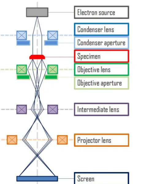
In contrast to X-rays, the electrons are negatively charged, and therefore it is possible to accelerate them using the electron guns, as well as to focus them using the electromagnetic fields of advanced systems of lenses (see Figure 3.1). Moreover, the electrons scattered by samples can be further collected by lenses to form true images in real space, in the sense that each point of the image in principle corresponds to a spe-cific point of the sample. In this respect, TEM is similar to optical mi-croscopes.
Importantly, for accelerating voltage of 1000 kV, the electron wave-lengths are of the order of 0.001 nm34, which should be more than
suffi-cient for the resolution with the atomic scale. Moreover, a combination of TEM with electron energy loss spectroscopy (EELS) allows one for a chemical analysis of the samples at the atomic scale.
Figure 3.1 Basic set up for transmission electron microscopy in the im-aging mode
In reality, there are limitations. Perhaps the most essential here are the so-called aberrations because the lenses are not perfect, and they create magnetic fields that affect the path of the electrons through the TEM. Because of this, the resolution of the TEM was limited to ~0.2 nm until middle of 1980s. Another important feature of the TEM is that it presents two-dimensional projections of three-dimensional specimens. However, modern TEM are equipped with devices that allow for the aberration corrections, which made it possible for the researchers to achieve sub-Ångström resolution (~ 0.5-Å resolution at 300 keV). In fact, the highest-magnification image obtained using a transmission electron microscope has just been reported and has a resolution of 0.39 Å35. Consequently, a sensitivity of state-of-the-art TEM allows for
only in the bulk, but also in nano-systems31. Moreover, taking series of
images each at different focus settings is believed to allow for a recon-struction of three-dimensional images 34.
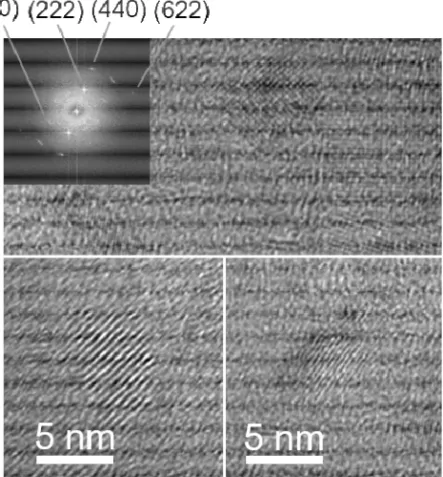
Figure 3.2 Transmission electron microscope images of pure Gd2O3
nanoparticles dialyzed for 1 day (lower left corner and upper overview), PEGylated Gd2O3 nanoparticles (GMP) (lower right corner) and a
cor-responding Fast Fourier Transform (FFT) for the PEGylated nanoparti-cles (GMP). For more discussion see Paper II.
TEM is efficient to study the size and crystallinity of nanoparticles samples. Figure 3.2 shows examples of TEM images of dialyzed Gd2O3
nanoparticles and Gd2O3 nanoparticles functionalized with PEG
(PEGylated Gd2O3 nanoparticles) investigated in detail in Paper II. The
former show crystalline particles. For the latter, it is possible to identify organic material, which is part of the samples. However, crystalline particles about 5 nm in size can still be found (Figure 3.2, the lower right corner). For further examples on Gd2O3 and Ce2O3 nanoparticles
3.2. Dynamic light scattering
Highly efficient light scattering experimental technique that allows to study particles with ~1 nm in diameter is Dynamic Light Scattering (DLS), sometimes also called as Photon Correlation Spectroscopy or Quasi-Elastic Light Scattering). DLS is an optical method useful for studies of nanoparticles, allowing for the analysis of their dynamic properties and size distribution36. The basic set up of the DLS
experi-ment is shown in Figure 3.3.
Figure 3.3 Schematic setup of Dynamic Light Scattering experiment.
A laser beam is used to illuminate a spatially limited volume within the sample, which for instance could be a colloidal suspension of nano-particles. The latter scatter the light, and the information is obtained from time-dependent fluctuations of the scattered light. In principle, scattered light waves spread out in all directions with intensity Is(t), which is time-dependent. Indeed, nanoparticles in the colloidal suspen-sion move randomly, or more correctly following the Brownian motion. Therefore, the interparticle distances fluctuate, leading to fluctuations of the phase relations of the scattered light. Moreover, the number of parti-cles within the scattering region is not constant as a function of time:
some particles can enter or leave the illuminated part of the sample. Consequently, constructive or destructive interference of the scattering waves, is stochastic, leading to fluctuations of Is. In DLS experiments, the fluctuations of Is are detected at a known scattering angle θ by a fast photon detector (Figure 3.3).
In order to analyze the fluctuations of Is(t) one derives the so-called intensity correlation function g2(t). This can be done either by electronic digital correlator, that is using special hardware, or by software that performs statistical analysis of Is(t). From g2(t) it is possible to obtain the diffusion coefficient D of the particles. It is related to the hydrody-namic radius R of the particles via Stokes-Einstein equation36:
D=kBT/(6πηR) , (3.1) where kB is the Boltzmann constant, T the temperature and η the viscosi-ty.
The mean hydrodynamic radius R is the main parameter, which is determined from the most straight-forward DLS experiments. Note that modern advanced multi-angle instruments can be used to accurately determine the distribution of the particle sizes.
3.3. Infrared spectroscopy
Infrared vibrational spectroscopy allows one to identify molecules by analysis of vibrational properties of their constituent bonds. IR spectros-copy is an excellent tool for analyzing molecular structures. Relatively small systems can be studied, for instance, molecules adsorbed on me-tallic clusters37. IR spectroscopy can be used as an analytical tool of the
surface composition and structure by making comparisons with surfaces of pure nanoclusters.
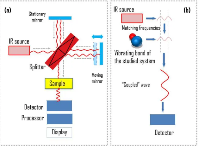
The infrared spectrum of a sample is recorded by passing a beam of infrared light through the sample, as shown schematically in Figure. 3.4a. The infrared light is used in the spectroscopy because it corre-sponds in energy to vibrational frequencies of most solids. There are two options to record the spectra. With the earliest spectrometers an IR spectrum could be measured by scanning the wavelength range using a monochromator. Today, the entire wavelength range is measured mo-mentary. Then a transmittance or absorbance spectrum is generated by means of a Fourier transform instrument.
Figure 3.4 Schematic illustration of (a) setup of IR experiment and (b) physical principle behind the IR spectroscopy.
The physical principle behind the IR spectroscopy is illustrated in Fig-ure 3.4(b). Each chemical bond in a studied system has its characteristic vibrational frequency (multiple modes are possible, of course, for group of atoms moving as a whole group). The technique is typically used for systems with covalent bonds. If the bonding in the system has an ionic component, vibrations of atoms lead to a modification of the dipole in the bond. Because of this, a photon with the frequency that matches the vibrational frequency will be absorbed by the system. The resonant fre-quencies are affected by the interactions between vibrating atoms, their masses, and the associated vibronic coupling. Therefore, analyzing the absorption spectra one sees how much energy was absorbed at each wavelength. In doing so one obtains valuable information about the chemical groups present in the sample, and consequently, about its composition.
Chapter 4
Hematopoietic cells for studies of nanoparticle-cell
interaction
Biocompatibility of synthesized and characterized nanoparticles is vital for their use in medical applications, e.g. as MRI contrast agents. In general, evaluation of biocompatibility is a complex process that in-volves efforts from several disciplines, medicine, biology, chemistry and physics. A promising path on this route is to start with studies of nanoparticle interactions with isolated cells. In the present thesis several different cell types, e.g. neutrophil granulocytes and macrophages and their interaction with nanoparticles have been analyzed. The aim is to rule out in this way the nanoparticles which would be avoid for test an-imals prior to potential animal tests. Because of this, studies of nanopar-ticles interactions with individual cell types are useful for screening and in that sense much more efficient in comparison to animal tests when the task is to improve biocompatibility of the nanoparticles via func-tionalization, different types of synthesis, by capping or doping with other elements. Also, this approach allows for rapid feedback and speeding up the development of new nanomaterials.
All the cellular elements of the blood, including the cells of the im-mune system, arise from pluripotent hematopoietic stem cells in the bone marrow38. Division of the pluripotent cells produces two types of
stem cells: a common lymphoid progenitor and a common myeloid pro-genitor. The former gives rise to the lymphoid lineage of white blood cells or leukocytes, which include the natural killer cells, as well as T and B lymphocytes. The latter gives rice to the myeloid lineage. The rest of leukocytes, including the monocytes, the dendritic cells and the granulocytes, as well as erythrocytes, and the megakaryocytes belong to the myeloid lineage. In this section I briefly describe cell types of inter-est for this study.
4.1. Polymorphonuclear neutrophils
Polymorphonuclear neutrophils (PMN) are a type of blood cells that belong to the granulocyte family of leukocytes. The granulocytes are characterized by multilobed nuclei and the presence of multiple, distinct granules within their cytoplasm. Besides neutrophils, the family also includes basophils and eosinophils39. PMNs develop and mature in the
bone marrow from pluripotent CD34+ stem cells. The rate of production is ~5-10 x 1010 cells per day over a 7-14 days period40. The cells
diame-ter within circulation is about 10 μm (see Figure 4.1). As a matdiame-ter of fact, they are the most abundant circulating leukocytes in our blood. Indeed, the concentration of PMNs in the blood is ~2.5-7.5 x 10⁹ cells/L.
In the peripheral blood the concentration of PMNs is fairly constant if one deals with a resting uninfected host, because the production and elimination of the cells are balanced in this case. On the other hand, the lifespan of neutrophils in the circulation is quite short: their half-life is 7-12 hours. Neutrophils undergo spontaneous apoptosis before their removal by macrophage in the lung, spleen, and lever 40.
Figure 4.1 Differential interference contrast (DIC) microscopy pictures of human neutrophils. The scale bar indicates 10 µm.
At the same time, the number of circulating neutrophils increases greatly, up to an order of magnitude, during pathological conditions caused, for instance by a bacterial infection. Indeed, the PMNs act as the first defense of the immune system against invading pathogens. This property of the PMNs make them especially valuable for studies of ef-fects of newly synthesized nanoparticles and their biocompatibility41-43.
Indeed, PMNs represent a valuable model system to study a remarkable array of generalized cellular functions, as well as specialized functions and molecules important to host defense against infection, the mediation and resolution of inflammation44. Using the neutrophil model system,
we may study several types of cellular process and biochemical path-ways, including phagocytosis and oxygen radical production. Neutro-phil activation by nanoparticles may lead to production and release of several different inflammatory mediators for example myeloperoxidase (MPO) into the extracellular space (into the buffer or medium). The cells may also express apoptosis (programmed cell death) and then for example shrink in size and express a lot of phosphatidylserine in their membrane44. The effects can be studied, e.g. by the cell clinic
tech-niques.
4.2. Isolation of polymorphonuclear neutrophils from human
blood
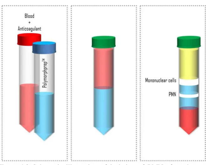
Isolation of PMNs from human blood employed in this work has been performed using a protocol adopted from the technique proposed by Böyum in 196845. Its schematic representation is shown in Figure 4.2.
Böyum proposed a technique for isolation of mononuclear cells from whole blood by one centrifugation, and of granulocytes by a two-steep procedure, combining centrifugation and a sedimentation. In short, anti-coagulated blood was layered on top of a mixture of Isopaque and ficoll and centrifuged, and the cellular elements were divided into two main fractions. Granulocytes and erythrocytes sedimented to the bottom of the tube, while mononuclear cells and platelets remained at the interface. Diluting the blood with saline before the initial centrifugation, Böyum found truly remarkable efficiency of separation of the mononuclear cells, with almost 100% yield. At the second stage, mononuclear cells are removed and mixed the cell mass in the bottom of the tube with plasma and dextran. He removed plasma layer containing the granulocytes when erythrocytes had settled by sedimentation at 1 x g gravity field.
Figure 4.2 Schematic illustration of isolation of PMNs from human blood following the protocol employed in this work. See text below for further details.
In more details, the following protocol has been adopted in this work: 1. Prepare 25 ml venous whole blood donated by apparently
healthy, non-medicated volunteers. Blood must be anti-coagulated with 10U/ml heparin and must be allowed to equili-brate to room temperature.
2. Layer the blood on an equal volume of a density gradient (Polymorphprep™, see Figure 4.2, middle panel)
3. Centrifuge at 480 x gfor 40 min at room temperature. Allow the rotor to decelerate with very weak braking.
4. Remove the top layer (plasma), the second layer (mononuclear cells) and the third layer (remained PolymorphprepTM), see
Fig-ure 4.2, right panel. Harvest the next layer (the lower band of PMNs) into a new 50 ml centrifuge tube.
5. Resuspend the harvested material with an equal volume of 0.45% NaCl solution. Add ca 20 ml of PBS equilibrated to room temperature. Turn the tube couple of times.
6. Centrifuge at 400 x gfor 10 min at room temperature.
7. Pour off the supernatant to harvest the PMNs and resuspend the pellet carefully.
8. Remove residual erythrocytes by brief (35 s) hypotonic lysis in 4.5 ml cold distilled water. Then add 1.5 ml PBS containing 3.4% NaCl. Next add 5 ml cold Hepes-buffer. All operations of (8) must be done on ice.
9. Centrifuge at 400 x gfor 5 min at temperature +40C.
10. Repeat operations described in (7)-(9).
11. Pour off the supernatant and resuspend the pellet carefully. Add 1 ml ice-cold Hepes-buffer. Take 10 μl of the cell suspension for a cell counting.
12. Dilute the cell suspension in Hepes-buffer to a needed concen-tration, in this work usually to 2*106 cells/ml.
4.3. ROS production
PMNs are phagocytic cells of the innate immune system. Although tra-ditionally considered as an innate immune cell, recent knowledge points towards a more complex role of these cells also involved in chronic in-flammatory processes and as a contributor in adaptive immune respons-es. Clearly, they are potent cells capable of releasing for example a range of immunomodulatory molecules46.
They react first to defend the host against invading pathogens, main-ly bacteria and fungi, but also viruses40. PMNs have powerful
microbi-cidal equipment, and they have been shown to play a large role in in-flammatory responses. For instance, neutrophils react quickly, within minutes to an hour of tissue injury, making themselves into a hallmark of acute inflammation. They are mobile, and they have a capacity to seek and destroy different targets following phagocytosis of an invading pathogen. Neutrophils at rest consume relatively small amounts of oxy-gen. However, in case of an agonist challenge, their consumption of oxygen increases dramatically. Neutrophils reduce oxygen to superox-ide anions through the activation of the enzymatic system which is unique for phagocytic cells, including neutrophils. Specifically, the pro-cess is mediated by nicotinamide adenine dinucleotide phosphate
(NADPH)-oxidase enzyme. The superoxide anions could further metab-olize to hydrogen peroxide (H2O2) and other even more toxic
substanc-es. , H2O2, singlet oxygen, and other products derived from the
me-tabolism of H2O2 generated by neutrophils following the uptake of
par-ticles or in response to inflammatory stimuli are referred to as reactive oxygen species (ROS). The process described above is also referred to as “the respiratory burst” defined as an increase in the oxidative me-tabolism40.
Moreover, the neutrophils antimicrobial activity essential for patho-gen killing also involves the release of their intracellular granules, in addition to ROS production. The granules contain lytic enzymes and antimicrobial polypeptides. At the same time, it is worth to point out that PMNs-derived respiratory burst could damage the surrounding tis-sues upon uncontrolled release of ROS. In fact, there are inflammatory diseases dominated by neutrophils. Therefore, neutrophils turnover should be controlled quite strictly.
To measure the ROS production, one can use the effect of chemilu-minescence. Indeed, simultaneously with generation of oxidative me-tabolites by neutrophils upon the respiratory burst, the light is also gen-erated, that is the chemiluminescence takes place. The light can be de-tected, and the sensitivity of the measurements can be increased via ad-dition of luminol or 2,7-dichlorofluorescein diacetate (DCF-DA, Sigma Aldrich, St Louis, MO, USA). Both substances can be excited by ROS, and then emit photons upon the transition to the ground state. The mole-cules can pass the biological membrane, and therefore they can be used for the measurements of both, intracellular and released oxygen radicals. However, if one needs to measure only released oxygen metabolites, it is possible to use isoluminol, which does not penetrate cells because of its chemical structure.
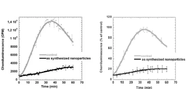
An example of chemiluminescence recordings of the reactive oxygen species (ROS) production from neutrophil granulocytes from Paper I is shown in Figure 4.3. Neutrophil granulocytes (2x10⁶ cells/ml) were
challenged with IgG-opsonized yeast particles (5x10⁶ yeast particles/ml)
after an initial exposure to as synthesized Gd2O3 nanoparticles (0.94
mM) for 15 minutes. Results obtained on control cells with only IgG-opsonized yeast are shown for comparison. Chemiluminescence was recorded, using luminol, in a Microplate Fluorescence Reader FL600. The ROS production was calculated as relative chemiluminescence