The involvement of degradation pathways and
neuron-to-neuron transmission in Alzheimer’s disease
Lotta Agholme
Department of Clinical and Experimental Medicine Faculty of Health Science, Linköping University
Linköping, Sweden
© Lotta Agholme 2012
Illustrations on p. 28 and p. 30 by Lotta Agholme. All other illustrations by Jakob Domert.
All previously published papers were reproduced with kind permission from the publishers.
Printed by LiU-tryck, Linköping 2012
ISBN: 978-91-7519-848-4 ISSN: 0345-0082
Don’t aim for the stars. There are so many stars, and they are so far away. Aim for the moon instead. The moon is large, clear, and within reach.
Martin Hallbeck, Associate Professor
Division of Pathology, Department of Clinical and Experimental Medicine, Faculty of Health Science, Linköping University
Co-supervisors
Katarina Kågedal, Associate Professor
Division of Experimental Pathology, Department of Clinical and Experimental Medicine, Faculty of Health Science, Linköping University
Jan Marcusson, Professor
Division of Geriatrics, Department of Clinical and Experimental Medicine, Faculty of Health Science, Linköping University
Faculty Opponent
Gunnar Gouras, Professor
Experimental Dementia Research Unit, Wallenberg Neuroscience Center, Department of Experimental Medical Science, Lund University
Committee Board
Sven Hammarström, Professor
Division of Cell Biology, Department of Clinical and Experimental Medicine, Faculty of Health Science, Linköping University
Maria Ankarcrona, Associate Professor
Division of Alzheimer's Disease Research Center, Department of Neurobiology, Care Sciences and Society, Karolinska Institutet
David Engblom, Associate Professor
Division of Cell Biology, Department of Clinical and Experimental Medicine, Faculty of Health Science, Linköping University
Abstract
Although the vast majority of Alzheimer’s disease (AD) cases are of the sporadic type, mutations causing the familial form have been the focus of AD research for decades. The disease is pathologically characterised by β-amyloid (Aβ) and tau protein aggregates in neuritic plaques and neurofibrillary tangles. Furthermore, it is known that AD pathology spreads throughout the brain, most often along the same anatomical pattern. However, so far no cause for the sporadic form of the disease has been found. Accumulation of protein aggregates as well as decreased activity of the protein degradation systems, lysosomes and proteasomes, is found in diseased brains. This indicates that defective degradation contributes to sporadic AD.
The aim of this thesis was to develop an improved neuronal model, and study the effects of decreased proteasome function on tau phosphorylation and axonal transport. In addition, the effects on Aβ accumulation and generation upon proteasome inhibition were investigated. Finally, the possibility that intracellularly accumulated Aβ oligomers could be transferred from one neuron to another was tested.
Differentiation of human SH-SY5Y neuroblastoma cells in an extracellular matrix gel, using a set of neurotrophic factors, resulted in cells with neuronal phenotype, expressing neuron specific markers and all six adult isoforms of tau. Within this neuronal model, we found that reduced proteasome activity inhibited neuritic transport, and caused tau phosphorylation in a c-Jun and ERK 1/2 dependent manner. Using proteasome inhibition in APP over-expressing cells, we found an autophagy dependent intralysosomal Aβ accumulation, together with elevation of intra- and extracellular concentrations of Aβ. Autophagy inhibition protected the cells from the toxicity induced by decreased proteasome activity. Finally, we could, as the first group, show that Aβ can be directly transferred from one neuron to another through connected neurites. Furthermore, accumulation of Aβ in the endo-lysosomal compartment of receiving cells caused toxicity and neurodegeneration.
We believe that cells not able to degrade accumulated Aβ, due to increased generation or reduced degradative capacity, instead tries to clear its content through transfer to connected neurons. If not properly degraded in the receiving cell, this can accelerate AD pathology and cause neuritic and neuronal degeneration spreading throughout the brain. Increasing the activity of the degradative systems, or inhibiting transmission of Aβ between neurons could therefore be novel treatments for AD.
Populärvetenskaplig
Sammanfattning
Alzheimers sjukdom finns i två varianter, ärftlig och sporadisk, där merparten (över 90%) tillhör den sporadiska. Alzheimers karaktäriseras av ansamlingar av ihopklumpade proteiner, β-amyloid och tau, i hjärnan samt nervcellsdöd, som leder till försämring av minnesfunktionerna. Nervceller har två ”renhållningssystem”, lysosomer och proteasomer, som bland annat tar hand om dessa ihopklumpade proteiner. Studier har visat på försämringar av båda dessa system vid Alzheimers sjukdom.
I den här avhandlingen har vi undersökt vad som händer när dessa renhållningssystem slutar fungera. Specifikt har vi studerat vad som händer när man minskar aktiviteten hos proteasomen och har sett att det leder till flera av de kända sjukliga förändringarna vid Alzheimers sjukdom. Dessa inkluderar ansamling av proteinet β-amyloid, skadliga förändringar av proteinet tau samt en försämrad transport längs nervcellernas långa utskott.
Vidare har vi, som första grupp i världen, visat att proteinet β-amyloid, när det klumpar ihop sig i cellerna, kan skickas vidare till nästa nervcell och där orsaka stor skada. Denna överföring, eller ”smitta”, sker bara där nervcellerna har kontakt med varandra. Överfört till människor skulle det betyda att hjärnans funktion och patientens minne försämras i takt med att nedbrytningen av nervceller sprids genom hjärnan. Detta skulle också kunna förklara det mönster som Alzheimers sjukdom vanligtvis sprids efter.
Ett möjligt händelseförlopp vid Alzheimers sjukdom kan vara att nervceller som får för mycket β-amyloid, till exempel på grund av ett dåligt renhållningssystem, försöker göra sig av med detta genom att skicka det till nästa cell. I den mottagande cellen orsaker β-amyloidet skador, men även ytterligare ansamlingar av proteiner som skickas vidare. På så sätt kan sjukdomen spridas genom hjärnan och orsaka minnesstörningar.
Utveckling av mediciner som förbättrar nervcellernas förmåga att bryta ner proteiner, eller förhindrar överföringen av β-amyloid mellan nervceller, kan förhoppningsvis bromsa eller hindra Alzheimers sjukdom från att spridas i hjärnan.
Table of contents
ORIGINAL PUBLICATIONS... 3
ABBREVIATIONS ... 5
INTRODUCTION... 7
FINDING THE CAUSE OF SPORADIC ALZHEIMER’S DISEASE... 7
APP AND Aβ ... 8
APP CLEAVAGE AND Aβ GENERATION 8 SITES FOR Aβ GENERATION AND INTRACELLULAR ACCUMULATION 9 Aβ SPECIES 10 Aβ CLEARANCE 10 Aβ IN NEURODEGENERATION 12 TAU IN HEALTHY BRAIN AND ALZHEIMER’S DISEASE...13
TAU ISOFORMS 13 TAU PHOSPHORYLATION AND AGGREGATION 13 TAU IN NEURODEGENERATION 14 INTERACTIONS BETWEEN Aβ AND TAU IN NEURODEGENERATION 15 AXONAL TRANSPORT AND MICROTUBULE...15
AXONAL TRANSPORT DEFICITS IN ALZHEIMER’S DISEASE 17 THE ROLE OF Aβ AND TAU IN AXONAL TRANSPORT DISRUPTION 17 CELLULAR DEGRADATION PATHWAYS...17
LYSOSOMES AND AUTOPHAGY 18 THE LYSOSOMAL SYSTEM IN ALZHEIMER’S DISEASE 18 PROTEASOMES 19 PROTEASOME FUNCTION IN ALZHEIMER’S DISEASE 19 SPREADING OF ALZHEIMER’S DISEASE PATHOLOGY...21
MODELLING ALZHEIMER’S DISEASE...22
AIM OF THE THESIS...25
METHODS ...27
CELLULAR MODELS AND CELL CULTURE...27
MESSENGER RNA EXPRESSION...27
PROTEIN EXPRESSION AND PROTEIN MODIFICATION...29
WESTERN BLOT 29 ENZYME-‐LINKED IMMUNOSORBENT ASSAY (ELISA) 29 ELECTROCHEMOLUMINESCENT IMMUNOSORBENT ASSAY 30 PROTEOME PROFILER PHOSPHO-‐KINASE ARRAY 31 COMPARISON OF DIFFERENT PROTEIN ANALYSIS ASSAYS 31 PROTEIN LOCALISATION AND CO-LOCALISATION...31
IMMUNOCYTOCHEMISTRY 32 ORGANELLE SPECIFIC DYES 32 FLUORESCENTLY TAGGED PROTEINS 32 COMPARISON OF THE DIFFERENT ASSAYS 33 NEURITIC TRANSPORT...33
Aβ OLIGOMER PREPARATION AND CHARACTERISATION...34
INHIBITION OF PROTEINS AND CELLULAR PROCESSES...34
SMALL MOLECULE INHIBITORS 34 SMALL INTERFERING RNA 35 CELL TOXICITY AND VIABILITY...35
MTT VIABILITY ASSAY 35 CRYSTAL VIOLET ASSAY 36 MICROTUBULE BEADING 36 ORGANELLE RUPTURE 36 COMPARISON OF DIFFERENT VIABILITY ASSAYS 37 RESULTS...39
PAPER I ...39
PAPER II...40
PAPER III ...42
PAPER IV...43
DISCUSSION AND CONCLUSIONS...45
A NEW MODEL FOR ALZHEIMER’S DISEASE RESEARCH...45
PROTEASOME ACTIVITY AND AXONAL TRANSPORT...46
PROTEASOME ACTIVITY IN Aβ GENERATION AND ACCUMULATION...47
NEURONAL TRANSMISSION OF Aβ ...48
GENERAL DISCUSSION...49
CONCLUSIONS...51
FUTURE PERSPECTIVES...53
ACKNOWLEDGEMENTS...55
Original publications
This thesis is based on the following original publications, which will be referred to by their Roman numerals.
I. Lotta Agholme, Tobias Lindström, Katarina Kågedal, Jan
Marcusson, Martin Hallbeck. An in vitro model for
Neuroscience: Differentiation of SH-SY5Y
neuroblastoma cells into cells with morphological and biochemical characteristics for mature neurons. Journal
of Alzheimer’s Disease, 2010. 20(4), 1069-1082.
II. Lotta Agholme, Sangeeta Nath, Jakob Domert, Jan Marcusson, Katarina Kågedal, Martin Hallbeck. Proteasome inhibition
induces stress kinase dependent transport deficits – implications for Alzheimer’s disease. Submitted to PLoS
ONE.
III. Lotta Agholme, Martin Hallbeck, Eirikur Benedikz, Jan Marcusson, Katarina Kågedal. Amyloid-β secretion,
generation and lysosomal sequestration in response to proteasome inhibition: Involvement of autophagy.
Journal of Alzheimer’s Disease. 2012. 31(2) 343-358.
IV. Sangeeta Nath, Lotta Agholme, Firoz Roshan Kurudenkandy, Björn Granseth, Jan Marcusson, Martin Hallbeck. Spreading of
neurodegenerative pathology via neuron-to-neuron transmission of β-amyloid. Journal of Neuroscience, 2012.
Abbreviations
Aβ β-amyloid
oAβ Oligomeric β-amyloid AD Alzheimer’s disease
AA Amino acids
Apo Apolipoprotein
APP Amyloid precursor protein BDNF Brain derived neurotrophic factor CDK Cyclin-dependent kinase
DIC Differential interference contrast ECM Extracellular matrix
EGFP Enhanced green fluorescent protein ELISA Enzyme linked immunosorbent assay ERK Extracellular-signal regulated kinases GSK Glycogen synthase kinase
HRP Horseradish peroxidase IDE Insulin degrading enzyme iPS Induced pluripotent stemcells JNK c-Jun N-terminal kinase KLC Kinesin light chain
MTT 3-(4,5-Dimethylthiazol-2-yl)-2,5-diphenyltetrazolium
bromide
NFT Neurofibrillary tangles NGF Nerve growth factor
NRG Neuregulin β1
PAGE Polyacrylamide gel PHF Paired helical filament
RT-PCR Reverse transcriptase polymerase chain reaction
RA Retinoic acid
SDS Sodium dodecyl sulfate siRNA Small interfering RNA TMR Tetramethyl rhodamine
Introduction
Over 100 years ago, Alois Alzheimer described the pathological hallmarks of the neurodegenerative disease later named after him, Alzheimer’s disease (AD) [1]. Since then, extensive research has been carried out, both within the general field of neuroscience, as well as within the Alzheimer’s field, to find the underlying cause for this dreadful disease. Despite all the great efforts, the cause for the most common form of AD, the sporadic form, is still not known. Nor is there a treatment available that can prevent, reverse or even halt the disease.
Alzheimer’s disease is the most common form of dementia, and it is estimated that more than 26 million people worldwide are affected today (reviewed in [2]). Aging is the single most important risk factor, and as the life expectancy increases, so will the number of people suffering from AD. The number of affected is estimated to quadruple until the year 2050, if the cause and a cure for AD are not found. The disease causes tremendous agony for the affected and their families, as well as a huge healthcare cost. It is therefore of utmost importance to find the cause of, and the treatment to AD.
Finding the cause of sporadic Alzheimer’s
disease
The pathological phenotype for AD, described by Dr Alzheimer already in 1907, includes extracellular amyloid plaques, intracellular neurofibrillary tangles (NFTs), dystrophic neurites and neuronal loss. Ever since the proteins behind plaques and tangles, β-amyloid (Aβ) and tau respectively, were discovered [3, 4], the importance of these two proteins for disease progression have been the focus of AD research.
Mutations resulting in increased production of Aβ are the cause of the familial, early onset, form of AD (reviewed in [5]). As the sporadic form of AD shares pathological hallmarks with the familial form; many believe that Aβ is the
driving force in this form as well. However, the possible causes for Aβ accumulation in sporadic AD are largely unknown.
The focus has now shifted from plaques and tangles, which are now believed to accumulate later in disease progression, to the soluble forms of Aβ and tau [6-8]. Furthermore, synaptic disruption and axonal transport deficits are suggested to be the early events in disease progression [9-11]. How Aβ and tau can contribute to these deficits is, to a large extent, unknown.
Genomic presence of the Apolipoprotein (Apo) E4 allele, a protein involved in lipid transport, is the most important genetic risk factor for sporadic AD [12]. Other risk factors include high cholesterol levels and other vascular factors, diabetes, as well as low physical and mental activity (reviewed in [13]).
APP and A
β
The 38-43 amino acid (AA) long Aβ peptide, the main component of extracellular amyloid plaques [3], is cleaved from the transmembrane glycoprotein amyloid precursor protein (APP). APP, a protein ubiquitously expressed throughout the body, is present in several isoforms. The 751 AA long variant is the most common in all tissues but the brain, where the 695 AA variant is dominant (reviewed in [14]). The normal function for APP has not been clearly elucidated. It is however suggested that APP has a role in cell adhesion as well as neuritic outgrowth and development [15, 16]. Different cleavage products of APP have also been suggested to have physiological roles, such as the intracellular C-terminal fragment regulating gene transcription [17], and the soluble cleavage product, APPα, having neuroprotective functions [18]. Aβ is also believed to have physiological importance, including regulation of synaptic function and having neurotrophic and neuroprotective effects [19].
APP cleavage and A
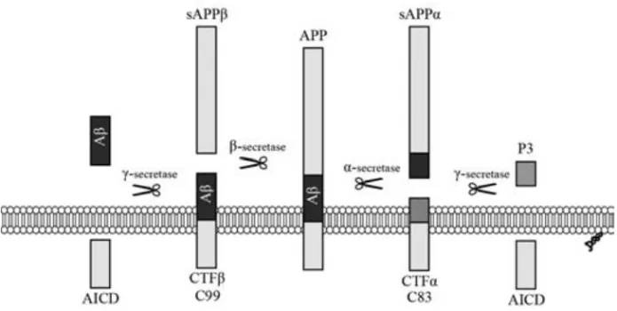
βgeneration
APP is cleaved by three secretases, resulting in two different forms of soluble APP, one intracellular domain, and two different peptides; p3 and Aβ (Fig. 1). Cleavage of APP by α-secretase, followed by γ-secretase generates the non-amyloidogenic (non-aggregating) peptide P3 (reviewed in [20]). Cleavage of APP by β-secretase followed by γ-secretase generates the amyloidogenic, aggregation-prone, peptide Aβ. Imprecise cleavage by γ-secretase results in peptides varying in length from 17 to 43 AA, where the 38-43 AA long peptides
are the most investigated [20, 21]. Aβ 1-40 is the most abundant form in human brain, whereas Aβ 1-42 is most prone to aggregate [22]. Aβ can also be N-terminally cleaved and modified [23], altogether contributing to the heterogeneity of Aβ species.
The familial form of AD is caused by mutations in the genes for APP, presenilin (PS)-1 or PS-2 (part of the γ-secretase complex), causing increased production of Aβ, increased Aβ42 to Aβ40 ratio, or more aggregation-prone Aβ [24-26]. If the cause for sporadic AD is increased Aβ generation is still not completely agreed on. It is however an intense search ongoing for the possible mechanisms causing the Aβ accumulation in sporadic AD.
Sites for A
βgeneration and intracellular
accumulation
APP cleavage and Aβ generation is thought to mainly take place at the plasma membrane and in the secretory pathway, resulting in extracellular release of Aβ (reviewed in [20]; Fig. 2). In neurons, the synapse is most likely a major site for APP cleavage, as several proteins in the APP cleavage machinery have been located there [27]. In addition, Aβ has been shown to regulate synaptic transmission, and synaptic activity can regulate Aβ production [28, 29].
Figure 1. APP can be cleaved by α-secretase or β-secretase followed by γ-secretse. The first pathway results in release of the non-amyloidogenic peptide P3. The latter results in production of the aggregation-prone peptide Aβ.
Several intracellular organelles have also been suggested as sites for Aβ production, including the ER and golgi, as well as endosomes, autophagosomes and lysosomes [30-32]. APP has been localised to all these organelles, which also contain both β- and γ-secretase. Upon intracellular APP cleavage, Aβ is released intra-luminally, resulting in intracellular Aβ accumulation (Fig. 2). In addition, re-uptake of extracellularly released Aβ occurs through binding to a number of different receptors (reviewed in [20]). Aβ was for a long time seen solely as an extracellular peptide, but after an intense debate, most researchers now agree on the presence of intraneuronal Aβ. The most compelling evidence is the staining of Aβ in brain tissue from AD patients, showing that Aβ localises to multivesicular bodies, an organelle originating from the endosomal pathway [33]. Aβ accumulation in endosomes and autophagosomes in AD neurons have also been shown [31, 34]. However, whether intracellular or extracellular Aβ is the major inducer of neurotoxicity still needs to be elucidated.
A
βspecies
The Aβ peptide is, due to its hydrophobic nature, highly fibrillogenic and can readily aggregate into oligomers, protofibrils, and fibrils. The transition from monomers to fibrils is executed through several steps, where the native conformation of Aβ (α-helical and unordered) is transformed into β-sheets. This promotes Aβ assembly into oligomers varying in size from dimers and trimers (2-3 Aβ molecules, 8-12 kDa) to 100-kDa molecules and larger [35]. These oligomers can aggregate further into protofibrils, and in the end mature fibrils, being the building block of amyloid plaques. The rate of Aβ aggregation depends on several factors including concentration, pH and temperature.
A
βclearance
Accumulation of Aβ, as seen in AD, could either be due to increased generation or decreased clearance. Aβ can be degraded and cleared from the intra- or extracellular compartments in several ways. Neprilysin and insulin degrading enzyme (IDE) are the two major enzymes responsible for Aβ degradation. Neprilysin is mainly responsible for degradation of aggregated Aβ associated with membranes. IDE on the other hand, is mainly responsible for degradation of intracellular, non-aggregated Aβ (reviewed in [14]). Aβ can also be cleared from the brain through interaction with lipoprotein receptors [36]. ApoE,
Figure 2. APP cleavage and Aβ generation has been suggested to take place mainly at the plasma membrane, in neurons most likely at the synapses. Here, Aβ is released extracellularly. However, intracellular APP cleavage in ER, golgi and the lysosomal pathway has also been suggested. This results in Aβ release intra-luminally.
being a lipid transporter, also has a role in the clearance of Aβ from the brain, and ApoE4 is shown to be less effective, compared to ApoE2 and ApoE3 variants [37]. Apart from direct digestion by enzymes, Aβ can also be degraded through the major cellular degradation pathways, lysosomes and proteasomes, as discussed below.
A
βin neurodegeneration
There are a massive number of studies exploring the effect of extracellularly administered Aβ to cells and slice cultures in vitro. They have shown that different forms of Aβ have a number of toxic effects including oxidative stress, synaptic dysfunction, and axonal transport deficits [38, 39]. Different animal models have shown that Aβ has effects on synaptic function and axonal transport, but also immune cell activation [40]. Several animal models have also confirmed the negative effect of Aβ on memory and cognition (reviewed in [41]). In addition, Aβ has repeatedly been shown to induce tau phosphorylation, which is believed to cause the aggregation of tau into NFTs [42, 43].
For a long time, it was thought that the fibrillar form of Aβ, found in the plaques, was the most toxic species. For instance, an in vitro study showed that exposure of cells to fibrillar Aβ caused toxicity by disrupting synapses [44]. However, it has later been suggested that the pre-fibrillar, oligomeric form of Aβ (oAβ) is the most toxic. Different oAβ forms (from dimers to larger spherical aggregates) are potent neurotoxins both in vitro and in vivo [45-48]. Although this might seem confusing, the different results obtained could be due to the ability of Aβ to constantly associate and dissociate, making the studies of specific species difficult.
A number of mechanisms and pathways for how Aβ induce neuronal dysfunction have been proposed. Aβ has been shown to bind to a variety of transmembrane receptors, including N-methyl-D-aspartate and nicotinergic acetylcholine receptors, as well as the cellular prion protein, thereby inducing reduction in long-term potentiation, dendritic spine density, and synaptic loss [49, 50]. Since Aβ aggregates can form pores in bilipid membranes, it is also postulated that oligomeric Aβ can cause leakage from intracellular organelles as well as the plasma membrane [51]. The mechanisms behind intracellular Aβ toxicity are unclear, but one possibility is inhibition of the cellular degradation systems [52]. This will be discussed in more detail below.
Tau in healthy brain and Alzheimer’s
disease
Ever since it was discovered that the tangles consisted of a hyper-phosphorylated form of the microtubule binding protein tau [4], phosphorylation of tau has been the main focus for tau research. Not only because hyperphosphorylated tau is found in neurofibrillary tangles, but also because it has been shown, both in vivo and in vitro, that Aβ is able to cause tau hyperphosphorylation [43, 53].
Tau is a member of a family of microtubule-associated proteins present in many cell types throughout the body. In neurons, tau is mainly located to axons [54]. There, it is thought to regulate microtubule assembly and stability [55-57], although this is only proven in vitro. Tau regulates this by binding more or less tightly to microtubules, where tau tightly bound to microtubules increases its stability and vice versa [58]. The binding strength of tau to microtubules can be regulated in two ways, by differentially splicing and by phosphorylation. Tau is also important for neurite polarity and development, as well as cell division [59-61].
Tau isoforms
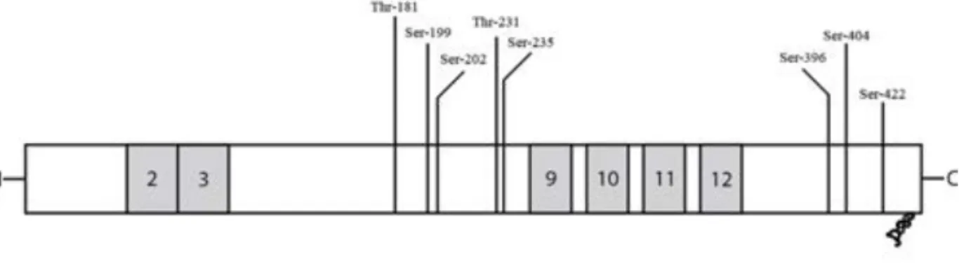
Tau can exist in six different isoforms, originating from one gene by alternative splicing of three exons. Two of the exons, 2 and 3, are situated in the N-terminal projection domain, giving rise to 0, 1 or 2N tau. The third is exon 10, one of four C-terminally located microtubule-binding domains, resulting in 3R or 4R tau (Fig. 3). Inclusion of exon 10 increases the binding strengths to microtubules [62, 63]. The role of the projection domain is less clear, but is believed to have a role in spacing between microtubules and interactions with the plasma membrane [64, 65]. The adult human brain contains all six isoforms, in a 1:1 ratio of 3R and 4R tau [66]. In the foetal brain, only the shortest isoform of tau is present [62, 67], and this might protect tau from aggregation even if heavily phosphorylated during development [68].
Tau phosphorylation and aggregation
The tau protein has 79 serine and threonine residues, many of them are readily phosphorylated by a number of kinases (reviewed in [69]). In healthy neurons, the level of phosphorylation regulates the binding strength to microtubules.
Figure 3. Tau protein can be alternatively spliced N-terminally resulting in exclusion of
both exon 2 and 3, inclusion of exon 2 or inclusion of both exon 2 and 3 (0, 1 or 2N). Furthermore, it can be alternatively spliced C-terminally causing inclusion or exclusion of exon 10, one out of four microtubule binding repeats (3 or 4R). There are many phospho-epitopes of tau, and eight sites thought to be relevant for AD are depicted above.
High levels of phosphorylation reduce the binding strength of tau, hence increasing microtubule dynamics [70].
In AD, tau is hyperphosphorylated by increased phosphorylation at regular epitopes as well as by de novo phosphorylation at AD-specific epitopes [69]. Some AD specific sites suggested include S199, T231 and S396 [71] (Fig. 3). Many kinases are able to hyper-phosphorylate tau in vitro. Glycogen synthase kinase (GSK) 3β is believed to be of great importance in AD, together with cyclin-dependent kinase (CDK) 5 (reviewed in [72]). Many other kinases have also been implicated to contribute to tau phosphorylation in AD, including those belonging to the c-Jun N-terminal kinase (JNK), extracellular-signal regulated kinase (ERK) and p38α pathways [73-75]. Hyperphosphorylation of tau causes it to detach from microtubules, and aggregate into paired helical filaments (PHF) that in the end stage form NFTs in the cell soma [76-78]. In addition, it is also believed that truncation of tau is important for aggregation into NFTs [79].
Tau in neurodegeneration
It is still not known how hyperphosphorylation of tau contributes to AD pathology. The working hypothesis for many years was that hyperphosphorylation of tau caused detachment from microtubules (Fig. 4). This would result in microtubule destabilisation, causing disturbed axonal transport and cell death [80]. Later, it has been shown that removal of tau in a transgenic mouse model does not impair cognition [81]. In addition,
overexpression of tau impairs axonal transport [80, 82], making this hypothesis questionable.
Instead, it has been suggested that hyperphosphorylated tau itself is toxic to neurons. Expression of tau mimicking hyperphosphorylation (pseudo-phosphorylated) causes toxicity both in cultured neurons and in drosophila models [83, 84]. The mechanisms behind the toxicity induced by hyperphosphorylated tau needs to be further investigated.
Interactions between A
βand tau in neurodegeneration
Although the number of extracellular plaques correlate poorly with the degree of AD dementia, and the amount of NFTs is a better correlate [85], it is believed that Aβ pathology lies upstream of tau. This has been proven both invitro, where exposure of cells in culture to different forms of Aβ causes tau
phosphorylation [43, 86], and in animal models where Aβ pathology appears earlier than the tau pathology [87]. In addition, exposing tau transgenic mice to Aβ induces tau phosphorylation and tangle formation [53]. It was established early on that Aβ has the potency to activate kinases such as GSK3β and CDK5, both being able to phosphorylate tau [88, 89]. However, the exact mechanism(s) by which Aβ induce tau phosphorylation still needs to be elucidated.
The importance for tau in Aβ-induced neurodegeneration has been firmly established in vitro and in animal models over the last ten years. Cells lacking tau are protected from Aβ-induced microtubule disruption and cellular degeneration [42, 90], and knocking out tau in an animal model rescues the animal from Aβ-induced impairments in memory and learning [81].
Axonal transport and microtubule
Neurons, with their long branched neurites, rely heavily on neuritic transport for distribution of energy, proteins, and organelles. Neurons are therefore especially vulnerable to disturbances in microtubule function, and many neurodegenerative diseases, including AD, share a phenotype involving axonal transport deficits [10].
Axonal and dendritic transport is facilitated by the motor proteins kinesin and dynein, travelling along microtubule tracks. The microtubule consists of
Figure 4. Anterograde axonal transport is facilitated by kinesins. Kinesin-1 attaches to
microtubule and kinesin light chains (KLC) attaches to cargoes such as mitochondria and vesicles. Microtubule stabilising protein tau detaches from microtubule when phosphorylated, which could lead to destabilisation. Tau can also interfere with kinesin assembly, and therefore directly inhibit transport. Aβ can activate kinases such as GSK3β and casein kinases (CK), responsible for phosphorylation of kinesins and KLCs. This causes detachment from microtubule and cargo, resulting in disturbed axonal transport.
assembled α- and β-tubulin, and has a plus and a minus end. The anterograde transport (towards the synapse) is regulated by kinesins, where Kinesin-1 connects with the microtubules, and the kinesin light chain (KLC) attaches to the cargo vesicle or organelle. See Figure 4 for an overview. The retrograde transport (towards the cell body) is facilitated by dyneins, where dynein heavy chains bind to microtubules and a complex of different dynein chains are responsible for the attachment to cargo (reviewed in [10]).
Axonal transport deficits in Alzheimer’s disease
It was suggested early on that disturbed axonal transport could be the cause of AD [91]. Decrease in axonal transport results in accumulation of organelles in neurites, a pathological sign of AD [92]. Reduced levels of kinesins increases Aβ generation [11], indicating that reduced axonal transport can start a cascade of events causing AD pathology. On the other hand, APP is suggested to be involved in regulation of axonal transport [93], and both Aβ and tau can disturb axonal transport, as discussed below. Thus, it remains to be elucidated if axonal transport deficits are a cause or a consequence of AD.
The role of A
βand tau in axonal transport disruption
Several in vivo studies have shown that Aβ can induce disturbances in axonal transport [38, 94, 95]. Different mechanisms have been proposed. Aβ can activate casein kinase 1 and 2, as well as GSK3β, causing phosphorylation of KLC and detachment of cargo vesicles [94] (Fig. 4). Other kinases activated by Aβ, such as JNKs, can phosphorylate Kinesin-1 resulting in a similar detachment of cargo from microtubules [96]. The above studies do not take into account the contribution of tau on Aβ induced transport deficits. It has been shown that Aβ-induced microtubule destabilisation and axonal transport deficits require the presence of tau [42, 97]. Therefore, direct actions of Aβ on transport proteins cannot be the whole explanation. Hyperphosphorylated tau has been shown to cause microtubule destabilisation [98], and it was recently shown that phosphorylated tau can disturb axonal transport by competing with proteins connecting cargo vesicles to the microtubules [99] (Fig. 4).Cellular degradation pathways
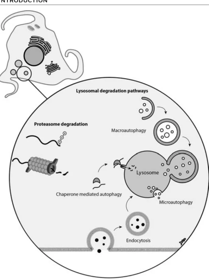
Protein turnover (synthesis and degradation) is an essential process for all cells. Protein degradation regulates the cell cycle, signalling events, and metabolism. It is also important for clearance of misfolded proteins, as well as damaged and worn out proteins and organelles. There are two major systems for protein degradation: the lysosomal and the proteasomal. Proteasomes and lysosomes are both capable of degrading long- and short-lived proteins, but they have different specific roles, which also can be cell type dependent (reviewed in [100]). For a schematic image of the two degradation pathways and the routes of delivery, see Figure 5.
Lysosomes and autophagy
Lysosomes are acidic organelles, surrounded by a single lipid bilayer, containing a number of proteases responsible for degradation of proteins and organelles. The material that is to be degraded can be transported to lysosomes by a number of different mechanisms. These include enclosement of material into autophagosomes, (macroautophagy), as well as delivery by endosomes (endocytosis), and secretory vesicles (crinophagy) (reviewed in [100]). Material can also be transported directly to the lysosomes, independent of vesicles, by microautophagy or chaperone mediated autophagy (Fig. 5).
Macroautophagy, hereafter called autophagy, is the main route for delivery of material to the lysosomes. Autophagy is mainly a non-specific degradation mechanism, responsible for the recycling of cellular components. It is upregulated in response to starvation, when there is an increased need for nutrients in the cells. In addition, selective degradation of certain proteins and specific degradation of worn out mitochondria is performed through autophagy. The generation of autophagosomes is orchestrated by a number of regulatory proteins, forming several large complexes. Autophagosomes are in the end fusing with lysosomes, releasing their content for degradation in the acidic environment.
The lysosomal system in Alzheimer’s disease
Neurons, as well as other post mitotic cells, rely heavily on autophagy and lysosomal degradation for removal of worn out proteins and organelles. During aging, the amount of oxidative stress increases, resulting in an increased accumulation of damaged proteins, demanding even more effective autophagy machinery.
As early as the 1960s, accumulation of lysosomes in dystrophic neurites of AD patients was described [101]. Later on, up-regulation and/or dysfunction of the endo-lysosomal system in AD brain, was confirmed [102, 103]. This seems to take place early in disease progression, before amyloid plaques are formed, at the time point where levels of oligomeric Aβ start to rise [104]. Lysosomal accumulation of Aβ has been shown in animal AD models [105], and exposure of neurons in culture to Aβ causes it to accumulate in lysosomes [106, 107]. The acidic pH of lysosomes can promote Aβ aggregation, and it has been suggested that oAβ can cause toxic lysosomal leakage [107]. In addition, it has
been shown that disruption of lysosomal degradation capability causes AD-like axonal dystrophy [108].
The lysosome is, at least in part, responsible for degradation of both Aβ, and tau [109, 110]. Impairment of lysosomal function could increase intracellular Aβ levels, and Aβ 1-42 has been shown to be more resistant to degradation than Aβ 1-40, possibly due to its aggregation properties [111]. In addition, phosphorylated tau is harder to degrade by the lysosomal protease calpain, than unphosphorylated tau [112]. Taken together, decreased lysosomal function causes accumulation of undegraded, toxic proteins. In addition, these proteins are harder to degrade by the lysosomal system.
Not only lysosomes have been shown to accumulate in AD neurons, but also autophagosomes. These autophagosomes contain undegraded material, indicating that the fusion with lysosomes and/or degradation of material is dysfunctional [92]. As described previously, Aβ can be generated within autophagosomes, and although not being important in healthy cells, the increased autophagic accumulation could contribute to increased Aβ generation.
Proteasomes
Proteasomes are large protein complexes localised both in the cytosol and nucleus (Fig. 5). The proteasome consists of a barrel-shaped core built up by α- and β- subunits (the 20S proteasome), with trypsin-like, chymoptrypsin-like and caspase-like activity. Addition of a regulatory subunit to the 20S proteasome results in the 26S proteasome. Proteasomes are especially important for degradation of defective proteins, such as those not being properly folded in the ER. They are also important for degradation of short-lived proteins, such as cyclins and transcription factors and thereby regulate cell cycle events and cell signalling (reviewed in [100]). Most proteins targeted for degradation by proteasomes are tagged by ligation of several ubiquitin molecules. However, the 26S proteasome can also degrade proteins in a non-ubiquitin dependent manner [113].
Proteasome function in Alzheimer’s disease
There is much evidence pointing at a disturbance of the proteasomal system in AD. Proteasome activity is decreased in the aged brain, and further decreased
Figure 5. The proteasome is a barrel-shaped protein complex present in the cytosol and
nucleus. Proteins that are to be degraded by the proteasome are targeted by addition of ubiquitin molecules, called ubiquitination. The lysosome is an acidic organelle, containing a number of proteases responsible for degradation. Proteins (and organelles) can be delivered to the lysosome through different autophagy mechanisms. Proteins can also be delivered to the lysosome via the endocytic pathway.
in AD brains compared to healthy controls [114, 115]. As proteasomes are important for degradation of misfolded proteins, decreased proteasome activity could increase the accumulation of toxic aggregates, contributing to AD pathology.
Both plaques and tangles consists of, not only Aβ and tau protein, but also ubiquitin [116], indicating that the proteasome cannot properly handle proteins targeted for degradation [117]. A mutated form of Ubiquitin B, UBB+1
is accumulated in AD and other neurodegenerative diseases [118]. High concentrations of UBB+1 inhibit the proteasome, and results in an AD
phenotype including memory deficits in mice [119]. In vitro studies have shown that reduced proteasome activity results in increased tau phosphorylation, neuritic degeneration, as well as neuronal death [120-122]. Reduced proteasome activity increases accumulation of pathogenic protein aggregates, but these aggregates also potentially inhibit proteasome function. Aβ in different forms has the capability of inhibiting the proteasome, and thereby causing AD relevant changes including tau pathology and neuronal toxicity [123, 124]. A vicious cycle of reduced degradation properties, accumulation of toxic proteins causing an even more severe inhibition of degradation, could therefore occur in AD neurons, leading to neuronal degeneration.
Spreading of Alzheimer’s disease pathology
The pathology of AD always starts at the same location, in the entorhinal cortex, spreading to hippocampus and then throughout the brain. The pattern of spreading is the same for most AD cases, following anatomical connections in the brain. The classification of disease stages is based on the spreading of tau pathology, described by Braak and Braak [85], as the appearance of plaques does not follow the same anatomical pattern. It has been suggested that this spread could be due to transfer of toxic proteins from one cell to another, connected anatomically.
Neuron-to-neuron transfer has been shown for other aggregate-prone proteins including Parkinson disease related protein α-synuclein [125]. Recently, it was also shown that aggregated tau can be transferred from one cell to another [126]. However, as tau pathology is thought to occur downstream of Aβ pathology, this does not explain the pattern of disease progression.
It has long been suggested that transfer of Aβ could also occur between connected neuronal pathways. Injected oligomeric Aβ in transgenic animals acts as a seed for further aggregation and spread of pathology [127]. Furthermore, expression of Aβ in a small area of the brain in transgenic mice resulted in spreading of pathology along connected neuronal circuits [128]. However, it has not been shown if the Aβ peptide has the ability of spreading itself, or if Aβ in one cell induces pathology and Aβ production in the next.
Modelling Alzheimer’s disease
To study AD in human brain is, for obvious reasons, not easy. As the pathological changes are predicted to start 10-30 years before clinical symptoms [5], it is even harder to study the early events in human brain. Access to human neurons is also scarce, and surrounded by ethical issues. Therefore, different methods and models have been developed for studying the cause of and treatments for AD.
Material from post mortem brains has been used both as a diagnostic tool, but also to investigate pathological changes. Initially, staining techniques were developed to investigate plaques and tangles, but later on changes in protein and RNA expression have also been investigated. However, as the post mortem brain is severely damaged after decades of progressing disease, it is hard to find the initiating pathology in these studies. Different imaging techniques, including magnetic resonance imaging and positron emission tomography, have therefore been developed to study brain function and pathological deposition [129]. This enables studies of the living brain, from early signs of disease to later stages. In addition, impact of new treatments can also be followed. However, the resolution is poor, and it is not possible to study intracellular events with these methods.
Discovery of the mutations causing familial AD, together with the development of transgenic techniques, have resulted in a number of animal models of AD. Most of them are mouse models, where mutated APP and/or PS, sometimes together with mutated tau are used to model AD [130]. These animals display plaque deposition, tau tangles, memory deficits and early mortality. In addition to studies on whole animals, neurons and neuronal slices from wild type and transgenic mice are often used. However, until this day, all promising AD treatments in animal models (i.e. immunisation or γ-secretase inhibitors)
have failed to show the same positive effects in humans. It is therefore questionable if these animals are valid models of sporadic AD.
Other animals have also been used as AD models, including zebra fish, drosophila flies and the nematode C. elegans [131]. The advantage of these models is a short lifespan and that genetic manipulations are relatively easy to perform. However, their nervous systems are simple, and the extrapolation of results to humans might not be straightforward. All animal models also lack human specific proteins, which could be of great importance in AD.
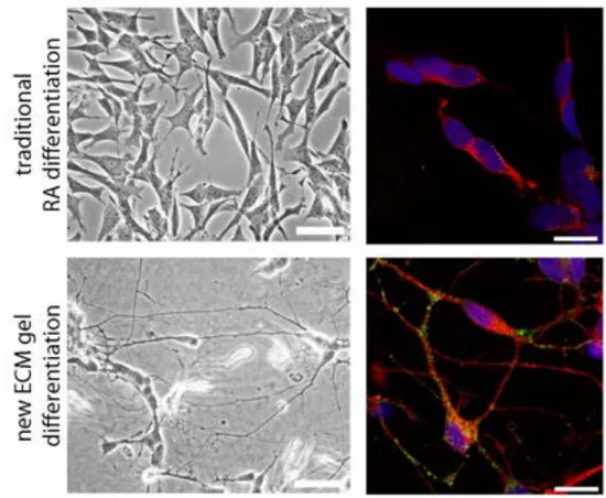
Human neuronal cell lines are widely used in AD research. These cells enable studies in cells with human proteins and function. Another advantage is the simplicity to manipulate the cells chemically and genetically for specific studies of proteins and functions. However, as they are of cancerous origin, they also lack many of the neuronal features. Therefore, different ways to differentiate these cells have been developed, but have still not generated cells with complete neuronal phenotype.
Aim of the thesis
The general aim of this thesis was to investigate how disturbances in intracellular degradation pathways, responsible for clearance of Aβ and tau, and the transmission of toxic protein aggregates, could contribute to AD pathology. The more specific aims of the four papers included in this thesis are listed below.
Paper I
The aim of paper I was to develop an improved neuronal cell model using human neuroblastoma cells. The goal was to find a differentiating protocol yielding cells with morphological and biochemical features of mature neurons, while expressing all isoforms of tau. The cell model should enable studies in the molecular biology and protein field, as well as whole- and live-cell experiments.
Paper II
The aim of paper II was to investigate if reduced proteasome activity, as seen in AD, affected tau phosphorylation and microtubule function. If so, we also aimed at finding a mechanistic explanation.
Paper III
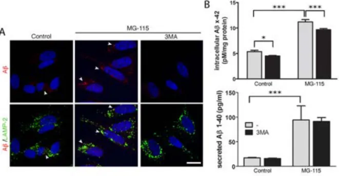
The aim of paper III was to investigate the effect of reduced proteasome activity on Aβ generation and accumulation, as well as neuronal viability. In addition, the mechanisms underlying these effects were investigated.
Paper IV
The aim of paper IV was to test the hypothesis that oligomeric Aβ could be transmitted from one neuronal cell to another. We also wanted to investigate the effect of Aβ on neuronal viability. The study also aimed at investigating the possible mechanisms behind Aβ transfer.
Methods
The results in this thesis are based on a variety of experimental procedures, to investigate effects on and changes in RNA, proteins, organelles and whole cells. A brief description of the key methods used in this thesis is presented together with the possibilities and limitations of the different methods.
Cellular models and cell culture
The studies in this thesis are based on cellular models, mainly using SH-SY5Y neuroblastoma cells. SH-SY5Y is a human neuroblastoma cell line, originally derived from a neuroblastoma metastasis. It has been subcloned several times since its first establishment in the 1970’s. The cells have a poor neuronal phenotype, and are often differentiated with retinoic acid (RA) to become more neuronal-like. In paper I an extra cellular matrix (ECM) gel differentiation model was developed to obtain neuron-like human cells from the SH-SY5Y cell line. This model was thereafter used in paper II and IV. In paper III, SH-SY5Y cells stably transfected with APP containing the Swedish mutation (APPSwe) or wildtype APP (APPWt), was used. In paper III and IV,
primary rat neurons were also used (approved by the ethical committee in Linköping no 101-08 and 72-11). Brain tissue was dissected from newborn rats, and neurons were selectively cultured and differentiated in vitro.
Using cell lines is convenient, as you easily have access to a lot of material. Cells are also easy to treat and manipulate, and the results can be interpreted without influences from other cell types, or the whole organism. However, most cell lines are of cancerous origin, and do not have the same characteristics as their non-cancerous counterpart. Primary murine neurons have the advantage of not being cancerous cells. However, many proteins and processes differ between humans and mice/rats, and results from mice cannot be directly translated to humans.
Messenger RNA expression
In paper I, expression of different tau mRNA splicing variants was investigated using reverse transcriptase (RT) PCR and quantitative (q), or real-time,
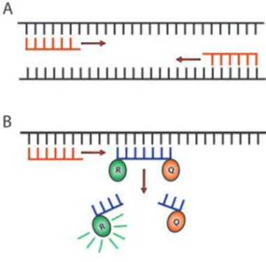
RT-PCR (qRT-RT-PCR). Both methods investigate the presence of mRNA, which for technical reasons is converted to a DNA copy (cDNA) enabling multiplication. RT-PCR is based on amplification of cDNA in a PCR reaction using primer pairs targeting the cDNA sequence of interest (Fig. 6A). The amplified cDNA is separated, regarding to size, on an agarose gel and visualised using the DNA binding dye, ethidium bromide.
qRT-PCR is performed using both primer pairs, and probes containing a reporter and a quencher molecule, targeting your cDNA sequence of interest. During replication, the probe is cleaved, resulting in the reporter probe dissociating from the quencher, becoming fluorescent (Fig. 6B). The fluorescent signal represents one replication cycle, and the initial amount of cDNA /mRNA can therefore be calculated from the number of cycles it takes to reach cycle threshold (Ct). To reduce errors based on unequal loading of
starting material, the amount of a stably expressed mRNA (house keeping gene) is also measured. The amount of cDNA (mRNA) is quantified in absolute numbers (the standard curve method) or in relation to a reference sample (the ΔΔCt method). In paper I, the ΔΔCt method was used to compare tau exon
expression in differentiated cells, where non-differentiated cells were used as a reference.
Figure 6. A. RT-PCR Primers
recognising a cDNA sequence of interest resulting in amplification by the PCR reaction. B. Primer and probe in qRT-PCR. During replication, the probe is released causing the reporter (R) molecule to be released from the quencher (Q) molecule, resulting in a fluorescent signal.
RT-PCR is not quantitative, and can therefore only answer the question if the mRNA sequence is expressed in cells or not. It can however be more sensitive than qRT-PCR. In both cases, one must remember that protein expression not always correlate to the mRNA expression.
Protein expression and protein
modification
In all papers included in this thesis, different methods to investigate protein expression, cleavage, and post-transcriptional modifications were used. There are many different methods for protein quantification; all of them having advantages and disadvantages. All the methods used in this thesis are based on antibody recognition, and issues with antibody specificity, epitope recognition and unspecific binding must be taken into account.
Western blot
Western blot has been used throughout this thesis to analyse protein expression, splicing and cleavage, and post-transcriptional modifications. In western blot, proteins are denatured and thereafter separated on a sodium dodecyl sulfate (SDS) polyacrylamide gel (PAGE), based on size (Fig. 7A). Thereafter, the proteins are transferred (blotted) to a membrane, and antibodies directed to the protein of interest are added. Antibodies directed towards a specific post-translational modification (phosphate or acetyl group) can also be used. Secondary antibodies conjugated with horseradish peroxidase (HRP) are then added. A developing solution, containing a substrate that becomes luminescent when cleaved by HRP is added, and the protein bands are visualised using a light-sensitive camera or photosensitive film. The amount of light produced is converted to a grey/black signal which correlates to the amount of protein in the sample. Densitometric analysis is used to quantify protein concentration, and a housekeeping protein, being constantly expressed, is used as a reference.
Enzyme-Linked ImmunoSorbent Assay (ELISA)
In paper III, enzyme-linked ImmunoSorbent Assay (ELISA) was used for protein quantification. Two antibodies, both specific for your protein of interest, are used in a “sandwich” setting with your protein of interest in the middle (Fig. 7B). The capture antibody is coated to the bottom of a well, and
after the addition of your protein sample, a HRP conjugated detection antibody is added. The detection solution contains a substrate which, when being cleaved by HRP, transforms into a coloured or luminescent product. The absorbance or emitted light, correlating to the amount of protein, is quantified and compared to a standard curve of known protein concentrations.
ElectroChemoluminescent ImmunoSorbent assay
In paper II and III, we used the MesoScale Discovery (MSD) system to quantify proteins and protein phosphorylation. The method is based on the same principle as ELISA, but can quantify several different proteins simultaneously in one sample. This is due to the detection method, where an electrical current is placed under the plate. This activates a molecule conjugated to the detection antibody resulting in emission of light. The amount of light emitted correlates to the amount of protein in the sample. The light Figure 7. Different methods for protein quantification. A. In western blot, proteins (dark
grey) are blotted onto a membrane after separation. Primary antibodies (ab; black) recognise the protein, and secondary ab (orange), conjugated with HRP (red), binds to the primary ab. B. In ELISA, capture ab (black) are coated to the bottom of a well (blue) and the protein of interest (grey) is recognised by the ab. A secondary ab conjugated to HRP (black and red) also recognises the protein of interest. C. In the MSD system, capture ab (black) recognising different proteins are coated to separate spots in one well (blue). Proteins in the sample (grey, red and green) are recognised by the different antibodies. A secondary ab, conjugated to an enzyme (black and yellow) also recognises the proteins of interest.
emission is constrained to a small area, and this allows for coating of several different capture antibodies in the bottom of one well (Fig. 7C).
Proteome profiler phospho-kinase array
In paper II, the proteome profiler human phospho-kinase array was used to screen 46 kinases for activation. The array is based on a membrane coated with antibodies directed against phosphorylation sites on all kinases. Each phospho-specific antibody is allotted its own “spot”, and one protein sample is added to each membrane. Secondary antibodies conjugated to HRP were used for detection, and the detection was performed as with western blot. By densitometric analysis, treated samples could be compared with a control sample, and the kinase activation could be semi-quantified.
Comparison of different protein analysis assays
One advantage with western blot is that you can analyse several different proteins in a few samples using small amounts of reagents and material. It also enables you to investigate several different cleavage products of the same protein. The method however, is only semi-quantitative and the difference in band density does not always directly correlate to the difference in protein content. The advantage of ELISA compared to western blot is that you get a quantitative value of the amount of protein in you sample. It is also more convenient when analysing a large amount of samples. However, you might need a larger amount of sample for analysis, especially if you want to investigate many different proteins. The advantage of the MSD system is that you can quantify several proteins simultaneously, requiring smaller sample size for each experiment. However, you have to be cautious of cross-reactions between the different antibodies in the same well.
Protein localisation and co-localisation
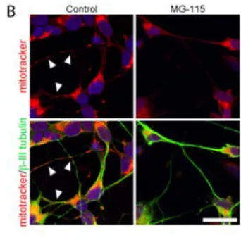
In all studies included in this thesis, presence, localisation, and co-localisation of protein and organelles have been investigated using immunocytochemistry and live cell imaging. Antibodies were used for visualisation in paper I-IV, and in paper IV, transfection of fluorescently tagged organelle-specific proteins was also performed. In addition, organelle specific dyes targeted towards mitochondria and lysosomes were used in paper II and IV.
Immunocytochemistry
Immunocytochemistry not only allows for investigation of protein expression, but also localisation of proteins, and co-localisation with different organelles. During cell fixation, proteins are cross-linked using paraformaldehyde, and the cells are then permeabilized to enable intracellular antibody binding. The cells are incubated with a blocking solution, to avoid unspecific antibody binding, before incubation with primary antibodies, followed by fluorescently tagged secondary antibodies. For nuclear staining, DNA binding dyes such as DAPI or ToPro3 is added before mounting the samples on slides for microscopy. All immunocytochemical analyses in this thesis have been performed using confocal microscopy.
Performing immunocytochemistry for detection of Aβ and tau, using conventional protocols, might have its drawbacks. It has been reported that fixing samples with formic acid can interfere with Aβ recognition by antibodies [132]. In addition, it has been shown that tau can be released from microtubule during fixation and blocking, which could hamper the interpretation of results [133].
Organelle specific dyes
In paper II and IV, organelle specific dyes, labelling mitochondria and lysosomes, were used. Mitotracker® Red accumulates in mitochondria due to
the membrane potential, and remains there after fixation. As the dye relies on the membrane potential for accumulation, this method requires functional mitochondria. However, the dye can be used both in live and fixed cells. Lysotracker® Green accumulates in acidic organelles, and therefore stains late
endosomes and lysosomes. Lysotracker do not always retain its fluorescence after fixation, and is therefore only suitable for live-cell experiments.
Fluorescently tagged proteins
In paper IV, we used transient transfection to express proteins directly conjugated to enhanced green fluorescent protein (EGFP). The transfection was performed using BacMam, a humanised insect viral system, suitable for cells that are hard to transfect, such as neurons. The transfection protocol resulted in about 70 % of the cells being transfected, and the localisation being comparable with endogenous proteins.
Another way of visualising proteins without using antibodies is to label already produced protein with small fluorescent molecules before adding them to the cells, as performed with Aβ in paper IV. In this case directly tetramethyl rhodamine (TMR) conjugated Aβ was purchased, or produced in the lab.
Comparison of the different assays
Using antibodies for protein localisation relies heavily on the specificity of the antibody and the compatibility with the secondary antibody. The advantage is that you are investigating the localisation of endogenously produced proteins and that it does not require transfection of proteins.
By using fluorescently tagged proteins, you do not have to rely on the specificity of antibodies. You can also visualise proteins and cellular structures in non-fixed living cells, which is advantageous. However, the disadvantage is that the overexpression of proteins might change the cellular localisation compared to endogenously expressed proteins. It can also alter the equilibrium with a number of other proteins. The small fluorescent molecules in pre-tagged recombinant proteins should not affect protein function as much as the larger fluorescent proteins. However, the protein of interest is added to the cell media, and must be taken up by the cells if intracellular localisation is to be investigated. The organelle specific dyes do not require transfection for organelle visualisation, and can be used both in living and fixed tissue. However, the specificity can be questionable and the dyes require, in many cases, functional organelles.
Neuritic transport
In paper II, investigation of neuritic transport was performed as a measurement of neuronal function and health. To investigate the general transport along neurites, labelling of vesicles is not necessary. By using differential interference contrast (DIC) microscopy, enhancing the contrast of your specimen, and a high-resolution objective, the vesicles can be visualised as black dots in the neurites. Time-lap imaging followed by analysis using the Image J software, and a manual tracking plugin, was used to calculate the velocity for each vesicle.
In paper IV, transport of endosomes/lysosomes and their content was visualised by labelling organelles with EGFP-tagged proteins and organelle
specific dyes. The analysis was thereafter performed using confocal microscopy and time-lap imaging.
The DIC microscopy method is simple, and do not require organelle staining. It is however not possible to distinguish between transport of different organelles. The use of fluorescently tagged proteins or organelle specific dyes enables investigation of transport of specific organelles/vesicles. However, this requires confocal microscopy for ECM gel cultured cells, where the narrow plane of focus can be of disadvantage.
A
β
oligomer preparation and
characterisation
In paper IV, oligomeric preparations of recombinant Aβ were used to investigate transfer between cells. Several protocols for oligomer preparation have been described, and a well-established protocol, where Aβ is left to aggregate into oligomers at 4°C for 24 hours [134], was used. To determine the aggregation state of Aβ, different methods were performed including both non-denaturing PAGE and SDS-PAGE followed by western blot, as well as electron microscopy and oligomer specific antibodies.
Manipulation of samples during preparation for most methods can affect Aβ oligomers, both by dissociation to monomers and aggregation to fibrils. It is therefore important to use several different methods for oligomer characterisation to make sure that your Aβ sample is of the right kind.
Inhibition of proteins and cellular
processes
To investigate the role of specific proteins and processes, it is necessary to have methods for inhibition of these proteins and processes. In this thesis, two different methods were used for protein inhibition, small molecule inhibitors and small interfering (si) RNA knockdown.
Small molecule inhibitors
Small molecule inhibitors were used in paper II-IV to inhibit processes such as proteasome degradation and autophagy, inhibiting activation of kinases, and blocking binding to certain receptors. Small molecule inhibitors have a
structure designed to bind to a site of the protein, regulating its effects. Optimal inhibitor concentrations were investigated, and the inhibitors were added before or together with other manipulations.
Small interfering RNA
In paper III, siRNA knockdown was used to inhibit the expression of a specific protein. This method uses a biological control system, activated to degrade double stranded mRNA [135]. By adding short sequences of double stranded RNA, constructed to match a sequence of the mRNA of interest, the mRNA is degraded before being translated to protein. The time it takes for down-regulation of a protein is dependent on the turnover of the protein of interest. The time of turnover also affects the time span where the protein is down-regulated.
Unspecific inhibition is a common problem with small molecule inhibitors, or that the selectivity is not high enough. The inhibitors are, on the other hand, easy to apply to many types of cells, including our ECM gel differentiation model. The advantage with siRNA is that it is more specific than small molecule inhibitors, even though off-target effects can be seen. The transfection protocol can, however, cause toxicity by itself, and side effects such as immune responses can also be seen. siRNA knockdown can also be harder to perform in cells that are difficult to transfect, such as primary neurons.
Cell toxicity and viability
In this thesis, different outcome measurements have been used to investigate cell viability. Different methods, detecting cellular toxicity or cell death in different ways, can be used to investigate early and later toxic events.
MTT viability assay
The MTT assay was used in paper II and III to determine viability upon proteasome inhibition. The method is based on the conversion of the yellow tetrazolium salt 3-(4,5-Dimethylthiazol-2-yl)-2,5-diphenyltetrazolium bromide (MTT) to a purple formazan salt [136]. Dissolved MTT salt is added to cells in culture, and is let to reduce by the mitochondria. Thereafter, the purple
formazan salt is dissolved, and the absorbance, correlating to the number of viable cells, is measured.
Crystal violet assay
The crystal violet assay was used in paper III to investigate cell viability upon proteasome inhibition and subsequent inhibition of autophagy. This method is based on staining of all attached cells, and does not take in to account the status of the remaining attached cells. Cells are fixed with PFA after washing, and thereafter a crystal violet solution is added which adheres to all remaining cells. After extensive washing, the remaining dye is resuspended in a SDS solution. The absorbance, correlating to the number of cells in the well is measured, and the values were compared to control experiments.
Microtubule beading
In paper II and IV, we also investigated tubulin beading as a sign of neuronal toxicity. Microtubule beading is thought to be an early sign of toxicity, preceding neuronal death [11, 137]. The disruption of microtubule, ending up in beaded structures (as pearls on a string) causes disruption of neuritic transport and can in turn lead to cell death. This was performed by immunocytochemistry using antibodies against tubulin, or by transfecting cells with EGFP-tagged tubulin protein. The advantage of this method is that the appearance of beaded microtubules indicates an early toxicity. However, this was only performed in a qualitative manner, not resulting in any measurable results.
Organelle rupture
In paper IV, the appearance of organelle rupture was used as an early sign of neuronal toxicity [138]. Disruption of organelle membranes, followed by leakage of organelle content is a sign of reduced cell function, and subsequently leads to cell death. We used the translocation of EGFP-tagged Rab5 from a spot-like pattern (localised in vesicles) to being distributed throughout the cell and having enhanced fluorescence, as a sign of cellular toxicity.
Comparison of different viability assays
The MTT assay is widely used for analysis of both proliferation and viability. However, it has been reported that Aβ interferes with the MTT salt and might therefore be unsuitable when toxicity of Aβ is analysed [139]. Other methods, based on the same principle, such as the XTT method might therefore be a better choice. The advantage with the crystal violet method, compared to MTT, is that Aβ does not interfere with the substrate. However, the crystal violet assay does not distinguish between living and dead attached cells, and can result in a false high number of living cells.
Both of the above mentioned methods only detect dead or dying cells. When earlier signs of toxicity are to be measured, tubulin or organelle disruption might be a better choice. There are also other methods to investigate early signs of toxicity, such as decrease in ATP content. The disadvantage of the utilised methods is that they are not easily quantified and a more qualitative approach must be used.
In all papers included in the thesis, we also analysed phase-contrast images of cells, as a complement to the viability assays. The visual control of cell viability always correlated well to the results obtained from more quantitative methods.