G. Edman Tynelius
L. Bondemark
E. Lilja-Karlander
A randomized controlled trial of
three orthodontic retention
methods in Class I four premolar
extraction cases
– stability after
2 years in retention
Authors' affiliation:
G. Edman Tynelius, L. Bondemark, E. Lilja-Karlander, Department of Orthodontics, University of Malm€o, Malm€o, Sweden
Correspondence to: Gudrun Edman Tynelius
E-mail: gudrunedmantynelius@gmail.com
Edman Tynelius G., Bondemark L., Lilja-Karlander E. A randomized controlled trial of three orthodontic retention methods in Class I four
premolar extraction cases– stability after 2 years in retention
Orthod Craniofac Res 2013; 16: 105–115. © 2013 John Wiley & Sons A/S. Published by Blackwell Publishing Ltd
Structured Abstract
Objective – To evaluate three different retention methods in compliant patients after 2 years of retention.
Design – Three group randomized controlled trial.
Materials and Methods – The sample was recruited from patients hav-ing their fixed appliance treatment between 2001 and 2007. Seventy-five patients (45 girls and 30 boys with a mean age of 14.4 years at start of retention) were randomized into three retention methods: vac-uum-formed retainer in the maxilla and bonded canine-to-canine retainer in the mandible (Group V-CTC), vacuum-formed retainer in the maxilla combined with stripping of the 10 proximal surfaces of the lower man-dibular anterior teeth (Group V-S) and prefabricated positioner covering the teeth in the maxilla and the mandible (Group P). The following linear measurements were performed: Little′s irregularity index (LII), interca-nine width, intermolar width, arch length, overjet, overbite and body height growth. Registrations were made before orthodontic treatment, at start of retention, after 12 and finally 24 months in retention. Differences in means between groups were tested by one-way analysis of variance (SPSS).
Results – After 2 years all three retention methods were successful in retaining orthodontic treatment results. The major part of relapse took place during the 1st year of retention.
Conclusions – All 3 types of retention methods were equally effective in controlling relapse to a clinically acceptable level.
Key words: orthodontics; RCT; retention methods; treatment outcome
Date:
Accepted 3 November 2012 DOI: 10.1111/ocr.12011
© 2013 John Wiley & Sons A/S. Published by Blackwell Publishing Ltd
Introduction
One of the biggest challenges of orthodontic treatment is to maintain the achieved treatment result (1–3). Massive efforts have been carried out to find the best strategies to retain the teeth in their new position after orthodontic treatment and to avoid relapse. Focus has been on maloc-clusion before treatment or after debonding to find any connection with stability and tooth posi-tion (4). Different retenposi-tion treatments have also been analysed with respect to tooth stability and in the last decade several well-designed random-ized controlled trials (RCTs) have been published (5–8). Small but significant differences have been reported when it comes to comparing retention appliance capacities. One study showed that vac-uum-formed retainers were more effective than Hawley retainers to retain teeth after 6 months (9). Furthermore, it has been shown that full-time and part-time wear of an Essix retainer had the same effect (10). In our previous study the effi-ciency of three retention methods was analysed (8). From a clinical perspective it was evident that all three retention methods had equal capacity to retain treatment results during the first year.
By tradition growth, initial crowding as well as cooperation have been pointed out as factors influencing orthodontic treatment stability (2, 4, 11, 12). Bacetti et al. (13) have demonstrated extensive increase of mandibular length during pubertal growth. As long as growth of maxilla and mandible is going on it may affect the position of teeth and, thus the result of retention treatment. Therefore the purpose of the present study was to analyse and compare three retention methods after 2 years of retention in a randomized con-trolled trial design, and also, to evaluate the retention capacity in relation to cooperation, growth, initial crowding and gender. The null hypothesis was that there would be no clinical difference in retention capacity between the three retention methods in compliant patients.
Subjects and methods
The Ethics Committee of Lund/Malm€o Univer-sity, Sweden, approved the protocol and the
informed consent form (LU515-01). Each patient and parent were given oral as well as written information and had to sign the written consent before being included in the trial. The study was carried out on patients referred to an orthodontic clinic in the National Health Service (NHS), Ystad, Sweden. The NHS clinic was responsible for treatment of malocclusions of patients in the southeast County Council of Scania. In Sweden free dental care including orthodontic treatment is offered to patients with a certain degree of malocclusion up to the age of 20 years. One experienced orthodontist treated all the patients (GET). The patients taking part in the study underwent orthodontic treatment between 2001 and 2007 and the following inclusion criteria were to be met: no previous experience of ortho-dontic treatment, permanent dentition, space deficiencies in both jaws, normal skeletal and dentoalveolar sagittal, vertical and transverse relationships, Class I molar relationship or 3 mm anterior or posterior deviation, and a treatment plan involving extraction of four premolars fol-lowed by fixed straight wire appliances (0.022 inch, MBT) in both jaws. The generation of ran-domisation sequence was performed in blocks of five to ensure that equal numbers of patients were allocated to each of the three retention groups. Fifteen paper sheets, five ballots with maxillary vacuum-formed retainer and bonded mandibular canine-to-canine retainer (V-CTC), five with maxillary vacuum-formed retainer and
mandibular interproximal enamel reduction
(V-S) and five with positioner (P) were placed in a basket. The patient then decided the retention treatment by picking a ballot from the basket.
Retention methods
The three retention methods of choice were:
•
A removable vacuum-formed retainer coveringthe palate and the maxillary anterior teeth from to-canine and a bonded canine-to-canine retainer in the lower arch (Group V-CTC), (Fig. 1A)
•
An identical maxillary vacuum-formedstripping of the lower anterior teeth and no lower retention device (Group V-S), (Fig. 1B)
•
A prefabricated positioner covering all erupted teeth in the maxilla and the mandible (Group P), (Fig. 1C)The vacuum-formed retainers were made of 2 mm Biolon (Dreve Dentamid GmbH, Unna, Germany) in a Scheu Ministar press (Scheu-Den-tal GmbH, Iserlohn, Germany). The canine retainers consisted of 0.7 mm spring hard wire (Dentaurum noninium, Dentaurum, Ispringen, Germany) bonded with Transbond LC (3M Uni-tek Orthodontic Products, Monrovia, CA, USA) to the lower canines.
Mechanical stripping of the lower incisors and canines was performed either by hand with single sided medium and fine metal blades (TP Ortho-dontics, La Porte, IN, USA) or with Ortho-Strips for the EVA system (GAC International, New York, NY, USA). The method of stripping was depen-dent on tooth form, non-triangular or triangular respectively, and was either performed on the visit 5–6 weeks prior to debonding or at debond-ing. The aim of stripping was to obtain small but distinct enamel flattening of the contact surfaces. The reduction of any contact point between two teeth amounted approximately to the thickness of the coarse blade of either system, i.e. 0.22 mm for hand stripping or 0.34 mm for EVA-stripping. At debonding all spaces were closed and no addi-tional stripping was performed.
The preformed positioner (Ortho-Tain
Posi-tioner, Ortho-Tain Inc., Toa Alta, Puerto Rico, USA) was a soft plastic device covering all erupted teeth.
All retention appliances were handed out within 1 h after debonding. The patients in groups V-CTC and V-S were instructed to wear the vacuum-formed retainer 22–24 h per day for 2 days and nights and then during the night for 12 months. In group P the positioner was to be worn for 30 min during the daytime and during sleep for 12 months. During the daytime wear, patients were instructed to actively chew into their positioners. The second year of retention patients wore their retainers every other night and visited the clinic twice for control of cooper-ation and appliances. At the end of the second year all retainers were taken off.
Documentation
Dental casts were obtained on four occasions, i.e. before orthodontic treatment, when the fixed A
B
C
Fig. 1. Vacuum-formed retainer in the maxilla and canine-to-canine retainer in the mandible (A), vacuum-formed retai-ner in the maxilla and stripping of the mandibular incisors and canines (B), and prefabricated positioner covering all erupted teeth in the maxilla and the mandible (C).
orthodontic appliance was removed, after 12 and finally after 24 months of retention. The follow-ing linear measurements were made: Little’s irregularity index (LII) of maxilla and mandible (14), intercanine width of maxilla and mandible, intermolar width of maxilla and mandible, arch length of maxilla and mandible, overjet and overbite. The same examiner (GET), who was blinded for the retention protocol, performed all measurements with an electronic digital calliper (Mauser Digital 6, Winterthur, Switzerland) to a precision of 0.01 mm. Study casts before ortho-dontic treatment, at retention start, after 12 and 24 months of retention were measured indepen-dently of each other.
Compliance was estimated on every visit to the clinic. Patients were asked how many nights per week they had used their retainers. Compli-ance was considered as excellent if the vacuum-formed retainer in groups V-CTC and V-S fitted well on the teeth and in group P if the positioner had changed its colour into less translucent.
Body height was measured on every visit to the clinic with a wooden standard apparatus mounted on the wall to a precision of 0.1 cm and with no shoes on and head and heels to the wall.
Successful retention was decided according to the Little Irregularity Index. Values < 3.5 mm were defined as successful (15).
Statistical analysis
The sample size for each group was calculated based on a significance level of 0.05 and 80 per cent power to detect clinically meaningful
differ-ence of 2.0 mm (SD = 2.0 mm) of the LII. The
power analysis showed that 16 patients in each group were sufficient. To compensate for drop-outs in future follow-up studies, 25 patients were enrolled in each group. The strategy of analysis is best described as per protocol (PP). Dropouts
were randomly spread in the randomized
groups. Arithmetic means and standard devia-tions (SD) on group level at times corresponding to pretreatment, start of retention and after one and finally 2 years of retention were calculated for each variable. Significant differences in
means between groups were tested by one-way analysis of variance (ANOVA) using the Statistical
Package for Social Sciences (version 14.0; SPSS Inc., Chicago, IL, USA). The relapse of LII between severe (>6.5 mm) and mild (<3.5 mm) initial crowding was tested by independent-sam-ples t-test. The degree of relapse in the lower front in relation to retention method, coopera-tion, growth, initial crowding and gender was tested by univariate analysis of variance. p-val-ues <5 per cent (p < 0.05) were considered sta-tistically significant.
Error of method
Twenty randomly selected dental casts were measured on two separate occasions with a 4-week interval by the same examiner (GET). The method error (16) did not exceed 0.45 mm for any of the 10 measurements. No significant mean differences between the two series of records were found using paired t-test. All results were tested for normality.
Results
After 2 years of retention the trial consisted of 69 patients out of 75 from the start. Four patients did not show up to the 2-year follow up (one girl in group V-CTC, two boys in group V-S and one boy in group P) and 2 boys in group P were excluded during the 1st year as they never used their retainers. In group V-CTC there were 24 patients (17 girls and 7 boys), in group V-S 23 patients (14 girls and 9 boys) and in group P 22 patients (13 girls and 9 boys) (Fig. 2). Altogether, the mean age at start of retention was 14.4 years (SD 1.5) and the mean active treatment time lasted 1.7 years (SD 0.4). There were no significant differences between any of the three retention groups regarding age at start of treatment, active treatment time, body height at retention start or gender. In addition, there
were no significant differences in linear
measurements on study casts between any of the retention groups before and after active treatment.
Retention capacity Maxilla
•
The only significant difference betweengroups was found after 2 years of retention for the mean change in maxillary intercanine
width between P ( 1.8 mm) vs. V-CTC
( 1 mm) and V-S ( 0.9 mm) (p < 0.01). Most
of this difference happened during the 1st year though differences were not significant then (Table 1).
Mandible
•
There was a significant difference in meanmandibular LII change between V-CTC
(0.6 mm) and P (1.6 mm) (p< 0.01). The
change was largest but not significant during the 1st year of retention but the change went on in the P group (Table 2).
•
There was a significant difference in meanin-tercanine width change between V-CTC
(0.2 mm), an increase, vs. V-S ( 1.0 mm) and P ( 1.1 mm) (p< 0.001), with decreases. The difference between V-CTC and V-S was seen already the 1st year (p< 0.001). During the 2nd year there was a significant mean interca-nine width change between P ( 0.4 mm) vs.
V-CTC (0.1 mm) and V-S (0.0 mm) (p< 0.01).
After 2 years the mean intercanine width dif-fered significantly between V-CTC (27.6 mm) and V-S (26.4 mm) (p< 0.05).
•
Intermolar width and arch length did notdif-fer significantly at the end of the 2 year fol-low-up period.
Overjet and overbite
•
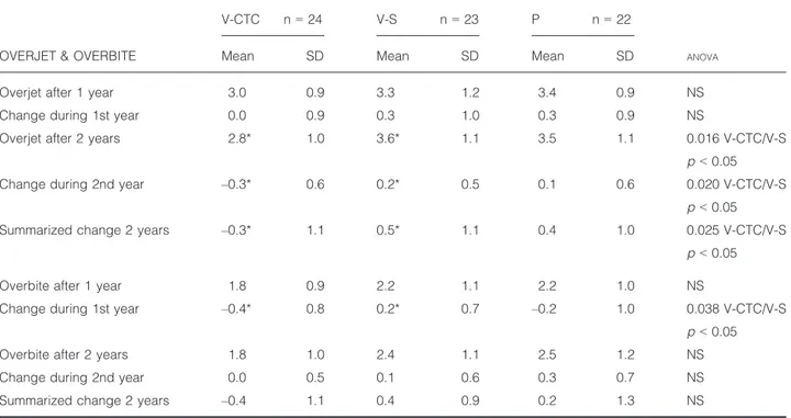
There was a significant difference in meanoverjet between V-CTC (2.8 mm) and V-S (3.6 mm) (p< 0.05). The mean overjet change was a reduction in V-CTC ( 0.3 mm) and an increase in V-S (0.5 mm) (p< 0.05; Table 3).
•
The mean overbite was reduced, albeit notsignificantly, in V-CTC ( 0.4 mm) and
increased in V-S (0.4 mm) and P (0.2 mm). Nevertheless, during the 1st year there was a significant difference in mean overbite change between V-CTC ( 0.4 mm) and V-S (0.2 mm) (p< 0.05).
Retention capacity and cooperation
Out of 69 patients 38 cooperated excellently and 31 were judged to cooperate well (Fig. 3). Figure 3 Fig. 2. The Consort diagram of the trial. Vacuum-formed retainer in the maxilla and canine-to-canine retainer in the mandible (group V-CTC), vacuum-formed retainer in the maxilla and stripping of the mandibular incisors and canines (group V-S), and positioner (group P).
shows a negative correlation in the group with excellent compliance between change in mandib-ular LII and growth but a positive correlation in the well cooperating group. This means that when patient cooperation was excellent LII did not change despite the fact that patients were growing. When tested with independent samples t-test there was a significant difference in mean mandib-ular change of LII with 0.7 mm in the excellently cooperating group and 1.3 mm in the well cooper-ating group (p < 0.05). There was no significant difference in cooperation between boys and girls.
During the 2 years of retention, 5 patients lost their vacuum-formed retainer and had to have it replaced with a new one. No patients lost their positioner. Out of 24 patients 17 kept their CTC
in situ. Three patients had their CTC rebonded once, 3 patients had it rebonded twice and one patient had his CTC rebonded four times. These complications did not have any significant influ-ence on the outcome of retention.
Retention capacity and body height growth
All patients were to be measured at start of retention and after 2 years of retention but 13 measurements were missed out, i.e. 56 patients were measured adequately (Fig. 4). In group V-CTC (14 girls and 5 boys) the mean body height increase was 1.4 cm (SD 1.8), in V-S (9 girls and 7 boys) 2.4 cm (SD 3.1) and in P (13 girls and 8 boys) 1.4 cm (SD 2.5). There were no Table 1. The mean measurements (mm) and the mean changes for the maxilla from start of retention to one and 2 years of retention in the three retention groups: removable vacuum-formed retainer and bonded lower canine-to-canine retainer (V-CTC), removable vacuum-formed retainer and lower anterior stripping (V-S), and positioner (P)
MAXILLA
V-CTC n= 24 V-S n= 23 P n= 22
ANOVA
Mean SD Mean SD Mean SD
LII after 1 year 1.1 1.1 1.0 1.0 1.3 1.1 NS
Change during 1st year 0.6 1.0 0.5 1.0 0.8 1.2 NS
LII after 2 years 1.0 0.9 1.4 1.1 1.5 1.3 NS
Change during 2nd year 0.0 0.5 0.3 0.8 0.2 0.6 NS
Summarized change 2 years 0.5 0.8 0.9 1.1 1.1 1.4 NS
Intercanine width after 1 year 35.9 1.8 35.9 2.2 35.1 3.1 NS
Change during 1st year 0.7 0.7 0.6 0.6 1.6 2.4 NS
Intercanine width after 2 years 35.6 1.8 35.5 2.1 34.8 2.1 NS
Change during 2nd year 0.3 0.5 0.4 0.5 0.1 2.0 NS
Summarized change 2 years 1.0** 0.8 0.9** 0.9 1.8** 1.5 0.018 V-CTC & V-S/P
p < 0.01
Intermolar width after 1 year 48.3 2.6 48.0 3.0 48.5 2.2 NS
Change during 1st year 0.8 1.7 0.8 1.0 1.0 1.1 NS
Intermolar width after 2 years 48.0 2.4 47.5 3.2 48.0 2.1 NS
Change during 2nd year 0.4 1.3 0.4 0.5 0.4 0.5 NS
Summarized change 2 years 1.1 1.5 1.2 1.3 1.4 1.2 NS
Arch length after 1 year 23.5 1.5 22.7 1.8 23.1 1.6 NS
Change during 1st year 0.3 1.1 0.1 0.4 0.3 2.5 NS
Arch length after 2 years 23.3 1.6 22.3 1.7 22.9 1.7 NS
Change during 2nd year 0.2 0.4 0.3 0.5 0.3 0.4 NS
Summarized change 2 years 0.0 1.0 0.2 0.7 0.1 2.7 NS
**p < 0.01. LII, Little′s Index.
significant differences between the groups dur-ing the 2 years. The boys grew significantly more than the girls, with a mean of 3.3 cm (SD 3.2) vs. a mean of 0.8 cm (SD 1.2) for the girls. However, boys and girls did not differ significantly in any other measurements. During the 2 years of retention 29 patients (14 girls and 15 boys) grew more than 1 cm and 4 (1 girl and 3 boys) of those 29 patients had a relapse of mandibular LII more than 3.5 mm with a maximum of LII
4.9 mm. No correlation was found between mean change of LII and body height growth. Retention capacity and initial crowding
After 2 years of retention there was no signifi-cant difference in mandibular LII between the group of 18 patients with initial mild crowding (LII< 3.5 mm) and the group of 28 patients with initial severe crowding (LII> 6.5 mm) (Table 4). Table 2. The mean measurements (mm) and the mean changes for the mandible from start of retention to one and 2 years of retention in the three retention groups: removable vacuum-formed retainer and bonded lower canine-to-canine retainer (V-CTC), removable vacuum-formed retainer and lower anterior stripping (V-S), and positioner (P)
MANDIBLE
V-CTC n= 24 V-S n= 23 P n= 22
ANOVA
Mean SD Mean SD Mean SD
LII after 1 year 1.0 0.6 1.1 1.1 1.5 1.1 NS
Change during 1st year 0.6 0.6 0.8 0.9 1.1 1.0 NS
LII after 2 years 0.9** 0.7 1.2** 1.0 2.0** 1.5 0.006 V-CTC&V-S/P
p < 0.01
Change during 2nd year 0.0 0.5 0.2 0.3 0.4 1.0 NS
Summarized change 2 years 0.6** 0.7 0.9 0.8 1.6** 1.4 0.005 V-CTC/P
p < 0.01
Intercanine width after 1 year 27.4 1.5 26.4 2.1 26.8 1.6 NS
Change during 1st year 0.0*** 0.4 1.0*** 0.9 0.7 1.0 0.001 V-CTC/V-S
p < 0.001
Intercanine width after 2 years 27.6* 1.3 26.4* 2.1 26.4 1.7 0.024 V-CTC/V-S
p < 0.05
Change during 2nd year 0.1** 0.4 0.0** 0.5 0.4** 0.5 0.002 V-CTC&V-S/P
p < 0.01
Summarized change 2 years 0.2*** 0.5 1.0*** 1.0 1.1*** 1.2 0.001 V-CTC/V-S&P
p < 0.001
Intermolar width after 1 year 42.2 1.5 41.7 2.5 42.2 1.9 NS
Change during 1st year 0.0 1.1 0.6 1.8 0.8 1.8 NS
Intermolar width after 2 years 42.0 1.7 41.2 2.5 41.6 1.6 NS
Change during 2nd year 0.2 0.6 0.5 0.5 0.4 0.6 NS
Summarized change 2 years 0.3 1.2 1.0 2.1 1.3 1.9 NS
Arch length after 1 year 18.6 1.3 17.7 1.4 18.2 1.4 NS
Change during 1st year 0.7* 1.0 0.0* 0.8 0.5 0.8 0.024 V-CTC/V-S
p < 0.05
Arch length after 2 years 18.3 1.7 17.4 1.6 17.8 1.4 NS
Change during 2nd year 0.3 0.7 0.2 0.4 0.4 0.5 NS
Summarized change 2 years 0.4 1.1 0.1 0.9 0.0 0.8 NS
*p < 0.05, **p < 0.01, ***p < 0.001. LII, Little′s Index.
Retention capacity and gender
After 2 years there were 44 girls and 25 boys in the study. No significant difference in any measurements between boys and girls were found.
Successful retention
After 2 years of retention all three retention methods had a good capacity to retain the ortho-dontic treatment results although there was a sig-nificant difference V-CTC (0.9) and V-S (1.2) vs. P (2.0). The major part of relapse took place in the lower front during the 1st year of retention.
Discussion
From a clinical point of view, it was found that the three retention methods after 2 years of retention had a good and successful capacity to retain the orthodontic treatment results. We did find some significant differences in stability but it is important to remember that these changes were small and of no clinical significance. Crowding located to one or two teeth are clini-cally significant but in this study we did not take into account how the crowding was allocated. Thus, the initially stated null hypothesis could not be rejected.
Table 3. The mean measurements (mm) and the mean changes for overjet and overbite from start of retention to one and 2 years of retention in the three retention groups: removable vacuum-formed retainer and bonded lower canine-to-canine retainer (V-CTC), removable vacuum-formed retainer and lower anterior stripping (V-S), and positioner (P)
OVERJET & OVERBITE
V-CTC n= 24 V-S n= 23 P n= 22
ANOVA
Mean SD Mean SD Mean SD
Overjet after 1 year 3.0 0.9 3.3 1.2 3.4 0.9 NS
Change during 1st year 0.0 0.9 0.3 1.0 0.3 0.9 NS
Overjet after 2 years 2.8* 1.0 3.6* 1.1 3.5 1.1 0.016 V-CTC/V-S
p < 0.05
Change during 2nd year –0.3* 0.6 0.2* 0.5 0.1 0.6 0.020 V-CTC/V-S
p < 0.05
Summarized change 2 years –0.3* 1.1 0.5* 1.1 0.4 1.0 0.025 V-CTC/V-S
p < 0.05
Overbite after 1 year 1.8 0.9 2.2 1.1 2.2 1.0 NS
Change during 1st year –0.4* 0.8 0.2* 0.7 –0.2 1.0 0.038 V-CTC/V-S
p < 0.05
Overbite after 2 years 1.8 1.0 2.4 1.1 2.5 1.2 NS
Change during 2nd year 0.0 0.5 0.1 0.6 0.3 0.7 NS
Summarized change 2 years –0.4 1.1 0.4 0.9 0.2 1.3 NS
*p < 0.05. 5.0 4.0 3.0 2.0 1.0 0.0 –1.0 0.00 3.00 6.00 9.00 12.00 Growth 2 years ChangeLII 2 years Cooperation Excellent cooperation Linear = 0.111 Excellent cooperation: R2
Good cooperation: R2 Linear
= 0.113 Good cooperation Excellent cooperation Good cooperation
Fig. 3. The scatter plot of mandibular LII change (mm) and body height growth (cm) for 56 patients. The patients were divided into two groups, excellent or good cooperation. The excellently cooperating group showed a negative correlation between growth and relapse and the well cooperating group showed a positive correlation.
This study implies that the use of any of the three tested retention methods was the most important factor in retaining the treatment result in the lower front during a 2-year retention per-iod during adolescence even when the results were adjusted for cooperation, initial crowding,
growth and gender. Our inclusion criteria
ensured that these three patient groups were representative of the main patient group
receiv-ing orthodontic treatment, i.e. bimaxillary
crowding treated with extraction of four premo-lars and fixed appliance.
Kuijpers–Jagtman (17) clearly stated that 50% of the relapse occurred within the first 2 years of retention. However, few studies have reported stability after a second year of retention. Our results showed that there were only small changes and differences during the 2nd year of retention and this implies that the 1st year of retention is the most important to retain the treatment result.
The differences in mean intercuspid distance in the maxilla revealed that a vacuum-formed retainer was more stable than a prefabricated positioner in soft material in keeping this dis-tance. On the other hand, both appliances seemed to keep the other variables in the max-illa in an equal manner. Our results also con-firm, like other studies, that part-time use of retainers is sufficient (6, 7, 10). Moreover, the
mean changes of mandibular intercuspid dis-tance and LII were similar to those confirmed by Renkema et al. (18). In a retrospective 5-year study they found that 94 out of 235 patients with a CTC had an increase in LII.
Discussions on cons and pros for CTC and stripping are ongoing. From this study it can be concluded that CTC’s often are superfluous. Pandis et al. (19) stated that CTC’s cannot be used without taking into account patient attitude toward oral hygiene. On a long-term basis of Fig. 4. The scatter plot of mandibular LII change (mm) and body height growth (cm) after 2 years of retention for 56 patients. Twenty-nine patients grew>1 cm but only 4 of those showed a change of the LII of 3.5 and 4.9 mm.
Table 4. The mean Little′s Irregularity Index in the mandi-ble for Group A with initial mild (LII< 3.5 mm) and Group B with initial severe (LII= >6.5 mm) lower front crowding at start of treatment, at start of retention and after 2 years of retention N Mean SD T-Test Start of treatment A 19 1.8** 1.0 0.004** B 31 9.4** 2.3 Start of retention A 19 0.3 0.3 0.513 B 31 0.4 0.4 2 years of retention A 18 0.9 0.5 0.063 B 28 1.4 1.3 **p < 0.01.
10 years they found a significant increase in cal-culus accumulation and probing depth of the teeth embraced by the CTC. Furthermore, Wat-ted et al. (20) found that bonded retainers had a negative impact on the damping properties of the periodontal tissues. In a prospective multi-centre study Tacken et al. (21) concluded that after 2 years of retention patients in the control group had significantly less gingival inflamma-tion, bleeding on probing and plaque index than patients in the three groups with different
bonded retainers. Despite this fact they
summarize that the multistranded lingual
retainer remains the gold standard. In addition, Zachrisson et al. (22) found in a retrospective 10-year follow-up study of patients given a com-bination of stripping and CTC that stripping per se did neither cause any iatrogenic damage to the lower front teeth nor cause any problems regarding caries and periodontal disease. More-over, Aasen and Espeland (23) presented a
retro-spective 3-year follow-up of stripping and
described the results as successful and this is in line with our results.
When it comes to relapse and growth in the adolescent period our results are in line with Fu-dalej et al. (12) who could not find any associa-tion between skeletal growth and long-term stability of incisor alignment. On the other hand, and contrary to our study Ormiston et al. (24) found in a retrospective study that male sex and
prolonged growth were associated with
increased instability.
From this study new questions arise, such as the long-term stability, which of the methods is to be preferred from a cost-effectiveness point of
view, root resorptions and a cephalometric anal-yses of the material? To accomplish this, new studies have been commenced and will be presented.
Conclusions
•
All 3 types of retention methods were equallyeffective in controlling relapse to a clinically acceptable level.
•
The major part of relapse took place duringthe 1st year of retention while small or negli-gible changes were found during the 2nd year.
Clinical relevance
As the major part of relapse takes place during the 1st year of retention it essential to have a successful start of the retention period. This study showed that retention with a vacuum-formed retainer in the upper jaw combined with either a CTC or stripping, and a prefabri-cated positioner were equally effective. This enables the clinician to avoid the routine use of a CTC and to choose between retention
appliances taking into account orthodontic
diagnosis, cooperation expectations and patient wishes.
Funding
This work was supported by the Scania County Council and the Swedish Dental Society.
References
1. Ten Cate AR. Physiological tooth movement: eruption and shedding. In: Ten Cate AR, editor. Oral Histol-ogy, Development, Structure and Shedding, 3rd edn Missouri, USA. The CV Mosby Company; 1989. pp. 275–98.
2. Nanda RS, Nanda SK. Consideration of dentofacial growth in long term retention and Stability: is active retention needed? Am J Orthod Dentofacial Orthop 1992;101:297–302.
3. Destang DL, Kerr WJS. Maxillary retention: is longer better? Eur J Orthod 2003;25:65–9.
4. Melrose C, Millett DT. Toward a per-spective on orthodontic retention? Am J Orthod Dentofacial Orthop 1998;113:507–14.
5. Littlewood SJ, Millett DT, Doubleday B, Bearn DR, Worthington HV. Retention procedures for stabilising tooth position after treatment with orthodontic braces. Cochrane Data-base Syst Rev 2006;3:CD002283.
6. Thickett E, Power S. A randomized clinical trial of thermoplastic retai-ner wear. Eur J Orthod 2010;32:1–5. 7. Shawesh M, Bhatti B, Usmani T,
Mandal N. Hawley retainers full - or part-time? A randomized clinical trial. Eur J Orthod 2010;32:165–70. 8. Edman Tynelius G, Bondemark L,
Lilja-Karlander E. Evaluation of orthodontic retention capacity after one year of retention - a randomized controlled trial. Eur J Orthod 2010;32:542–7.
9. Rowland H, Hichens L, Williams A, Hills D, Killingback N, Ewings P, et al. The effectiveness of Hawley and vacuum-formed retainers: A single-center randomized controlled trial. Am J Orthod Dentofacial Orthop 2007;132:730–7.
10. Gill DS, Naini FB, Jones A, Tredwin CJ. Part-time versus full-time wear following fixed appliance therapy: a randomised prospective controlled trial. World J Orthod 2007;8:300–6. 11. Kuc-Michalska M, Baccetti T.
Dura-tion of Pubertal Peak in Skeletal Class I and Class II Subjects. Angle Orthod 2010;80:54–7.
12. Fudalej P, Rothe LE, Bollen AM. Effects of posttreatment skeletal maturity measured with the cervical vertebral maturation method on incisor alignment relapse. Am J Orthod Dentofacial Orthop 2008;134:238–44.
13. Baccetti T, Franchi L, McNamara JA. An improved version of the cervical vertebral Maturation (CVM) method for assessment of mandibular growth. Angle Orthod 2002;72:316–23. 14. Little RM. The Irregularity Index: a
quantitative score of mandibular
anterior alignment. Am J Orthod 1975;68:554–63.
15. Little RM, Wallen TR, Riedel RA. Stability and relapse of mandibular anterior alignment– first premolar extraction cases treated by tradi-tional edgewise orthodontics. Am J Orthod 1981;80:349–65.
16. Dahlberg G. Statistical Methods for Medical Biological Students. London: Allen and Unwin; 1940. pp. 122–32. 17. Kuijpers-Jagtman AM. Repair and
revision 8. Relapse of lower incisors: retreatment?. Nederland Tijdschrift Tandheelkd 2002;109:42–6. 18. Renkema AM, Al-Assad S,
Bronk-hurst E, Weindel S, Katsaros C, Lisson JA. Effectiveness of lingual retainers bonded to the canines in preventing mandibular incisor relapse. Am J Orthod Dentofacial Orthop 2008;134:179–80.
19. Pandis N, Vlahopoulos K, Madianos P, Eliades T. Long term periodontal status of patients with mandibular lingual fixed retention. Eur J Orthod 2007;29:471–6.
20. Watted N, Wieber M, Teuscher T, Schmitz N. Comparison of incisor mobility after insertion on
canine-to-canine lingual retainers bonded to two or to six teeth. A clinical study. J Orofac Orthop 2001;62:387– 96.
21. Tacken MP, Cosyn J, De Wilde P, Aerts J, Govaerts E, Vannet BV. Glass fibre reinforced versus multistran-ded bonmultistran-ded orthodontic retainers: a 2-year prospective multi-centre study. Eur J Orthod 2010;32:117–23. 22. Zachrisson BU, Ny€oygaard L,
Moba-rak K. Dental health assessed more than 10 years after interproximal enamel reduction of mandibular anterior teeth. Am J Orthod Dentofa-cial Orthop 2007;131:162–9.
23. Aasen OA, Espeland L. An approach to maintain orthodontic alignment of lower incisors without the use of retainers. Eur J Orthod 2005;27:209– 14.
24. Ormiston JP, Huang GJ, Little RM, Decker JD, Seuk GD. Retrospective analysis of long-term stable and unstable orthodontic treatment out-come. Am J Orthod Dentofacial Orthop 2005;128:568–74.