ANNUAL TRANSACTIONS OF THE NORDIC RHEOLOGY SOCIETY, VOL. 19, 2011
ABSTRACT
The viscoelastic properties of stimulated and unstimulated whole human saliva
(HWS) and human
submandibular-sublingual saliva (HSMSL) as well as unstimulated human parotid saliva (HPS) were characterized using small amplitude oscillatory shear with a narrow gap parallel plate system. All saliva samples formed weak gels within 20 minutes. Stimulation resulted in saliva with lower moduli.
INTRODUCTION
Saliva has many important functions related to oral health where the major functions are to protect hard and soft oral
tissues from wear, dehydration,
demineralisation, chemical insult and microbial imbalance1. The importance of saliva is apparent in the case of salivary gland dysfunction, a common side-effect of drugs, which can lead to hyposalivation and xerostomia (subjective perception of dry mouth) with symptoms including difficulty with speech and mastication2.
It is particular salivary glycoproteins such as mucins and proline-rich proteins that have structural features that correlate to the
protective function of masticatory
lubrication. Mucins, of both high-molecular weight (MUC5B) and low-molecular weight
(MUC7), are secreted from the
submandibular-sublingual salivary glands while the proline-rich glycoproteins emanate
from the parotid glands1. To understand the function of saliva from different glands in the masticatory process it is of interest to study their viscoelastic properties.
Saliva is a dilute viscoelastic polymer solution with very low shear modulus and is therefore difficult to characterize experi-mentally. The saliva is present only in small volumes thus further limiting the experi-mental techniques available. Davies and Stokes have developed an experimental technique which combines small sample volumes with high strains and strain rates and demonstrated its applicability for saliva viscoelasticity3. The method utilizes a parallel plate system with narrow gaps down to 5 µm. The advantages of this new tech-nique as compared to previous measure-ments using oscillating capillary flow4 and resonant oscillation5,6 are a wide selection of frequencies and a well defined strain. THEORY
Small amplitude oscillatory shear with a narrow gap parallel plate system was used to be able to measure the viscoelastic properties of such a dilute system as saliva.
The technique has been thoroughly
described by Davies and Stokes7 and utilizes the high strains and strain rates produced in narrow gaps. In order to obtain meaningful measurements numerous gap errors have to be accounted for. The most prominent error is the unavoidable misalignment of the
Effect of Acid Stimulation on the Dynamic Rheological Properties of Human
Saliva
Daniel Johansson1, Mats Stading1,2, Christina Diogo-Löfgren3, Cecilia Christersson3
1
SIK – The Swedish Institute for Food and Biotechnology, Göteborg, Sweden
2
Chalmers University of Technology, Göteborg Sweden
3
parallel plates which produces an underestimation in the measurements at gaps less than a few tenths of a millimetre. A typical gap error is 5-30 µm7. The gap error ∆h is determined by measuring the viscosity ηm of a Newtonian liquid for
decreasing set gap heights hs. A plot of hs/η
vs. hs true s true m s h h h
η
η
η
∆ + = 1 (1)will give a straight line with slope 1/ηtrue and
an intercept of ∆h/ηtrue where ηtrue is the true
viscosity of the Newtonian fluid. The gap error can then be used to give a corrected modulus through s s measured true h h h G G = +∆ (2)
where G refers to either G’ or G”. MATERIALS AND METHODS
Stimulated and unstimulated HWS and HSMSL as well as unstimulated saliva from the right side parotid gland of one healthy male individual were collected. Whole saliva was collected by allowing the donor to drool into a cup. A custom made collector for the HSMSL saliva was individually adapted to the floor of the mouth of the donor by silicone impression material (Provil®). A Lashley cup was utilised to collect saliva from the parotid gland. Unstimulated saliva was passively collected by allowing the donor to undisturbed drool into a test tube. Gustatory stimulation was achieved by applying 2 % citric acid on the sides of the tongue with a cotton swab every 30 second until the test volume was collected.
An ARES-G2 rheometer (TA
Instruments, New Castle, DE, USA) equipped with a parallel plate measuring system (40 mm diameter plates) was used. The gap error was first determined to be
∆h=2.8 µm using a Newtonian calibration oil with a viscosity of 318.7 mPas at 37°C.
220 µl of the saliva was pipetted to the centre of the bottom plate. The upper plate was lowered to a gap of 100 µm and an oscillatory time sweep with frequency 0.1 Hz was started immediately. Data was recorded for 20 minutes after which a mechanical spectrum was immediately recorded in the interval 0.01-1 Hz. The sample was covered with a thin layer low viscous paraffin oil to avoid drying of the sample. The measurements were performed in the linear region using an applied strain of 5%. All measurements were performed at 37°C.
RESULTS AND DISCUSSION
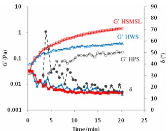
The changes in the viscoelastic properties of HWS, HSMSL and HPS over time were studied with time sweeps, shown in Figure 1, during the first 20 minutes after collection. All three samples displayed an increase in moduli and elasticity during the measurement time with gelling occurring within the first minute for HWS and HSMSL and after slightly less than 5 minutes for HPS. This confirms previous results which have shown an increase in elasticity and moduli in saliva with time8.
Figure 1. Time sweeps of HWS (triangles, solid lines), HSMSL (squares, solid lines) and HPS (circles, dashed lines). G’ in open symbols and phase angle in filled symbols.
G’ HSMSL G’ HWS
G’ HPS
Figure 2 shows mechanical spectra recorded after 20 minutes. The different kinds of saliva all form weak gels with approximately the same degree of elasticity but with varying strength. This indicates a difference in the density of crosslinks or entanglements in the samples which could be a result of different concentrations of structure forming proteins. Unstimulated HSMSL, with the supposed highest mucus content had the highest G’, indicating a higher density of crosslinks, while saliva produced in the parotid gland, with no production of mucins and the supposed lowest mucus content, had the lowest G’. This could reflect the difference in physiological functions of saliva produced by the submandibular-sublingual glands compared to the saliva produced by the parotid glands. Whole saliva had a G’ in between that of HSMSL and HPS which is expected considering it is a mixture of saliva from different glands consisting mostly of HSMSL and HPS.
Figure 2. Mechanical spectra of unstimulated HWS (triangles, solid lines),
HSMSL (squares, solid lines) and HPS (circles, dashed lines) recorded after 20 min. G’ in open symbols and phase angle in filled
symbols.
After acid stimulation a decrease in moduli was expected due to production of more dilute saliva. This could be seen for both whole saliva and
submandibular-sublingual saliva. Time sweeps of
unstimulated and stimulated HWS and HSMSL showed that the G’ still increased over time and there was no apparent change in kinetics. In both cases the final modulus was lower after stimulation (Figure 3 and Figure 4). Stimulated HSMSL however, had a slightly higher initial G’ than unstimulated HSMSL. This is in line with previous results8.
Figure 3. Time sweeps of unstimulated (squares, solid lines) and stimulated (triangles, solid lines) HWS. G’ in open symbols and phase angle in filled symbols.
Figure 4. Time sweeps of unstimulated (squares, solid lines) and stimulated (triangles, solid lines) HSMSL. G’ in open symbols and phase angle in filled symbols.
G’ HSMSL G’ HWS G’ HPS δ Unstimulated Stimulated G’ δ G’ Unstimulated Stimulated G’ δ G’
Figure 5. Mechanical spectra of unstimulated (squares, solid lines) and stimulated (triangles, dashed lines) HSMSL
recorded after 20 min. G’ in open symbols and phase angle in filled symbols.
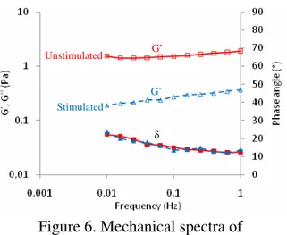
Figure 6. Mechanical spectra of unstimulated (squares, solid lines) and stimulated (triangles, dashed lines) HWS recorded after 20 min. G’ in open symbols
and phase angle in filled symbols. Mechanical spectra of stimulated HWS and HSMSL recorded after 20 minutes show that there was a decrease in G’ compared to unstimulated saliva in both cases. Both, however, still formed gels. Stimulated HSMSL displays the largest decrease in G’ but retained the same degree of elasticity as the unstimulated saliva in the frequency range measured (Figure 6). Whole saliva had smaller relative decrease in G’ when stimulated but at frequencies below 0.1 Hz a slight decrease in elasticity can be detected
(Figure 5). This could be due to a change in the composition of the whole saliva after stimulation with not only a lower concentration of structure forming protein but with a lower average molecular weight of the structure forming proteins.
CONCLUSIONS
The characterization of viscoelastic properties of human saliva by small amplitude oscillatory shear with a narrow gap parallel plate system showed that
• Saliva from both the submandibular-sublingual glands and the parotid gland as well as whole saliva forms weak gels over time scales at the order of 20 minutes.
• Saliva from the
submandibular-sublingual glands formed
significantly stronger gels compared to whole saliva and saliva from the parotid gland indicating higher crosslink density.
• Acid stimulated HWS and HSMSL
still formed gels, but with
significantly lower moduli indicating a decrease in crosslinking density. • Stimulated HWS had a decreased
elasticity at lower frequencies which could be a result of a lower average molecular weight of the structure
forming proteins compared to
unstimulated HWS. REFERENCES
1. Schipper, R.G., Silletti, E. and Vingerhoeds, M.H. (2007) “Saliva as research material: Biochemical, physico-chemical and practical aspects”, Arch. Oral
Biol., 52, 1114-1135.
2. Harris, M., Edgar, M. and Meghji S. (1998) ”Clinical oral science” Butterworth-Heinemann, Oxford, pp. 179-189. Unstimulated Stimulated G’ δ G’ Unstimulated Stimulated G’ δ G’
3. Davies, G.A. and Stokes, J.R. (2007) “Viscoelasticity of human whole saliva collected after acid and mechanical stimulation”, Biorheology, 44, 141-160
4. van der Reijden, W.A., Veerman, E.C.I. and van Nieuw Amerongen, A. (1993)
“Shear rate dependent viscoelastic
behaviour of human glandular salivas”,
Biorheology, 30, 141-152.
5. Christersson, C.E., Lindh, L. and Arnebrandt, T. (2000) “Film forming properties and viscosities of saliva substitutes and human whole saliva”, Eur. J.
Oral Sci., 108, 418-425
6. van der Reijden, W.A., Veerman, E.C.I. and van Nieuw Amerongen, A. (1994) “Rheological properties of commercially available polysaccharides with potential use in saliva substitutes”, Biorheology, 31, 631-642.
7. Davies, G.A. and Stokes, J.R. (2008) “Thin film and high shear rheology of multiphase complex fluids”, J.
Non-Newtonian Fluid Mech., 148, 73–87.
8. Stading, M., Johansson, D., Diogo-Löfgren, C. and Christersson, C.E. (2009) ”Viscoelastic properties of saliva from different glands”, Ann. Trans. Nordic Rheol.