Contents lists available atScienceDirect
Electrochemistry Communications
journal homepage:www.elsevier.com/locate/elecomFull communication
Electrochemistry of a high redox potential laccase obtained by
computer-guided mutagenesis combined with directed evolution
Olga Aleksejeva
a, Ivan Mateljak
b, Roland Ludwig
c, Miguel Alcalde
b, Sergey Shleev
a,⁎ aDepartment of Biomedical Science, Health and Society, Malmö University, 20560 Malmö, SwedenbDepartment of Biocatalysis, Institute of Catalysis, CSIC, Cantoblanco, 28094 Madrid, Spain
cDepartment of Food Sciences and Technology, VIBT – Vienna Institute of Biotechnology, BOKU – University of Natural Resources and Life Sciences, A-1190 Vienna,
Austria
A R T I C L E I N F O
Keywords:
Laccase
Oxygen bioelectroreduction Direct electron transfer Graphite electrode Gold nanoparticles T1 site redox potential
A B S T R A C T
Electrochemical characterization of the GreeDo variant of a high redox potential fungal laccase obtained by la-boratory evolution together with computer-guided mutagenesis, in comparison to its parental variety (the OB-1 mutant), is presented. Both laccases, when immobilized on graphite electrodes either by direct physical adsorption or covalently attached via gold nanoparticles, were capable of both non-mediated and mediator-based bioelec-troreduction of molecular oxygen at low overpotentials. GreeDo exhibited higher open circuit potential values and onset potentials for oxygen bioelectroreduction compared to OB-1. However, even though in homogeneous cat-alysis GreeDo outperforms OB-1 in terms of turnover numbers and catalytic efficiency, when exposed to high redox potential substrates, direct electron transfer based bioelectrocatalytic currents of GreeDo and OB-1 modified electrodes were similar. High operational stability of freely diffusing GreeDo and also the immobilized enzyme in the acidic electrolyte was registered, in agreement with high storage stability of GreeDo in acidic solutions.
1. Introduction
Laccase (p-diphenol:oxygen oxidoreductase, EC 1.10.3.2) is a multi-copper enzyme, facilitating the oxidation of different organic and in-organic compounds with concomitant reduction of molecular oxygen (O2) to water (H2O) [1]. Since the enzyme catalyzes the oxidation of a large variety of substrates, it has become the subject of significant in-terest in many fields of research, such as development of bioelectronic devices (biosensors, and biocathodes for biological power sources), biodegradation of xenobiotics, bioremediation, food processing, and organic synthesis [2–4].
Laccases (Lcs) contain four copper ions of three types, defined as T1, T2 and T3. The substrate oxidation takes place at the T1 site, whereas O2is reduced at the T2/T3 cluster formed by T2 and T3 copper ions [1,5]. An important characteristic of Lcs is the standard redox potential of the T1 site (ET1), which values that lie between 0.43 V and 0.79 V vs. NHE for different Lcs [6]. The catalytic efficiency, i.e. the kcat/KMvalue,
for certain reduced substrates is known to be dependent on ET1[7,8]. Therefore, Lcs with high ET1 values, i.e. ca. 0.73–0.79 V [9], are of particular interest in some specialized branches of biotechnology, e.g., bleaching and bioremediation processes [2]. Moreover, high redox potential Lcs are usually capable of direct electron transfer (DET)
reactions, which offer several advantages for fabrication of enzyme-based bioelectronic devices, first of all, biodevices which electroreduce O2at low overpotentials. Specifically, the lowest overpotential for O2 reduction reaction, viz. 0.06 V, was reported for the high redox poten-tial Lc from the basidiomycete Trametes hirsuta [10].
Very recently, we reported the GreeDo mutant [11], a laboratory evolved laccase with increased ET1and improved pH and thermal sta-bility, which was engineered from an already high-redox potential laccase mutant, the OB-1 variant, originally evolved for heterologous functional expression in yeast [12]. The GreeDo mutant was obtained by combining computational design and directed evolution methods, and represents the first and so far the only experimental evidence on the enhancement of E0′
T1 with concomitant improvement of activity and stability in high redox potential Lcs. Below, a comparative electro-chemical characterization of the GreeDo mutant and Lc OB-1 mutant, is presented.
2. Materials and methods
2.1. Chemicals and materials
BF4Bu4N, 4-nitrobenzenediazonium tetrafluoroborate, sodium
https://doi.org/10.1016/j.elecom.2019.106511
Received 27 April 2019; Received in revised form 5 August 2019; Accepted 7 August 2019 ⁎Corresponding author.
E-mail address:sergey.shleev@mah.se(S. Shleev).
Available online 07 August 2019
1388-2481/ © 2019 The Authors. Published by Elsevier B.V. This is an open access article under the CC BY-NC-ND license (http://creativecommons.org/licenses/BY-NC-ND/4.0/).
nitrite, 1-(3-dimethylaminopropyl)-3-ethylcarbodiimide (EDC), 2,2′-azinobis-(3 ethylbenzothiazoline-6-sulfonic acid) diammonium salt (ABTS), sodium acetate, hydrochloric acid, acetic acid, N-hydro-xysuccinimide (NHS), 6-mercapto-1-hexanol (MH), NaIO4, NaF, KCl, NaOH, HAuCl4, and morpholino-ethanesulfonic acid (MES) were pur-chased from Sigma-Aldrich (St. Louis, MO, USA). Tetrakis(hydro-xymethyl) phosphonium chloride (THPC) was obtained from Fluka Chemie AG (Buchs, Germany). Absolute ethanol (EtOH) was obtained from CCS Healthcare AB (Borlänge, Sweden). Acetonitrile (HPLC grade) was purchased from RdH Laborchemikalien GmbH & Co. (Seelze, Germany). NaClO4was obtained from Merck (Darmstadt, Germany). All aqueous solutions were prepared using water purified with the PURELAB UHQ II system from ELGA Labwater (High Wycombe, United Kingdom). Argon (Ar) and oxygen (O2) gases were supplied by AGA Gas AB (Sundbyberg, Sweden). Spectrographic graphite electrodes (GEs) of 3.05 mm in diameter were obtained from Alfa Aesar (Ward Hill, MA, USA).
2.2. Enzymes
Two variants of basidiomycete Lc PM1, GreeDo mutant (9 mg mL−1), and parental OB-1 mutant (10 mg mL−1), in 20 mM bis-tris buffer pH 6.0 were used. The Lc from basidiomycete PM1 with high thermostability was isolated from the western Mediterranean region and submitted to directed evolution for functional expression in yeast, yielding the parental OB-1 variant [12]. A new mutant version, GreeDo, was obtained from OB-1 by combined computational and directed evolution to improve ET1. Both enzymes were purified to homogeneity and thoroughly characterized, as described in Ref. [11].
2.3. Biomodification of GEs with Lcs by physical adsorption
GEs were polished on a silicon carbide grinding paper (Grit 1000/P 2500, Buehler, USA), rinsed thoroughly with ultrapure H2O, and dried. For biomodification, 10 μL of either GreeDo or OB-1 solution were dropped onto the GE surface and allowed to adsorb for 20 min. 2.4. Covalent attachment of Lcs onto GEs modified with gold nanoparticles
Gold nanoparticles (AuNPs) were synthesized for electrode mod-ification. 5 ± 3 nm AuNPs freshly prepared in aqueous media by ad-dition of 1 mM HAuCl4 to 60 mM NaOH solution containing 1 mM THPC under vigorous stirring [13]. The resulting AuNPs were filtered with a PTFE 0.200 μm pore size filter from Corning Incorporated (Kaiserslautern, Germany). The AuNPs size was determined by UV–Vis spectroscopy using a numerical approximation method [14].
Prior to surface modification, GEs were polished on a silicon carbide grinding paper (Grit 1000/P 2500, Buehler, USA), sonicated for 5 min in ultrapure water, and dried [15]. Thereafter, polished and cleaned GEs were modified with 4-aminophenyl groups, as described in Ref. [15]. Then, the modified electrodes were immersed during 2 h in a 14.5 mM NaNO2and 0.5 M HCl solution in order to convert the aro-matic amino groups on the electrode surface into diazonium groups. Subsequently, the electrodes were incubated into an aqueous dispersion of AuNPs during 3–72 h. Afterwards, two cyclic voltammograms (CVs) were recorded between 0.6 V and −0.6 V at 0.2 V s−1with the GE/ AuNPs in 50 mM acetate buffer, pH 4.2, containing 100 mM NaClO4at room temperature [16].
The GE/AuNP electrodes then were immersed in an acetonitrile solution containing 2 mM p-nitrophenyldiazonium tetrafluoroborate and 100 mM BF4Bu4N. One CV from 0.6 V to −0.6 V with 0.2 V s−1 scan rate was recorded. Afterward, the electrodes were taken into a previously deoxygenized 9:1 H2O/EtOH, 0.1 M KCl solution, and elec-trochemical reduction of nitro groups on the GE/AuNP surface was carried out by running two CVs from 0 V to −1.4 V at 0.2 V s−1scan rate. The amino-terminated GE/AuNP electrodes were then immersed
overnight in a water solution containing 1 mM MH [16].
Five microliters of either GreeDo (9 mg mL−1) or OB-1 (10 mg mL−1) solution were placed into 55 μL of 47 mM NaIO
4solution for 30 min. Afterward, 90 μL of 100 mM Na2HPO4 was added to the mixture. The modified GE/AuNP electrodes were then incubated in either GreeDo or OB-1 solution for 90 min. The GE/AuNP/GreeDo or GE/AuNP/OB-1 electrodes were rinsed with 10 mM MES buffer, pH 6.0. Finally, the electrodes' surface was covered with 10 μL of 10 mM MES, pH 6.0, buffer solution containing 36 mM EDC and 17 mM NHS, and covered to avoid evaporation, letting the reaction take place for 2 h [16].
2.5. Electrochemical measurements
Electrochemical experiments were performed using a μAutolab Type III/FRA2 potentiostat/galvanostat from Metrohm Autolab B. V. (Utrech, The Netherlands) in an electrochemical cell with a total volume of 40 mL thermostated at 25 °C. Ag|AgCl| KClsat (0.199 V vs. NHE), Pt wire, and biomodified GEs were used as a reference, counter, and working electrodes respectively. Three different electrolytes were used, viz. 0.1 M phosphate buffer pH 8.0, 0.1 M phosphate buffer pH 6.0, and 0.1 M acetate buffer pH 4.0 containing 100 mM NaClO4. Cyclic vol-tammograms (CVs) were recorded in O2 saturated electrolytes in a potential window from 1.0 V to 0.2 V vs. NHE with a scan rate of 0.025 V s−1. Averaged OCP and current density values are based on at least three measurements with the highest relative standard deviations of 2% and 14% for OCPs and current densities, respectively. All the reported potentials are given vs. NHE.
2.6. Operational stability of GreeDo in homogenous solution
GreeDo was incubated in the solution of 10 mM K4Mo(CN)8in 0.1 M acetate buffer, pH 4.0 at a concentration of 0.1 mg mL−1 under con-tinuous stirring. 10 μL of enzyme was taken from the solution and in-jected into an Oxygraph Clark-type electrode from Hansatech Ltd. (Norfolk, England), containing 0.25 mL of 10 mM K4Fe(CN)6in air sa-turated 0.1 M acetate buffer, pH 4.0. Rates of O2uptake, measured at 25 °C under continuous stirring, were assessed by Oxygraph Plus soft-ware provided by Hansatech Ltd. (Norfolk, England).
3. Results and discussion
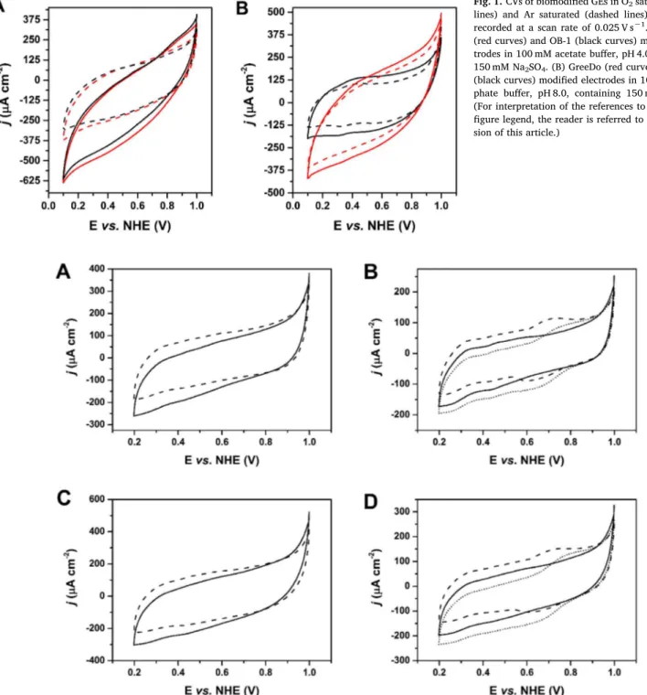
Initially, the two Lcs were physisorbed on GEs, following a simple method for physical adsorption of blue multicopper oxidases (MCOs) on porous spectroscopic graphite electrodes [9,17]. CVs of GreeDo/GEs and OB-1/GEs were recorded in acidic and alkaline electrolytes, re-vealing well-pronounced bioelectrocatalysis at pH 4.0, but indicating complete absence of bioelectrocatalytic reduction of O2, at least at low overpotentials, at pH 8.0 (Fig. 1). These results are in good agreement with data obtained for homogeneous biocatalysis, where the absence of enzymatic activity for both Lcs at pH 8 was observed [11]. The obtained catalytic current densities were about 230 μA cm−2(here and below the bioelectrocatalytic currents were measured at an applied potential of 0.2 V, i.e. at the highest overpotential without non-enzymatic O2 elec-troreduction to H2O2and the standard deviations are calculated based on three measurements) for both enzymes (cf. dotted and solid lines in Fig. 1A). Despite the well-pronounced bioelectrocatalytic process on biomodified GEs, the recorded CVs were far from ideal. No sigmoidal shape was observed, whereas a well-pronounced potential dependence of current outputs was registered, suggesting a sluggish DET (DET-rate limited process in which there is a superposition of different electro-chemical rate constants [18]) along with a random orientation of both enzymes on the electrode surface. Thus, for an efficient DET commu-nication between graphite electrodes and Lcs, a previously developed nanotechnological approach was tried. Specifically, instead of random adsorption, oriented covalent binding of both enzymes to small gold
nanoparticles was tested. Porous GEs allow covering a large surface area with AuNPs, which can be tailored by surface chemistry to im-prove biomolecule orientation and therefore enhance DET [16,19]. Moreover, contrary to large NPs [20], AuNPs of a size comparable to the dimensions of biomolecules are known to perform as electronic bridges between electrodes and enzyme redox centers, thus enhancing the rate of DET reactions [16,21,22].
The CVs of AuNPs decorated GEs with adsorbed Lcs demonstrated bioelectrocatalytic reductive current in the absence of a redox mediator (Fig. 2). The onset potential in the case of GreeDo/AuNPs/GEs was
Fig. 1. CVs of biomodified GEs in O2saturated (solid lines) and Ar saturated (dashed lines) electrolytes recorded at a scan rate of 0.025 V s−1. (A) GreeDo (red curves) and OB-1 (black curves) modified elec-trodes in 100 mM acetate buffer, pH 4.0, containing 150 mM Na2SO4. (B) GreeDo (red curves) and OB-1 (black curves) modified electrodes in 100 mM phos-phate buffer, pH 8.0, containing 150 mM Na2SO4. (For interpretation of the references to color in this figure legend, the reader is referred to the web ver-sion of this article.)
Fig. 2. CVs of biomodified AuNP/GEs in O2saturated 50 mM acetate buffer, pH 4.0, containing 100 mM NaClO4recorded at a scan rate of 0.025 V s−1. (A) GreeDo modified electrodes in the absence (solid line) and presence (dashed line) of 50 mM NaF. (B) GreeDo modified electrodes in the absence (solid line), presence (dotted line) of 0.1 mM ABTS without NaF, and after the addition of 50 mM NaF (dashed line). (C) OB-1 modified electrodes in the absence (solid line) and presence (dashed line) of 50 mM NaF. (D) OB-1 modified electrodes in the absence (solid line) and presence (dotted line) of 0.1 mM ABTS without NaF, as well as after the addition of 50 mM NaF (dashed line).
Table 1
The T1 site redox potentials and open circuit potential values for graphite electrodes modified with GreeDo mutant and OB-1 parental type Lcs in O2 sa-turated electrolytes, as well as the equilibrium redox potentials of O2/H2O couple at the same conditions.
ET1, pH 6 (V) OCP, pH 4.0 (V) OCP, pH 6.0 (V) OCP, pH 8.0 (V)
GreeDo 0.79 0.89 0.79 0.76
OB-1 0.74 0.86 0.75 0.70
higher compared to OB-1/AuNPs/GEs, i.e. 0.83 V vs. 0.79 V, in agree-ment with the 0.05 V E0′
T1difference of the enzymes (Table 1). However, contrary to biomodified GEs, the obtained catalytic current densities were ca. 5 times lower for both GreeDo/AuNPs/GEs and OB-1/AuNPs/ GEs. Moreover, the bioelectrocatalytic current output from GreeDo/ AuNPs/GEs was lesser compared to OB-1/AuNPs/GEs, i.e. 40 and 50 μA cm−2, respectively. Two hydrophobic substitutions surrounding the T1Cu site of GreeDo, viz. Ala162Val and Ala458Leu mutations, could be responsible for these effects. Specifically, much more enzyme was adsorbed on hydrophobic GEs compared to AuNPs/GEs, as well as less specific orientation of hydrophobic GreeDo was achieved on AuNPs/GEs compared to OB-1.
Admixture of a mediator, i.e. ABTS, did enhance the
bioelectrocatalysis, i.e. the current density values reached 60 and 90 μA cm−2. These results are in agreement with data from homo-geneous assays using ABTS as an enzyme substrate, where higher cat-alytic constants and lower Michaelis constants were determined for the parental enzymes [11]. However, in homogeneous catalysis in the case of high redox potential electron donors, i.e. violuric acid and mo-lybdenum cyanide, the biocatalytic process is strictly limited by the oxidation of the reduced substrate at the T1 site and it is highly de-pendent on ET1. Indeed, for these substrates, catalytic constants and efficiency of the GreeDo mutant were substantially higher compared to the OB-1 Lc [11]. The significant difference between DET and mediated ET current outputs (Fig. 2B and D), as well as a strong potential de-pendence of current outputs (Fig. 2A and C), still pointed to a random
Fig. 3. Open circuit potential measurements of GreeDo/GEs in O2saturated electrolytes before (dashed lines) and after (solid lines) applied potential of 0.3 V. (A) pH 4.0. (B) pH 8.0. Inserts: Amperometric responses recorded at 0.3 V.
orientation of both Lcs on the electrode surface despite surface nanos-tructuration and covalent attachment of enzymes. The electrocatalytic signals were absent upon addition of NaF to the electrolyte, confirming the bioelectrocatalytic origin of the currents obtained. Fluoride ions are well-known inhibitors of bio(electro)catalytic reduction of O2, because of their binding to the T2/T3 cluster of MCOs [23], preventing the intramolecular electron transfer (IET) between the T1 site and the T2/ T3 cluster [24,25].
Since neither DET enhancement was achieved, nor were bioelec-trocatalytic current outputs improved by complex enzyme im-mobilization procedure and advanced electrode modification, addi-tional studies were done, again using simple bare GEs. Taking into account the fact that electrocatalysis is not actually related to current increase but rather to a decrease of the O2 electroreduction over-potential, open circuit potential (OCP) measurements of GreeDo and OB-1 modified electrodes were performed. OCP values of GreeDo/GEs and OB-1/GEs were measured at pH 4.0, 6.0, and 8.0, giving on average 0.03–0.06 V higher values for the GreeDo mutant variant compared to the OB-1 enzyme and being in agreement with E0′
T1difference, i.e. 0.05 V (Table 1). The OCP values decreased with increasing pH, following the trend of ca. 0.04 V/pH, which is precisely in between the T1 site and T2/T3 cluster pH dependencies (about 0.02 and 0.06 V/pH [26]). Moreover, OCP values obtained at pH 6.0 were almost identical to the E0′
T1of both Lcs (Table 1). Similar results (the dependence of OCP on ET1, as well as identical OCP and ET1values at pH 6.0) were noticed pre-viously for other high redox potential fungal Lcs [9]. Furthermore, quite low overpotentials for O2bioelectroreduction were obtained in all electrolytes, but with zero difference between the initial OCP of GreeDo/GEs and the equilibrium redox potential of the O2/H2O couple at pH 8.0 (Table 1). Last but not least, the addition of 50 mM NaF to the pH 8.0 electrolyte resulted in 16% OCP drop. The only report in the literature regarding O2electroreduction by a high redox potential lac-case at pH > 7, revealed bioelectrocatalysis at high overpotential and low current (< 50 μA cm−2 below 0.35 V vs. NHE) [27]. Thus, just above mentioned experimental results, in addition to small bioelec-trocatalytic currents at high overpotentials registered at GreedDo/GEs (cf. solid and dashed curves in Fig. 1B at applied potentials below 0.2 V), pointed to the fact of bioelectrocatalytic reduction of O2by the enzyme in alkaline electrolyte with zero overpotential.
To prove or disprove such a strong statement, i.e. Lc based over-potential-less bioelectrocatalysis at pH 8.0, additional OCP and am-perometric measurements were performed at pH 4.0 and 8.0. Contrary to the pH 4.0 circumstances, the OCP of GreeDo/GE was decreasing with time at pH 8.0 owing to OH−inhibition of the enzyme (cf.Fig. 3A
and B). Moreover, the adsorbed enzyme was fully reduced in just 15 s when a potential of 0.3 V was applied, and close to zero currents were registered after that. Furthermore, the initially observed OCP value of 0.76 V was never again achieved. Thus, as evident from the results presented in Fig. 3, a bioelectrocatalytic process at pH 8.0 was not observed and the obtained results could be explained, at least to a certain extent, using the “control center” designation in the electro-chemistry of MCOs [28]. At pH 4.0 the IET is fast and the T1 site is not the control center. Thus, ET1influences but does not define the potential of bioelectrocatalysis. Indeed, high OCP values reflect a high redox potential of the T2/T3 cluster, rather than the ET1. At pH 6.0, the IET is the rate determining slow step of the overall bioelectrocatalytic reac-tion and the T1 is the control center, which defines the potential of bioelectrocatalysis. At pH 8.0, the IET rate between the T1 site and the T2/T3 cluster is close to zero, and the measured OCP values reflect the potential of the T1 site. Additional confirmation comes from the fact that the potential dependence between pH 4.0 and pH 6.0 was 0.05 V/ pH (close to the T2/T3 potential/pH dependence), whereas between pH 6.0 and pH 8.0 it was 0.02 V/pH (close to the T1 site). One can also speculate that the OCP decrease in the case of fluoride inhibition at pH 8.0 relies on a T2/T3 → T1 IET process, or in other words, reduction of the T1 site by a low redox potential pro tem T2/T3 cluster formed by the attachment of OH−and F−ligands.
Finally, the operational stability of GreeDo in solution and in ad-sorbed state was investigated in the acidic electrolyte, pH 4.0, using an aliquot of the enzyme containing solution and by single-potential am-perometry, respectively. The enzyme operating in solution containing the high redox potential substrate was relatively stable, whereas GreeDo/GEs were even more stable, retaining ca. 60% of the initial activity after 2 h of continuous operation (cf. dotted and solid curves in Fig. 4). One could suggest that hydrophobic-hydrophobic interaction between GreeDo molecules and GE surface stabilized the enzyme. When the storage stability of GreeDo at acidic pH was measured, the mutant retained roughly 80% of its initial activity after a 96 h incubation at pH 4.0, whereas the activity of OB-1 was almost negligible. These re-sults are in agreement with the behavior of the enzymes in homo-geneous solutions [11]. On the one hand, it is a widely held view that the stability of fungal laccases is pH-dependent such that they are tol-erant of neutral-alkaline pH, but rapidly become inactive at acidic pH [11]. On the other hand, it is also known that some fungal laccases are fairly stable at acidic pH [29,30], including the parental enzyme of GreeDo, PM1 laccase [31].
4. Conclusions
The biocatalytic and bioelectrocatalytic properties of GreeDo and OB-1 Lcs are in good agreement. Both enzymes were capable of redu-cing O2at low overpotentials in the presence and absence of the redox mediator, ABTS. The onset and open circuit potentials for DET based O2 bioelectroreduction was slightly higher for GreeDo Lc, in agreement with the ET1 values of the enzymes. MET based bioelectrocatalytic currents were higher for OB-1 modified electrodes compared to GreeDo bioelectrodes, following the trend for homogeneous catalysis in the case of substrates with redox potentials below the ET1. High operational stability of GreeDo was registered in 0.1 M acetate buffer, pH 4.0, in agreement with the high storage stability of the enzyme in acidic so-lutions.
Acknowledgements
The work was financially supported by the Swedish Energy Agency (P44707-1).
References
[1] F. Xu, W. Shin, S.H. Brown, J.A. Wahleithner, U.M. Sundaram, E.I. Solomon, Fig. 4. Operational stability of GreeDo in solution (dotted line with squares)
Biochim. Biophys. Acta 1292 (1996) 303–311.
[2] O.V. Morozova, G.P. Shumakovich, S.V. Shleev, Y.I. Yaropolov, Appl. Biochem. Microbiol. 43 (2007) 523–535.
[3] K. Brijwani, A. Rigdon, P.V. Vadlani, Enzyme Res. (2010),https://doi.org/10.4061/ 2010/149748.
[4] T. Senthivelan, J. Kanagaraj, R.C. Panda, Biotechnol. Bioprocess Eng. 21 (2016) 19–38.
[5] O.V. Morozova, G.P. Shumakovich, M.A. Gorbacheva, S.V. Shleev, A.I. Yaropolov, Biochem. Mosc. 72 (2007) 1136–1150.
[6] S. Shleev, J. Tkac, A. Christenson, T. Ruzgas, A.I. Yaropolov, J.W. Whittaker, L. Gorton, Biosens. Bioelectron. 20 (2005) 2517–2554.
[7] F. Xu, Biochemistry 35 (1996) 7608–7614.
[8] F. Xu, J.J. Kulys, K. Duke, K. Li, K. Krikstopaitis, H.J. Deussen, E. Abbate, V. Galinyte, P. Schneider, Appl. Environ. Microbiol. 66 (2000) 2052–2056. [9] S. Shleev, A. Jarosz-Wilkolazka, A. Khalunina, O. Morozova, A. Yaropolov,
T. Ruzgas, L. Gorton, Bioelectrochemistry 67 (2005) 115–124. [10] S. Shleev, V. Andoralov, D. Pankratov, M. Falk, O. Aleksejeva, Z. Blum,
Electroanalysis 28 (2016) 2270–2287.
[11] D. Mate, C. García-Burgos, E. García, A. Ballesteros, S. Camarero, M. Alcalde, Chem. Biol. (2010) 17.
[12] I. Mateljak, E. Monza, M. Lucas, G. V., O. Aleksejeva, R. Ludwig, D. Leech, S. Shleev, M. Alcalde, ACS Catal., 9 (2019) 4561–4572.
[13] D.G. Duff, A. Baiker, P.P. Edwards, Langmuir 9 (1993) 2301–2309. [14] W. Haiss, N.T.K. Thanh, J. Aveyard, D.G. Fernig, Anal. Chem. 79 (2007)
4215–4221.
[15] C. Vaz-Dominguez, S. Campuzano, O. Ruediger, M. Pita, M. Gorbacheva, S. Shleev, V.M. Fernandez, A.L. De Lacey, Biosens. Bioelectron. 24 (2008) 531–537. [16] C. Gutierrez-Sanchez, M. Pita, C. Vaz-Dominguez, S. Shleev, A.L. De Lacey, J. Am.
Chem. Soc. 134 (2012) 17212–17220.
[17] A.I. Yaropolov, A.N. Kharybin, J. Emneus, G. Marko-Varga, L. Gorton, Bioelectrochem. Bioenerg. 40 (1996) 49–57.
[18] C. Leger, A.K. Jones, S.P.J. Albracht, F.A. Armstrong, J. Phys. Chem. B 106 (2002) 13058–13063.
[19] M.S. Thorum, C.A. Anderson, J.J. Hatch, A.S. Campbell, N.M. Marshall, S.C. Zimmerman, Y. Lu, A.A. Gewirth, J. Phys. Chem. Lett. 1 (2010) 2251–2254. [20] D. Pankratov, R. Sundberg, D.B. Suyatin, J. Sotres, A. Barrantes, T. Ruzgas,
I. Maximov, L. Montelius, S. Shleev, RSC Adv. 4 (2014) 38164–38168. [21] M. Kizling, M. Dzwonek, A. Wieckowska, R. Bilewicz, Curr. Opin. Electrochem. 12
(2018) 113–120.
[22] M. Kizling, M. Dzwonek, A. Wieckowska, R. Bilewicz, ChemCatChem 10 (2018) 1988–1992.
[23] B.R.M. Reinhammar, Biochim. Biophys. Acta Bioenerg. 275 (1972) 245–259. [24] A. Naqui, S.D. Varfolomeev, FEBS Lett. 113 (1980) 157–160.
[25] C. Di Bari, N. Mano, S. Shleev, M. Pita, A. L. De Lacey, J. Biol. Inorg. Chem. 22 (2017) 1179–1186.
[26] S. Shleev, V. Andoralov, M. Falk, C.T. Reimann, T. Ruzgas, M. Srnec, U. Ryde, L. Rulisek, Electroanalysis 24 (2012) 1524–1540.
[27] D.M. Mate, D. Gonzalez-Perez, M. Falk, R. Kittl, M. Pita, A.L. De Lacey, R. Ludwig, S. Shleev, M. Alcalde, Chem. Biol. 20 (2013) 223–231.
[28] L. dos Santos, V. Climent, C.F. Blanford, F.A. Armstrong, Phys. Chem. Chem. Phys. 12 (2010) 13962–13974.
[29] M. Dubernet, P. Ribereau-Gayon, H.R. Lerner, E. Harel, A.M. Mayer, Phytochemistry 16 (1977) 191–193.
[30] O.V. Koroljova-Skorobogat'ko, E.V. Stepanova, V.P. Gavrilova, O.V. Morozova, N.V. Lubimova, A.N. Dzchafarova, A.I. Jaropolov, A. Makower, Biotechnol. Appl. Biochem. 28 (1998) 47–54.
[31] P.M. Coll, J.M. Fernandex-Abalos, J.R. Villanueva, R. Santamaria, P. Perez, Appl. Environ. Microbiol. 59 (1993) 2607–2613.