Idiopathic bone cavity of the jaws: an updated analysis of the cases reported in the literature Short title: Idiopathic bone cavity: a review
Bruno Ramos Chrcanovic 1* Ricardo Santiago Gomez 2
1 Department of Prosthodontics, Faculty of Odontology, Malmö University, Malmö, Sweden. bruno.chrcanovic@mau.se; brunochrcanovic@hotmail.com
2
Department of Oral Surgery and Pathology, School of Dentistry, Universidade Federal de Minas Gerais, Belo Horizonte, Brazil. rsgomez@ufmg.br
DEPARTMENT OF PROSTHODONTICS, FACULTY OF ODONTOLOGY, MALMÖ UNIVERSITY, MALMÖ, SWEDEN; DEPARTMENT OF ORAL SURGERY AND PATHOLOGY, SCHOOL OF DENTISTRY, UNIVERSIDADE FEDERAL DE MINAS GERAIS, BELO HORIZONTE, BRAZIL
* Corresponding author:
Bruno Ramos Chrcanovic. Department of Prosthodontics, Faculty of Odontology, Malmö University, Carl Gustafs väg 34, SE-214 21, Malmö, Sweden. bruno.chrcanovic@mau.se; brunochrcanovic@hotmail.com Mobile: +46 725 541 545 Fax: +46 40 6658503
Funding/grant support: none
KEYWORDS
Idiopathic bone cavity; simple bone cyst; traumatic bone cyst; clinical features; persistence following treatment; systematic review
ABSTRACT
The purpose of the present systematic review was to compare the clinic-radiological features of solitary with multiple idiopathic bone cavity (IBC) reported in the jaws. We also aimed to identify possible features that may have some influence on the frequency of persistence of IBC following treatment. An electronic search was undertaken in August/2018. Eligibility criteria included publications having enough clinical/radiological/histological information to confirm the diagnosis. A total of 284 publications reporting 1253 IBCs were included. Multiple IBCs affect older patients and more frequently women in comparison to solitary IBCs. While trauma is more commonly found in solitary IBC, scalloping around teeth, bone expansion and persistence of the cavity following treatment were more significantly associated with multiple lesions. The most relevant factors that are suggested to influence the persistence of the cavity are ‘surgical access only’ in comparison to ‘curettage’, presence of scalloping around teeth, patients with multiple IBCs, and increase in size of the lesion. Solitary and multiple IBC differ in some clinical and radiological aspects and show distinct rates of persistence following treatment. Curettage is the treatment of choice for IBC compared to surgical access only.
KEYWORDS
Idiopathic bone cavity; simple bone cyst; traumatic bone cyst; clinical features; persistence following treatment; systematic review
INTRODUCTION
Idiopathic bone cavity (IBC), also called of traumatic, simple, solitary, hemorrhagic, extravasation, or unicameral bone cyst, is an intraosseous cavity that is not really a true cyst, because it lacks an epithelial lining. The cavity can be either empty or filled with serous or sanguineous fluid1. The wide variety of names shows the lack of understanding of the etiology and pathophysiology of this lesion2. The aim of the systematic review was to compare the clinical and radiologic features of solitary with multiple IBC reported in the jaws, as well as to try to identify possible features that may have some influence on the frequency of persistence following treatment of these lesions.
MATERIALS AND METHODS
This systematic review followed the PRISMA Statement guidelines3.
Research questions
The present study was aiming to answer the following questions: (1) Is there any difference in the clinical features between solitary and multiple IBCs?; (2) What is the best treatment of choice for IBCs?; (3) Is there any factor that can possibly increase the rate persistence following treatment of IBCs?
Search strategies
An electronic search without time restrictions was undertaken in August 2018 in the following databases: PubMed/Medline, Web of Science, Science Direct, J-Stage, and Lilacs. The following terms were used in the search strategies:
(“idiopathic bone cavity” OR “solitary bone cyst” OR “traumatic bone cyst” OR “simple bone cyst” OR “hemorrhagic bone cyst” OR “haemorrhagic bone cyst” OR “unicameral bone cyst” OR “extravasation bone cyst” OR “idiopathic bone cavities” OR “solitary bone cysts” OR “traumatic bone cysts” OR “simple bone cysts” OR “hemorrhagic bone cysts” OR “haemorrhagic bone cysts” OR “unicameral bone cysts” OR “extravasation bone cysts”) AND (mandible OR mandibular OR maxilla OR maxillary OR jaw OR jaws)
Google Scholar was also checked. A manual search of all related oral pathology, maxillofacial and specialist dental and oral journals was performed. The reference list of the identified studies and the relevant reviews on the subject were also checked for possible additional studies. Publications with lesions identified by other authors as being IBC, solitary bone cyst, traumatic bone cyst, or
simple bone cyst, even not having these terms in the title of the article, were also re-evaluated by an author (R.S.G.) of the present study.
Inclusion and Exclusion Criteria
Publications reporting cases of IBC with enough clinical, radiological and histological information to confirm the diagnosis were included. The definitions and criteria of the World Health Classification of Tumors – Head and Neck Tumors book 1, were used to diagnose a lesion as IBC. IBCs associated with cemento-osseous or fibrous dysplasia were not considered for this study, as they may behave differently from the classic IBC. Cases in which a surgical approach to the suspected IBC for definitive biopsy was not performed were not included, as it would not possible to absolutely rule out a large number of cysts and tumors that may occur as differential diagnosis.
Study selection
The titles and abstracts of all reports identified through the electronic searches were read independently by the authors. For studies appearing to meet the inclusion criteria, or for which there were insufficient data in the title and abstract to make a clear decision, the full report was obtained. Disagreements were resolved by discussion between the authors. The clinical and radiological aspects, as well as the histological description of the lesions reported by the publications were thoroughly assessed by one of the authors (R.S.G.) of the present study, an expert in oral pathology, in order to confirm the diagnosis of IBC.
Data extraction
The following data were then extracted: patient’s sex, age and race, duration of the lesion previously to treatment, history of previous (local) facial trauma, lesion location, lesion size (largest diameter), perforation of cortical bone, locularity appearance in radiological exams (unilocular/multilocular), presence of pathological fracture, presence of solitary or multiple lesions in
the same patient (the presence of more than one cavity would classify the patient as having multiple lesions), triangular or cone shape of the anterior margin 4, scalloping around the roots of adjacent teeth, tooth displacement and/or tooth root resorption due to lesion’s growth, vitality of teeth adjacent to/involved by the IBC, bone expansion, presence of clinical symptoms, treatment performed, surgical finding (empty cavity or fluid), persistence following treatment, and follow-up period. Contact with authors for possible missing data was performed.
Analyses
The mean, standard deviation (SD), and percentages were presented as descriptive statistics. Kolmogorov–Smirnov test was performed to evaluate the normal distribution of the variables, and Levene’s test evaluated homoscedasticity. The performed tests for two independent groups were Student’s t-test or Mann-Whitney test, depending on the normality. Pearson’s chi-squared or Fisher’s exact tests were used for categorical variables, depending on the expected count of events in a 2x2 contingency table. The probability of persistence of the cavity following treatment was calculated for some variables, whenever possible, in odds ratio (95% confidence interval). The degree of statistical significance was considered p < 0.05. All data were statistically analyzed using the Statistical Package for the Social Sciences (SPSS) version 25 software (SPSS Inc., Chicago, IL, USA).
RESULTS
Literature search
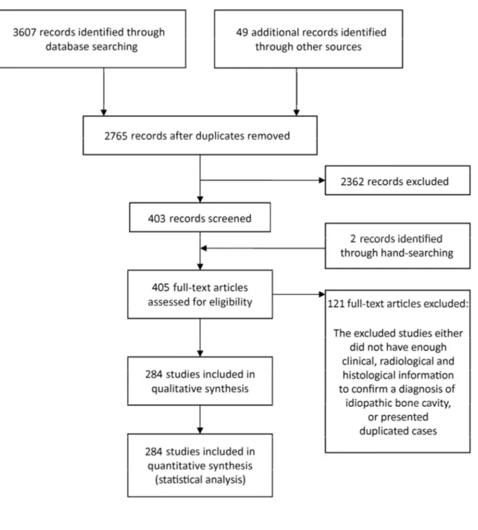
The study selection process is summarized in Figure 1. The search strategy in the databases resulted in 3607 papers (PubMed/Medline, n=326; Web of Science, n=881; Science Direct, n=2069; J-Stage, n=102; Lilacs, n=229), 49 additional eligible papers were found in Google Scholar, and 2 papers through hand-searching. At the end, a total of 284 publications were included (readers interested in the list should contact the first author for details).
Description of the Studies and Analyses
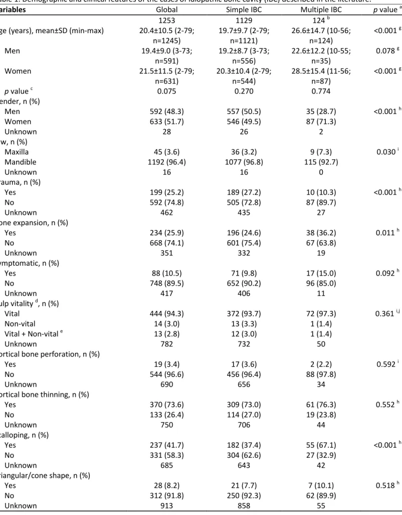
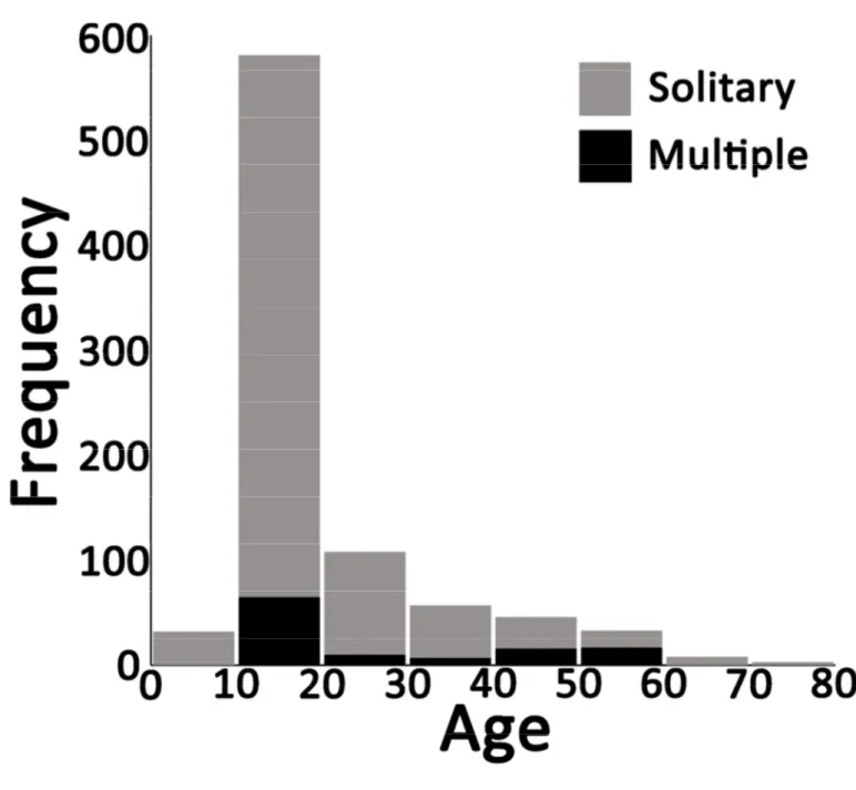
Table 1 presents demographic and clinical features of all 1253 IBSs – 1129 IBCs as solitary cases, 124 IBCs as multiple cases in 58 patients. IBCs were equally prevalent between the sexes, except when only cases of multiple IBCs are considered, with a higher prevalence in in women. The mean age of the patients was 20.4±10.5 (min-max, 2-79), with higher mean age for women and for patients presenting multiple IBCs. Figure 2 shows the distribution of the lesions according to age, with the highest prevalence in the second decade of life. The lesions were far more prevalent in the mandible in comparison to the maxilla, at the posterior region in comparison to the anterior region (Figures 3-5).
The common features of IBCs include mandibular location, cortical bone thinning, unilocular appearance in radiological exams, and pulp vitality of teeth adjacent to/involved by the IBC. A fluid inside the cavity was observed in nearly half of the IBCs. Scalloping around the roots of adjacent teeth was observed in about 40% of the lesions, being more prevalent in patients with multiple IBCs. History of trauma was more frequently observed in solitary IBC compared to multiple lesions. On the other hand, multiple IBC were more associated with bone expansion, scalloping of teeth and persistence after treatment.
Treatment of the lesions was known in 964 cases, the most commonly performed being curettage, followed by surgical access only. Time of follow-up was informed for 487 lesions, with a
mean±SD of 22.8±25.3 months (min-max, 1-216). The interval from initial treatment to the detection of the first persistent lesion following treatment ranged from 3 to 98 months, with a mean interval of 36.1±25.3 months.
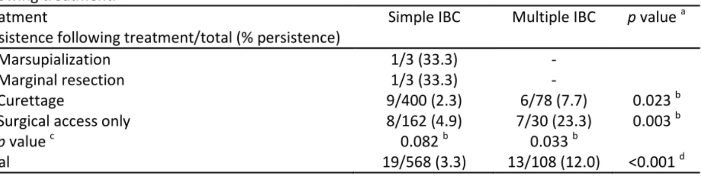
Table 2 shows the rate of persistence after treatment according to the treatment performed. A higher rate of persistence after treatment was observed after the performance of ‘surgical access only’ in comparison to ‘curettage’, and in cases of multiple IBCs in comparison to solitary IBCs.
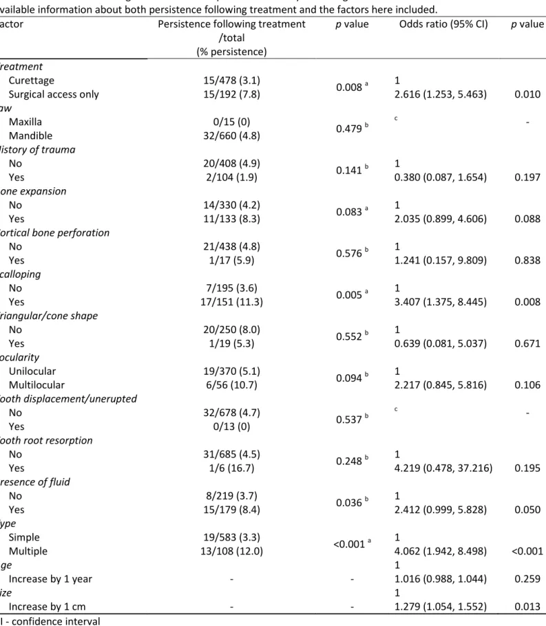
Table 3 shows the rate of persistence after treatment according to the some variables. According to the results, it is suggested that the following factors may have an influence on the rates of persistence of the lesion after treatment: ‘surgical access only’ in comparison to ‘curettage’, presence of scalloping, patients with multiple IBCs, and increase in size of the lesion.
DISCUSSION
In the present systematic review we performed an integrated analysis of the available data published in the literature on IBC. As there is scarce information in the literature comparing clinical-radiological features of solitary and multiple IBC, this analysis was initially performed, followed by the study of the factors that are associated with persistence following treatment. A review of pathological lesions is important because it provides information that can improve diagnostic accuracy, allowing pathologists and surgeons to make informed decisions and refine treatment plans to optimize clinical outcomes5-10. In the present review we observed that most of the IBCs occur in mandibles, are asymptomatic, do not present bone expansion, present vital teeth that have roots adjacent to/involved by the cavity, are not preceded by a local trauma, do present cortical thinning (but not cortical bone perforation), are unilocular, have a higher prevalence in the posterior mandible, and are discovered in the second decade of life. Moreover, IBCs are equally prevalent in males and females, and about 40% of the IBCs show scalloping around the roots of adjacent teeth. These observed clinical-radiological features are in accordance to the current knowledge about this lesion and are important to be considered during the working diagnosis of bone lesions.
There are many published cases of supposed IBC in which the diagnosis was incorrect. There is also a considerable amount of reports that have not differentiated IBC from the cement-osseous dysplasia associated with IBC. Moreover, the diagnosis of IBC relies on clinical, radiographic, and ultimately, surgical findings. As IBC radiographic presentation can mimic other bone lesions, only cases confirmed by biopsy were included. Therefore, some possibly IBCs that were not surgically explored were excluded11-18.
The etiology of the IBC remains unclear2. According to the one of the earliest hypothesis on its etiology, a traumatic event would incite medullary hemorrhage and a subsequent failure of the hematoma to organize and be replaced with tissue would result in an IBC19. This theory is questioned by the fact that in most of the cases there is no history of trauma. The present study observed that, when the information was available, only one of every four IBCs was preceded by a traumatic event.
Moreover, the incidence of trauma in patients with IBC is no greater than in the general population20. We do not have a plausible explanation for the highest frequency of trauma in solitary IBC and we are not sure about the biological relevance, if any, of this finding. However, the hypothesis that solitary and multiple IBC present distinct etiopathogenesis should be further explored in the future.
When we compared multiple with solitary IBC we found other interesting data. Multiple IBC were more associated with bone expansion and persistence following treatment, which suggests that this clinical form is more aggressive. Multiple IBC also present higher frequency of scalloping of teeth detected in the radiographic exam, but this can be explained by a highest number of teeth affected when more than one lesion is found.
The present review identified 7 cases that were discovered after a pathologic fracture of the jaws, which represents only 0.6% of the cases reported in the literature. According to a review21, about 80% of orthopedic IBCs are discovered because of pathologic fracture, whereas IBC in the maxillofacial region are usually noted incidentally on panoramic radiographs. This great discrepancy of the diagnosis of an IBC after pathologic fractures between long bones and the jaws is probably due to the fact that children routinely undergo radiography of the jaws for orthodontic treatment or other dental reasons.
Although some IBCs show spontaneous healing, resolution after aspiration of the cavity or surgical exploration without curettage, our study supports curettage as the first treatment option. IBCs that were surgically accessed only showed a higher rate of persistence following treatment when compared to lesions that were submitted to curettage. This was especially evident for multiple lesions. This can be explained by the fact that curettage promotes the formation of a blood clot that is necessary for subsequent bone repair. In fact, curettage is the most widely accepted approach to the management of patients with IBC. Periodic observation of suspected IBCs without surgical intervention is not suggested, because IBCs may continue to enlarge if left untreated, and may eventually result in pathologic fracture of the affected bone22.
In addition to curettage and multiple lesions, other clinical-radiological factors were also associated with persistence after treatment. It is interesting that there is a higher chance of persistence after treatment for larger lesions and scalloping teeth. Larger lesions or those with scalloping teeth could make the formation of a sufficient blood clot difficult for bone repair in some circumstances, which can explain the persistence of the lesion.
A study23 suggested that cases with absent lamina dura, scalloped margins, nodular bone expansion, internal radiopaque masses, and/or multiple cavities may indicate more likelihood of persistence following treatment. We did not evaluate some of these radiographic features due to the retrospective nature of the present review, and also due to the low quality that radiological images usually present on printed paper, which does not allow a proper identification of these features in many cases. For example, only good-quality periapical radiographs would allow an adequate evaluation of the absence of lamina dura.
The results of our study have to be interpreted with caution because of its limitations. First, all included studies were retrospective reports, which inherently result in errors, with incomplete records. Secondly, many of the published cases had a short follow-up, which could have led to an underestimation of the actual rate of persistence following treatment. However, it is hard to define what it would be considered a short follow-up period to evaluate the persistence following treatment of IBCs Thirdly, many of the cases described were published as isolated case reports or small case series.
CONCLUSIONS
Solitary and multiple IBC differ in some clinical and radiological aspects and show distinct rates of persistence following treatment. Curettage is the treatment of choice for IBC surgical management compared to surgical access only.
ACKNOWLEDGEMENTS Funding/grant support
This research received no specific grant from any funding agency in the public, commercial, or not-for-profit sectors.
Declaration of conflicting interests
There are no conflicts of interest to declare.
We would like to thank the following people who provided us some articles: Dr. Kirsten Bechtel, Dr. Marcio Ajudarte Lopes, Dr. Aparna Naidu, Dr. Gina D. Roque-Torres, Dr. Mohammad Hosein Kalantar Motamedi, Dr. Steven Zijderveld, Dr. Kumar Nilesh, Dr. Jadbinder Seehra, Dr. Bogumił Lewandowski, and Dr. John K. Brooks. Last but not least, we would like to thank the librarians of Malmö University (with a special thanks to Ms. Anneli Svensson), who helped us to obtain some articles. RSG is a research fellow at CNPq, Brazil.
REFERENCES
1. WHO. World Health Organization Classification of Head and Neck Tumours. 4th edition ed. Lyon: IARC Press; 2017.
2. Kuhmichel A, Bouloux GF. Multifocal traumatic bone cysts: case report and current thoughts on etiology. J Oral Maxillofac Surg 2010;68:208-12.
3. Moher D, Liberati A, Tetzlaff J, Altman DG, Grp P. Preferred Reporting Items for Systematic Reviews and Meta-Analyses: The PRISMA Statement. Ann Intern Med 2009;151:264-9, W64. 4. Copete MA, Kawamata A, Langlais RP. Solitary bone cyst of the jaws: radiographic review of
44 cases. Oral Surg Oral Med Oral Pathol Oral Radiol Endod 1998;85:221-5.
5. Chrcanovic BR, Gomez RS. Ameloblastic Fibrodentinoma and Ameloblastic Fibro-Odontoma: An Updated Systematic Review of Cases Reported in the Literature. J Oral Maxillofac Surg 2017;75:1425-37.
6. Chrcanovic BR, Gomez RS. Calcifying epithelial odontogenic tumor: An updated analysis of 339 cases reported in the literature. J Craniomaxillofac Surg 2017;45:1117-23.
7. Chrcanovic BR, Gomez RS. Cementoblastoma: An updated analysis of 258 cases reported in the literature. J Craniomaxillofac Surg 2017;45:1759-66.
8. Chrcanovic BR, Gomez RS. Squamous odontogenic tumor and squamous odontogenic tumor-like proliferations in odontogenic cysts: An updated analysis of 170 cases reported in the literature. J Craniomaxillofac Surg 2018;46:504-10.
9. Chrcanovic BR, Gomez RS. Melanotic neuroectodermal tumour of infancy of the jaws: an analysis of diagnostic features and treatment. Int J Oral Maxillofac Surg 2018.
10. Chrcanovic BR, Gomez RS. Ameloblastic fibrodentinosarcoma and ameloblastic fibro-odontosarcoma: A systematic review. J Stomatol Oral Maxillofac Surg 2018;119:401-6. 11. Damante JH, Da SGEN, Ferreira Jr O. Spontaneous resolution of simple bone cysts.
Dentomaxillofac Radiol 2002;31:182-6.
13. Poyton HG, Morgan GA. The simple bone cyst. Oral Surg Oral Med Oral Pathol 1965;20:188-97.
14. Ribeiro EB, Bisol FCT, Iwaki LCV, Silva MCS, Tolentino ES. Cisto ósseo simples: relato de casos clínicos. RFO UPF 2014;19:359-63.
15. Sapone J, Hansen LS. Traumatic bone cysts of jaws: diagnosis, treatment, and prognosis. Oral Surg Oral Med Oral Pathol 1974;38:127-38.
16. Sapp JP, Stark ML. Self-healing traumatic bone cysts. Oral Surg Oral Med Oral Pathol 1990;69:597-602.
17. Shah KM, Mistry JD. Traumatic bone cyst or solitary bone cyst. BMJ Case Rep 2013;2013. 18. Szerlip L. Traumatic bone cysts. Resolution without surgery. Oral Surg Oral Med Oral Pathol
1966;21:201-4.
19. Olech E, Sicher H, Weinmann JP. Traumatic mandibular bone cysts. Oral Surg Oral Med Oral Pathol 1951;4:1160-72.
20. Kaugars GE, Cale AE. Traumatic bone cyst. Oral Surg Oral Med Oral Pathol 1987;63:318-24. 21. Lokiec F, Wientroub S. Simple bone cyst: etiology, classification, pathology, and treatment
modalities. J Pediatr Orthop B 1998;7:262-73.
22. Naidu A, Chesley LD. Oral and maxillofacial pathology case of the month. Traumatic bone cyst. Tex Dent J 2008;125:544-5, 54-5.
23. Suei Y, Taguchi A, Nagasaki T, Tanimoto K. Radiographic findings and prognosis of simple bone cysts of the jaws. Dentomaxillofac Radiol 2010;39:65-71.
Table 1. Demographic and clinical features of the cases of idiopathic bone cavity (IBC) described in the literature.
Variables Global Simple IBC Multiple IBC p value a
n 1253 1129 124 b
Age (years), mean±SD (min-max) 20.4±10.5 (2-79; n=1245) 19.7±9.7 (2-79; n=1121) 26.6±14.7 (10-56; n=124) <0.001 g Men 19.4±9.0 (3-73; n=591) 19.2±8.7 (3-73; n=556) 22.6±12.2 (10-55; n=35) 0.078 g Women 21.5±11.5 (2-79; n=631) 20.3±10.4 (2-79; n=544) 28.5±15.4 (11-56; n=87) <0.001 g p value c 0.075 0.270 0.774 Gender, n (%) Men 592 (48.3) 557 (50.5) 35 (28.7) <0.001 h Women 633 (51.7) 546 (49.5) 87 (71.3) Unknown 28 26 2 Jaw, n (%) Maxilla 45 (3.6) 36 (3.2) 9 (7.3) 0.030 i Mandible 1192 (96.4) 1077 (96.8) 115 (92.7) Unknown 16 16 0 Trauma, n (%) Yes 199 (25.2) 189 (27.2) 10 (10.3) <0.001 h No 592 (74.8) 505 (72.8) 87 (89.7) Unknown 462 435 27 Bone expansion, n (%) Yes 234 (25.9) 196 (24.6) 38 (36.2) 0.011 h No 668 (74.1) 601 (75.4) 67 (63.8) Unknown 351 332 19 Symptomatic, n (%) Yes 88 (10.5) 71 (9.8) 17 (15.0) 0.092 h No 748 (89.5) 652 (90.2) 96 (85.0) Unknown 417 406 11 Pulp vitality d, n (%) Vital 444 (94.3) 372 (93.7) 72 (97.3) 0.361 i,j Non-vital 14 (3.0) 13 (3.3) 1 (1.4) Vital + Non-vital e 13 (2.8) 12 (3.0) 1 (1.4) Unknown 782 732 50
Cortical bone perforation, n (%)
Yes 19 (3.4) 17 (3.6) 2 (2.2) 0.592 i
No 544 (96.6) 456 (96.4) 88 (97.8)
Unknown 690 656 34
Cortical bone thinning, n (%)
Yes 370 (73.6) 309 (73.0) 61 (76.3) 0.552 h No 133 (26.4) 114 (27.0) 19 (23.8) Unknown 750 706 44 Scalloping, n (%) Yes 237 (41.7) 182 (37.4) 55 (67.1) <0.001 h No 331 (58.3) 304 (62.6) 27 (32.9) Unknown 685 643 42 Triangular/cone shape, n (%) Yes 28 (8.2) 21 (7.7) 7 (10.1) 0.518 h No 312 (91.8) 250 (92.3) 62 (89.9) Unknown 913 858 55
Locularity, n (%) Unilocular 503 (86.0) 435 (87.0) 67 (79.8) 0.077 h Multilocular 82 (14.0) 65 (13.0) 17 (20.2) Unknown 669 629 40 Tooth displacement/unerupted, n (%) Yes 13 (1.1) 12 (1.1) 1 (0.8) 0.597 i No 1169 (98.9) 1046 (98.9) 123 (99.2) Unknown 71 71 0
Tooth root resorption, n (%)
Yes 10 (0.8) 8 (0.8) 2 (1.6) 0.283 i No 1172 (99.2) 1050 (99.2) 122 (98.4) Unknown 71 71 0 Pathological fracture, n (%) Yes 7 (0.6) 7 (0.6) 0 (0) 0.481 i No 1246 (99.4) 1122 (99.4) 124 (100) Unknown 0 0 0 Presence of fluid, n (%) Yes 325 (46.0) 283 (46.2) 42 (44.7) 0.777 h No 381 (54.0) 329 (53.8) 52 (55.3) Unknown 548 517 30 Treatment, n (%) Marsupialization 3 (0.3) 3 (0.4) 0 (0) Curettage 641 (66.5) 557 (66.0) 84 (70.0) 0.462 h,k
Surgical access only 315 (32.7) 279 (33.0) 36 (30.0)
Marginal resection 3 (0.3) 3 (0.4) 0 (0)
Segmental resection f 2 (0.2) 2 (0.2) 0 (0)
Unknown 289 285 4
Persistence following treatment, n (%)
Yes 32 (4.6) 19 (3.3) 13 (12.0) <0.001 h
No 659 (95.4) 564 (96.7) 95 (88.0)
Unknown 562 546 16
Follow-up time (months), mean±SD (min-max) 22.8±25.3 (1-216; n=487) 23.5±26.3 (1-216; n=396) 19.8±20.7 (1-99; n=91) 0.162 g Lesion size (cm), mean±SD (min-max) 3.1±1.8 (0.5-14.0;
n=380) 3.0±1.7 (0.6-14.0; n=337) 3.4±2.1 (0.5-12.0; n=43) 0.178 g SD – standard deviation a
Comparison between simple and multiple IBCs b
Here the ‘number of IBCs’ is considered as the statistical unit. There were 124 IBCs in 58 patients: 45 patients presented two IBCs, 11 patients with three IBCs, and 2 patients with many (uncountable) IBCs, which here were considered as only one IBC
c Comparison of the mean age between men and women (Mann-Whitney test) d It concerns tooth(teeth) directly adjacent to/involved by the idiopathic bone cavity e
Concomitant presence of vital and non-vital teeth adjacent to/involved by the idiopathic bone cavity f
Resection with continuity defect g
Mann-Whitney test h
Pearson’s chi-squared test i Fisher’s exact test
j Comparison between ‘vital’ and ‘non-vital’ k
Table 2. Persistence following treatment– for the lesions with available information about treatment and persistence following treatment.
Treatment
Persistence following treatment/total (% persistence)
Simple IBC Multiple IBC p value a
Marsupialization 1/3 (33.3) -
Marginal resection 1/3 (33.3) -
Curettage 9/400 (2.3) 6/78 (7.7) 0.023 b
Surgical access only 8/162 (4.9) 7/30 (23.3) 0.003 b
p value c 0.082 b 0.033 b
Total 19/568 (3.3) 13/108 (12.0) <0.001 d
a
Comparison between simple and multiple IBCs b
Fisher’s exact test c
Comparison between ‘curettage’ and ‘surgical access only’ d
Table 3. Persistence following treatment for idiopathic bone cavity according to different factors – for the lesions with available information about both persistence following treatment and the factors here included.
Factor Persistence following treatment
/total (% persistence)
p value Odds ratio (95% CI) p value
Treatment
Curettage 15/478 (3.1)
0.008 a 1
Surgical access only 15/192 (7.8) 2.616 (1.253, 5.463) 0.010
Jaw Maxilla 0/15 (0) 0.479 b c - Mandible 32/660 (4.8) History of trauma No 20/408 (4.9) 0.141 b 1 Yes 2/104 (1.9) 0.380 (0.087, 1.654) 0.197 Bone expansion No 14/330 (4.2) 0.083 a 1 Yes 11/133 (8.3) 2.035 (0.899, 4.606) 0.088
Cortical bone perforation
No 21/438 (4.8) 0.576 b 1 Yes 1/17 (5.9) 1.241 (0.157, 9.809) 0.838 Scalloping No 7/195 (3.6) 0.005 a 1 Yes 17/151 (11.3) 3.407 (1.375, 8.445) 0.008 Triangular/cone shape No 20/250 (8.0) 0.552 b 1 Yes 1/19 (5.3) 0.639 (0.081, 5.037) 0.671 Locularity Unilocular 19/370 (5.1) 0.094 b 1 Multilocular 6/56 (10.7) 2.217 (0.845, 5.816) 0.106 Tooth displacement/unerupted No 32/678 (4.7) 0.537 b c - Yes 0/13 (0)
Tooth root resorption
No 31/685 (4.5) 0.248 b 1 Yes 1/6 (16.7) 4.219 (0.478, 37.216) 0.195 Presence of fluid No 8/219 (3.7) 0.036 b 1 Yes 15/179 (8.4) 2.412 (0.999, 5.828) 0.050 Type Simple 19/583 (3.3) <0.001 a 1 Multiple 13/108 (12.0) 4.062 (1.942, 8.498) <0.001 Age 1 Increase by 1 year - - 1.016 (0.988, 1.044) 0.259 Size 1 Increase by 1 cm - - 1.279 (1.054, 1.552) 0.013 CI - confidence interval a
Pearson’s chi-squared test b
Fisher’s exact test
c On at least one case, the value of the weight variable was zero. Such cases are invisible to statistical procedures and graphs which need positively weighted cases
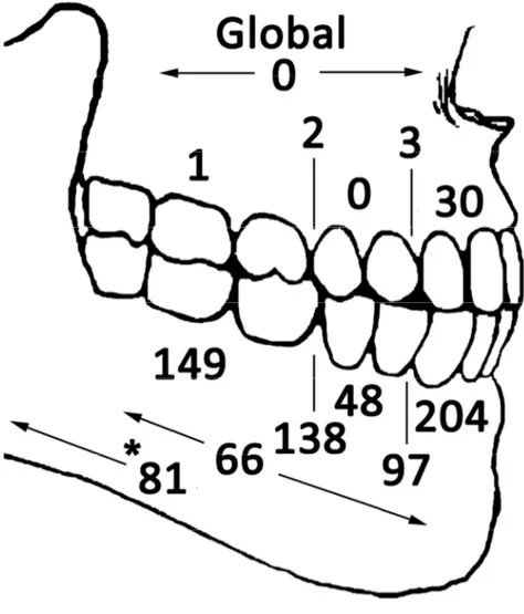
Figure 3. Topographical distribution of the known precise locations (n=819) of IBCs. Cases involving multiple regions (or an entire quadrant) are indicated between arrows. Numbers at the top and bottom of the lines indicate cases involving both adjoining regions: anterior/premolar, premolar/molar. One asterisk (*) indicates the number of lesions from the mandibular body that reached the angle and/or ramus. For the rest of the lesions (n=434), the location was the ‘maxilla’ (n=8), ‘mandible’ (n=181), ‘posterior maxilla’ (n=1), ‘posterior mandible’ (n=79), ‘mandibular body’ (n=71), ‘mandibular angle’ (n=11), ‘mandibular ramus’ (n=42), ‘mandibular condyle’ (n=24) (3 other IBCs also affected the mandibular condyle, but were mainly affecting the mandibular ramus), maxillary-zigomaticus suture (n=1), and not informed (n=16). A number of 129 IBCs crossed the midline. Six IBCs also affected the coronoid process, but mainly affecting the mandibular ramus.
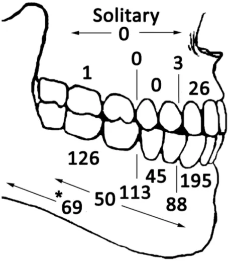
Figure 4. Topographical distribution of the known precise locations (n=716) of IBCs in patients with solitary IBCs. Cases involving multiple regions (or an entire quadrant) are indicated between arrows. Numbers at the top and bottom of the lines indicate cases involving both adjoining regions: anterior/premolar, premolar/molar. One asterisk (*) indicates the number of lesions from the mandibular body that reached the angle and/or ramus. For the rest of the lesions (n=413), the location was the ‘maxilla’ (n=5), ‘mandible’ (n=172), ‘posterior maxilla’ (n=1), ‘posterior mandible’ (n=75), ‘mandibular body’ (n=69), ‘mandibular angle’ (n=10), ‘mandibular ramus’ (n=40), ‘mandibular condyle’ (n=24) (3 other IBCs also affected the mandibular condyle, but were mainly affecting the mandibular ramus), maxillary-zigomaticus suture (n=1), and not informed (n=16). A number of 116 IBCs crossed the midline. Six IBCs also affected the coronoid process, but mainly affecting the mandibular ramus.
Figure 5. Topographical distribution of the known precise locations (n=103) of IBCs in patients with multiple IBCs. Cases involving multiple regions (or an entire quadrant) are indicated between arrows. Numbers at the top and bottom of the lines indicate cases involving both adjoining regions: anterior/premolar, premolar/molar. One asterisk (*) indicates the number of lesions from the mandibular body that reached the angle and/or ramus. For the rest of the lesions (n=21), the location was the ‘maxilla’ (n=3), ‘mandible’ (n=9), ‘posterior mandible’ (n=4), ‘mandibular body’ (n=2), ‘mandibular angle’ (n=1), and ‘mandibular ramus’ (n=2). A number of 13 IBCs crossed the midline.