Voltage dependent anion
channels (VDAC) in
plasma membrane
induces apoptosis
Nesar Akanda
Voltage-dependent anion channels (VDAC)
in the plasma membrane induce apoptosis
Department of Biomedicine and Surgery Division of Cellbiology, Faculty of Health Sciences
Linköping University SE-581 85 Linköping, Sweden Institute of Environmental Medicine Division of Toxicology and Neurotoxicology
Karolinska Institutet SE-171 77 Stockholm, Sweden
I dedicate this book to my parents and beloved Shoma and Summit
ABSTRACT
Apoptosis, or programmed cell death, is essential for proper development and functioning of the body systems. During development, apoptosis plays a central role to sculpt the embryo, and in adults, to maintain tissue homeostasis by eliminating redundant, damaged or effete cells. Therefore, a tight regulation of this process is essential. Cell shrinkage associated efflux of K+ and Cl– through plasma membrane ion channels is an early event of apoptosis. How-ever, little is known about these fluxes. The aim of this thesis was to investigate ion channels in the plasma membrane of neurons undergoing apoptosis. We studied differentiated (the mouse hippocampal cell line HT22, the human neuroblastoma cell line SK-N-MC, and rat primary hippocampal neurons) and undifferentiated (rat primary cortical neural stem cells cNSCs) cells with the patch-clamp technique. All cell types displayed a low electrical activity under control conditions. However, during apoptosis in differentiated neurons, we found an activation of a voltage-dependent anion channel. The conductance of the channel is 400 pS, the voltage dependence of the opening is bell shaped with respect to membrane voltage with a maximum open probability at 0 mV, and the Cl− to cation selectivity is >5:1. These biophysi-cal properties remind about the voltage-dependent anion channel normally found in the outer mitochondrial membrane (VDACmt). Hence, we call our apoptosis-inducing plasma mem-brane channel VDACpl. The molecular identity of the channel was corroborated with the spe-cific labelling of different anti-VDAC antibodies. Block of this channel either with antibodies or with sucrose prevented apoptosis, suggesting a critical role for VDACpl in the apoptotic process. VDACpl is a NADH (-ferricyanide) reductase in control cells. We found that the en-zymatic activity is altered while the VDACpl channel is activated during apoptosis. Surpris-ingly, in cNSCs we did not find any activation of VDACpl, no VDACpl-specific labelling, no enzymatic activity, and no prevention of apoptosis with VDACpl-blocking strategies. Instead, we found an activation of a voltage-independent 37 pS ion channel, and that the Cl– channel blocker DIDS prevented apoptosis in cNSCs. Our finding that activation of VDACpl is criti-cal for apoptosis in differentiated neurons hopefully can lead to new strategies in the treat-ment of several diseases related to apoptosis.
LIST OF PUBLICATIONS
I. F Elinder, N Akanda, R Tofighi, S Shimizu, Y Tsujimoto, S Orrenius and S Ceccatelli. 2005. Opening of plasma membrane voltage-dependent anion channels (VDAC) precedes caspase activation in neuronal apoptosis induced by toxic stimuli. Cell Death Differ. 12:1134-1140
II. N Akanda, R Tofighi, J Brask, C Tamm, F Elinder and S Ceccatelli. 2006. Voltage-dependent anion channels (VDAC) in the plasma membrane play a critical role in apoptosis in differentiated hippocampal neurons but not in neuronal stem cells (submitted for publication)
III. N Akanda and F Elinder. 2006. Biophysical properties of the apoptosis-inducing plasma membrane voltage-dependent anion channel. Biophys J. 90:4405-4417
IV. N Akanda and F Elinder. 2006. Sucrose reduces the current through plasma membrane voltage-dependent anion channels (VDACpl) mainly by reducing the open probability. (manuscript)
CONTENTS
INTRODUCTION 9 1. Ion channels 9 1.1. General aspects 9
1.2. Regulation of membrane potential 10 2. Cell death 11
2.1. What is apoptosis? Why is it so important? 11 2.2. Two major apoptotic pathways 13
2.2.1. The death-receptor pathway (extrinsic) 13 2.2.2. The mitochondrial pathway (intrinsic) 14
3. The role of ion channels in apoptosis: Molecules for life, disease, and death 15
AIMS 17
MATERIALS AND METHODS 18 1. Cell culture 18
1.1. HT22 and SK-N-MC cell lines 18 1.2. Primary hippocampal neurons 18 1.3. Primary cortical neuronal stem cells 19 2. Induction and prevention of apoptosis 19 3. Evaluation of apoptotic cells 19
4. Electrophysiology 20
4.1. The patch-clamp method 20
4.2. Electrodes, amplifiers, softwares, and solutions 21 4.3. The voltage protocol 22
4.4. Equations 22
5. Immunoblotting and immunocytochemistry 23
5.1. Detection of VDACs in paraformaldehyde fixated cells 23 5.2. Detection of VDACs in glutaraldehyde fixated cells 24 6. Quantification of intracellular molecules 24
6.1. ATP determination 24
6.2. NADH-ferricynade reductage activity 25
RESULTS 26
1. Activation of plasma membrane ion channels during apoptosis 26
2. The large anion selective single-channel current is the voltage-dependent anion channel Contents
2.2. Immunocytochemistry 27
3. A deeper biophysical analysis of VDACpl 28
3.1. Main and subconductances of VDACpl versus VDACmt 28 3.2. The open probability of VDACpl versus VDACmt 29 3.3. The selectivity of VDACpl versus VDACmt 29 4. Pharmacology of VDACpl 29
4.1. The effect of physiological intracellular molecules on VDACpl 30 4.2. The effect of non-physiological molecules on VDACpl 30 5. VDACpl plays a key role in the apoptotic process 31
5.1. VDACpl plays a crucial role in the early stage of apoptosis 31 5.2. VDACpl plays a dual role 32
5.3. The role of VDAC differs during development 32
DISCUSSION 34
1. The Voltage-dependent anion channel (VDAC) 34 1.1. The VDAC family 34
1.2. The VDAC structure and the size of the pore 35 2. The role of VDACpl in apoptosis 36
2.1. The VDACpl activation changes during cellular ontogenesis 36 2.2. Is VDACpl and VDACmt the same protein? 38
3. Intracellular ion concentration plays a critical role to regulate apoptosis 39
3.1. Normal intracellular concentration of K+ acts as a safeguard against apoptosis 39 3.2. Anions bind to cytocrome c and prevent apoptosis 39
3.3. The role of VDACpl for reduction of intracellular anions and cations 40
CONCLUSION AND FUTURE DIRECTION: VDACpl − TO LIVE OR TO DIE 41
APPENDIX 42
Historical notes on ion channels 42
REFERENCES 44
INTRODUCTION
The brain is the most complex vital organ of the human body, responsible for the central control of bodily functions, and for the generation of cognitive processes. During its de-velopment, it passes a series of structural changes where unwanted or damaged cells are promptly eliminated by phagocytosis without harming neighbour cells (e.g. formation of cavities). This elimination process is called programmed cell death or apoptosis and is observed in all types of tissues: healthy and neoplastic, adult and embryonic (Wyllie, 1987, Clarke, 1990, Rudin and Thompson, 1997, Aukrust et al., 1999, Best et al., 1999, Saikumar et al., 1999, Danial and Korsmeyer, 2004). During apoptosis, the building blocks of the brain, the neurons, undergo a number of biochemical changes in its plasma membrane as well as intracellular organelles, which decide its fate. Some of these changes activate ion fluxes through the biological membranes. These fluxes lead to criti-cal changes in intracellular ionic concentrations, eventually leading to cell shrinkage and cell death. However, surprisingly little is known about these ion fluxes which are medi-ated by transmembrane ion channels, present in all living cells. The ion channels are responsible for generating and orchestrating the electrical signals passing through the thinking brain, the beating heart, and the contracting muscle. Regular physiological activities depend upon the proper functioning of ion channels and any disturbance in its operation can cause disease (Ashcroft, 2000).
In this thesis work, I will investigate the role of plasma membrane ion channels during neuronal apoptosis by electrophysiological and immunocytochemical techniques. 1. Ion channels
1.1. General aspects
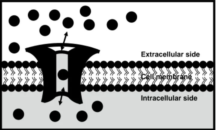
Ion channels are transmembrane proteins present in all living cells, from simple bacteria to highly specialized neurons, and these are essential for important physiological processes such as sensory transduction, action-potential generation, and muscle contraction. The channels act as a doorway that allow the passage of ions (e.g. K+, Na+, Cl–, and Ca2+) through the hydrophobic membrane. But they are not simple holes; ion channels are sophisticated machines that can conduct ions with exquisite specificity, at speeds close to the limit of diffusion (up to 100 million ions per second), under very tight regulation (Figure 1). Channels differ with respect to the ions they allow through, and the
way they regulate the flow of different ions.The physiological importance of the ion channels is reflected by their ubiquity and crucial role in numerous basic physiological processes (e.g. membrane potential, signal transduction, cell volume regulation, secretion, and absorption). Indeed, when their function goes errant, there can be serious consequences, including life-threatening diseases (e.g. cancer and neurodegenerative diseases). However, not much is known about the role of ion channels in many of such diseases. Nevertheless, ion channels are target to the drug design for the treatment of many non-curable chronic diseases like – Alzheimer’s disease, Parkinson’s disease, can-cer, epilepsy, and cystic fibrosis (Jalonen et al., 1997, Menzaghi et al., 1997, Fraser et al., 2000, Hubner and Jentsch, 2002, Pardo et al., 2005, Alarcon et al., 2006).
Figure 1. A general model of an ion channel
1.2. Regulation of membrane potential
The most abundant cellular ions are Na+, K+, and Cl–. They have significant effects in our normal physiology. If Na+ channels are open at neuronal resting membrane potential (–70 mV), Na+ ions will go into the cell until it reaches its equilibrium potential (+60 mV). In fact, the process in reality is more complicated – a positive feed back mechanism will generate an all-or-nothing action potential (Hodgkin and Huxley, 1952). Block of Na+ channels with different pharmacological or toxicological compounds, such as local anaesthetics and TTX (tetrodotoxin – from the puffer fish), prevent the entry of Na+ ions into the cell and prevent action potentials. In contrast to Na+ channels, opening of K+
Extracellular side
Intracellular side Cell membrane
channels lead to K+ ions efflux until the equilibrium potential (–90 mV) is reached. K+ channels can be blocked with different pharmacological or toxicological compounds, such as TEA (tetraethylammonium) and dendrotoxins, which can depolarized the cell and increase excitability. Likewise, if Cl– channels are open at neuronal resting membrane potential (e.g. GABAA receptors either with ethanol or general anaesthetics), Cl
–
ions will go out until it reach its equilibrium potential (–60 mV) and prevent neuronal activity. Cl– channels can be blocked with different pharmacological or toxicological compounds, such as DIDS (4,4´-diisothiocyanostilbene-2,2´-disulfonic acid) and NPPB (5-nitro-2-3-phenylpropylamino benzoic acid). During development, the intracellular Cl–ions concentration is very high (60 mM), but goes down in adult cells (15 mM) (Ben Ari et al., 1989). Consequently, the equilibrium potential of Cl– ions is also changed from –20 to – 60 mV, leading to different action of Cl– channel opening depending on the developmental stage of the cell.
2. Cell death
Cell death is an integral part of life. There are two major types of cell death: apoptosis, which is a regulated process that happens actively, and necrosis that is an accidental and less orderly process initiated by injurious stimuli (e.g. toxin, physical, and ischemia). Apoptosis is distinctly different from necrosis.
2.1. What is apoptosis? Why is it so important?
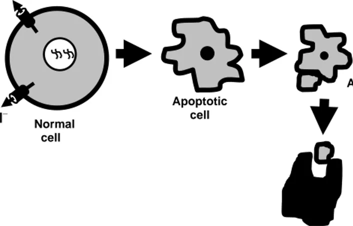
The word apoptosis, derived from the Greek word used for “falling off” – meaning fal-ling leaves from trees or petals from flowers, was first used to describe the new form of cell death distinct from necrosis in the early 1970s (Kerr et al., 1972). Apoptosis is de-fined by specific biochemical and morphological features, such as cell shrinkage (by ef-flux of ions), nuclear condensation, membrane blebbing, fragmentation into membrane-bound apoptotic bodies and eventually phagocytosis (Figure 2). Such stereotyped se-quences of changes allow the cell to die without adversely affecting its neighbors (Kerr et al., 1972). In contrast to apoptosis, necrosis is morphologically characterized by swelling of the cytoplasm and organelles, including mitochondria and eventually loss of cellular membrane integrity, resulting in cell lysis, and release of noxious cellular contents into the extracellular space with subsequent inflammation (Table 1).
Figure 2. A cartoon of apoptotic cell death
Apoptosis is not an incidental part of life, but a highly controlled, genetically pro-grammed, and medically important event. It is critical for the normal development and to maintain tissue homeostasis. In every adult human being about a hundred thousand cells are produced every second by mitosis and a similar number die by apoptosis. It is there-fore of vital importance that these events are tightly regulated. However, abnormal apop-tosis plays a significant role in the pathogenesis and progression of diseases. In a simpli-fied manner, increased apoptosis has been described in many diseases such as Alz-heimer’s, Parkinson’s, and AIDS (acquired immunodeficiency syndrome). In contrast, decreased apoptosis also cause many diseases such as cancer, viral infection (e.g. adeno and baculoviruses), and autoimmune diseases (e.g. myasthenia gravis and SLE – systemic lupus erythematosus). Apoptotic cell Apoptotic body Phagocytosis K+ Cl– Normal cell
Table 1. Key features distinguishing apoptosis from necrosis
Key features Apoptosis Necrosis
Cell volume Reduced Increased Plasma membrane Remains intact Disrupted
Nucleus Condensed Swelled and ruptured DNA breakdown Early, internucleosomal pattern Late, randomized Cellular energy Dependent Independent
Tissue reaction Apoptotic bodies, Cell lysis, inflammation
no inflammation, phagocytosis
2.2. Two major apoptotic pathways
The initiation and execution of apoptosis may occur via several alternative pathways. Among them the death receptor and the mitochondrial pathways are the two major ones. 2.2.1. The death-receptor pathway (extrinsic)
Death receptors exist in the plasma membrane of cells and initiate activation of caspases when triggered by their cognate ligand with consequent induction of apoptosis. Death receptors are members of the tumor necrosis factor (TNF) receptor superfamily, which possesses a so called death domain (DD). One of the best known death receptors is CD95/Fas/Apo1, which is a member of the death receptor super family (Trauth et al., 1989, Yonehara et al., 1989, Ashkenazi and Dixit, 1998, Hengartner, 2000). Binding of CD95 ligand (death ligand) to CD95 forms a death domain, which induces receptor clustering (Hengartner, 2000, Ashkenazi and Dixit, 1998). A Fas associated death domine (FADD) is recruited by an activated CD95 death domain, which in turn associates with pro-domains of caspase-8. This complex is referred to as the DISC (death-inducing signaling complex) (Budihardjo et al., 1999). Upon further clustering of procaspases-8 at the DISC, procaspase-8 is autocatalytically cleaved into active caspase-8 (caspase-8 activation can be blocked by recruitment of the degenerated caspase homologue c-FLIP) that further cleaves procaspase-3 to active caspase-3, and subsequent apoptotic cell death. Active caspase-8 can also cleave Bid to tBid (truncated Bid), which induces oligomerization of Bax, followed by the insertion into the mitochondrial membrane. However,both Bcl-2 and Bcl-XL can inhibit the Bax oligomarization (Figure 3).
2.2.2. The mitochondrial pathway (intrinsic)
The mitochondria is not only the cell’s power house but also the integrator of cell death pathways. Mitochondria sequester a potent cocktail of pro-apoptotic proteins. Most prominent among these is the cytochrome c. The release of apoptogenic factors from mitochondria is a crucial event which can lead to caspase activation. The precise mechanism behind the release of apoptogenic factors is unclear. However, mitochondrial homeostasis could be influenced by extracellular causes, internal insults such as DNA damage, or directly by the Bcl-2 family members, which cause modulation of proteins in the mitochondrial membrane (e.g. VDACmt – the voltage-dependent anion channel in the mitochondrial outer membrane) (Shimizu et al., 1999, Shimizu and Tsujimoto, 2000, Vander Heiden et al., 2001, Rostovtseva et al., 2004).
Cytochrome c Bcl-XL Bcl-2, Bax Caspase-8 Procaspase-3 Caspase-3
Apoptotic cell death Cell membrane Extracellular side Intracellular side Procaspase-8 CD95 FADD CD95L Apoptosome C-FLIP Bid DISC VDAC Procaspase-9 Caspase-9 Apaf-1 tBid
Figure 3. Schematic view of the biochemical process of apoptosis (the details are explained in the text)
VDACmt is a large channel to facilitate the release of apoptogenic factors, especially the cytochrome c from mitochondria to the cytosol, as it is a subunit of the mitochondrial permeability transition pore (PTP). Opening of the PTP quickly leads to cytochrome c
release into the cytosol (Petit et al., 1998, Gross et al., 1999, Shimizu et al., 1999, Shimizu et al., 2000, Godbole et al., 2003, Rostovtseva et al., 2004, Sade et al., 2004). Cytocrome c in the cytosol interacts with Apaf-1 (apoptotic protease-activating factor-1) via a specific CARD (caspase recruitment domain). CARD also recruits procaspase-9 to form a complex known as apoptosome (Li et al., 1998). Within this complex caspase-9 is autoactivated and initiates activation of caspase-3. Eventually, that causes apoptotic cell death (Figure 3).
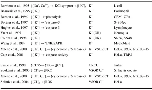
3. The role of ion channels in apoptosis: Molecules for life, disease, and death The plasma membrane’s ion channels play a critical role in apoptotic cell death, including neuronal apoptosis (Heidenreich, 2003). The earliest striking characteristic of apoptosis is cell shrinkage, which is associated with an increased efflux of K+ and Cl– ions (Barbiero et al., 1995, Beauvais et al., 1995, Benson et al., 1996, Bortner and Cidlowski, 1996, Bortner et al., 1997, Yu et al., 1997, Colom et al., 1998, Wang et al., 1999, Maeno et al., 2000, Souktani et al., 2000) (Table 2). Altered intracellular ionic concentrations pro-foundly influences the alteration of protein structure (e.g. cytocrome c, Apaf-1 – apoptotic protease-activating factor-1,TK – tyrosine kinase, and PKC – protein kinase C) and the activation of different intracellular enzymes (e.g. proteases and nucleases) (Strickland et al., 1991, Wondrak et al., 1991, Polgar and Patthy, 1992, Di Paolo et al., 1995, Polgar, 1995, Vukelic et al., 1995, Adamska et al., 1996, Hughes et al., 1997) (Ta-ble 2). K+ ions inhibit the activation of caspases by abrogating Apaf-1 oligomerization and apoptosome formation, suggesting a key role of normal intracellular concentration of K+ ions for the cellular survival (Cain et al., 2001). Furthermore, stimulation of CD95 (Fas/Apo1) receptor has been shown to increase the efflux of Cl– ions by triggering the activation of outwardly rectifying chloride channels (ORCC) (Lepple-Wienhues et al., 1998). Thus, the efflux of K+ and Cl– ions are fundamental for the apoptotic cell shrink-age and subsequent apoptotic cell death. However, still, very little is known about the mechanism of cell volume loss and the relation of the ionic efflux with the death machin-ery during apoptosis. This thesis work aims to investigate the role and the relation of ion channels with the death machinery during the apoptotic process.
Table 2. Studies discussing the involvement of plasma membrane’s ion channels in apoptosis
Study Mechanism Channel Cell type Barbiero et al., 1995 ↑[Na+, Ca2+]i →↑KCl symport→↓[ K+]i K+ L cell Beauvais et al., 1995 ↓[ K+]i K+ Eosinophil Benson et al., 1996 ↓[ K+]i →↑proteolysis K+ CEM–C7A Bortner et al., 1997 ↓[ K+]i →↑caspase-3 K+ S49 Neo Hughes et al., 1997 ↓[ K+]i →↑caspase-3 K+ Lymphocyte Yu et al., 1997 ↓[ K+]i K+(OR) Neuroglia Colom et al., 1998 ↓[ K+]i K+(DR) SN56, SN48 Wang et al., 1999 ↓[ K+]i →↑JNK/SAPK K+ Myeloblast
Maeno et al., 2000 ↓[ K+, Cl–]i →↑cytocrome c,↑caspase-3 K+, VSOR Cl– HeLa, U937, NG108–15 Cain et al., 2001 ↓[ K+]i →↑caspase activity K+ HeLa, TRP.1
Szabo et al., 1998 ↑CD95→↑TK→↓[Cl–]i ORCC Jurkat
Souktani et al., 2000 ↓[Cl–]i→↓PKC VSOR Cl– X. laevis oocyte Maeno et al., 2000 ↓[ K+, Cl–]i →↑cytocrome c,↑caspase-3 K+, VSOR Cl– HeLa, U937, NG108–15 Shimizu et al., 2004 ↓[Cl–]i→↑ROS VSOR Cl– HeLa
OR, outward rectifier; DR, delayed rectifier; ORCC, outwardly rectifying chloride channel; VSOR Cl–, volume-sensitive outwardly rectifying Cl– channel; TK, tyrosine kinase; PKC, protein kinase C; CD95, cell death 95; JNK/SAPK, jun N-terminal kinase/stress-activated protein kinase; ROS, reactive oxygen species.
AIMS
The general aim of this thesis work was to characterize the electrophysiological properties of apoptotic neurons. The specific aims were to study:
1) The electrophysiological properties of differentiated apoptotic neuronal cell lines and primary hippocampal neurons undergoing apoptosis (paper I and II).
2) The electrophysiological properties of undifferentiated apoptotic primary cortical neural stem cells undergoing apoptosis (paper II)
3) The biophysical and pharmacological properties of the apoptosis-inducing plasma membrane voltage-dependent anion channel (VDACpl) (paper III and IV). Aims
MATERIALS AND METHODS 1. Cell culture
Animals were handled according to Karolinska Institutet’s and Linköpings Universitet’s guidelines and experiments were performed with permission from Stockholm’s “Norra djurförsöksetiska nämnd” and from “Linköpings djurförsöksetiska nämnd”, local ethical committee.
1.1. HT22 and SK-N-MC cell lines (Paper I, III, and IV)
The cells were routinely seeded at a density of 3 000 and 40 000 cells/cm2,respectively, in CO2-independent medium (Invitrogen, LifeTechnologies/Gibco BRL, Grand Island, NY, USA, catalogue number: 18045-054) supplemented with 10% fetal calf serum (FCS), 4 mM L-glutamine, 100 U/ml penicillin and 100 µg/ml streptomycin (Dare et al., 2002, Tofighi et al., 2006). The cell culture flasks were locked and kept in the incubator at 37°C with proper humidity for 24 h before exposure to the apoptotic stimuli. All chemicals for cell culture were supplied by Life Technologies (Invitrogen, LifeTechnolo-gies/Gibco BRL, Grand Island, NY, USA).
1.2. Primary hippocampal neurons (Paper II)
Sprague-Dawley rats (B&K Universal AB, Sollentuna, Sweden) were kept under stan-dard laboratory conditions, were sacrificed on the 18th gestational day using carbon diox-ide, and the hippocampi dissected from the foetuses. Briefly, the cultures were prepared as follow: the dissected hippocampi were incubated at 37°C for 15 min in 0.1% trypsin (Invitrogen, LifeTechnologies/Gibco BRL, Grand Island, NY, USA) diluted in Ca2+ and Mg2+ free Hank’s Balanced Salt Solution (pH 7.3) and subsequently triturated through a narrowed pasteur pipette. Cell suspensions were then seeded into 35 mm tissue culture dishes (Corning, New York, NY, USA) at a cell density of 0.17 × 10⁵ cells/cm2. Prior to seeding the dishes were coated with 0.1 mg/ml poly-L-lysine hydrobromide (MW 3-7 × 10⁴; Sigma, Chemical Co., St. Louis, MO, USA) and subsequently washed twice in dis-tilled water. The cells were grown in 2 ml Neurobasal medium supplemented with B27, 1:50, (NB B27), 15 μg/ml gentamicin and 2 mM L-glutamine (all from Invitrogen, LifeTechnologies/Gibco BRL, Grand Island, NY, USA). The cultures were maintained in
an incubator providing 5% CO2at 37°C. The growth medium was not changed and no re-feeding was done during the experimental period.
1.3. Primary cortical neural stem cells (Paper II)
Stem cells were obtained from embryonic cortices dissected in Hanks' Balanced Salt So-lution (HBSS) (Invitrogen, LifeTechnologies/Gibco BRL, Grand Island, NY, USA) from timed-pregnant Sprague-Dawley rats (B & K, Sollentuna, Sweden) at E15 (E1 was de-fined as the day of copulatory plug) (Tamm et al., 2006). The tissue was gently mechani-callydispersed; meninges and larger cell clumps were allowed to sediment for 10 min. The cells were plated at a density of 0.6 × 10⁶ cells per 100 mm cell culture dish pre-coated with poly-L-ornithine and fibronectin (both from Sigma, Chemical Co., St. Louis, MO, USA). Cells were maintained in enriched N2 medium (Bottenstein and Sato, 1979) with 10 ng/ml basic fibroblast growth factor (bFGF) (R & D Systems, Minneapolis, MN, USA) added every 24 h and the medium changed every other day to keep cells in an un-differentiated and proliferative state. When still subconfluent, cells were passaged by de-taching by incubation with HBSS and subsequent scraping. Afterwards, the cells were gently mixed in N2 medium, counted, and plated at the desired density. The cells were used for experiments 48 h after the first passage.
2. Induction and prevention of apoptosis (Paper I, II, III and IV)
To induce apoptosis, cells were exposed to 1 μM of the pan-caspase inhibitor, stauro-sporine (STS) (Gorman et al., 2000), 30 μM 2,3-dimethoxy-1,4-naphthoquinone, 4 μM methylmercury, or 0.3 mM styrene 7,8-oxide (Dare et al., 2002), for 1.5 to 6.5 h. To pre-vent apoptosis, cells were pre-incubated with anti-VDAC antibodies - Ab25 (1:200), Ab20 (1:200), (Shimizu et al., 2001), and anti-Porin 31 HL Ab-2 (1:100) (Calbiochem, Darmstadt, Germany), or the pan-caspase inhibitor zVADfmk (20 μM) (Peptide Institute, Osaka, Japan). As a negative control we used an unrelated antibody the Neurofilament (DSHB, Iowa, USA). In some experiments, cells were pre-incubated with sucrose (240 mM), which prevented apoptosis.
3. Evaluation of apoptotic cells (Paper I, II)
The occurrence of apoptosis was evaluated on fixed or living cells. Cells grown on cover-slips, were fixed with ice-cold methanol/water (8/2 = v/v) and stained with
impermeable propidium iodide (PI) or cell-permeable Hoechst 33358 to visualize nuclear condensation. Apoptotic cells were identified by the irregular shape of the cell, the smaller size of the nucleus, and the brighter intensity of the stained chromatin. For vital stainings, cells grown on coverslips and incubated with a solution of Annexin V-FITC (0.5 µg/ml), which binds to phosphatidylserine (PS), PI (1 µg/ml) and cell-permeable Hoechst 33358 (1 µg/ml) in a buffer containing 10 mM HEPES/NaOH pH 7.4, 140 mM NaCl, 2.5 mM CaCl2. Cells were analyzed with an Olympus BX60 fluorescence micro-scope (Olympus, Tokyo, Japan) equipped with a C4742-95-10sc digital camera (Hamamatsu Photomics Norden AB).
4. Electrophysiology (Paper I, II, III, and IV) 4.1. The patch-clamp method
The electrophysiological properties of ion channels were studied with the patch-clamp technique based on the voltage-clamp method (Cole, 1949, Hodgkin et al., 1952). The patch-clamp technique is a highly sensitive voltage-clamp method, which permits the measurement of ionic currents passing through a single ion channel, while a small part of the membrane is tightly sealed against the electrode (Neher and Sakmann, 1976).
The glass pipette touches the cell membrane by a downward movement of the pipette using the micromanipulator (Figure 4a). In this position, a mild suction can cause the cell to form a tight seal (GΩ) with the pipette (Figure 4b). This configuration is called cell-attached and is used to study single-channel currents under voltage-clamp conditions. From the cell-attached configuration, we can alter the recording configuration to either whole-cell or inside-out patch type. The whole-cell configuration is obtained when a strong suction removes a piece of the cell membrane (Figure 4c) and can be used to measure the whole-cell current. However, if the electrode is quickly retracted from the cell surface by the micromanipulator, a piece of the membrane is excised and we obtained an inside-out patch configuration (Figure 4d). In this configuration, the inside of the membrane is exposed to the bath solution, giving us the possibility to change the bath solutions if necessary (Table 3) (e.g. for the ion selectivity and the block of channel) and study the single channel current.
Figure 4. The patch-clamp method
4.2. Electrodes, amplifiers, softwares, and solutions
The patch pipettes were made of borosilicate glass. The diameter of the tip of the patch pipette was about 1 μm and the resistance was 4-6 MΩ with the solutions I used. A silver wire coated with silver chloride connected the solution in the electrode with the recording equipment. Both in the cell-attached and in the inside-out recordings, the pipettes were filled with the extracellular solution and the extracellular solution was also used in the bath (Table 3). To perform the electrophysiological investigations an EPC-7 (HEKA Electronics, Lambrecht/Pfalz, Germany), or an Axopatch 200B (Axon Instruments, Fos-ter City, CA, USA) patch-clamp amplifier, and pClamp softwares (Axon Instruments, Foster City, CA, USA) were used.
c b a d Whole-cell Inside-out Mild suction Tight contact between pipette and membrane Cell-attached Excision Strong suction Air Water
Table 3. Composition of solutions
Solutions K+ Na+ Cl– Ca2+ Mg2+ Hepes Sucrose EGTA (mM) (mM) (mM) (mM) (mM) (mM) (mM) (mM) E.C.S. 5 140 150.6 1.8 1 10 23 – I.C.S. 140 4 147 0.5 1 10 – 5 Sucrose, 300 mM 5 1.5 12.1 1.8 1 10 300 – Sucrose, 244.6 mM 1 28 30.1 0.36 0.2 2 244.6 – Sucrose, 200 mM 5 51.5 62.1 1.8 1 10 200 – Sucrose, 100 mM 5 101.5 112.1 1.8 1 10 100 – Sucrose, 50 mM 5 126.5 137.1 1.8 1 10 50 – Sucrose, 0 mM 5 151.5 162.1 1.8 1 10 0 –
E.C.S., extra cellular solution; I.C.S., intracellular solution.
4.3 The voltage protocol
To perform the patch-clamp recordings (cell-attached and excised), I used a voltage-clamp protocal of 11 pulses from +100 to –100 mV separated by 20 mV, while the holding voltage was 0 mV (VH = 0 mV). Each pulse is 100 ms long and between the pulses the membrane voltage was kept at 0 mV for 0.4 s. The leakage currents and the capacitive currents were corrected proportionally for the whole family or by subtraction of corresponding traces with no channel activity. The current was always denoted as posi-tive for currents from the intracellular side towards the extracellular pipette side.
4.4 Equations
To calculate the conductance, we used:
G = I/V, (1) where G is conductance, I is current, and V is voltage.
For the analysis of the selectivity, we used:
where (ΔVrev) is the shift of the reversal potential. R, T, and F have their normal thermo-dynamic significances, z is the valence of the ion X, and where [X] is the concentrations of the ion X in different solutions.
For the single-channel current amplitude histograms we used one to three Gaussian curves:
N = A exp(−0.5((i-imean)/s)
2) / (s(2π)0.5
), (3) where N is the number of events, A is the area of the curve, i is the single-channel current,
imean is the mean current, and s is the standard deviation.
To calculate the current (IS) through a channel depending on the voltage (V) and the extra ([S]o) and intracellular concentrations ([S]i) of the ion S, we used the Goldman-Hodgkin-Katz equation (Goldman, 1943, Hodgkin and Goldman-Hodgkin-Katz, 1949, Hille, 2001):
IS = PS zS2 VF2R-1T-1 ([S]i - [S]o exp(-zSFVR-1T-1)) / (1-exp(-zSFVR-1T-1)), (4)
where PS is the permeability of the channel and zS is the valence of the ion S.
5. Immunoblotting and immunocytochemistry (Paper I and II)
To monitor the release of mitochondrial cytochrome c into the cytosol, the cytosolic frac-tions from control and exposed cells were separated from the mitochondria (Robertson et al., 2002). Cytochrome c was detected by immunoblotting with a primary mouse anti-body (1:2500, BD-Pharmingen, San Diego,CA) and with a goat anti mouse secondary an-tibody, horseradish peroxidase (dilution 1:20000, Pierce Rockford, IL, USA), conjugated according to methods previously described (Robertson et al., 2002). Immunoblot bands were quantified with a LKB Ultrascan XL laser densitometer.
5.1. Detection of VDACs in paraformaldehyde fixated cells (Paper I)
Immunocytochemistry was performed on unfixed or fixed cells (4 % paraformaldehyde). In order to prevent endocytosis of any added antibodies, living cells were blocked with BSA-PBS for 5 min at 4°C. Fixed or unfixed cells were then incubated overnight at 4°C with two different anti-VDAC antibodies, one raised in rabbit (Ab25) (1:200) (Shimizu et
al., 2001) and the other one in mouse (anti-Porin 31 HL Ab-2) (1:100) (Calbiochem, Darmstadt, Germany). After several washes with PBS, fluorescein isothiocyanate (FITC)-conjugated goat anti-rabbit or donkey anti-mouse (Jackson, GTF, Sweden) antibodies were added as secondary antibodies for 30 min at 4°C. For control purpose, cells were also incubated with the secondary antibodies alone. In some experiments, living cells were pre-incubated with the MitoTracker Red (100 nM) (Invitrogen, Molecular Probes, Grand Island, NY, USA) for 30 min, fixed and stained as above. Stained cells were ana-lyzed with a fluorescence microscope and images captured as described above, or with a confocal microscope BioRad Radiance Plus.
5.2. Detection of VDACs in glutaraldehyde fixated cells (Paper II)
Cells were fixed under unpermeabilized conditions in PBS (pH 7.4), containing 2% para-formaldehyde, 1% glutaraldehyde, and 120 mM sucrose, these are known to preserve plasma membrane integrity and thereby avoid intracellular antibody leakage. Unperme-abilized cells were washed in PBS and incubated with 50 mM ammonium chloride for 1 h at room temperature to reduce the generation of free aldehyde groups. Fixed cells were then washed again in PBS and incubated overnight at 4°C with anti-VDAC antibodies, raised in mouse (anti-Porin 31 HL Ab-2) (1:100 in PBS and supplemented with 0.5% BSA). After several washes with PBS, fluorescein isothiocyanate (FITC)-conjugated goat anti-mouse antibody (Alexa Fluor 488, Invitrogen, Molecular Probes, Grand Island, NY, USA) (1:200) was added as secondary antibody for 1 h at room temperature. Cells were further incubated with Hoechst 33358 for 5 min before mounting with the coverslips onto glass slides with PBS/glycerol (1/9 = v/v) containing 0.1% (w/v) phenylendiamine. For control purpose, cells were also incubated with the secondary antibody alone. Stained cells were analyzed with Zeiss LSM 510 Meta confocal microscope (Zeiss, Jena, Ger-many).
6. Quantification of intracellular molecules (Paper I and II)
6.1. ATP determination (Paper I)
ATP concentrations were determined in a luminometric assay using the ATP dependency of the light-emitting luciferase-catalyzed oxidation of luciferin (Boehringer Mannheim, Mannheim, Germany) according to the manufacturer’s protocol. Briefly, cells (5.0×10⁵) were resuspended in 50 µl PBS and 450 µl of boiling lysing buffer (100 mM Tris, 4 mM
EDTA, pH 7.75). Samples were incubated for another 2 min at 100°C and 100 µl were taken out to a 96-well plate. Prior to measurement, 100 µl of luciferase were added to each well and the plate was analyzed in a luminometer (Berthold, R-Biopharm AG, Ger-many).
6.2. NADH-ferricyanide reductase activity (Paper I and II)
Cells (1.5 – 4x10⁶) were harvested and incubated in 1 ml buffer, containing 50 mM Tris-HCl, pH = 8.0 and 250 µM β-NADH for 5 min at 37°C. The reaction was started by addi-tion of 250 µM potassium ferricyanide (Sigma, Chemical Co., St. Louis, MO, USA) to the reaction buffer leading to reduction of ferricyanide to ferrocyanide. After 10 min, cells were spun down and the concentration of remaining ferricyanide was assessed, us-ing a UNICAM 5625 spectrophotometer at 420 nm. Ferricyanide reductase activity was calculated as nmol ferricyanide reduced per min per 106 cells.
RESULTS
The results are presented in detail in the published papers and the manuscripts, appended with this thesis. Here, I will present an overview of the results.
1. Activation of plasma membrane ion channels during apoptosis (Paper I and II) To study electrophysiological changes during apoptosis, I investigated neuronal cell lines (the mouse hippocampal HT22 and the human neuroblastoma SK-N-MC cells), primary hippocampal neurons, and primary cortical neural stem cells (cNSCs) with the patch-clamp technique. All cell types displayed a low electrical activity under control condi-tions in cell-attached, isolated membrane patches as well as in whole-cell recordings. However, during apoptosis, we found a K+ current of delayed-rectifier type in whole-cell recordings and a large Cl– selective single-channel current in excised inside-out mem-brane patches of neurons. The K+ current was voltage-dependent (opened at more posi-tive voltages than –20 mV) and blocked by tetraethylammonium (TEA). The activation of K+ currents during apoptosis has been reported by many investigators (Table 2) and will not be studied further here. However, the activation of the large anion current during apoptosis seems to be a novel finding, and therefore, this channel will be in focus in the present thesis work.
2. The large anion selective single-channel current is the voltage-dependent anion channel (VDAC) current (Paper I, II, III, and IV)
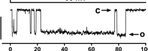
The activation of the large single-channel current was significant in excised inside-out membrane patches from apoptotic cells (from 4 % to 48 % of investigated membrane patches in control and STS respectively) (Figure 5).
Figure 5. A typical large square-like current trace at –60 mV, O denotes open and C denotes closed states
0 20 40 60 80 100 0 mV -60 mV 20 p A o
c
Time (ms)To investigate the identity of this current (Figure 5), we performed more extensive elec-trophysiological and immunocytochemical experiments.
2.1. Electrophysiology of the open probability, the conductance, and the selectivity
The channel was voltage dependent and opened over a relatively narrow membrane po-tential range (+20 mV to –20 mV) and closed rapidly at more positive and more negative membrane potentials than +20 mV and –20 mV respectively. Thus, the open probability curve of the channel is bell-shaped in relation to voltage, with the maximum open prob-ability at 0 mV. The conductance of the channel is about 400 pS in a physiological solu-tion (140 mM NaCl). To characterize the selectivity of the channel, we diluted the bath solution (NaCl) to 1/5 of its original concentration (140 mM NaCl). This decreased the current from the pipette solution (extracellular) to the bath solution (intracellular), while leaving the current in the opposite direction essentially unchanged. The reversal potential was shifted from 0 to –41 ± 6 mV. This suggests that the large-conductance channel is predominantly Cl– selective. All our electrophysiological findings so far (large-conductance about 400 pS, the bell-shaped open probability, and Cl– ion selectivity) sug-gest that the large-conductance channel in the plasma membrane of the apoptotic neuron is similar to the voltage-dependent anion channel (VDAC) normally found in the mito-chondrial outer membrane, where it is involved in certain forms of apoptotic cell death.
2.2. Immunocytochemistry
To further establish the identity of the large-conductance channel, we performed experi-ments with different anti-VDAC antibodies recognizing different epitopes. Antibodies showed VDAC-like immunoreactivity in the plasma membrane (thus VDACpl). Using the same antibodies on fixed cells preincubated with MitoTracker Red, we found dot-like cytoplasmic VDAC immunoreactivity localized in mitochondria (thus VDACmt), sug-gesting the same identity of the channels. Furthermore, VDACpls were detected in both control and apoptotic cells, suggesting that the channels are constitutively present in the plasma membrane, but activated only during apoptosis.
Taken together, the electrophysiological and immunocytochemical data suggest that the large-conductance channel is VDACpl, normally found in the mitochondrial outer mem-brane (VDACmt). However, whether or not VDACpl and VDACmt are identical has been debated (Yu and Forte, 1996, Thinnes and Reymann, 1997, Bathori et al., 2000). To Results
resolve this problem, we performed a detailed biophysical analysis of our VDACpls dur-ing apoptosis in Paper III with other VDACpls (reported by other investigators) and the VDACmts.
3. A deeper biophysical analysis of VDACpl (Paper III)
VDAC or VDAC-like channels in the plasma membrane have been reported sporadically in several tissues (Table 2 in Paper III). Whether or not VDACpl and VDACmt are simi-lar channels is heatly debated. To csimi-larify this, we performed a detailed quantitative bio-physical analysis of our apoptosis-inducing VDACpl, and found a strong correlation with data from VDACmts (Colombini et al., 1996), and the other VDACpls reported by other investigators (Yu and Forte, 1996, Thinnes et al., 1988, Bathori et al., 2000,), suggesting the same identity of the channel.
3.1. Main and subconductances of VDACpl versus VDACmt
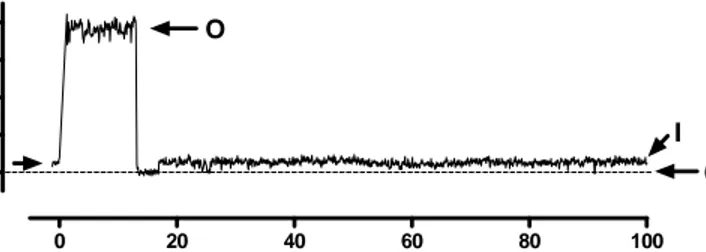
The conductance of the apoptosis-inducing VDACpl is about 400 pS in physiological so-lutions (140 mM NaCl), which is close to what has been reported for other VDACpls in similar solutions. The VDACmt is in most cases studied in lipid bilayers with bath solu-tions of 1 M KCl, resulting in a very high conductance (about 4 nS). However, if the VDACpl and the VDACmt are studied in the same solution, they have the same conduc-tance. Besides the main-conductance level (400 pS), we have also found different sub-conductance levels (220 and 28 pS) (Figure 6). Because of their rare existence, it was dif-ficult to get detailed information about the kinetics, selectivity, and pharmacological properties of those sub-conductance levels. The 220 pS level was more prevalent shortly after excision, when the channel is transformed from a closed state to a fully mature open state. Other reports on the VDACpl also show a similar sub-conductance state at either positive or negative voltages. The 28 pS sub-conductance state was seen after the main-conductance state at the most positive and most negative voltages and was linked to inac-tivation of the VDACpl. Thus, inacinac-tivation coincides with the low sub-conductance cur-rent. The VDACmt also has sub-conductance states that are seen in the 1 M KCl solu-tions in the lipid bilayers at either positive or negative voltages and they are called lower-conducting or closed states (Colombini et al., 1996).
Figure 6. The inactivated state of the VDACpl (the low sub-conductance current), just after a brief opening of VDACpl from a cell attached patch. The potential (p) is here defined as bath potential minus pipette po-tential. There is a small steady current in the beginning of the pulse at 0 mV. O denotes open, C denotes close, and I denotes inactivated state of the VDACpl.
3.2. The open probability of VDACpl versus VDACmt
VDACmt is open at membrane potentials close to zero, and closes rapidly at voltages above +20 mV and below –20 mV respectively. That is, the open probability curve is bell shaped in relation to voltage, which is similar to that of our apoptosis-inducing VDACpl as well as to that of other VDACpls have been reported.
3.3. The selectivity of VDACpl versus VDACmt
In physiological solution (140 mM NaCl) our apoptosis-inducing VDACpl is more per-meable to anions than cations (>5:1), which has also been reported for other VDACpls and VDACmts in similar solutions (Schein et al., 1976, Colombini et al., 1996). How-ever, the selectivity for VDACmt in 1 M KCl is relatively less for Cl– ions; anion versus cation permeability is about 2:1. The selectivity depends on ionic strength. Thus, the VDACpl and the VDACmt both are highly anion selective in physiological solutions. 4. Pharmacology of VDACpl (Paper I, III, and IV)
To further ascertain the identity of the apoptosis-inducing VDACpl, we investigated the effect of several physiological intracellular molecules (ATP, cAMP, Ca2+) and non- physiological molecules (Gd3+, citrate3–, sucrose). We performed inside-out patch-clamp experiments, while the intracellular side perfused with 4 mM ATP, 100µM cAMP, 500
0 20 40 60 80 100 0 10 20 30 40 O C I +100 mV (p) C u rr e n t (p A ) Results
µM Ca2+, 30 µM Gd3+, 46.7 mM citrate3–, and sucrose (in concentrations from 0 to 300 mM; Table 3)
4.1. The effect of physiological intracellular molecules on VDACpl (Paper I)
We measured the ATP concentration of cells exposed to staurosporine (STS) and found a significant decrease in the ATP level in apoptotic neurons. This declined ATP level is compatible with apoptotic cell death (Leist et al., 1997). To investigate the hypothesis that declined ATP level could activate VDACpl, we perfused the intracellular side of an excised inside-out membrane patch and found that 4 mM ATP reversibly blocks the channel activity, while cAMP and Ca2+ ions had no effect. Thus, probably the loss of ATP could be a triggering event of VDACpl during apoptosis.
4.2. The effect of non physiological molecules on VDACpl (Paper I, III, and IV)
It has been reported that lanthanides such as the trivalent La3+ completely blocks the cur-rent of VDACmt (Gincel et al., 2001). We found that 30 µM the trivalent lanthanide Gd3+ blocked the current of apoptosis-inducing VDACpl almost completely, reversibly, and voltage independently when applied from the intracellular side of an excised inside-out membrane patch. We also found that the trivalent citrate3– ion in the bath solution in-stead of the Cl– ion reduced the current but did not eliminate it, suggesting that the citrate ion can pass through the channel. Therefore, the ion-conducting pore must be wider than the citrate3– ion (>7-8 Å). Taken together, our findings and other reports suggest that the VDACpl and the VDACmt is the same channel.
Interestingly, we found that 240 mM sucrose added to the diluted 1/5 NaCl solution (to maintain the osmolarity of the solution) blocked the single-channel current by 90%, without affecting the reversal potential. In line with the hypothesis that the blocking of VDACpls with sucrose also can prevent apoptosis, we incubated the cells with the same concentration of sucrose 30 min prior to STS exposure and found a significant decrease in the number of apoptotic cells. This last finding that sucrose can block VDACpl sug-gests a simple and cheap tool to regulate apoptosis. However, the blocking mechanism and concentration dependence was not known. It has also been reported in a few studies that glucose has a role during apoptosis and cellular redox regulation (Sagone et al., 1983, Olejnicka et al., 1997). In the biophysical study of VDACpl (Paper III), we found that the VDACpl channel exists in several closed and inactivated conformations. The
in-activated state was surprisingly found to be conducting. The conductance was 7% of the main conductance level. This suggested that the sucrose-blocked channel might be an in-activated state of VDACpl.
To investigate the mechanism of sucrose block we studied the single-channel conduc-tance and the open probability of VDACpl for different concentrations of sucrose be-tween 0 and 300 mM (Table 3). The single channel current reduction had a Kd value close to 300 mM. In contrast the open probability was more affected with a Kd value close to 50 mM. Because, its relatively long recovery time to high open probability after sucrose exposure we suggest that the low open probability caused by sucrose is due to in-activation of VDACpl. Thus, the 90% reduction we reported in Paper I is probably be-cause of a sucrose-induced inactivation of the channel but not a pore block.
5. VDACpl plays a key role in the apoptotic process (Papers I and II)
To investigate the functional role of the finding of VDAC-like immunoreactivity in the plasma membrane of apoptotic cells, we performed experiments to occlude the channel with different VDAC blockers and other agents. Pre-incubation of cells with anti-VDAC antibodies or sucrose (240 mM) for 30 min prior to STS exposure, blocked plasma mem-brane VDAC activation and drastically reduced the number of apoptotic cells, suggesting an essential and a critical role of the plasma membrane VDAC during apoptosis.
5.1. VDACpl plays a crucial role in the early stage of apoptosis (paper I and II)
Whether or not the activation of the VDACpls is an early or a late stage event of the apoptotic process was not clear to us. To ascertain this, we performed electrophysiologi-cal investigations with the cells exposed to the pan-caspase inhibitor zVAD-fmk 30 min prior to STS. However, pre-treatment with the caspase inhibitor did not prevent STS-induced VDACpl current, suggesting that the VDACpls play a critical role during the early stages of apoptosis.
5.2. VDACpl plays a dual role (paper I and II)
It has been reported that the VDAC1 protein in the plasma membrane of control cells work as NADH (–ferricyanide) reductase (Baker et al., 2004), which is involved in transmembranous redox regulation. To investigate this hypothesis, we measured NADH Results
(–ferricyanide) reductase activity in control and in STS treated cells. Interestingly, we found a time-dependent increase in NADH (–ferricyanide) reductase activity in cells ex-posed to 1 µM STS. Increased NADH (–ferricyanide) reductase activity was inhibited by preincubation of cells with anti-VDAC antibodies 30 min prior to exposure to STS. Thus, it appears that both the channel formation and the NADH (–ferricyanide) reductase activ-ity of the VDAC protein are stimulated in apoptotic cells, and both activities can be pre-vented by anti-VDAC antibodies.
5.3. The role of VDAC differs during development (Paper II)
Whether or not VDAC plays a critical role in the neuronal plasma membrane at differen-tial stages of its development is not clear. To investigate this, we studied the rat primary hippocampal neurons and the primary cortical neural stem cells (cNSCs) from rat during apoptosis with immunocytochemical and electrophysiological techniques. We found VDACpl in hippocampal neurons, where it showed similar characteristics to what we found in Paper I. To our surprise, VDACpl was only occasionally present in the cNSCs (3 in 179 cells) with no correlation to the apoptotic process and with an atypical character (sigmoidal instead of bell-shaped open probability curve). Instead of VDACpl, we found a significant increase (from 16 to 44% of excised membrane patches) of a voltage inde-pendent 37 pS conductance channel in apoptotic cNSCs (Figure 7), suggesting different apoptotic strategies for undifferentiated and differentiated neurons. In line with these electrophysiological findings, we found an increased activity of NADH (–ferricyanide) reductase in the primary hippocampal neurons but not in the primary cNSCs during apop-tosis. Anti-VDAC antibodies prevented apoptosis in the differentiated hippocampal neu-rons but not in the undifferentiated cortical neural stem cells (cNSCs). Therefore, the ex-pression of the VDAC protein in the plasma membrane is probably a matter of a devel-opmental process, which depends on the maturity of the channel protein from the undif-ferentiated to the difundif-ferentiated stage.
Figure 7. The 37 pS voltage-independent channel from an inside-out membrane patch, at +100 mV, VH = 0, typical for cNSCs apoptosis. 0, 1, and 2 are the number of open channels respectively.
To summarize the experimental data, we have shown the activation of VDACpl in differ-ent cell lines and in primary hippocampal neurons during apoptosis. The immunocyto-chemical experiments tell us that VDAC is constitutively present in the plasma mem-brane of control cells, where it works as an enzyme (the NADH–ferricyanide reductase) and it becomes activated during apoptosis and acts as an ion channel. We have also found an increased activity of NADH (–ferricyanide) reductase during apoptosis. Anti-VDAC antibodies or sucrose blocks VDACpl and prevents NADH (–ferricyanide) reductase ac-tivity and reduced the number of apoptotic cells significantly. Therefore, the activation of VDACpl is an inevitable phenomenon during the early stage of neuronal apoptosis. To our surprise, we found very few and atypical VDAC in the plasma membrane of the neu-ral stem cells in our electrophysiological experiments. Instead, we found another channel which is activated significantly during apoptosis. Probably, this channel is playing the same role during apoptosis in undifferentiated neural stem cell as VDACpl is playing in differentiated neurons. 0 1 2
0
mV
+100 mV
5 p
A
20 ms
ResultsDISCUSSION
The major finding in my thesis work is that the voltage-dependent anion channel in the plasma membrane (VDACpl) is activated during neuronal apoptosis and that this has a critical role for the apoptotic process. VDAC is normally found in the mitochondrial outer membrane, where it is involved in apoptotic cell death (Shimizu et al., 2001). 1. The voltage-dependent anion channel (VDAC)
One of the first ion channels recorded at the single-channel level is the voltage-dependent anion channel in the outer mitochondrial membrane (VDACmt) (Schein et al., 1976, Co-lombini et al., 1996). This channel is open at membrane potentials close to zero, and closed at voltages above +20 mV and below –20 mV respectively. That is, the open prob-ability curve is bell shaped in relation to voltage. Furthermore, in a physiological solution (140 mM NaCl) the channel is more permeable to anions than cations (5:1) (Schein et al., 1976, Colombini, 1989, Colombini et al., 1996). VDACmt is involved in the early stages of certain forms of apoptotic cell death (Shimizu et al., 2001, Godbole et al., 2003, Zheng et al., 2004).
1.1. The VDAC family
In mammals, three VDAC isoforms (VDAC1, VDAC2, and VDAC3) have been identi-fied by cDNA cloning and sequencing (Reymann et al., 1995, Sampson et al., 1997, Xu et al., 1999, Cesar Mde and Wilson, 2004). VDAC1 and VDAC2 have been identified in human (HVDAC1, and HVDAC2) (Blachly-Dyson et al., 1993). All three mammalian isoforms are present in the mitochondrial outer membrane – VDAC1 is most abundant representing 5% of total protein content (Bathori et al., 2000). Both VDAC1 and VDAC2 have pore-forming characteristics, while VDAC3 plays a major physiological role by regulating the functions of other proteins (Sampson et al., 1998). Furthermore, phyloge-netic analysis indicates that VDAC3 is the primordial VDAC gene in mammals, suggest-ing that other isoforms (VDAC1 and VDAC2) are derived from gene duplication and di-vergence events of VDAC3 (Sampson et al., 1996). Only the VDAC1 is present in the plasma membrane (Thinnes et al., 1989, Dermietzel et al., 1994, Jakob et al., 1995, Bathori et al., 1999, Buettner et al., 2000, 2006). Hence, when it is present in the plasma membrane, we call it VDACpl. However, if VDAC exists in the plasma membrane and if
VDACpl is identical to VDACmt is debated (Yu and Forte, 1996, Thinnes and Reymann, 1997, Bathori et al., 2000).
1.2. The VDAC structure and the size of the pore
The wild-type single VDAC channel protein is formed by a roughly 30-kDa polypeptide of about 285 amino acid residues (Mannella, 1987, Thomas et al., 1991, Peng et al., 1992, Colombini et al., 1996, Blachly-Dyson and Forte, 2001). VDAC has approximately 12 to 16 transmembrane β strands and one α helix located at the N-terminus of the molecule but it is not structurally determined (Blachly-Dyson et al., 1990, Blachly-Dyson et al., 1993, Thomas et al., 1993, Reymann et al., 1995, Thinnes and Reymann, 1997, Song et al., 1998, Bay and Court, 2002). However, the closely related bacterial porins have been described as β barrel structures. The β strands are connected by short and long loops, each loop playing a specific role in channel gating, ion selectivity, pore size, and struc-tural support. The long loops fold into the pore lumen and specially contribute to the ion selectivity and the gating of the channel. The short loops contribute to structural support by linking with the adjacent β-strands (Jap and Walian, 1996, Koebnik et al., 2000, Bay and Court, 2002) (Figure 8).
Figure 8. Crystal structure of a bacterical porin (PDB 1BT9) has served as reference for VDAC structure that displays high β-strand content (Cowan et al., 1992). (A) Side view. (B) Top view.
In VDACpl, the α-helical N-terminus of the molecule is facing to the surface of the cell while, in VDACmt, the α-helical N-terminus of the molecule is facing towards the inner Discussion
membrane of the mitochondria (Thinnes et al., 1989, Thinnes and Reymann, 1997, Bay and Court, 2002). The pore is cylindrical, approximately circular in cross-section, and the size of the pore is about 25 to 30 Å in diameter (Mannella et al., 1992, Colombini et al., 1996, Thinnes and Reymann, 1997, Song et al., 1998). The large size of the channel and the plasma-membrane localization allow large and small molecules and ions to escape from the cell during apoptosis, which eventually will lead to cell shrinkage and cell death. 2. The role of VDACpl in apoptosis
We found that VDAC is expressed in the plasma membrane of control and apoptotic cells by immunocytochemical investigations with different anti-VDAC antibodies (Paper I and II). VDACpl in control cells works as an enzyme (NADH–ferricyanide reductase), which regulates the cellular free radicals (e.g. O2·-, OH·, NO·) (Baker et al., 2004). Production of free radicals occurs by a chemical reaction called redox, which is involved in the transfer of electrons between two chemical species. A high concentration of free radicals in the cell is risky for its survival, which causes many diseases such as – neurodegeneration (Parkinson’s and Alzheimer’s diseases), atherosclerosis, diabetes mellitus, and cancer (Adams and Odunze, 1991, Bankson et al., 1993, Omar and Pappolla, 1993, Fabryova and Cagan, 1995, Cestaro et al., 1997, Koutsilieri et al., 2002, Tappel and Tappel, 2004). However, we have found that during apoptosis the VDACpl protein activates and works as an ion channel, and transports important cellular ions into the extracellular space. Eventually, that causes the cell shrinkage and finally the apoptotic cell death. Further-more, we have also found that during apoptosis the activation of NADH (–ferricyanide) reductase is increased in a time-dependent manner, while cells are incubated with the pan-kinase inhibitor staurosporine (STS). Thus, it appears that in the plasma membrane of apoptotic cells, VDACpl has a dual role: enzymatic and ion transporting. Moreover, anti-VDAC antibodies block VDACpl and reduce apoptosis, as well as NADH (– ferricyanide) reductase activities, suggesting a key role during neuronal apoptosis.
2.1. The VDACpl activation changes during cellular ontogenesis
VDACpl plays the similar role for apoptosis in primary hippocampal neurons, as it plays in several cell lines (Paper I and II). In contrast, activation of VDACpl is hardly seen in the cortical neural stem cells (cNSCs) (Paper II). In few experiments, VDACpl was found in cNSCs, but it showed an atypical behaviour: sigmoidal voltage dependence, being open at positive voltages, in contrast to the bell-shaped voltage dependence normally
seen (Paper I and III). Hence, we could speculate that VDACpl gains a key role in the in-duction of apoptosis in differentiated neurons.
The absence of VDACpl activation in the undifferentiated neural stem cells during apop-tosis led us to the hypothesis of an alternative mechanism of apopapop-tosis in the undifferen-tiated cells. Indeed, we have found such a mechanism: There is a three-fold increase of a voltage-independent 37 pS conductance ion channel in the apoptotic cNSCs. The identity of the channel is not determined but the Cl– channel blocker DIDS blocked the channel and prevented apoptosis in cNSCs. This suggests that in both undifferentiated and differ-entiated neurons a Cl– selective channel plays a critical role for the induction/regulation of apoptosis.
Why is the strategy to induce apoptosis changing from a voltage-independent Cl– channel with specific selectivity to a voltage-dependent anion channel with less specific selectiv-ity? We speculate that this depends on a developmental change in intracellular Cl– con-centration. It is high (60 mM) in embryonic cells but decreases in adult life (15 mM) (Ben Ari et al., 1989) and consequently the Cl– ions equilibrium potential is changing from about –20 to –60 mV. A requirement for apoptosis is a loss of intracellular K+ ions (see section 3 below). To reach this loss, a Cl− efflux must accompany the K+ efflux until the Cl and K equilibrium potentials are equal. Opening of a Cl– channel at normal resting potential in undifferentiated neurons will lead to an efflux of Cl– ions and a depolariza-tion of the cell membrane. This in turn will activate voltage-gated K+ channels leading to a loss of intracellular K+ ions. If we assume that the concentration in the extracellular space is not affected by the efflux and if we assume that each cation will be followed by an anion to retain electroneutrality the following equation is easily derived:
[Cl−]
o / [K+-X]i = [Cl−-X]i / [K+]o (5)
If the intracellular and extracellular Cl− concentrations are 60 and 140 mM respectively, and if the intracellular and extracellular K+ concentrations are 140 and 5 mM respec-tively, the efflux X will be 52 mM. Thus the intracellular K+ concentration will drop from 140 to 88 mM. The same mechanism in differentiated cells with [Cl−]
i = 15 mM would only lead to an efflux of 10 mM. Thus the intracellular K+ concentration will drop from 140 to 130 mM. However, if the apoptosis channel is a VDACpl, which also letting other larger anions to pass then the efflux is not limited to the low Cl− concentration. This will allow a much larger K+ efflux and a consequently lower intracellular K+ concentration. Discussion
2.2. Is VDACpl and VDACmt the same protein?
As mentioned before, VDAC is normally found in the mitochondrial outer membrane, where it is involved in certain forms of apoptotic cell death, but a VDAC-like protein has also been suggested to exist in the plasma membrane. Whether or not the plasma mem-brane VDAC is identical to the mitochondrial VDAC has been debated (Yu and Forte, 1996, Thinnes and Reymann, 1997, Bathori et al., 2000). In our study, we found pro-found similarities between the VDACpl and VDACmt both with immunocytochemical (with different anti-VDAC antibodies) and electrophysiological investigations (Paper I and II). Furthermore, in a quantitative biophysical analysis of VDACpl, we found that data from the plasma membrane’s VDACs correlate strongly with data from mitochon-drial VDACs (e.g. the voltage-dependence, the open probability, the conductance, and the subconductances) (Paper III), suggesting the same identity of the channels.
Some studies have reported on a maxi-anion channel activity in the plasma membrane, which has some similar properties to VDACpl (Blatz and Magleby, 1983, Gray et al., 1984, Hals et al., 1989, Jalonen, 1993, Guibert et al., 1998). However, it is also suggested by some authors that it is different from VDAC. In a recent report (Sabirov et al., 2006), some critical discrepancies between the maxi-anion channel and the mitochondrial VDAC protein in wild-type mouse embryonic fibroblasts (WT-MEFs) were presented. They showed that the deletion and/or silencing of the VDAC genes in the WT-MEFs do not eliminate the channel activity. They assayed the release of ATP through the channel induced by hypotonicity from VDAC deficient WT-MEFs, nevertheless, found a time-dependent release of ATP into the extracellular milieu. Thus, they conclude that the maxi-anion channel and the mitochondrial VDAC are unrelated proteins. However, they did not exclude the fact that the VDAC proteins can be targeted to the plasma membrane, where they can perform other functions, like a trans-plasma membrane NADH (– ferricyanide) reductase. In line with this hypothesis, we found NADH (–ferricyanide) re-ductase activity in normal and apoptotic neurons (Paper I and II). We also found that VDAC protein functions as an active ion channel and this activation is only seen in apop-totic neurons (Paper I and II). Another difference between VDACpl and maxi-anion channel is that Gd3+ ions blocks VDACpl from the intracellular side of excised inside-out membrane patches (Paper III), while it has no effect on maxi-anion channel activity (Sabirov et al., 2006). Therefore, we suggest that VDAC is also present in the plasma
membrane. In normal cells it works as an enzyme, NADH (–ferricyanide) reductase but during apoptosis it is an ion channel.
3. Intracellular ion concentrations plays a critical role to regulate apoptosis
Apoptosis is accompanied with perturbation of the cell volume. It has been reported that ion channels are activated during apoptosis and play an important role of volume regula-tion (Benson et al., 1996, Bortner et al., 1997, Maeno et al., 2000, Souktani et al., 2000, Shimizu et al., 2004). Increased efflux of K+ ions concomitant with activation of anion channels during rapid volume decrease is necessary to maintain the electro neutrality (Okada and Maeno, 2001, Okada et al., 2004). Furthermore, stimulation of the CD95 (Fas/ Apo1) receptor activates the efflux of Cl– ions by triggering the activation of out-wardly rectifying chloride channels (ORCC) (Lepple-Wienhues et al., 1998). Therefore, intracellular concentration of both cations and anions are important to control the apop-totic process.
3.1. Normal intracellular concentration of K+ ions acts as a safeguard against apoptosis
Cell shrinkage is an early morphological alteration during apoptosis. K+ is the predomi-nant intracellular cation. Therefore, during apoptosis, efflux of K+ is mandatory to reduce the cell size. It has been reported that intracellular normal concentration of K+ inhibits the activation of caspases by abrogating Apaf-1 oligomerization and apoptosome formation (Purring-Koch and McLendon, 2000, Cain et al., 2001). Positively charged cytochrome c binds to negatively charged Apaf-1 in a 2:1 stoichiometry with a high affinity. However, in the presence of normal intracellular K+ concentration this binding affinity is markedly reduced. Thus, it appears that the positive K+ ions compete for the same binding sites on Apaf-1 as cytochrome c does. Interestingly, K+ has also been found to regulate the apop-totic process by suppressing the caspase activation and DNA fragmentation (Hughes et al., 1997). Taken together, these reports support the hypothesis that during apoptosis the decrease in intracellular K+ ions concentration (1) aggravates the binding of cytochrome c to Apaf-1 and apoptosome formation, and (2) activates caspases, and subsequent apop-totic cell death.
3.2. Anions bind to cytochrome c and prevent apoptosis
Anions directly bind to positively charged cytochrome c in its polylysine-binding pocket and prevent it to bind with Apaf-1 and apoptosome formation (Hampton et al., 1998). On the basis of this report, we can ask: which intracellular anion (Cl–, dATP or ATP) is the best candidate to perform this function. The intracellular concentration of Cl– is 5–15 mM, which could be the major candidate to bind to cytochrome c. The normal intracellu-lar concentration of dATP (deoxy ATP) is around 50 µM and is therefore, unlikely to be a good candidate. In contrast, ATP is present at a concentration of 2-10 mM and could thus very well prevent cytochrome c to activate the caspases (Hampton et al., 1998). Thus, both Cl– and ATP could be the candidates to bind to cytochrome c. It appears that during apoptosis reduction of cell size by efflux of K+ ions and concomitant with Cl– ions concentration facilitates the binding of cytochrome c to Apaf-1 and apoptosome forma-tion. Furthermore, it is well known that ions influence protein structure and can pro-foundly alter the activity of many enzymes including proteases and nucleases (Strickland et al., 1991, Wondrak et al., 1991, Polgar and Patthy, 1992, Polgar, 1995, Vukelic et al., 1995, Adamska et al., 1996). Hence, it is clear that efflux of ATP and Cl– ions, in addi-tion to the K+ ion efflux can cause the activation of caspases and subsequent apoptotic cell death.
3.3. The role of VDACpl for reduction of intracellular anions and cations
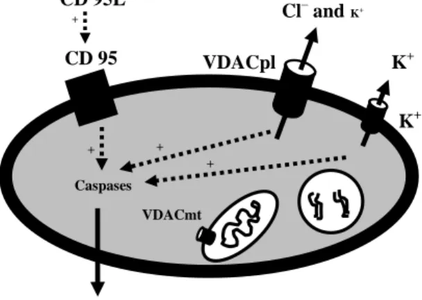
We found in different cell lines (HT22 and SK-N-MC cells) and primary hippocampal neurons, a clear activation of a K+ current of delayed-rectifier type in whole-cell experi-ments and VDACpl in excised inside-out membrane patches during apoptosis. We also found that the ATP concentration went down during apoptosis. Therefore, the findings from other reports (efflux of K+ and Cl– ions) (Table 2) and our investigations merge to-gether, and we can make a hypothesis that the apoptotic trigger cause enormous activa-tion of VDACpl and concomitant activaactiva-tion of a K+ channel (delayed rectifier type) (Fig-ure 9). Furthermore, activation of VDACpl is an early event of apoptosis (Paper I), which cause the loss of cell volume (by efflux of Cl– and K+ ions, 5:1), apoptotic cascade, and finally apoptotic cell death.