DISSERTATION
AGE-DEPENDENT DECLINE IN Kv4 CHANNELS, UNDERLYING MOLECULAR MECHANISMS, AND POTENTIAL CONSEQUENCES FOR COORDINATED MOTOR
FUNCTION
Submitted by Maximiliano Jose Vallejos Department of Biomedical Sciences
In partial fulfillment of the requirements For the Degree of Doctor of Philosophy
Colorado State University Fort Collins, Colorado
Spring 2019
Doctoral Committee:
Advisor: Susan Tsunoda Gregory Amberg
Gerrit Bouma Donald Mykles Michael Tamkun
Copyright by Maximiliano Jose Vallejos 2019 All Right Reserved
ABSTRACT
AGE-DEPENDENT DECLINE IN Kv4 CHANNELS, UNDERLYING MOLECULAR MECHANISMS, AND POTENTIAL CONSEQUENCES FOR COORDINATED MOTOR
FUNCTION
The voltage-gated potassium channel, Kv4, is widely expressed in the central nervous system and it is responsible for a highly conserved rapidly inactivating A-type K+ current. K
v4 channels play a role in the regulation of membrane excitability, contributing to learning/memory and coordinated motor function. Indeed, recent genetic and electrophysiological studies in
Drosophila have linked Kv4 A-type currents to repetitive rhythmic behaviors. Because a deterioration in locomotor performance is a hallmark of aging in all organisms, we were interested in examining the effects of age on Kv4/Shal channel protein.
In this dissertation, I use Drosophila as a model organism to characterize an age-dependent decline in Kv4/Shal protein levels that contributes to the decline in coordinated motor performance in aging flies. Our findings suggest that accumulation of hydrogen peroxide (H2O2) is amongst the molecular mechanisms that contribute to the age-dependent decline of Kv4/Shal. We show that an acute in vivo H2O2 exposure to young flies leads to a decline of Kv4/Shal protein levels, and that expression of Catalase in older flies results in an increase in levels of Kv4/Shal and improved locomotor performance. We also found that the scaffolding protein SIDL plays a role in maintaining Kv4/Shal protein levels and that SIDL mRNA declines with age, suggesting that an age-dependent loss of SIDL may also lead to Kv4/Shal loss. In behavioral studies, we found that a knockdown of SIDL resulted in a lethal phenotype, leading to a large decline in Drosophila
eclosion rates, an event that requires coordinated peristaltic motions. Expression of SIDL or
Kv4/Shal in this SIDL knockdown genetic background resulted in a partial rescue; these results are
consistent with a model in which SIDL and Kv4/Shal play a role in coordinated peristaltic motions and are required for successful eclosion.
The results presented in this dissertation provide new insight into the possible molecular mechanisms that underlie an age-dependent decline in Kv4/Shal protein. We identify two contributing factors: 1) ROS accumulation, and 2) the interacting protein SIDL. Our data also suggests that this age-dependent decline in Kv4/Shal levels is likely to be conserved across species, at least in some brain regions. Because Kv4/Shal channels have been implicated in the regulation of long-term potentiation and in repetitive rhythmic behaviors, the loss of Kv4/Shal may contribute to the age-related decline in learning/memory and motor function.
ACKNOWLEDGEMENTS
This work would have been impossible without the support of two very important women: my wife Coreen Frawley, and my PI Susan Tsunoda. Thank you for providing me with the environment and tools for success. Without you, this would have never happened.
During my PhD training, there were many positive and negative experiences which I think are truly important for character building and for the making of a scientist. There were many friendships as well; some were transient, and some will last a life time. There are many people who were directly or indirectly involved in the making of me as a scientist – they are my building blocks (wide-ranging from students to advisors and mentors). They provided me the extensive support (scientific, technical, emotional, etc.) necessary for moving forward during good and bad times.
Tuesday Salsa Dancing at the Rio Grande Restaurant was the first “out” I had from the stresses of graduate school. Many great supportive friendships were born there, including Hiro Gosden and countless others. I thank you for your support.
The Fort Collins Judo Club and the Colorado Judo League were another “out” that helped me through this career. As Judoka, we learn the ways of the Samurai in which courage and self-discipline are key, and surrendering is dishonorable. During my PhD training, these values became very engraved in my life; they helped me push through during the toughest times. Special thanks to my team, specially to Luis Briceño, Marcos Batan, and Galen Lyle (they are more than friends to me).
To my family, Helga Perez, Jose Vallejos, Geronimo Vallejos, and Catalina Vallejos thank you for your support during the years and for constantly telling me this was truly possible. To my
grandmothers Helga Cavieres and Gilda Vignolo, who were always asking me to solve your vision problems or your aging with my PhD, I thank you.
To my peers in graduate school that had a very positive impact and I am sure we will keep in touch for many years to come, thanks y’all!!! I chose not to write names here. You know who you are, and our friendship will be preserved through the many years to come. We have had a great run!
To Nicolas and Kilian, thank you for giving me a reason to finish this. I want you, and whoever comes thereafter, to know that the Vallejos-Frawley finish what they start. You and your mom are my life and my purpose to do what I do.
TABLE OF CONTENTS
ABSTRACT ... ii
ACKNOWLEDGEMENTS ... iv
CHAPTER 1. INTRODUCTION ... 1
1.1 Overview ... 1
1.2 Age-Effects on Locomotor Performance ... 1
1.3 Cellular Functions Impacted by Age ... 2
1.4 Oxidation During Aging ... 5
1.4.1 ROS effects on nucleic acids ... 5
1.4.2 ROS effects on protein ... 6
1.4.3 Regulation of intracellular ROS levels ... 8
1.4.4 ROS effects at the organismal level ... 11
1.5. Channel Proteins Affected by Age ... 13
1.5.1. AMPA and NMDA ionotropic glutamate receptors are affected with age ... 13
1.5.2. Effects of age on GABAA ionotropic receptors ... 15
1.5.3. Decline in Ca2+-activated K+ channels during aging in myocytes ... 16
1.6. Voltage-Gated Potassium Channels Known To Be Affected By Age ... 17
1.6.1. Historical perspective on voltage-gated K+ currents... 17
1.6.2. Structural characteristics of Kv channels ... 18
1.6.3. Age-related decline in Kv1/Shaker levels ... 24
1.6.4. Age-related decline in Kv2/Shab levels ... 25
1.6.5. Age-related decline in Kv3/Shaw levels... 27
1.6.6. Age-related effects on KCNQ/Kv7-type Channels ... 28
1.7. Kv4/Shal Channels ... 29
1.7.1. Kv4/Shal expression ... 29
1.7.2. Kv4/Shal neuronal subcellular localization and trafficking ... 31
1.7.3. Kv4/Shal channel function ... 35
1.7.4. Physiological roles of Kv4/Shal channels ... 38
1.7.5. Kv4/Shal accessory proteins ... 41
1.7.6. Age-related pathophysiology of Kv4/Shal ... 45
1.8. Overview of this dissertation ... 49
CHAPTER 2. MATERIALS AND METHODS ... 51
2.1. Drosophila strains ... 51
2.2. Immunoblotting ... 51
2.3. Mouse Brain Experiments ... 53
2.3.1. Mouse caring and sample storage ... 53
2.3.2. Sample preparation and immunoblot ... 54
2.3.3. Data collection and statistical analysis ... 54
2.4 RT-qPCR ... 55
2.4.1. RNA extraction ... 55
2.4.3. qPCR ... 57
2.5. ROS Fluorescence Detection ... 59
2.6. Drosophila locomotor activity assay ... 60
2.7. Drosophila longevity testing ... 60
2.8. Immunocytochemistry ... 61
2.8.1. Embryonic Neuronal Culture Preparation... 61
2.8.2. Testing Different Experimental Conditions ... 61
CHAPTER 3. AGE-DEPENDENT CHANGES IN KV4 CHANNEL LEVELS AND THEIR CONTRIBUTION TO LOCOMOTOR PERFORMANCE ... 63
3.1. Overview ... 63
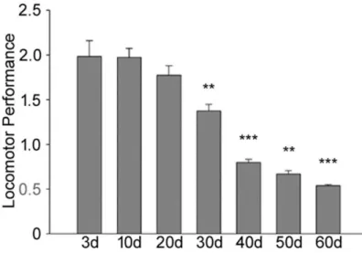
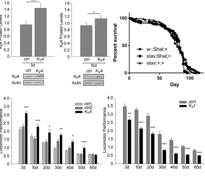
3.2. Age-Dependent Decline in Locomotor Performance In Drosophila ... 64
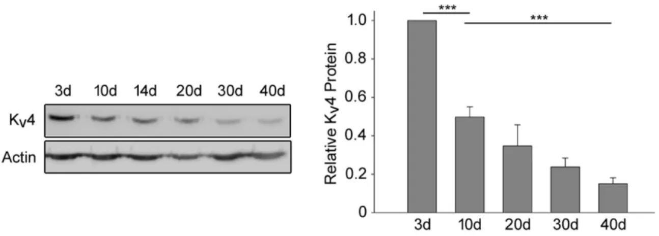
3.3. Age-Dependent Decrease In Drosophila Kv4 Channels ... 67
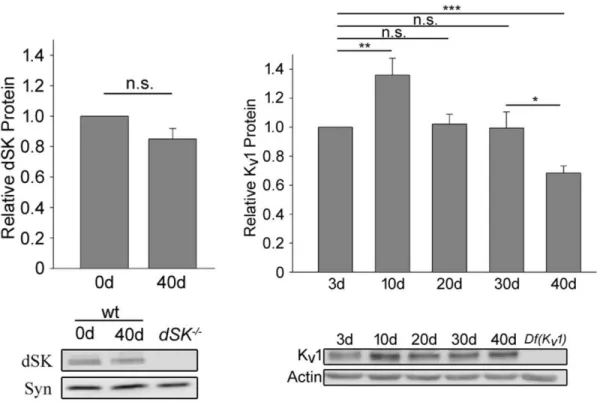
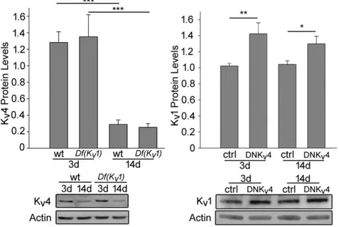
3.4. The Decline In Kv4 protein Is Likely Specific For Kv4 ... 68
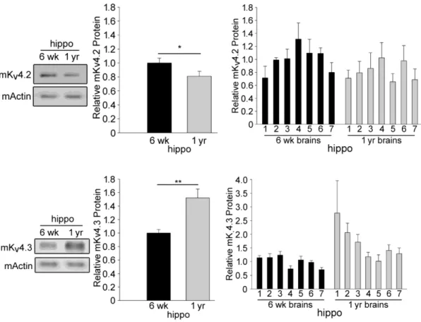
3.5. Age-Effects On Mouse Kv4 Protein Levels ... 71
CHAPTER 4. EFFECTS OF ROS ON Kv4 ION CHANNEL LEVELS ... 77
4.1 Overview ... 77
4.2 Effects of exposing Drosophila to H2O2... 78
4.3. Effects of Exposing Cultured Neurons To H2O2 ... 79
4.4. ROS Levels Increase With Age In Drosophila ... 85
4.5. Overexpression And Knockdown Of Enzymes That Regulate ROS Levels ... 86
CHAPTER 5. ROLE OF THE SCAFFOLDING PROTEIN SIDL ON Kv4 LEVELS DURING AGING, AND ITS CONTRIBUTION ON DROSOPHILA ECLOSION ... 91
5.1. Overview ... 91
5.2. SIDL mRNA declines with age ... 92
5.3. SIDL plays a role in Kv4 protein stabilization ... 93
5.4. Kv4 and SIDL are implicated in Drosophila eclosion ... 94
CHAPTER 6. DISCUSSION ... 97
6.1. Overview ... 97
6.2. Kv4 Channels Are Implicated In Locomotor Performance During Aging ... 98
6.3. Age-Related Kv4 Protein Decline Effects On Learning And Memory ... 101
6.4. ROS Accumulation During Aging Leads To Lower Kv4 Channel Levels ... 104
6.5. Oxidation And Neurodegenerative Diseases ... 109
6.6. The Scaffolding Protein SIDL Contributes To Maintaining Kv4 Levels ... 111
6.7. Conclusion ... 114
REFERENCES ... 116
CHAPTER 1. INTRODUCTION
1.1 Overview
As a species, the average human lifespan is 85 years of age. Life expectancy, however, has risen from the mid-40s to the mid-70s just during this past century1,2. Along with a rising number of individuals surpassing 65 years, there has been an associated increase in chronic brain-related ailments affecting the population3–5. The topic of aging is quite broad. Research areas include understanding cognitive abilities, anatomy, physiology, cellular regulation, and molecular changes with the goal of understanding the effects of aging on an organism6–10. During aging, there are a series of physiological changes that lead to brain-related conditions including sarcopenia, age-related dementia, Parkinson’s, and Alzheimer’s diseases7,9. Together, these age-related ailments affect locomotion, learning, and memory performances at the organismal level. These diseases are typically attributed to changes in the brain, both structural and functional2. Ion channels and receptors choreograph brain synaptic activity through the movement of ions. In this chapter, I provide background on the effects of age on locomotor performance and on cellular function, and the consequences of age-related intracellular oxidation. I provide background on a series of ion channels that undergo age-related effects in protein levels and modulation. I also present a historical perspective on voltage-gated potassium currents, and I introduce Kv4 channels, which are the proteins I examined during aging in this dissertation.
1.2 Age-Effects on Locomotor Performance
The age-dependent decline in locomotor performance is a phenomenon that is well-established across species11. Age-driven changes in the neuromuscular system have been
described to be, in part, the culprit. In humans, the nervous system experiences age-related effects that result in a decline of neuronal and motor nerve fiber densities, which contributes to a decrease in the effectiveness of neurotransmitter signaling and slower nerve conduction velocities12. Rhesus monkeys have shown an age-dependent decline in walking and jumping13. In laboratory rodents, age-dependent decline in physical activity is well-known11. More specifically, 2-year old mice were found to have significantly less locomotor performance than 1-year old mice in wheel running experiments14. In rats, a 50% reduction in locomotor performance has been measured between 6 and 32 months of age while testing exploratory activities13. In other laboratory rodent aging studies, the Mongolian gerbil and the deer mouse have been found to also have decreased locomotor performances in wheel-running and home-cage activity experiments, respectively15,16. In the invertebrate world, an age-dependent decline in locomotor movement has been observed in nematodes, houseflies, and fruit flies17–21. More specifically, in a comparative Drosophila study, researchers found that adult flies from two different populations – Congo and France – had a decline in walking speed when measured between 2 and 13 days of age22. This decline in motor function is likely to be triggered by a decrease in nervous system function, which can contribute to a possible loss of muscle density. Indeed, in a recent study measuring motor activity in C.
elegans, researchers found that a progressive decline in motor neuron function contributed to the
age-dependent loss of motor function even before any loss of muscle mass could be measured23.
1.3 Cellular Functions Impacted by Age
Cellular DNA mutations accumulate during aging. DNA mutations in germ cells are the basis for the evolutionary process and mutations in somatic cells can lead to detrimental effects on the function of the organism. Some mutations can occur during DNA replication where errors are
accidentally made by the molecular machinery24. Other mutations can arise from external sources such as UV radiation and chemical exposure25. In general, these endogenous and exogenous factors can lead to a wide variety of DNA damages including point mutations, single- and double-strand breaks, epigenetic alterations, genomic transpositions, chromosomal aberrations, and telomere shortening, all of which can affect cellular performance26. The accumulation of DNA damage to the point where repair is unsustainable is, in part, a hallmark of aging25–27.
Gene expression is affected throughout development and aging. The variations in expression display themselves in the form of phenotypic changes that occur in organisms as they reach adulthood and as they reach old age. Particularly, gene expression can increase, decrease, or stay unchanged28. Researchers have performed comparative analyses from cDNA libraries derived from mRNA of young and old rats to identify genes that are upregulated or downregulated throughout aging28. Studies in rats have shown that the amount of mRNA molecules transported from the nucleus to the cytoplasm is negatively impacted by age, probably due to decreased mRNA synthesis, a lack of proper polyadenylation of mRNA, and errors in the trafficking of mRNA from the nucleus through the nuclear pore29–31. Furthermore, an increase in transcriptional noise – a process that yields a heterogeneous transcriptional response across cells of the same genetic composition to the same stimulus – increases with age leading to a wider variety of transcriptional responses which might be detrimental to an organism26,32,33. Two highly conserved signaling pathways play an important role in aging: Insulin/IFG-1 Signaling (IIS) and target of rapamycin (TOR). These pathways are responsible for regulating gene expression through aging in response to stress and nutrient availability34,35.
Proteostasis is a term used to describe the maintenance of properly functioning protein. Protein turnover is critical for proper proteostasis36. A decline in proteostasis during aging is
characterized by the emergence of protein aggregates due to a disruption in the proteostasis network which includes the machinery for translation, chaperone proteins, and the principal protein degradation systems – proteasome and lyzosome37. Chaperone proteins, most of them from the heat-shock family of proteins (HSP), are at the vanguard of monitoring and ensuring proper protein folding. One important role of chaperone proteins is to aid other proteins to achieve a proper functional conformation, with the goal of counteracting aggregation of nascent protein38,39. There is also evidence of protein refolding by chaperones when misfolding occurs40. When a protein has been misfolded or incorrectly modified, and cannot be refolded, it is the role of chaperones to target the non-functional protein for degradation41,42. Unfortunately, both the ubiquitin-proteasome and autophagy-lysosome systems experience an age-dependent decline in activity43–45.
Age-dependent mitochondrial dysfunction has also been described. The “mitochondrial damage-energy loss” hypothesis of aging was described by Medvedev in 199046. In this hypothesis, age-dependent cellular injury by reactive oxygen species (ROS) occurs on the mitochondrial membrane and DNA, and it occurs especially in neurons47. This would lead to a decline in properly functioning mitochondria, and a decrease in readily available ATP molecules. With an increase in age, there is a reduction in mitochondria biogenesis as muscle studies have demonstrated in comparisons between young and 50-year plus men47. In a rat study, the enzymatic activity of heart mitochondrial oxidase was measured and found to be significantly diminished in older subjects48. Analyses of primate neocortex enzymatic activity of mitochondrial complex I and IV have shown that they also decline in activity with age49. These studies on the enzymatic activity of mitochondrial complexes were confirmed in mice, in which the activity of complex V – ATP synthase – was also found to decrease with age47. The age-dependent decrease in activity of
multiple critical mitochondrial enzymes is likely what leads to its dysfunction. Furthermore, in a recent study, Takihara and coworkers (2015) used mouse retinal ganglion cells (RGCs) as a model for measuring mitochondrial axonal transport in the central nervous system (CNS)50. Their findings reveal that mitochondrial axonal transport decreases with age; suggesting that loss of proper mitochondrial transport in CNS might be involved in the age-dependent dysfunction of mitochondria.
1.4 Oxidation During Aging 1.4.1 ROS effects on nucleic acids
DNA base damage was first proposed as the root of aging in 196751. As an organism ages, there is an accumulation of DNA damage that is both exogenous and endogenous in origin, and that this damage is likely to interfere with transcription52. Internal sources of this damage include genome reorganization53, genomic instability24, and improper DNA repair25. ROS have been largely considered a principal source of general DNA damage during aging. The primary site of ROS production is the mitochondria, where ROS are a byproduct of oxidative phosphorylation, even though other sources, such as from peroxisomes and cytochrome p450 enzymes, have also been described54–56. Because mitochondrial DNA (mtDNA) is closest to the source of ROS, it is thought that mtDNA damage occurs at faster rates than nuclear DNA damage, leading to the age-dependent mitochondrial dysfunction which causes a decline in ATP synthesis47,55,57,58. This is not to say, however, that nuclear genomic DNA does not undergo any oxidative damage, which was originally proposed in 1956 by Harman and experimentally confirmed in 2004 by Hartman et
al.56,59. Indeed, 30 genes involved in synaptic plasticity, ranging from ion channels to
and coworkers (2004). Their results show that, although there is some minor damage to exons, DNA damage had a higher occurrence in the promoter region of many of the genes expressed in the prefrontal cortex after 40 years of age, and was greatest in the promoter region of all measured brain genes after 70 years of age, leading to an age-dependent decrease in mRNA levels58 . This age-dependent decline in mRNA levels of various genes is conserved across species. In a microarray analysis of over 6,000 mouse genes, Lee and coworkers (2000) found that 10-15% of those in neocortex and cerebellum have lower mRNA levels with age62.
Moreover, results showing that some mRNAs increase with age have been published. In a study of over 11,000 genes focusing in the hypothalamus and cerebral cortex where researchers found that there is indeed an alteration in the expression levels of many genes with an increase in age; while mRNA coding for DNA-repair related proteins decreased with age, mRNA coding for protein degradation increased with age63. Their results also suggest that a measured increase in mRNA coding for mitochondrial enzymes involved in ATP production in the hypothalamus leads to an increase in ROS and, therefore, a greater effect of oxidative stress on the cells. In hippocampal studies, Blalock and coworkers (2003) found, in rat microarray analyses correlating gene expression to memory-related task performance, that almost half of the measured genes decreased with age, while the other half increased with age and negatively impacted memory performance64.
1.4.2 ROS effects on protein
The age-dependent increase in ROS, likely caused by a deteriorating mitochondrial electron transfer, also leads to an increase in the probability of protein oxidation. This, along with the aforementioned decline in activity of the protein degradation machinery, results in an
accumulation of oxidized protein with age. Carney and coworkers (1991) were the first to observe an accumulation of oxidized protein in the cortex of gerbils. They administered a daily dosage of the spin-trapping chemical N-tert-butyl-a-phenylnitrone, a short-lived free radical interactor, with the goal of decreasing the amount of oxidized protein in the brain of aged gerbils. They observed a decline in the amounts of oxidized protein along with an increase in temporal and spatial memory performance in aged animals65. Today, it is well known that the age-dependent increase in ROS lead to oxidation of proteins which can cause them to become dysfunctional. Typical oxidative effects involve peptide bond cleavage catalyzed by free hydroxyl radical interaction with the carbonyl carbon, amino acid residue side chain oxidative modifications such as carbonylation, and disulfide bridge interactions via cysteine sulfhydryl group oxidation, leading to protein misinteraction and aggregation66. The accumulation of these aggregates leads to an increase in the cellular stress response and likely degradation of the toxic structures from protein aggregates. Unfortunately, as mentioned above, both proteolytic – proteasome and lysosome – systems become dysfunctional with age43–45.
Indeed, work published by Friguet and colleagues (2000) has shown that the age-dependent decline in proteasome activity is, in part, due to oxidative modifications67–69. In neuronal studies, Keller et al. (2000) have described how chemically increasing oxidative damage on the spinal cord of young rats triggers a decline in proteasome activity similar to that measured in normal aging rats. Their results support the idea that the decline in proteolytic activity due to ROS exposure leads to its dysfunction, and possibly also contributes to neuronal cell death70. In regards to the lysosome, it was almost 50 years ago that it was first described that the lysosomal membrane is quite sensitive to oxidative damage71. Truly, these age-dependent oxidative effects are likely to cause changes in the pH within lysosomes affecting their stability and activity72. Kurz and
coworkers (2008) described how many of the proteins degraded by the lysosome are iron-containing. Iron, being highly susceptible to oxidation from the highly diffusible ROS, leads to the slow formation of lipofuscin – non-degradable pigment granules which increase in volume with age. This age-dependent lysosomal saturation with lipofuscin compromises the activity of the degradative activity of the machinery73.
1.4.3 Regulation of intracellular ROS levels
There are a variety of enzymes that play a critical role in the regulation of reactive oxygen species (ROS) described in the literature. Superoxide dismutase 1 & 2, and catalase participate in ROS degradation while nicotinamide adenine dinucleotide phosphate (NADPH) oxidases (NOX) generate ROS.
Superoxide dismutase (SOD) was first described in the late 1930s as a copper-containing protein. While attempting to gain an understanding of the role of copper in erythrocytes, Mann and Keilin (1939) found that a copper-containing protein was present in blood74. It was not until 20 years later that Markowitz and colleagues (1959) were able to isolate and purify this copper-containing protein75. A decade after, McCord and Fridovich (1968, 1969) published two reports characterizing this copper-containing protein. They described that the enzyme can catalyze the dismutation superoxide anion radicals into molecular oxygen and hydrogen peroxide (H2O2), and re-named this enzyme superoxide dismutase76,77.
Today, we know of three members that comprise the family of SOD enzymes. SOD1 is a copper and zinc containing enzyme that is found in the cytosol of cells, SOD2 is a manganese containing enzyme that is found in the mitochondria of cells, and SOD3 is a copper and zinc containing enzyme that is exclusively targeted extracellularly and which is the least studied thus
far78,79. Studies in Saccharomyces cerevisiae have shown that a mutation in either or both
SOD1/SOD2 causes a significant decrease in cell viability80. In Drosophila, a SOD1-null mutant
(cSODn108) was observed to have impaired performance in metabolizing superoxide anion, which correlated with reduced longevity81. In another report, Kirby et al. (2002) used the daughterless-GAL4 driver to express SOD2-RNAi, which resulted in undetectable levels of SOD2 in immunoblot experiments using whole adult males. It was described that this successful RNA interference caused high levels of mitochondrial oxidative stress and an enhanced onset of adult fly mortality82. A SOD2 missense mutant, named SOD2bewildered, has also been characterized in flies that results in detrimental effects during neurodevelopment and an anomalous brain morphology, and a reduced lifespan83. Overexpression of both SOD1 and SOD2 under the control of the constitutive actin5C promoter also resulted in a decreased life-span84. In contrasting studies, the overexpression of human SOD1 in, specifically, motor neurons lead to an increase in fly life span by 40%85. The results of these studies reflect the importance of this family of enzymes organismal viability and suggests that an age-dependent increase in ROS levels may have detrimental effects.
Catalase was first recognized as a ubiquitous enzyme by Loew as early as 190086. The role of catalase is to dismutate harmful H2O2 into water and molecular oxygen87. While these enzymes’ role is to reduce the levels of intracellular ROS, there are other enzymes that produce ROS as a by-product or for purposes of intracellular signaling.
The role of the nicotinamide adenine dinucleotide phosphate (NADPH) oxidases (NOX) family of enzymes is to produce ROS. NOX-dependent ROS production is quite varied and still under study; though originally thought to be only damaging, ROS are just beginning to be understood as a group of highly reactive molecules that are also involved in cellular signaling. The
NOX family is comprised of at least 7 different members identified so far: NOX1-5 and DUOX1-2. Much of what is currently known comes from studies using mouse knockout models of NOX protein function where different phenotypes such as loss of balance and hypothyroidism were described88. More specifically, NOX2 has been described to contribute to antimicrobial activity during phagocytosis89, while NOX3 has been shown to be essential for proper development of components in the vestibular system90–92. DUOX has also been described to play a role in defense from microbial defenses, but in the airway epithelium; it has been characterized as necessary for the production of hydrogen peroxide which is used for iodination of the thyroid hormone as well88.
The expression of the various oxidases from the NOX family in a variety of tissues, allows for them to play an important role in the upkeep of the vascular system from angiogenesis to tone93. In neuronal in vitro studies, researchers have made use of N-acetylcysteine, a ROS scavenger, to measure the effects of a decrease of ROS on a variety of neuronal development signaling pathways. Their results suggest that ROS generated by NADPH oxidases might play a role in neuronal cell differentiation94–96. In a study targeting NOX directly, Nitti and colleagues (2010) used the chemical DPI to block NOX activity to demonstrate that the product of the enzyme, possibly hydrogen peroxide, is involved in neuroblastoma cell differentiation97. In vivo mouse studies have reported that ROS produced by NADPH oxidases are present in the hippocampus and their localization responds to stimulation of hippocampal slices, suggesting that the ROS produced by these enzymes plays a role in LTP induction98. More specifically, NOX has been recently described to act as a regulator of the cytoskeletal organization in the hippocampus, by maintaining ROS physiological levels in vivo; this organization is critical for maintaining neuronal polarity and proper axonal length99. In Drosophila, a recent report published by the Landgraf laboratory (2017) uses the neuromuscular junction (NMJ) of larvae as a model to understand the role of H2O2 in
neuronal structural plasticity. Their results suggest a direct relationship between ROS levels and signaling involved in synaptic terminal growth at the NMJ100.
1.4.4 ROS effects at the organismal level
Physiological ROS levels must be maintained as they play an important role in learning and memory, lifespan, and locomotor performance. An excess or a scarcity of these molecules can have a detrimental effect in any of the aforementioned behavioral functions. Indeed, in their recent paper, Haddadi et al. (2014) measured an increase in Drosophila ROS with age which correlated to an age-dependent loss of memory retention in adult flies. They suggested that this increase in ROS is possibly due to a decrease in the enzymatic activity of the antioxidant enzymes catalase and superoxide dismutase101. In addition, in vitro studies in mammalian hippocampus treated with superoxide scavenging molecules or with overexpression of SOD have shown impairments in LTP induction suggesting that it is the superoxide anion that is involved in processes related to learning and memory102–105.
In mice, one study has been published where a Catalase knockout mouse was generated. The researchers reported that, though this mouse has no evident health problems up to 1 year of age and shows no apparent signs of clinical acatalasemia, they are highly susceptible to oxidative tissue injury, including deficiencies in oxidative phosphorylation after brain trauma106. Contrasting studies in which transgenic mice overexpressed Catalase showed increased lifespan; researchers also reported that the onset of cardiac and visual age-related diseases were also delayed107,108.
Earlier studies on the effects of ROS on Drosophila lifespan have mostly focused on the detrimental oxidative effects of ROS at the organismal level and usually go in hand with the free-radical theory of aging109–112. In Drosophila, a study isolating and characterizing Catalase
mutant flies reported that, when catalase activity is decreased by 97%, lifespan is significantly reduced113. Interestingly, though overexpression of Catalase confers greater resistance to oxidative stress, and showed increased lifespan in mice, increased lifespan in Drosophila occurs only if catalase is co-expressed with SOD114–116. Research has suggested that, in tandem with SOD enzymes, the role of catalase is to catalyze the peroxide from SOD reactions into harmless products. Moreover, when both enzymes are co-expressed in motor neurons to above wildtype levels, there is typically an increase in life-span117,118. Some contradicting reports, however, have been recently published. Sanz and coworkers (2010) noticed that, though an increase in mitochondrial ROS (mtROS) correlates to a decreased lifespan, it does not necessarily affect fly longevity directly. In their report, they expressed the enzyme alternative oxidase from the urochordate Ciona intestinalis to decrease mtROS but they were unable to measure an increase in lifespan119. In a similar study, Scialò et al. (2016) determined that Drosophila expression of NDI1 dehydrogenase leads to an enhanced reduction of coenzyme-Q which causes a reverse electron transport in complex I of the mitochondria, culminating in increased levels of mtROS. Their results show that increased mtROS production via complex I causes an increase in Drosophila lifespan120. ROS have also been described to influence Drosophila locomotor performance. Long and colleagues (2009) fed an antioxidant containing grape extract to flies to determine how ROS play a role in a Drosophila model for Parkinson’s disease. In their report, the extract improved the loss of locomotor performance of the model, suggesting that ROS are probably involved in this neurodegenerative disease121. In another study, Jimenez-del-Rio and coworkers (2010) restored
Drosophila locomotor performance, in flies with high paraquat-induced ROS levels, by
administering polyphenol antioxidants122. In a more recent study, a genetic screen was carried out to identify candidate genes linked to oxidative stress and locomotor phenotypes. Researchers used
the molecule menadione sodium bisulfite (MSB) which, unlike paraquat or H2O2, is a milder yet persistent inducer of chronic oxidative stress. Though their goal was to identify genes, their procedure led to the confirmation that oxidative stress in Drosophila leads to their loss of locomotor performance123. Taken altogether, the age-dependent increase in ROS has detrimental effects on Drosophila physiology and reports suggest that administration of neuroprotectant antioxidants ameliorate the reduced locomotor performance. I propose that ROS have an effect on Kv4 which, in turn, may have an effect on Drosophila locomotion. Chapter 4 of this dissertation describes the effects of ROS on Kv4 protein levels and describes the involvement of the enzyme catalase on Kv4 protein levels and an amelioration of locomotor performance in older flies.
1.5. Channel Proteins Affected by Age
1.5.1. AMPA and NMDA ionotropic glutamate receptors are affected with age
AMPA (-amino-3-hydroxy-5-methyl-4-isoxazole propionate) and NMDA (N-methyl-D-aspartate) receptors respond to pre-synaptic vesicle release of the amino acid L-glutamate by opening and allowing post-synaptic cation permeability124,125. Functionally, AMPA receptors typically allow for the influx of the monovalent cation Na+ into the cell which causes localized depolarization. In response to this depolarization, the Mg2+ ion blocking the channel of neighboring NMDA receptors is dislodged, permitting influx of Ca2+ 125–127. These two channels control the majority of mammalian neuronal excitatory transmission and have been found to decline in levels with age58,128–133.
Interestingly, scientists initially attempted to understand the effects of age on the levels of the amino acid L-glutamate rather than the levels of receptors themselves. Reports showed controversial results. Indeed, while some papers indicated an age-dependent decline of this amino
acid by ~12-17% in different regions of Fisher 344 rats134,135, others reported that L-glutamate does not change in level with age as measured in different brain regions of both Wistar rats and post-mortem human brain tissue136,137. To test the effects of age on cerebral cortex mRNA, Carpenter and coworkers (1992) injected mRNA from either 24 or 3-month old rats into Xenopus oocytes and measured voltage-gated currents to determine if there were any changes associated with mRNA from different aged rats. They found decreased L-glutamate induced currents in those oocytes with older mRNA, suggesting that mRNA from older rats coding for these channels might lead to lower expression of neurotransmitter receptors in the cortex128. In human studies, Lu and others (2004) used DNA microarray analyses on the prefrontal cortex of 30 post-mortem individuals, aged between 26 and 106 years, to compare mRNA levels of 12,000 genes; they reported that mRNA coding for the GluR1 AMPA subunit and R2A NMDA subunit both show a 2-fold decrease in subjects 40 years of age or older58.
Studies attempting to understand the effects of age on AMPA receptors in different mammalian model systems were later published. In a recent review, Henley and Wilkinson (2013) suggest that the age-related decline in AMPA receptors could be caused by an age associated defect in receptor trafficking, and that this may have an effect on long-term potentiation (LTP) and depression (LTD) in the hippocampus, which are involved in learning and memory138. In rat hippocampal studies, a report showed that, though GluR-1 mRNA levels were not affected by age, the levels of the GluR-1 subunit of AMPA receptors decline with age as measured in 24-month old animals130. Also in the hippocampus, but using mice as a model organism, Magnusson and Cotman (1993) used autoradiographic density analyses of AMPA receptors ligand binding and reported that levels of AMPA receptors decrease with age in BALB/c and C57B1 mice strains139. Bahr et al. (1992) reported that in the brain telencephalon of BALB/c mice there is an
age-dependent decline in GluR AMPA subunit levels when compared to mice at 3 and 25 months of age129.
Most of what is known about the effects of age on L-glutamate-dependent ionotropic channels, however, has been studied with NMDA receptors. In rodents, early reports described an age-dependent decline in NMDA receptors in different brain regions as a reason for age-related cognitive impairments140,141. Magnuson and coworkers (2002) later found that both NR1 and NR2B transcripts showed decreased levels in aged mice. They expanded these results by also measuring protein levels of NMDA subunits NR1, NR2A and NR2B, and found that they all had lower levels in the cerebral cortex of older C57B1/6 mice, while only NR1 and NR2B subunits were decreased in the hippocampus131. Other studies have shown that age has a negative effect on the levels of the obligatory NR1 and NR2B subunits of the NMDA receptors in the hippocampus which results in a decline of spatial memory abilities in older rats142–144. Even in the brain of primates, a decline in NMDA has been measured; Wenk and coworkers (1991) described an age-dependent decrease in NMDA levels between young (~7-year old) and aged (~30-year old) monkeys132. Regarding motor function, Ossowska and coworkers reported that the age-dependent decline in NMDA receptors leads to a loss in muscle tone of rat leg muscles. Their data suggest that this loss in muscle tone is a reason for the poor performance of rats in their T-maze experiments133.
1.5.2. Effects of age on GABAA ionotropic receptors
In the developed brain, -aminobutyric acid (GABA) is the major inhibitory neurotransmitter145. This neurotransmitter acts on GABA receptors, GABA
A and GABAB. Here, I describe the known effects of age on GABAA ionotropic receptor levels. Originally, measurements of GABAA receptor levels have been contradicting. While some studies reported a decline in
detectable GABAA in different brain regions of aged rats, others reported that GABAA remained unchanged in similar or other regions of the rat brain during aging132,146–148. Soon after, transcriptional studies in Fisher rats revealed that GABAA receptor subunit mRNA levels decreased by 70% in the cerebral cortex when comparing 6 month and 24-month old animals149. This report led Gutiérrez and coworkers (1994) to perform further testing on mRNA and protein levels of different subunits of the GABAA receptors. They reported that in the inferior colliculus of Sprague-Dawley and Fisher 344 rat brain both mRNA and protein levels of 2, 3, 2S, 2L, and 1 subunits declined with age150. Further testing in the same rats revealed no mRNA changes in the cerebral cortex 151, and that, though there are no changes in protein expression levels of
1, 2, or 3 subunits, mRNA levels coding for these three subunits decrease with age in the cerebral cortex152.
1.5.3. Decline in Ca2+-activated K+ channels during aging in myocytes
In the heart, Ca2+-activated K+ channels play a critical role in regulating membrane potential and regulating muscle contractility by intracellular free Ca2+ 153. Although, not much has been done to understand the effects of age on these channels in myocytes, Marijic et al. (2001) reported that these channels, in both Fisher 344 rats and humans, decreases with age in coronary smooth muscle which could be an explanation for some heart problems in the aging population154. Another group, using 2 year old Fisher 344 rats, reported that low-intensity exercise training can partially reinstate the levels of Ca2+-activated K+ channels155.
1.6. Voltage-Gated Potassium Channels Known To Be Affected By Age 1.6.1. Historical perspective on voltage-gated K+ currents
Voltage-dependent K+ currents were first described by Hodgkin and Huxley in 1952 in
Loligo giant axons. The purpose of these delayed-rectifier K+ currents is to restore the membrane
to resting potential so the cell is able to propagate another action potential156. Almost a decade later, Hagiwara and coworkers (1961) were the first group to define a third conductance during action potentials; they described this conductance as very transient, hyperpolarizing, and not a part of delayed membrane rectification, an event that lasts much longer157. This third conductance was later characterized first by Connor and Stevens (1971) in marine gastropod Anisodoris’ neuronal cell bodies and named the current A-type (IA). They described the activation potential of this transient outward current to occur when the membrane potential changes from its resting state of -70mV to the range of -35 to -50 mV at lower temperatures158. This current was measured in cell bodies of snail neurons and recorded to lasts ~200-400 milliseconds159. Pharmacologically, a hallmark characteristic of transiently activating potassium channels is that they are sensitive to 4-aminopyridine (4-AP) and generally unresponsive to tetraethylammonium (TEA), unlike other potassium channels160,161.
The first K+ gene identified was from a mutant fly that exhibited a leg shaking phenotype under mild ether anesthesia, and was named Drosophila Kv1/Shaker162–164. Three research groups reported the cloning of this channel in Drosophila165–167 and, soon after, Tempel et al. (1988) successfully cloned the first mammalian version168. These discoveries led to a series of other studies that resulted in the identification of three more Drosophila Shaker-like Kv genes: Kv2/Shab, Kv3/Shaw, and Kv4/Shal169–171. The series of cloning studies in mammals and other systems revealed that, these genes, each represented a distinct family of ion channels conserved across
species172–177. Today there are 12 known subfamilies of Kv channels: Kv1-12178–189. In this dissertation, I focus on Kv4 channels.
1.6.2. Structural characteristics of Kv channels
Voltage-gated potassium channels (Kv) are the largest family of all ion channels and are coded by 40 genes in humans182,189,190. Phylogenetically, these families are subdivided into four major family groups: a) Kv1-Kv4, b) Kv5, Kv6, Kv8, Kv9, c) Kv7, d) Kv10-Kv12189. The functional Kv channel is assembled in the ER membrane in which four nascent -subunits of the same family form a tetramer by interacting via an N-terminal T1 tetramerization domain of ~130 amino acids191–197. Below, I focus on the structural characteristics of the channels Kv1-Kv4 family of channels.
Originally, in an attempt to describe the secondary and tertiary protein structure of the opening of voltage-dependent channels, Guy and Seetharamulu (1986) performed computational models to identify what today is known as the S5-S6 linker and the P-loop198, Figure 1.1. With these modeling results, scientists focused on mutational analyses of the pore region in an attempt to characterize the P-loop of voltage-gated K+ channels. MacKinnon and Miller (1989) published the first report, in which the pore-blocker peptide inhibitor charybdotoxin (CTX) from the scorpion
Leiurus quinquestriatus was used to determine if the amino acid glutamate at position 422 was
near the conduction pathway of Kv1199. Soon after, MacKinnon and Yellen (1990), by the use of CTX and the open-channel interactor chemical TEA, reported that residues 431 and 449 were likely involved in the ion permeation properties of Kv1, as previously proposed by the earlier computer model from Guy and Seetharamulu (1986)198,200. MacKinnon and coworkers (1990) then further characterized this region and identified other amino acid residues that altered toxin
interaction to determine that the region connecting the S5-S6 linker also matched the proposed computational model representing the pore region201. Yool and Schwarz (1991), performed additional site-directed mutagenesis and functional studies in this showing that, indeed, this region allows for passage of K+ ions202. To confirm this, Hartmann and colleagues (1991) then created a chimera, in which the 21-amino acid pore region was transplanted into another potassium channel with different conductance and TEA sensitivity. Their results showed that this 21-amino acid replacement gave their channel the higher conductance and higher affinity to TEA, indicating that this 21-amino acid span controls the biophysical properties of the pore in these Shaker-like channels203. Further experiments by Yellen and colleagues (1991), using site-directed mutagenesis and internal TEA blocking, revealed that residues 431-449 are part of a reentrant loop between the S5-P-loop-S6 region204,205. Work by Heginbotham and coworkers (1992, 1994) later showed that the highly conserved 19-amino acid stretch is critical for the high K+ selectivity of these channels206,207. All these discoveries were subsequently confirmed by crystallographic evidence (see below).
Today, we know that the general structure of a single -subunit of the Shaker-like Kv1-Kv4 channels have both hydrophilic N- and C- termini located in the intracellular space. These termini flank the core region – an area of the -subunit that has about 40% identity between Kv1-Kv4, which is composed of 6 -helical transmembrane segments (S1-S6) with a potassium-selective pore (P) between S5 and S6182,191,208–210, Figure 1.1. The tertiary structure of the selectivity filter has been confirmed through studies on KcsA, a prokaryotic potassium channel that has a similar amino acid sequence to the S5-P-loop-S6 region of Shaker-like subfamilies of channels211,212. These prokaryotic channels have been described to have a high structural similarity to the eukaryotic version, suggesting that the potassium selectivity pore structure and function are quite
conserved213–215. When closed, the structure of this pore forms a funnel-like structure with a diameter of 12 Å in the outer area constricting to 4 Å, the equivalent of a van der Waals interacting distance between two interacting atoms, at the most inner region of the pore. When open, allosteric interactions between the -helices increase the inner region diameter to 12 Å215. The amino acid sequence of the selectivity filter is Gly-Tyr-Gly, a sequence highly conserved amongst Kv channels189,206,216–219. Four, linearly arranged, coordinating interactions occur in the selectivity filter with the carbonyl oxygens of the pore side chain; here, K+ enter in an alternating fashion with water molecules. More specifically, Zhou and coworkers (2001) were able to use X-ray crystallography to show that at lower concentrations of K+, the conformation of the selectivity filter appears to be in a closed state with ions being absent at positions 2 and 3218, Figure 1.2. The coordination chemistry of these carbonyl oxygens at the pore mimics that of the hydration shell of K+ in solution, allowing for K+ to diffuse freely through the pore. The coordination chemistry with Na+ is not the same with the constrictions of the pore, making the selectivity for K+ 1000-fold higher220.
In Kv channels, the pore opens in response to a membrane voltage change. The S4 transmembrane domain was first identified as the key voltage sensor of Shaker-like Kv channels, and the movement of this domain led to conformational changes that opened the pore. The original reasoning was that this domain contained basic residues such as lysine and arginine at every third position, with non-polar amino acids in-between, that provide it with an overall positive charge allowing it to detect changes in membrane potential that are below the action potential threshold208,219,221–224. The structure of S4 was found to be highly conserved across all voltage-gated channels in different species and it was a collection of studies in different types of voltage-gated ion channels that led to the understanding of the membrane potential detecting
capabilities of this domain. Stühmer and coworkers (1989) were the first to investigate the role of these positive charges in membrane potential sensing by experimenting with rat voltage-gated sodium channels in Xenopus oocytes; by replacing the positively charged amino acids in S4 by neutral or negatively charged ones, they were able to modify the activation potential of a voltage-gated sodium channel. Their results showed that the removal of positive charges results in a decrease of the slope for voltage-dependent activation225. Surprisingly, a similar study by Auld et
al. (1990) demonstrated that the positively charged amino acids were not the only ones responsible
for the selective gating of the channel. Indeed, by mutating the non-polar amino acid leucine, at position 860, they measured a shift in activation of the channel to more positive potentials226. Soon after, analogous experiments were performed on Kv1/Shaker channels in Drosophila227,228, and
mammalian Kv1.1 channels229. These results confirmed that the highly conserved S4 domain has voltage sensing capabilities and that modifications to polar or non-polar amino acids lead to shifts in the voltage-sensing and gating of voltage-gated ion channels.
Later, it was found that the transmembrane domain S2 was also involved in the voltage-sensing capabilities through an acidic residue, in contrast to the basic amino acids from S4; this presented the idea that negatively charged residues interacting with S4 could also play a role in voltage sensing230–232. Indeed, the work of Li-Smerin and coworkers (1998) showed that a tarantula toxin consistently interacts with S2-S4 regions of different voltage-gated ion channels which resulted in changes on their voltage-gating properties. Moreover, Lu and colleagues (2001) created a chimera by fusing Shaker S1-S4 to KcsA which resulted in KcsA gaining voltage-sensing capabilities. Altogether, these studies suggested that S1-S4 might be involved in the voltage-sensing. Although S4 performs most of the voltage-sensing, S1-S4 seems to be a voltage-sensing module224,231–235. Campos and coworkers (2007) used a mutagenesis approach to uncover a
relationship between the S1-S4 and the S2-S4 domains. They replaced three isoleucine residues with cysteine (I241C, I287C and R362C) to allow the formation of disulfide bridges between I241C or I287C with the S4 residue R362C which constrained the closed-state position of the S4 segment; their data suggests that there are stabilizing hydrophobic interactions given by I241 and I287 in the channel’s closed state236. When the module detects a change of +10 mM, the voltage-sensor undergoes conformational changes which results in the movement of the S6 domain, that possesses the conserved Pro-Val-Pro sequence allowing for segment mobility, leading to the opening of the pore231,235,237–242.
Shaker-like Kv channels are responsible for a wide variety of currents which include currents that are rapidly inactivating, non-inactivating, and slowly inactivating171. These A-type currents have since been described to play a physiological role in various types both non-excitable, such as epithelia, and excitable cells182,219,243–251.
Fast inactivation of A-type Kv channels is critical for modulating action potential firing in neurons. There are multiple of inactivation that have been described for Shaker-like channels252– 255. The N-type, or ball-and-chain, inactivation occurs in the millisecond scale via a domain that interacts with the intracellular portion of the open pore256–258. This amino acid sequence may be present in the N-terminal domain of the channel or in an interacting -subunit259–261. Both positive electrostatic charges and van der Waals contacts have been described to play a direct role in the interaction of this blocking amino acid sequence and the open pore blocking K+ conduction262,263. C-type inactivation is generally a slower process that likely involves a structural change culminating in the pinching of the pore and is independent from N-type inactivation; the exact mechanisms that lead to C-type inactivation are still not fully understood252,253,255,264,265. The overall mode of inactivation has been proposed to involve allosteric mechanisms that couple both
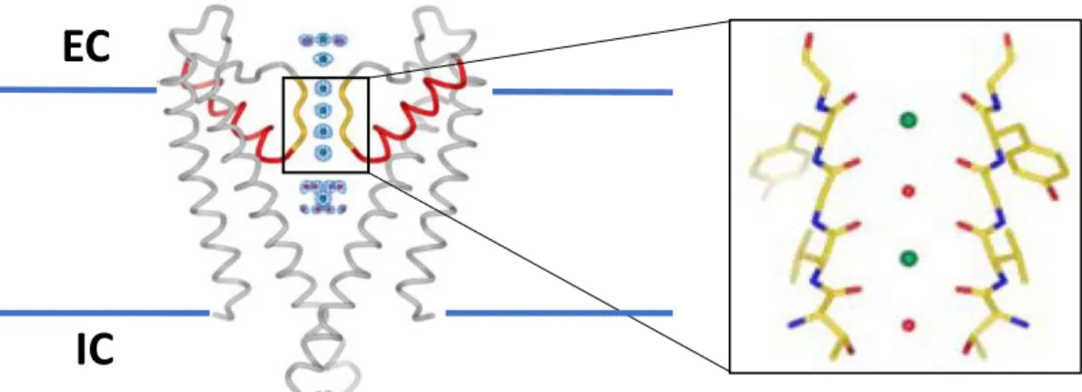
Figure 1.1. Structure of Kv channels. There are six transmembrane domains (S1-6, green)) area flanked by both cytosolic N- and C-termini (red and blue, respectively). The P-loop ion selectivity filter is located between S5 and S6 (purple). The T1 tetramerization domain is situated in the N-terminal end of the polypeptide. S4 contains positively charged residues giving it the voltage-sensing capabilities.
Figure 1.2. K+ pore and selectivity filter. Left, homologous domain of KcsA representing the equivalent S5-P-S6 motif from two of the four interacting -subunits is shown. Pore helices are in red and selectivity filter in yellow. Circles in blue mesh represent the electron density of ions entering through the filter. EC and IC are extracellular and intracellular domains, respectively. Right, enlarged section of the selectivity filter shows alternating K+ (green, positions 1 and 3) and H
2O (red, positions
2 and 4) ions as they enter the cell when the channel is open (adapted from MacKinnon 2003 with permission from John Wiley and Sons, License 4490440979830)220.
EC
N- and C-type inactivation processes. This was first noticed when Hoshi and coworkers found that a domain in the N-terminal of Shaker interacted directly with the open channel260. Along with this idea, Baukrowitz and Yellen have also described how N-type inactivation positively modulates C-type inactivation266.
.
1.6.3. Age-related decline in Kv1/Shaker levels
Kv1 is a voltage-gated potassium channel that is highly conserved across species with 70% identity between Drosophila and mice267. While in mammals there are at least twelve variants of Kv1 genes identified, there is only 1 representative of the Kv1 family in Drosophila178,267 and it is found in both muscles and neurons268,269. In mammals, variants K
v1.1, Kv1.2, and Kv1.5 have been reported to play a role in myogenic control270,271, while variants K
v1.4 and also Kv1.2 have been described to specifically localize to axons and nerve terminals221. The axonal localization of K
v1 in CNS is important for its function, where it likely plays a role in regulating neurotransmitter release221.
The first publication reporting any effects of age on Kv1 channels surfaced in 2001. In this report, age was described to affect expression Kv1.1 and Kv1.2 in rat cerebellum. Levels of these two proteins were found to be increased in the cell bodies of cerebellar output neurons in ~20-months old rats when compared to ~5-month old rats272, suggesting that changes in cerebellum function occur with age. These results were interesting as age-dependent effects on the functional activation and morphology have been reported, more recently in humans, to lead to a decline in locomotor performance273–275. Hearing loss is another ailment that is prevalent in the aging population and Kv1 has been described to undergo changes in the cochlear nuclei of rats. Specifically, Kv1.1 protein was found to be enriched with age, as it was previously described when
show that changes in Kv1 levels are not constant. Indeed, while transcriptional studies show that
Kv1.4 increases significantly with age277, immunoblot analyses of ventricular tissue revealed that the levels of Kv1.2 decrease significantly in 2-year old mice when compared to 3-month old mice278. More recently, a study in rats reported that Kv1.3 mRNA levels undergo an age-dependent increase. This transcriptional increase led to an upregulation of Kv1.3 protein levels which correlated to the measured age-dependent spontaneous increase in rat hypertension279.
1.6.4. Age-related decline in Kv2/Shab levels
While there is only one representative of the Kv2/Shab family of channels in Drosophila, there are two known members in mammals, Kv2.1 and Kv2.2267,280,281. In mouse cardiomyocytes, Kv2.1 localization and mobility has been reported to be responsible for the slow potassium currents responsible for regulating the QT interval282,283. In neurons, though K
v2 channels were originally thought to only localize to the soma and proximal dendrites284–286, reports have described Kv2 to also localize to the axon initial segment of cortical and hippocampal pyramidal neurons287. These channels are responsible for the slowly inactivating current in Drosophila embryonic neurons171 and the delayed-rectifier voltage-gated potassium currents in mammalian cortical and hippocampal pyramidal neurons281. K
v2/Shab also plays a role in regulating myogenic response in cerebral arteries288. K
v2.1 has been reported to be present at high levels in mammalian central neurons and a major contributor to the delayed-rectifier potassium currents responsible for regulating action potential firing frequency and backpropagation of action potentials287,289–292. Because of this, it has been proposed that, at least in CA1 hippocampal neurons, the Kv2.1 localization in proximal dendrites works as a resistor and has the function of depressing neuronal intrinsic excitability281
Mammalian Kv2.1 channels have been described to be modified by the age-related increase in oxidant species. Indeed, Sesti and coworkers have published a series of papers over the past six years describing how the age-dependent increase in ROS lead to oxidation of the Kv2.1 channel which results in neurodegenerative diseases293–296. In transgenic mouse models of Alzheimer’s disease, a neuropathy condition partially characterized by the age-dependent increased levels of oxidative stress, Kv2.1 was found to have high levels of oxidative damage which resulted in its aggregation, initiating apoptosis296. Interestingly, they also described that mutating a highly conserved oxidation-prone cysteine to alanine lead to neuroprotection in a mouse Alzheimer’s model and also in C. elegans expressing the same mutation in their Kv2.1 homologue in presence of A1-42 295. When studying the oxidation-dependent importance of this highly conserved cysteine residue, Sesti and colleagues found that its oxidation caused oligomerization of Kv2.1 subunits via disulfide bridges formed by these cysteines. They suggested that this sulfhydryl-dependent Kv2.1 subunit interaction caused a decline in Kv2.1 internalization resulting in Kv2.1 membrane accumulation; these aggregates disrupted the neuronal lipid raft membrane structure which led to cell apoptosis and a decline in brain neuronal density296. Traumatic brain injuries lead to a significant increase in ROS levels in the affected area. Sesti and Coworkers (2016) showed that mice expressing the oxidation-resistant Kv2.1 cysteine-to-alanine mutation resulted in mice with better locomotor performance than those expressing wildtype Kv2.1294. Altogether, their reports suggest that there is an age-dependent oxidative disruption of Kv2.1 channel homeostasis which can contribute to neuropathies such as Alzheimer’s disease.
1.6.5. Age-related decline in Kv3/Shaw levels
Four members make up the mammalian voltage-gated potassium family of Kv3/Shaw channels: Kv3.1, Kv3.2, Kv3.3, and Kv3.4267,297,298. In Drosophila, there is one representative of the Kv3/Shaw family of channels which has 55% identity with its mammalian counterpart267. In
Drosophila, Kv3/Shaw channels have been described to have low voltage sensitivity and single channels open for a relatively short periods of time in embryonic neurons171. In mammals, Kv3/Shaw subunits localize mostly to the central nervous system, though its presence in skeletal muscle was also described, and it is responsible for enabling neurons to fire action potentials at high frequency298,299.
Some reports have described opposing effects of age on Kv3 channel levels in different areas relating to the auditory system. In situ hybridization studies have revealed that Kv3.1 channel
mRNA levels increase during the development granule cells in the cerebellum300, an area of the brain which receives input from the auditory system301. On the other hand, Jung and colleagues (2005) reported that Kv3.1 channel levels underwent an age-dependent decline in the posterior ventral cochlear nucleus of the rat auditory nuclei; they suggested that, because Kv3.1 levels decrease in the cochlea, this could be a reason to the age-dependent deterioration of hearing276. Moreover, the b-subtype Kv3.1 (Kv3.1b) was described to undergo an age dependent decline in the mouse medial olivocochlear feedback system, a component of the auditory system302.
In an Alzheimer’s related study, Boda and colleagues (2012) reported that, during aging, the levels of Kv3 transcript were unchanged in several different brain regions of mice – olfactory
bulb, septum, neocortex, hippocampus, brainstem and cerebellum – with the exception of Kv3.1 and Kv3.4. They reported that, while Kv3.1 increased in the olfactory bulb, Kv3.4 decreased in the septum and neocortex. Interestingly, when they measured levels of Kv3 mRNA in their
Alzheimer’s mouse model, their results showed that transcript and protein levels for Kv3.1
decreased significantly by 12 months of age in the neocortex and the hippocampus, suggesting that this age-related disease leads to a decline in Kv3.1 levels which causes a neuronal impairment in repetitive action potential firing303.
1.6.6. Age-related effects on KCNQ/Kv7-type Channels
The KCNQ/Kv7 family of voltage-gated potassium channels is composed of five members – Kv7.1, Kv7.2, Kv7.3, Kv7.4, and Kv7.5. While Kv7.1 is mostly present in cardiac myocytes, Kv7.2-Kv7.5 are primarily expressed in neuronal cells. Kv7 channels have been described to co-localize with sodium channels in the axon initial segment and at nodes of Ranvier to regulate action potential threshold, hence dampening neuronal excitability304,305. Its dysfunction has been reported to generate a variety of diseases including short and long QT syndrome, familial atrial fibrillation, benign familial neonatal seizures and autosomal dominant type 2 deafness305.
Age-related dysfunctions of Kv7 channels have been described to be associated with some of the diseases mentioned above. Okada and coworkers (2003) investigated the connection between hippocampal Kv7 channels and benign familial neonatal convulsions in a rat model. They found that, within the first seven days of life, Kv7 channels strongly regulate action potential firing in rat hippocampal CA1 regions, while in mature neurons Kv7 channels were excluded from this role, likely through a decline in Kv7 current density306. However, in other studies, researchers reported an increase in Kv7 mRNA levels during the first week of life307,308. No reports, however,
on protein levels of Kv7 in this system are available to help in determining how age impacts this channel. Ocorr and colleagues (2007) performed an analysis of the Drosophila Kv7 channel in flies with the goal of understanding its role in the age-dependent arrhythmias of fly hearts. Their results
show that heart Kv7 mRNA levels decrease with age which correlated to the age-related increase
in Drosophila arrhythmias. When they studied a Kv7 mutant fly, young flies displayed an early
onset of arrhythmia, suggesting that the Kv7 mediated K+ current influences fly heart repolarization309. In auditory studies, Lv et al. (2010) performed electrophysiological studies in 2-week and 17-month old mice to determine the differences in Kv7-mediated currents in cochlear spiral ganglion neurons (SGN). They found that the K+ current contribution by K
v7 was higher in 17-month old mice when compared to 2-week old ones in both apical and basal SGN310. Their results suggest that, with increasing age, Kv7 dependent currents required for proper function of the cochlear SGN play an increasingly more important role. In memory-related studies, Cavaliere
et al. (2013) reported that Drosophila Kv7 mRNA levels undergo a progressive age-dependent
decline which correlates to a decline in short term memory abilities. They also tested short term memory in Kv7-null flies and found that their short-term memory abilities were eradicated. Their
data suggests that the age-dependent decline in Kv7 channels plays an important role in the age-dependent memory impairment measured in flies311. Altogether, these studies show various age-related effects on Kv7 mRNA levels. Though levels of Kv7 mRNA increase or decrease in different
organ systems with age, these reports illustrate the importance of maintaining proper ion channel levels in different cell types.
1.7. Kv4/Shal Channels
1.7.1. Kv4/Shal expression
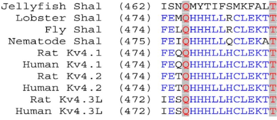
Kv4/Shal is a voltage-gated potassium ion channel whose sequence and function is conserved from jellyfish to humans312,313. Soon after the first cloning of these channels in
Kv4.3314. Since then, general studies on Kv4 channels have focused on its role in cellular excitability in the heart315–321, smooth muscle322–325 and lungs326,327, as well as in mammalian and
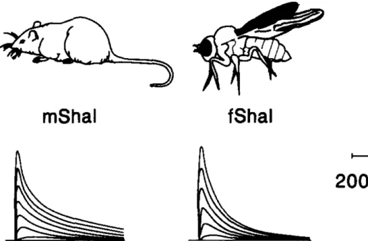
Drosophila neurons171,191,221,267,328–331. The mammalian Kv4 family of channels shares about 80% identity with Drosophila Kv4 (Shal), the only representative of Kv4 channels in fruit flies173,174,191,267,312,332. In the fly, the Shal gene contains two splice variants, Shal1 and Shal2, where the latter has a shorter C-terminus173.
Much of the work performed characterizing expression of Kv4 channels in neurons has been performed in mammalian systems, and most neuron-related expression studies have focused on Kv4.2 and Kv4.3 channels which show strong expression levels in the mammalian brain300,330,333–340. In 1998, Serôdio and Rudy (1998) reported a thorough examination of Kv4.1, Kv4.2, Kv4.3 mRNA levels and localization in the rat brain. Kv4.1 was found to have very low levels of transcript and protein expression in the central nervous system (CNS). Interestingly, when comparing expression of Kv4.2 and Kv4.3, they seem to have differing expression patterns in
separate areas of the brain. Kv4.2 was found to be present at high levels in the granule cells of the
olfactory bulb, in most of the basal ganglia, CA1 pyramidal and granule cells of the hippocampus, the paraventricular nucleus of the thalamus, the granular cell layer of the cerebellum, and in the pontine nucleus of the brain stem. Kv4.3 was measured to have high levels of expression in the
neocortex, in stratum interneurons and granule cells of the hippocampus, in the ventroposterior complex and laterodorsal nuclei of the thalamus, in Purkinje cells of the cerebellum, and in the substantia nigra and superior colliculi of the brain stem330. In Drosophila, Tsunoda and Salkoff (1995) determined that Kv4/Shal currents made up virtually all of the A-type currents present in the soma of embryonic neurons171.