Research
Report from SSM’s scientifi c council on
ionizing radiation within oncology, 2011
2012:20
SSM perspective Background
In 2009, the Swedish Radiation Safety Authority (Strålsäkerhetsmyn-digheten, SSM) appointed a scientific council on ionizing radiation within oncology. The council consists of scientific experts in the fields of oncology, radiobiology and medical physics. Their task is to annually review and evaluate scientific developments in radiotherapy and to give SSM advice in issues where a scientific examination of different views is necessary. The council began its work in the autumn of 2009 and this is the second report presented.
Objectives
The scientific council is obliged to produce an annual report on radioth-erapy issues. The report will summarize recent scientific knowledge.
Results
Many cancer patients are treated in accordance with written guidelines or clinical trial protocols. The scientific council states that the radioth-erapy part in those guidelines and protocols is less well specified than other therapies such as surgery and chemotherapy. This report identifies the key aspects of modern radiotherapy from international radiotherapy organisations and scientific papers in order to develop written radioth-erapy guidelines and clinical trial protocols.
Associated relevant information to be listed in protocol templates are analyzed and discussed. The report provides a framework for the des-cription of the entire radiotherapy process in both clinical care pro-grammes and trial protocols based on clinical as well as physical aspects. The framework includes preparatory imaging, specification of treatment prescription, relations to other therapies and treatment planning. The results are applicable in protocols for palliative care as well as for advan-ced treatments.
The report discusses the importance of clinical evaluation in clinical trials and in routine care as well as the importance to follow quality management guidelines when writing protocols and treatment program-mes. Finally the report also discusses special considerations for brachy therapy. The scientific council recommends SSM to promote the deve-lopment of protocol templates to use when writing the radiotherapy part in care programmes and clinical trial protocols.
Project information
Contact persons at SSM: Catarina Danestig Sjögren and Peter Björk Reference: SSM 2009/3757
2012:20
Authors: SSM´s scientific council on ionizing radiation within oncology
Report from SSM’s scientific council on
ionizing radiation within oncology, 2011
This report concerns a study which has been conducted for the Swedish Radiation Safety Authority, SSM. The conclusions and
view-Content
1. Introduction ... 3
2. Development of radiotherapy and review of current guidelines .... 5
3. Aim of the present report ... 7
4. Analysis of the modern radiotherapy process with suggestions for new guidelines ... 7
4.1. Preparatory imaging ... 7
4.2. Specification of treatment prescription ... 9
4.3. Relation to other therapies ... 13
4.4. Treatment planning ... 14
4.4.1. Treatment technique and equipment ... 14
4.4.2. Dose computation ... 14
4.4.3. Treatment plan optimisation and evaluation ... 15
4.5. Special considerations for brachytherapy ... 16
4.6. Clinical evaluation ... 17
4.7. Quality management ... 19
5. Conclusions ... 20
6. References... 21
1. Introduction
Advances in many aspects of diagnosis, staging and treatment have resulted in improved results with more patients living disease-free for long time periods.
Technical and computer improvements have facilitated this develop-ment in imaging, surgery and radiotherapy. At the same time new drugs with activity in at least sub-populations of many cancers have been developed. Combinations of treatments are used for more and more patients since these have shown superiority in clinical trials. The evidence-base is continuously increasing.
The development has improved outcome but also increased complexi-ty. The demands on written guidelines describing all relevant steps in the radiotherapy process therefore increase. In order to keep up with the increasing knowledge base and to secure high and equal quality for patients wherever they live, it has been realised that the guidelines must be (at least) national, although regional or local adaptations can be required. In Sweden, the six Regional Cancer Centres (RCCs) have the duty to co-ordinate this work. Regional or national guidelines (Care Programmes) have existed since decades, but have increased in numbers substantially during the past few years. The number of na-tional quality registries has increased in parallel. These allow evalua-tions of the quality of the intervenevalua-tions described in the Care Pro-grammes but are also a rich source for outcome research (1). Clinical trial protocols have sometimes also been integrated into the Care Pro-grammes, although most of them are kept separate.
Today many cancer patients are thus treated in accordance with writ-ten guidelines or clinical trial protocols. However, in combined treat-ment regimens, it is common that the radiotherapy part is less well specified than other therapies. Several very important parts of the ra-diotherapy process are not sufficiently well dealt with and thus open for local variations. A well-specified study protocol/clinical guideline is important, as it is fundamental for the evaluation of the outcomes, including patient-reported outcomes such as quality of life, and hence the development of future radiotherapy. It is known that protocol ad-herence is associated to better outcome in several malignancies (2-5). Well-specified treatment guidelines are important from a radiation protection point of view. Suboptimal radiotherapy routines may not only decrease the probabilities of tumour control but also increase the absorbed dose burden for the treated patients. Well-defined treatment protocols facilitate optimisation and evaluation of treatment planning and may therefore shorten the radiotherapy process, besides
improv-ing safety. Therefore, SSM’s scientific council has decided to focus on the writing of guidelines in this year’s report, to be applicable both for clinical guidelines/care programmes and for trial protocols. To our knowledge, there are no general guidelines for how to describe mod-ern advanced radiotherapy in protocols. The latest report on this topic was until recently the European Organisation for Research and Treat-ment of Cancer (EORTC) guidelines published by Bolla et al. more than 15 years ago (6), and, although it was detailed and has been use-ful for conventional radiotherapy, it is not use-fully applicable for modern techniques. The Radiation Oncology Group within EORTC realised this and recently published an update of guidelines on how to write clinical trial protocols involving advanced radiation therapy tech-niques (7).
The members of the scientific council on ionising radiation within oncology producing this report were as follows:
Professor Klas Blomgren, paediatric oncologist
Barncancercentrum, Drottning Silvias barn- och ungdoms-sjukhus, Göteborg
Associate professor Crister Ceberg, medical physicist
Avdelningen för Medicinsk Strålningsfysik, Lunds Universitet, Lund
Associate professor Giovanna Gagliardi, medical physicist
Avdelningen för sjukhusfysik, Karolinska Universitetssjukhu-set, Stockholm
Professor Bengt Glimelius, oncologist (chairman)
Onkologiklinikerna, Akademiska sjukhuset, Uppsala och Karo-linska Universitetssjukhuset, Stockholm
PhD Mikael Johansson, oncologist (secretary)
Cancercentrum Norrlands Universitetssjukhus Umeå
Associate professor Elisabeth Kjellén, oncologist
Skånes onkologiska klinik, Skånes Universitetssjukhus Lund
Professor Per Nilsson, medical physicist
Skånes Onkologiska klinik, Skånes Universitetssjukhus Lund
Professor Sten Nilsson, oncologist
2. Development of radiotherapy and
re-view of current guidelines
The continuous and fast advancements in radiation therapy and imag-ing technology, together with a growimag-ing body of knowledge and evi-dence in the field of clinical radiobiology, are constantly changing the radiotherapy world. Several new diagnostic modalities are available, and a whole new area has emerged related to image registration. New hybrid imaging modalities for target definition, new devices able to combine advanced imaging techniques and dose delivery have been developed. New questions regarding the management of heterogene-ous and moving targets have received increasing attention. New rec-ommendations from the International Commission on Radiation Units and Measurements (ICRU) have been issued for prescribing and re-porting intensity modulated radiotherapy (8).
Perhaps the most dramatic change, however, lies in the introduction of inverse treatment planning. This requires that treatment objective and constraints are specified and prioritised with more care and in greater detail than before. For the realisation of inverse optimised treatment plans, many new delivery modalities such as intensity-modulated ra-diation therapy (IMRT), volumetric modulated arc therapy (VMAT) and tomotherapy are available. Some of the novel beams have uncon-ventional spectral characteristics, and there are other new dose compu-tation and optimisation issues as well, that need to be specified in more detail in modern radiotherapy protocols. Due to the complicated dose- and volume prescription required for an inverse optimised treatment plan, also the plan evaluation procedure must be revised accordingly. Different priority orders and objective weighting factors may need to be evaluated in parallel by specially devised procedures. Possibilities to invoke radiobiological models for tumour control and normal tissue complication probabilities are also developed. A new area is also image-guided radiotherapy (IGRT). This brings along many variables that require specification in order to ensure the desired level of precision throughout the course of the treatment. Finally, the substantially increased complexity of the radiotherapy implies that aspects on quality assurance (QA) require significant updates.
Changes come also from other fields; independently of the technolog-ical development, a paradigm shift was introduced about twenty years ago in radiation oncology by the so-called stereotactic radiotherapy (9, 10). This brought new knowledge about tumour and normal tissue response when treating with extreme hypofractionated treatment schedules. This has strongly affected treatment choices in several di-agnoses (9-12). As a result of this technical solution, an increasing number of hypofractionation treatment protocols are nowadays ap-plied in routine clinical practice and in clinical trials. The
develop-ment is also supported by new data about radiation and fractionation sensitivity of tumour cells, which helps to tailor fractionation proto-cols according to specific parameters (13, 14). Inhomogeneous dose prescription to the target has become a general concept, to be obtained during one treatment course or as a result of the combination with a boost. Furthermore, combined modalities, e.g. external radiation ther-apy and brachytherther-apy, are also getting increased interest.
In some cases, radiotherapy protocols for specific pathologies are ac-cessible via the internet, e.g. the Cancer institute NSW, Australia (https://www.eviq.org.au/); most of them, however, are prepared ac-cording to local features. At another level societies and organisations are acting to adapt general radiation therapy guidelines to the new era. This is more a necessity than a need; when the present intention of most centres is to increase patient accruals in trials the harmonisation among both methods and contents of prescription and reporting is a condition for realising this. The work of framing the description of radiation therapy in general programmes, regional, national or interna-tional, is as usual a major challenge. Although not a general guideline, a prescription template for protocols from the Radiation Therapy On-cology Group (RTOG) is available at their website
(http://www.rtog.org). This document focuses on dose prescription aspects, and includes a discussion on the balance between dose pre-scription requirements and patient accrual.
The only general guidelines that we know of are those of the EORTC. The first EORTC guidelines from 1995 are divided into several sec-tions describing the radiotherapy process in subsequent steps, includ-ing preparation (positioninclud-ing of the patient, patient data acquisition, and volumes of interest), treatment planning (treatment technique, normal tissue sparing, dose computation, and dose specification), sim-ulation (simsim-ulation procedure), and delivery (equipment, treatment verification) (6). A separate section on brachytherapy is also included, as well as a chapter on QA. The EORTC guidelines follow the treat-ment preparation procedure and the treattreat-ment process, which makes it still useful for conventional radiotherapy. For modern advanced radio-therapy, however, it needs to be updated. Since this report was writ-ten, significant changes have been introduced in many links of the radiotherapy chain as briefly outlined above. EORTC has, in parallel to the development of our report, also recognised the lack of modern guidelines and their second report (7) was published at the time of completion of our report. They mostly focus on the description of clin-ical trial protocols involving advanced radiation techniques and then mainly discuss the clinical aspects in the trial protocols. Implementa-tion of QA procedures for advanced radiotherapy is also briefly de-scribed.
3. Aim of the present report
The purpose of this report is to identify important aspects of modern radiotherapy in order to assist in writing of radiotherapy protocols and clinical guidelines. The modern radiotherapy process and relevant issues to be listed in protocol templates are analysed and discussed. This report provides support and is applicable in the writing of proto-cols for both simple treatments used, e.g. in palliative situations, to advanced treatments where the highest requirements should be used or tested.
4. Analysis of the modern radiotherapy
process with suggestions for new
guide-lines
This section describes the modern radiotherapy process in a way suit-able for writing the radiotherapy part in guidelines/clinical trial proto-cols. The radiotherapy process is analysed, relevant issues are ad-dressed, and in some cases the implementation of new approaches are suggested.
Factors that must be established before the patient is referred to the radiotherapy department (e.g. patient prognostic factors such as age, co-morbidities, performance status; tumour prognostic factors such as differentiation, molecular characteristics, staging; and treatment inten-tion) have generally been addressed at multi-disciplinary conferences, and will not be discussed further. The choice of proper diagnostic tools for determining these factors is also not within the scope of this presentation. However, the entire radiotherapy process is based on these pre-therapeutic decisions, and it is essential that consistency is preserved through all following steps. Procedures for preparatory im-aging, treatment planning, delivery and evaluation must all be de-signed in alignment with the intentions.
4.1. Preparatory imaging
The preparatory process is aimed to provide the basis for target defini-tion, treatment planning and image guidance procedures during the course of the treatment. In order to support co-registration of data from different imaging modalities, and to provide a rigid frame for image guidance, it is essential for all procedures that the patient im-mobilisation and positioning are well defined. Target definitions are generally performed on a computer tomography (CT) data set, with or without contrast agents, and supplemented with patient-specific clini-cal information. This data set is generally also the foundation for the
absorbed dose calculations performed in the treatment planning sys-tem. Since imaging parameters may have great influence on both vol-ume delineation and absorbed dose calculations, it is important that all such information is properly chosen and specified.
In case additional imaging (e.g. magnetic resonance imaging (MRI) or positron emission tomography (PET)) is required, careful attention should be given to co-registration procedures, in particular if the data originates from separate machines (15-17). Rigid or deformable regis-tration procedures may have different requirements (18, 19).
In cases where motion needs to be taken into account, time-dependent data (4D) are crucial (20, 21). The proper method of 4D imaging de-pends on the intended type of treatment delivery motion management (22-25). In its simplest form, the 4D data set can be used to apply nec-essary margin expansion to the target volume(s) and organs at risk. More advanced forms of motion management include gating and tracking, which require time resolved image data sets (26-29). It is important to realise that the achievable margins are strictly related to the type and the frequency of the treatment verification procedure (see Ch 4.6).
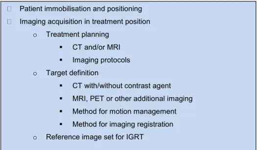
Figure 1. Summary of preparatory imaging
Patient immobilisation and positioning Imaging acquisition in treatment position
o Treatment planning CT and/or MRI Imaging protocols o Target definition
CT with/without contrast agent MRI, PET or other additional imaging Method for motion management Method for imaging registration o Reference image set for IGRT
4.2. Specification of treatment prescription
A specific statement of a radiation treatment objective includes infor-mation on both geometry (volume specification) and dosimetry (ab-sorbed dose prescription).
Volume specifications and absorbed dose prescriptions should be specified in accordance with the recommendations by the ICRU (8, 30-32). It should be stated how the gross tumour volume (GTV) and organ-at-risk volumes (OAR) are delineated, and which diagnostic tools are used. The delineation in three-dimensions of normal tissues is often affected by large inter-observers variability. This is also due to indistinctness of instructions, beyond the lack of an adequate imaging platform, as also the Quantitative Analysis of Normal Tissue Effects in the Clinic (QUANTEC) review (33) has underlined. In order to decrease this variability and to promote consistency in normal tissue delineation among observers and among centres several atlases have been prepared (34, 35).
If the GTV is considered as composed of several sub-volumes, this should be stated specifically. For the clinical target volume (CTV) and internal target volume (ITV), instructions for the delineation should be included. It is important to state the grey scale window settings to be used when defining the different target volumes. The planning target volume (PTV) and planning organ-at-risk volumes (PRV) are assumed to account also for motion related uncertainties, most commonly by applying an uncertainty margin to the CTV, determining the PTV (and similar for the PRV). The margins applied depend on equipment and patient immobilisation, and should be stated. The size of the margin can be based on direct or indirect observations of target motion in real patients, and statistical models have been developed for use in clinical practice (36).
The naming of target and OAR volumes should be specified in the protocol. Suggestion for a standardised naming convention for use in radiation therapy was recently proposed by a number of international radiotherapy societies (37).
There are alternative possibilities to account for motion related uncer-tainties. One way is to include the spatial distribution into the treat-ment planning calculations by convolving the dose calculation with a motion function, with the result that larger fields are required (38). In any case, the additional margin for motion uncertainties leads to larger irradiated volumes, and potentially an increased risk for unwanted effects on surrounding tissues. In the case of breathing motions, the required margins become particularly large, and as a consequence, other alternatives have been developed (23).For instance, the patient can be asked to hold breath during the time of irradiation (39). Active
breath-hold techniques are designed to support the patient not to breathe for prolonged times (40). Another alternative, which is more comfortable for the patient, is to gate the irradiation for certain parts of the respiratory cycle in order to minimise the influence of the mo-tion during the time of irradiamo-tion (41). This approach also has the advantage that the irradiation window can be chosen for a phase in the respiratory cycle when the target is in a favourable position relative to nearby risk organs (42). The most advanced alternative for motion management is to use tumour tracking. This can be realised by using a linac mounted on a robotic arm (43), or, on a conventional treatment unite by utilising the multileaf collimator dynamically to conform to the target motion in real time (44, 45). This method has the advantage of both higher delivery efficiency and less residual target motion than the breath-hold and gating techniques.
As mentioned above, a complete prescription includes both volume and dose specifications, preferably in terms of composite dose-volume objectives for each structure. The situation is further complicated by the fact that the two main objectives of radiotherapy, i.e. to treat the target tissue and to spare the healthy tissue, are always mutually con-flicting. Therefore, the relative importance of these two objectives must be stated explicitly in the treatment prescription. If this is not the case, the same prescription would not be able to discriminate between, on the one hand, a plan that emphasises good target coverage at the expense of somewhat higher doses to the risk organs, and on the other hand, a plan that emphasises a low dose to the risk organs at the ex-pense of less target coverage (within clinically acceptable limits). In-deed, there is often more than one organ of interest, and sometimes there are also differentiated target volumes. In such cases, the trade-off between the different treatment objectives will have multiple di-mensions, which makes it even more challenging to pin down a well-specified treatment prescription (the American Association of Physi-cists in Medicine (AAPM) Summer School 2011). Generally speak-ing, a complete IMRT treatment prescription will require specified objectives for close to every region exposed to the treatment. The rea-son is that any region that is not explicitly constrained may be used freely by the optimiser, in which case unexpected results may occur (46). Two plans may look nearly equivalent, except perhaps for a hotspot that may have occurred outside any constrained region of in-terest, and which could easily be overlooked in the treatment plan evaluation.
One possible way to address this issue may be to device criteria de-scribing what to prioritize when producing, evaluating and comparing plans. This could mean, for example, having CTV coverage as a pri-mary priority, then to uphold dose restrictions to critical organs as a
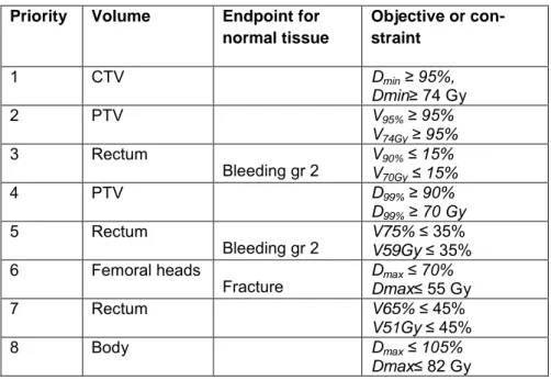
OARs as a fourth, and finally dose conformity. An example of such a prescription priority is presented in Table 1. These absorbed dose ob-jectives and constraints could be based on published guidelines e.g. QUANTEC data (47).
Table 1. Example of dose prescription Priority Volume Endpoint for
normal tissue Objective or con-straint 1 CTV Dmin ≥ 95%, Dmin≥ 74 Gy 2 PTV V95% ≥ 95% V74Gy ≥ 95% 3 Rectum Bleeding gr 2 V90% ≤ 15% V70Gy ≤ 15% 4 PTV D99% ≥ 90% D99% ≥ 70 Gy 5 Rectum Bleeding gr 2 V75% V59Gy ≤ 35% ≤ 35% 6 Femoral heads
Fracture DDmaxmax ≤ 70% ≤ 55 Gy
7 Rectum V65% ≤ 45%
V51Gy ≤ 45%
8 Body Dmax ≤ 105%
Dmax≤ 82 Gy
Dose prescription for a prostate cancer case. Observe that this is not a specific recommendation but an example of a dose prescription with prioritised objec-tives.
An emerging alternative is to prescribe a radiation treatment in terms of radiobiological effect parameters, such as a minimal required tu-mour control probability (TCP) and maximum tolerable normal tissue complication probabilities (NTCPs) for the exposed healthy organs (48). The risk for treatment-induced cancer is also an important side effect of radiotherapy that may be specified in the prescription of a radiation treatment in the future (49).
Finally, the temporal aspects of the treatment delivery, i.e. time and fractionation scheme is an important part of the radiotherapy prescrip-tion, in particular if unconventional fractionation schemes are used, e.g. two fractions per day. In this case it is important to specify the minimum required time between the two fractions to allow for an as complete as possible repair, e.g. at least 7h (50, 51). Note that the con-straints used for conventional fractionation should not be applied to other fractionation schedules without considering correction for frac-tionation effects (47, 52).
The importance of keeping the total treatment time and its influence on tumor control has been described by several authors (53-56). There should therefore be a plan for the management of unintended interrup-tions (57). Well-described examples are available by Dale et al. 2002 and Jones et al. 2007 (58, 59).
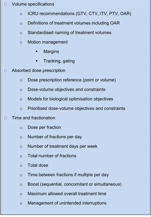
Figure 2. Summary of specification of treatment prescription (volume and dose)
Volume specifications
o ICRU recommendations (GTV, CTV, ITV, PTV, OAR) o Definitions of treatment volumes including OAR o Standardised naming of treatment volumes o Motion management
Margins
Tracking, gating Absorbed dose prescription
o Dose prescription reference (point or volume) o Dose-volume objectives and constraints o Models for biological optimisation objectives o Prioritised dose-volume objectives and constraints Time and fractionation
o Dose per fraction
o Number of fractions per day
o Number of treatment days per week o Total number of fractions
o Total dose
o Time between fractions if multiple per day o Boost (sequential, concomitant or simultaneous) o Maximum allowed overall treatment time
4.3. Relation to other therapies
Radiotherapy is one of several modalities used in the management of most cancers, in which case the relevant timing in relation to surgery and/or chemotherapy (or other drugs) and potential interactions be-tween the therapy modalities must be stated.
Surgery is often part of the cancer therapy. Radiotherapy may then be used preoperatively to allow surgery (down-sizing or down-staging) or minimise the risk of recurrence (neo-adjuvant) or postoperatively to minimise the risk of loco-regional recurrence (adjuvant). Occasional-ly, intraoperative therapy is used. In the case of preoperative radio-therapy, the treatment intention should be stated, i.e. neo-adjuvant treatment, down-staging or downsizing (60). Since it is important to keep the times between the radiation treatment and surgery, whether given pre- or postoperatively, time limits should be properly defined. Preoperatively there may be specific time windows to minimise tox-icity and prevent tumour repopulation and good co-operation between the different involved departments is required. Allowing too little time after surgery makes wound healing difficult, and on the other hand, too protracted time gaps will increase the probability of tumour recur-rence.
If concomitant chemotherapy or other drugs are given the timing of the radiation treatments and the drug administration should be careful-ly stated. When using drugs in combination with radiotherapy unex-pected toxicity may occur. In the evolution of new drugs, especially targeted therapies, little is known of combinatory effects. Special care must thus be considered when using known constraints for normal tissues, when combining radiotherapy and drugs (33, 49, 61, 62). Reporting of late effects are scarce, and in particular when radiothera-py and drugs are combined (33, 49). It is preferable that the protocol includes reporting of late damage with prolonged follow-up of the patients. Better routines how to facilitate long-term follow-up, also outside trial protocols, are of great importance to increase knowledge about late effects from combined treatments, and should be developed.
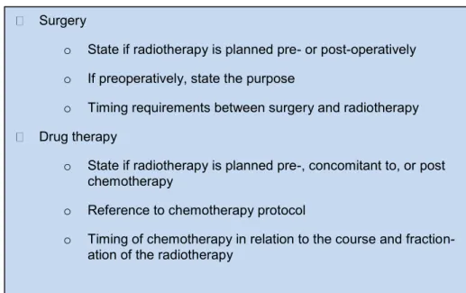
Figure 3. Combined treatments
4.4. Treatment planning
Treatment planning is a central part of the radiotherapy process and needs to be clearly specified in clinical guidelines and study protocols. The process may be subdivided in discretion of treatment technique and equipment, dose computation and treatment plan optimisation and evaluation.
4.4.1. Treatment technique and equipment
If the treatment objectives are complete and well specified, the choice of treatment technique and equipment is subordinated. However, due to the vastly different treatment techniques available today, it may in some situations be desirable to specify allowed alternatives.
4.4.2. Dose computation
It is important to ensure and uphold traceability to international do-simetry standards. This is generally achieved by following interna-tional reference dosimetry protocols, e.g. the Internainterna-tional Atomic Energy Agency (IAEA) TRS-398 (63). Participation in dosimetry au-dits are highly recommended in order to ensure high quality (64-66). The dose computation model in the treatment planning system may have limitations, regarding for instance heterogeneity correction mod-els. It is mandatory to specify the type of dose computation algorithm (67) etc., and to be aware of its applicability in clinically relevant
Surgery
o State if radiotherapy is planned pre- or post-operatively o If preoperatively, state the purpose
o Timing requirements between surgery and radiotherapy Drug therapy
o State if radiotherapy is planned pre-, concomitant to, or post chemotherapy
o Reference to chemotherapy protocol
o Timing of chemotherapy in relation to the course and fraction-ation of the radiotherapy
ometries (64, 67-72). Preferably calculation uncertainties should be estimated (23).
Guidelines should include requirements on the size and spatial resolu-tion of the dose matrix, since the evaluaresolu-tion of dose volume histo-grams (DVH) and DVH-parameters may be greatly distorted if the dose matrix is not adequately adjusted to the dose gradients and the size of the smallest volumes of interest.
4.4.3. Treatment plan optimisation and evaluation
In principle, a rigorous evaluation of a treatment plan would require a thorough review of the dose distribution in full 3D, i.e. in all the avail-able 2D sections. However, this is not practical. Instead, the infor-mation of the dose distribution is generally condensed into voxel sta-tistics (DVH) without providing spatial information (73). In the inter-est of having a single quantitative measure of the objective, DVHs information are often further condensed into percentiles (such as Dvol or Vdose), arithmetic means, generalised means, e.g. equivalent uni-form dose (EUD).This reduction may be necessary in order to obtain manageable quantitative measures, but one should be aware of the fact that, potentially critical information may be lost in the process. Objec-tives expressed in terms of radiobiological models are likely to be used clinically more often in the future, but the same principles should apply. However, it is important to note that in order to measure the actual degree of objective fulfilment, the treatment plan should be evaluated in terms of the same parameters used in the prescription. An NTCP evaluation is therefore only relevant if also the prescription is stated in terms of NTCP.
As discussed in section 4.2 above, inverse treatment planning requires that the constraints and optimisation objectives are prescribed in great detail and that their relative importance is specified accordingly (Ta-ble 1). In practice this is a matter of determining the proper weighting factors for the optimisation algorithm. Given that there are multiple constraints and optimisation objectives, the optimisation algorithm can only produce so called optimal results (74-76). A
Pareto-optimal solution is Pareto-optimal for a specific set of weighting factors, whereas another set of weighting factors gives another Pareto-optimal result. One objective may then be improved, although it will be at the expense of another. In order to obtain a treatment plan fulfilling the intentions, it is advisable to systematically vary the involved
weighting factors. This approach allows the whole trade-off between the prescribed objectives to be considered when selecting the final treatment plan.
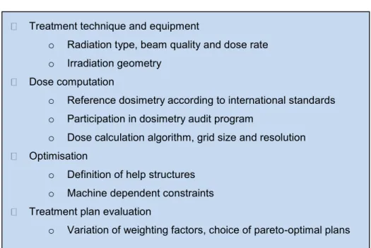
Figure 4: Summary of treatment plan optimisation
4.5. Special considerations for brachytherapy
Brachytherapy (BT) is increasingly being used in modern curative treatment of cancer. It can be applied either as monotherapy or as a boost to part of a larger volume treated with external beam therapy. BT can be used as an after-loading technique with high dose-rate (HDR), low dose-rate (LDR) or pulsed dose-rate (PDR) mode. The technique can also be used as a permanent implant. BT is a multifac-eted technique, where clinicians and physicists have developed rec-ommendations and guidelines for each and one of the different tu-mor/organ areas. Examples are for prostate cancer (77-80), gynecolog-ical malignancies (81-87), breast (88), and head and neck cancers (89).
BT requires a multimodal team work where oncologists, radiothera-pists, medical physicists, anesthesiologists and dedicated radiotherapy nurses are actively involved. It is also important to involve diagnostic radiologists and the modern radiological imaging techniques for delin-eation of tumor extension.
Similar types of anatomy-based algorithms as the ones used for dose calculation in external beam radiotherapy are now being increasingly applied in BT. The treatment planning systems are based on modern imaging techniques such as ultrasound, MRI and CT. The dose plan-ning can be performed prior to therapy, thus giving information on the precise placement of applicators/needles in the tumor/organ. On-line
Treatment technique and equipment
o Radiation type, beam quality and dose rate o Irradiation geometry
Dose computation
o Reference dosimetry according to international standards o Participation in dosimetry audit program
o Dose calculation algorithm, grid size and resolution Optimisation
o Definition of help structures o Machine dependent constraints Treatment plan evaluation
tems, been more frequently used during the last years in treatment of malignancies such as e.g. prostate cancer. This has streamlined the process and at the same time reduced the risk of repeating the pre-planning procedure due to unforeseen anatomic obstacles such as in-terfering parts of the skeleton or adjacent organs. It is important that the physician in charge assures before start of treatment that the dose prescribed to the planning target volume is obtained and that dose constraints in organs at risk are kept.
Over the years, separate guidelines have, as mentioned above, been developed for various tumor diagnoses. These guidelines have all formed a major step forward in defining how and when to use the dif-ferent BT techniques described above. The majority of the guidelines focus on practical aspects related to a specific tumour diagnosis. The-se also include important aspects on topics such as radiation protec-tion, quality assurance and education. Still, there is a need, as in exter-nal beam radiotherapy, for further development of guidelines how to write protocols for clinical trials of BT. Such work is currently ongo-ing.
4.6. Clinical evaluation
In clinical radiotherapy assessment of acute and late toxicity and effi-cacy is essential to evaluate the treatment to improve outcome for fu-ture patients. To define criteria for radiotherapy follow-up is important not only in clinical trials but also in routine care.
During the course of the treatment acute side effects should be moni-tored and adequate actions taken. This is routine at all radiotherapy departments and may result in treatment modifications. In chemother-apy, dose modification after monitoring and grading of acute toxicity is routine and well described in protocols since decades. This ap-proach may be of relevance also in dose-intense radiotherapy, in par-ticular when combined with drugs and requires the use of pre-defined toxicity assessment scales such as common terminology criteria for adverse events (CT-CAE), EORTC/RTOG.
In adaptive radiotherapy the treatment is adjusted during the course according to tumour response or toxicity in order to maximise tumour control and/or minimise toxicity in the individual patient (90).For this approach a more extensive monitoring than presently used is required during treatment. Early assessment (within days or a few weeks after treatment start) of response in order to change treatment schedule or modality during the radiotherapy course is one example of adaptive radiotherapy. This requires specifications of early response assessment modality and timing of the assessment. Presently much research is devoted to repeated PET or MRI imaging during therapy. Actions
tak-en to insufficitak-ent response could be modified radiotherapy (increased dose, changed fractionation or modified target volume) or a change of treatment modality (like surgery rather than radio-chemotherapy, as presently explored in oesophageal cancer).
Follow-up of tumour response is essential for evaluation of radiother-apy. It should be stated in the protocol the timing between the end of therapy and first evaluation as well as the timing of subsequent evalu-ations. It is also important to state the method for evaluation, e.g. MRI, CT, PET-scan, clinical evaluation with biopsy and pathological assessment depending on the disease in addition to the intention of the radiotherapy. Response criteria should follow international recom-mendations and disease progression should be recorded as local, re-gional or distant progression. In radiotherapy local control is in most cases a much better outcome measure than grading of response ac-cording to tumour shrinkage as assessed by imaging. Quality of life, if considered to be of relevance, should be assessed at pre-defined time-points using validated questionnaires. In case of reporting to quality registries, the actually given therapy should be reported, which some-times deviates from the originally intended therapy.
Recording of late toxicity is essential in radiotherapy but is often ne-glected due to lack of formal follow-up schedules in everyday prac-tice. Appropriate assessment of late toxicity is seldom done in clinical trials due to the long follow-up times needed. As previously stressed (49) this is particularly important using new radiotherapy techniques such as IMRT alone or in combinations with drugs. The reporting of late toxicity should therefore be included in future quality registries where radiotherapy is a treatment option. A fundamental requirement for description of late toxicity is an adequate description of given ra-diotherapy and a systematic follow-up of patients. Follow-up of late toxicity in clinical trials may require a more detailed description of late toxicity with special consideration to OAR function. In such cases a description of assessment method and timing of evaluation is neces-sary.
In Sweden there is today no central registry for reporting serious un-expected adverse events of radiotherapy. To improve safety this should be initiated by the regulatory authorities. Reporting to such a registry should be mandatory as is the case for drug side effects (49). In order to increase the likelihood of early detection of potential mis-treatment, it is strongly advisable to be observant to any complaints patients may have during or after the treatment session (91). All inci-dents and acciinci-dents should be reported via established local and/or national systems.
4.7. Quality management
Existing quality management guidelines should be followed when writing radiotherapy study protocols and treatment programmes. Gen-eral quality management tools, in addition to process mapping and risk analysis (92), contain parts that should be specified in care pro-grammes and study protocols.
This includes dummy run procedures that are performed before the start of a new radiotherapy study, or the commissioning of a new treatment modality. Dummy runs can also be performed during on-going trials in order to check and further improve the protocol compli-ance.
A pretreatment dummy run should be performed when a new trial/care program is initiated. The main purposes of the dummy runs are to as-sure compliance to the study and to find any ambiguities in the guide-lines. The dummy run often consists of two parts, i.e. segmentation of structures (targets and organs at risk) and treatment planning (93-97). For dosimetry (measurements and independent calculations) and IGRT procedures (on-line and off-line), both pre- and during treat-ment, a strategy has to be defined to identify and act about the correc-tion of the systematic deviacorrec-tion as compared to the treatment plan. This means, for instance, that acceptance criteria for the evaluation should be stated. The additional absorbed dose contribution due to IGRT procedures shall be estimated (49). For international guidelines, reports and other related documents, see e.g.:
AAPM reports at:
http://www.aapm.org/pubs/reports/
American Society for Radiation Oncology (ASTRO) reports at:
https://www.astro.org/ ClinicalPractice/Guidelines/Index.aspx
European Society for Radiotherapy & Oncology (ESTRO) guidelines at:
http://www.estroeducation.org/publications/Pages/ESTROPhysicsBoo klets.aspx
IAEA documents at:
https://rpop.iaea.org/RPOP/RPoP/Content/InformationFor/HealthProfe ssionals/2_Radiotherapy/index.htm
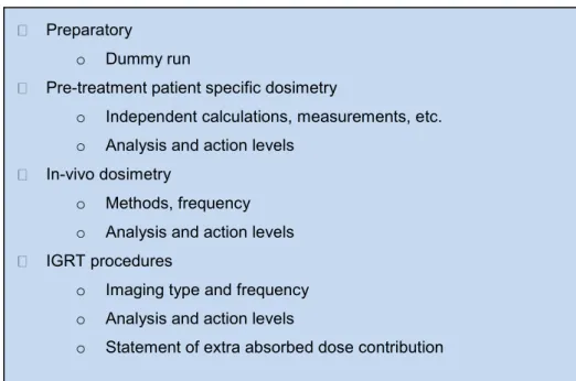
Figure 5. Summary of quality control
5. Conclusions
New guidelines for writing protocols for modern radiotherapy are re-quired. The scientific council suggests that SSM promotes the devel-opment of protocol templates to be used when writing the radiothera-py part in care programmes and clinical trial protocols. The recently published EORTC guidelines mainly focus on the clinical aspects of clinical trial protocols. In the present report our intention has been to provide a detailed framework for the entire radiotherapy process in-cluding clinical as well as physical aspects for the description of the radiotherapy process in both clinical care programmes and trial proto-cols.
Preparatory
o Dummy run
Pre-treatment patient specific dosimetry
o Independent calculations, measurements, etc. o Analysis and action levels
In-vivo dosimetry
o Methods, frequency o Analysis and action levels IGRT procedures
o Imaging type and frequency o Analysis and action levels
6. References
1. Rosén M uldgru an i h lso-och s u rden. S eriges o uner och Landsting, Stockholm), pp 1-284.
2. Abrams RA, et al. (2012) Failure to adhere to protocol specified radiation therapy guidelines was associated with decreased survival in RTOG 9704--a ph9704--ase III tri9704--al of 9704--adjuv9704--ant chemother9704--apy 9704--and chemor9704--adiother9704--apy for patients with resected adenocarcinoma of the pancreas. International
journal of radiation oncology, biology, physics 82(2):809-816.
3. Gebski V, Lagleva M, Keech A, Simes J, & Langlands AO (2006) Survival effects of postmastectomy adjuvant radiation therapy using biologically equivalent doses: a clinical perspective. J of the National Cancer Institute 98(1):26-38.
4. Miralbell R, et al. (1997) Pediatric medulloblastoma: radiation treatment technique and patterns of failure. International journal of radiation
oncology, biology, physics 37(3):523-529.
5. Peters LJ, et al. (2010) Critical impact of radiotherapy protocol compliance and quality in the treatment of advanced head and neck cancer: results from TROG 02.02. Journal of Clinical Oncology 28(18):2996-3001.
6. Bolla M, et al. (1995) EORTC guidelines for writing protocols for clinical trials of radiotherapy. Radiotherapy and Oncology 36(1):1-8.
7. Fairchild A, et al. (2012) Development of clinical trial protocols involving advanced radiation therapy techniques: The European Organisation for Research and Treatment of Cancer Radiation Oncology Group approach.
European journal of cancer (Oxford, England : 1990) 48(7):1048-1054.
8. ICRU (2010) Prescribing, recording, and reporting photon-beam intensity-modulated radiation therapy (IMRT) (ICRU report 83). Journal of the
ICRU 10(1):1-112.
9. Blomgren H, Lax I, Näslund I, & Svanström R (1995) Stereotactic high dose fraction radiation therapy of extracranial tumors using an accelerator. Clinical experience of the first thirty-one patients. Acta Oncologica 34(6):861-870.
10. Lax I, Blomgren H, Näslund I, & Svanström R (1994) Stereotactic
radiotherapy of malignancies in the abdomen. Methodological aspects. Acta
Oncologica 33(6):677-683.
11. Timmerman R, et al. (2011) Stereotactic body radiation therapy: normal tissue and tumor control effects with large dose per fraction. Frontiers of
radiation therapy and oncology 43:382-394.
12. Timmerman R, Heinzerling J, Abdulrahman R, Choy H, & Meyer JL (2011) Stereotactic body radiation therapy for thoracic cancers: recommendations for patient selection, setup and therapy. Frontiers of
radiation therapy and oncology 43:395-411.
13. Valdagni R, et al. (2011) Long-term biochemical control of prostate cancer after standard or hyper-fractionation: Evidence for different outcomes between low-intermediate and high risk patients. Radiotherapy and
Oncology 101(3):454-459.
14. Yarnold J & Haviland J (2010) Hypofractionated Adjuvant Whole Breast Radiotherapy: Progress and Prospects. Acta Oncologica 49(8):1288-1292. 15. Daisne J-F, et al. (2003) Evaluation of a multimodality image (CT, MRI
patients: accuracy, reproducibility and consistency. Radiotherapy and
Oncology 69(3):237-245.
16. Li G, et al. (2008) Accuracy of 3D volumetric image registration based on CT, MR and PET/CT phantom experiments. Journal of applied clinical
medical physics 9(4):2781.
17. Mutic S, et al. (2001) Multimodality image registration quality assurance for conformal three-dimensional treatment planning. International journal
of radiation oncology, biology, physics 51(1):255-260.
18. Brock KK, et al. (2008) Improving image-guided target localization through deformable registration. Acta Oncologica 47(7):1279-1285. 19. Castadot P, et al. (2008) Comparison of 12 deformable registration
strategies in adaptive radiation therapy for the treatment of head and neck tumors. Radiotherapy and Oncology 89(1):1-12.
20. Li G, et al. (2008) Advances in 4D medical imaging and 4D radiation therapy. Technology in cancer research and treatment 7(1):67-81.
21. Low D (2011) 4D imaging and 4D radiation therapy: a New Era of therapy design and delivery. Frontiers of radiation therapy and oncology 43:99-117.
22. Ezhil M, et al. (2008) Comparison of Rigid and Adaptive Methods of Propagating Gross Tumor Volume Through Respiratory Phases of Four-Dimensional Computed Tomography Image Data Set. International
Journal of Radiation OncologyBiologyPhysics 71(1):290-296.
23. AAPM (2006) The management of Respiratory Motion in Radiation Oncology (AAPM Report 91). (AAPM, College Park), pp 1-80. 24. Potters L, et al. (2010) American Society for Therapeutic Radiology and
Oncology (ASTRO) and American College of Radiology (ACR) Practice Guidelines for Image-Guided Radiation Therapy (IGRT). International
journal of radiation oncology, biology, physics 76(2):319-325.
25. Rietzel E, Pan T, & Chen GTY (2005) Four-dimensional computed tomography: image formation and clinical protocol. Medical Physics 32(4):874-889.
26. Falk M, et al. (2010) Real-time dynamic MLC tracking for inversely optimized arc radiotherapy. Radiotherapy and Oncology 94(2):218-223. 27. Knudtsen IS, et al. (2011) Dynamic respiratory gated 18FDG-PET of lung
tumors – a feasibility study. Acta Oncologica 50(6):889-896.
28. Korreman S, Persson G, Nygaard D, Brink C, & Juhler-Nøttrup T (2012) Respiration-correlated image guidance is the most important radiotherapy motion management strategy for most lung cancer patients. International
journal of radiation oncology, biology, physics.
29. Verellen D, et al. (2010) Gating and tracking, 4D in thoracic tumours.
Cancer radiothérapie 14(6-7):446-454.
30. ICRU (1999) Prescribing, Recording and reporting Photon Beam Therapy (ICRU report 62). (ICRU, Bethesda, MD).
31. ICRU (2004) Prescribing, recording, and reporting electron beam therapy (ICRU report 71). Journal of the ICRU 4(1):1-100.
32. ICRU (2007) Prescribing, Recording, and Reporting Proton-Beam Therapy (ICRU report 78). Journal of the ICRU 7(2):1-210.
33. Marks LB, et al. (2010) Use of normal tissue complication probability models in the clinic. International journal of radiation oncology, biology,
physics 76(3 Suppl):S10-19.
34. Feng M, et al. (2011) Development and validation of a heart atlas to study cardiac exposure to radiation following treatment for breast cancer.
35. Kong FM, et al. (2011) Consideration of dose limits for organs at risk of thoracic radiotherapy: atlas for lung, proximal bronchial tree, esophagus, spinal cord, ribs, and brachial plexus. International journal of radiation
oncology, biology, physics 81(5):1442-1457.
36. Van Herk M (2004) Errors and margins in radiotherapy. Seminars in
Radiation Oncology 14(1):52-64.
37. Santanam L, et al. (2012) Standardizing Naming Conventions in Radiation Oncology. International journal of radiation oncology, biology, physics. 38. McCarter SD & Beckham WA (2000) Evaluation of the validity of a
convolution method for incorporating tumour movement and set-up variations into the radiotherapy treatment planning system. Physics in
medicine and biology 45(4):923-931.
39. Pedersen AN, Korreman S, Nyström H, & Specht L (2004) Breathing adapted radiotherapy of breast cancer: reduction of cardiac and pulmonary doses using voluntary inspiration breath-hold. Radiotherapy and Oncology 72(1):53-60.
40. Wong JW, et al. (1999) The use of active breathing control (ABC) to reduce margin for breathing motion. International journal of radiation
oncology, biology, physics 44(4):911-919.
41. Korreman SS, Pedersen AN, Nottrup TJ, Specht L, & Nystrom H (2005) Breathing adapted radiotherapy for breast cancer: comparison of free breathing gating with the breath-hold technique. Radiotherapy and
oncology : journal of the European Society for Therapeutic Radiology and Oncology 76(3):311-318.
42. Korreman SS, et al. (2006) Reduction of cardiac and pulmonary
complication probabilities after breathing adapted radiotherapy for breast cancer. International Journal of Radiation OncologyBiologyPhysics 65(5):1375-1380.
43. Dieterich S & Gibbs IC (2011) The CyberKnife in clinical use: current roles, future expectations. Frontiers of radiation therapy and oncology 43:181-194.
44. Keall PJ, Siebers JV, Arnfield M, Kim JO, & Mohan R (2001) Monte Carlo dose calculations for dynamic IMRT treatments. Physics in medicine and
biology 46(4):929-941.
45. Poulsen PR, Fledelius W, Cho B, & Keall P (2012) Image-based dynamic multileaf collimator tracking of moving targets during intensity-modulated arc therapy. International journal of radiation oncology, biology, physics 83(2):e265-271.
46. Webb S (2004) Contemporary IMRT (Institute of Physics Publishing, London) p 492.
47. Marks LB, Ten Haken RK, & Martel MK (2010) Guest editors introduction to QUANTEC: a users guide. International journal of radiation oncology,
biology, physics 76(3 Suppl):S1-2.
48. Nahum AE & Kutcher J (2007) Biological evaluation of treatment plans.
Handbook of Radiotherapy, eds Mayles WPM, Nahum AE, & Rosenwald
J-C (Taylor and Francis), pp 731-788.
49. SSM (2011) Report from SSMs scientific council on ionizing radiation within oncology (SSM Report 2011:25). ed Glimelius B (Swedish Radiation Safety Authority, Stockholm), pp 1-64.
50. Ang KK (1998) Altered fractionation trials in head and neck cancer.
Seminars in Radiation Oncology 8(4):230-236.
51. Chen J, van de Geijn J, & Goffman T (1991) Extra lethal damage due to residual incompletely repaired sublethal damage in hyperfractionated and continuous radiation treatment. Medical Physics 18(3):488-496.
52. Bentzen S, et al. (2010) Quantitative Analyses of Normal Tissue Effects in the Clinic (QUANTEC): An Introduction to the Scientific Issues.
International journal of radiation oncology, biology, physics 76(S):S3-S9.
53. Bese NS, Hendry J, & Jeremic B (2007) Effects of prolongation of overall treatment time due to unplanned interruptions during radiotherapy of different tumor sites and practical methods for compensation. International
journal of radiation oncology, biology, physics 68(3):654-661.
54. Tarnawski R, et al. (2002) How fast is repopulation of tumor cells during the treatment gap? International journal of radiation oncology, biology,
physics 54(1):229-236.
55. Wyatt RM, Beddoe AH, & Dale RG (2003) The effects of delays in radiotherapy treatment on tumour control. Physics in medicine and biology 48(2):139-155.
56. Wyatt RM, Jones BJ, & Dale RG (2008) Radiotherapy treatment delays and their influence on tumour control achieved by various fractionation schedules. British Journal of Radiology 81(967):549-563.
57. RCR (2008) The timely delivery of radical radiotherapy: standards and guidelines for the management of unscheduled treatment interruptions. (Royal College of Radiologists, London), pp 1-37.
58. Dale RG, et al. (2002) Practical Methods for Compensating for Missed Treatment Days in Radiotherapy, with Particular Reference to Head and Neck Schedules. Clinical Oncology 14(5):382-393.
59. Jones B, Hopewell JW, & Dale RG (2007) Radiobiological compensation for unintended treatment interruptions during palliative radiotherapy.
British Journal of Radiology 80(960):1006-1010.
60. Valentini V, et al. (2009) Multidisciplinary Rectal Cancer Management: 2nd European Rectal Cancer Consensus Conference (EURECA-CC2).
Radiotherapy and Oncology 92(2):148-163.
61. De Ruysscher D (2006) Time Between the First Day of Chemotherapy and the Last Day of Chest Radiation Is the Most Important Predictor of Survival in Limited-Disease Small-Cell Lung Cancer. Journal of Clinical
Oncology 24(7):1057-1063.
62. Pijls-Johannesma M, et al. (2007) Timing of chest radiotherapy in patients with limited stage small cell lung cancer: a systematic review and meta-analysis of randomised controlled trials. Cancer Treatment Reviews 33(5):461-473.
63. Andreo P, et al. Absorbed Dose Deter ination in External Bea Radiotherapy. (IAEA, Vienna), pp 1-242.
64. Ibbott G, Ma CM, Rogers DWO, Seltzer SM, & Williamson JF (2008) Anniversary Paper: Fifty years of AAPM involvement in radiation dosimetry. Medical Physics 35(4):1418.
65. Knöös T, Medin J, & Persson L (2010) Standards, applications and quality assurance in medical radiation dosimetry (IDOS). in Preliminary results
from a dosimetric audit performed at swedish radiotherapy centres (IAEA,
Vienna), pp 365-376.
66. Palmer A, Mzenda B, Kearton J, & Wills R (2011) Analysis of regional radiotherapy dosimetry audit data and recommendations for future audits.
British Journal of Radiology 84(1004):733-742.
67. Morgan AM, Knöös T, McNee SG, Evans CJ, & Thwaites DI (2008) Clinical implications of the implementation of advanced treatment planning algorithms for thoracic treatments. Radiotherapy and Oncology 86(1):48-54.
68. Aarup LR, et al. (2009) The effect of different lung densities on the accuracy of various radiotherapy dose calculation methods: Implications for tumour coverage. Radiotherapy and Oncology 91(3):405-414. 69. Panettieri V, Barsoum P, Westermark M, Brualla L, & Lax I (2009) AAA
and PBC calculation accuracy in the surface build-up region in tangential beam treatments. Phantom and breast case study with the Monte Carlo code penelope. Radiotherapy and Oncology 93(1):94-101.
70. Panettieri V, et al. (2010) Influence of dose calculation algorithms on isotoxic dose-escalation of non-small cell lung cancer radiotherapy.
Radiotherapy and Oncology 97(3):418-424.
71. ESTRO (2004) Quality assurance of treatment planning systems practical examples for non-imrt photon beams (ESTRO booklet 7). (ESTRO, Brussels), pp 1-104.
72. ESTRO (2008) Guidelines for the verification of IMRT (ESTRO booklet 9). (ESTRO, Brussels), p 136.
73. Allen Li X, et al. (2012) The use and QA of biologically related models for treatment planning: Short report of the TG-166 of the therapy physics committee of the AAPM. Medical Physics 39(3):1386.
74. Ottosson RO, et al. (2009) The feasibility of using Pareto fronts for comparison of treatment planning systems and delivery techniques. Acta
Oncologica 48(2):233-237.
75. Pettersson D, et al. (2010) Interim analysis of the Stockholm III trial of preoperative radiotherapy regimens for rectal cancer. The British Journal of
Surgery 97(4):580-587.
76. Boyd S & Vandenberghe L (2004) Convex optimization (Cambridge University Press, Cambridge) p 730.
77. Ash D, et al. (2000) ESTRO/EAU/EORTC recommendations on permanent seed implantation for localized prostate cancer. Radiother Oncol 57(3):315-321.
78. Kovacs G, et al. (2005) GEC/ESTRO-EAU recommendations on temporary brachytherapy using stepping sources for localised prostate cancer. Radiother Oncol 74(2):137-148.
79. Rosenthal S, et al. (2011) American Society for Radiation Oncology (ASTRO) and American College of Radiology (ACR) practice guideline for the transperineal permanent brachytherapy of prostate cancer. Int J
Radiat Oncol Biol Phys 79(2):335-341.
80. Salembier C, et al. (2007) Tumour and target volumes in permanent prostate brachytherapy: a supplement to the ESTRO/EAU/EORTC recommendations on prostate brachytherapy. Radiother Oncol 83(1):3-10. 81. Dimopoulos JC, et al. (2012) Recommendations from Gynaecological
(GYN) GEC-ESTRO Working Group (IV): Basic principles and parameters for MR imaging within the frame of image based adaptive cervix cancer brachytherapy. Radiother Oncol 103(1):113-122. 82. Haie-Meder C, et al. (2005) Recommendations from Gynaecological
(GYN) GEC-ESTRO Working Group (I): concepts and terms in 3D image based 3D treatment planning in cervix cancer brachytherapy with emphasis on MRI assessment of GTV and CTV. Radiother Oncol 74(3):235-245. 83. Hellebust TP, et al. (2010) Recommendations from Gynaecological (GYN)
GEC-ESTRO Working Group: considerations and pitfalls in
commissioning and applicator reconstruction in 3D image-based treatment planning of cervix cancer brachytherapy. Radiother Oncol 96(2):153-160. 84. Lee LJ, et al. (2012) American Brachytherapy Society consensus guidelines
for locally advanced carcinoma of the cervix. Part III: low-dose-rate and pulsed-dose-rate brachytherapy. Brachytherapy 11(1):53-57.
85. Potter R, et al. (2006) Recommendations from gynaecological (GYN) GEC ESTRO working group (II): concepts and terms in 3D image-based treatment planning in cervix cancer brachytherapy-3D dose volume parameters and aspects of 3D image-based anatomy, radiation physics, radiobiology. Radiother Oncol 78(1):67-77.
86. Viswanathan AN, et al. (2012) American Brachytherapy Society consensus guidelines for locally advanced carcinoma of the cervix. Part II: high-dose-rate brachytherapy. Brachytherapy 11(1):47-52.
87. Viswanathan AN & Thomadsen B (2012) American Brachytherapy Society consensus guidelines for locally advanced carcinoma of the cervix. Part I: general principles. Brachytherapy 11(1):33-46.
88. Polgar C, et al. (2010) Patient selection for accelerated partial-breast irradiation (APBI) after breast-conserving surgery: recommendations of the Groupe Europeen de Curietherapie-European Society for Therapeutic Radiology and Oncology (GEC-ESTRO) breast cancer working group based on clinical evidence (2009). Radiother Oncol 94(3):264-273. 89. Mazeron JJ, et al. (2009) GEC-ESTRO recommendations for
brachytherapy for head and neck squamous cell carcinomas. Radiother
Oncol 91(2):150-156.
90. Duma MN, Kampfer S, Schuster T, Winkler C, & Geinitz H (2012) Adaptive radiotherapy for soft tissue changes during helical tomotherapy for head and neck cancer. Strahlentherapie und Onkologie 188(3):243-247. 91. Leveson N & Turner C (1993) An Investigation of the Therac-25
Accidents. Computer 26(7):18-41.
92. SSM (2011) Riskanalyser inom extern strålbehandling (SSM Report 2011:27). ed Arvidsson M (Swedish Radiation Safety Authority, Stockholm), pp 1-128.
93. Djärv E, et al. (2006) Dummy run for a phase II study of stereotactic body radiotherapy of T1-T2 N0M0 medical inoperable non-small cell lung cancer. Acta Oncologica 45(7):973-977.
94. Kouloulias VE, et al. (2004) Quality assurance of the 22961 EORTC trial. A phase III study of the optimal combination of hormonal adjuvant treatment by LHRH analogue and radiation therapy for the management of locally advanced prostate cancer: the dummy run. Radiotherapy and
Oncology 73(1):11-20.
95. Matzinger O, et al. (2009) Quality assurance in the 22991 EORTC ROG trial in localized prostate cancer: Dummy run and individual case review.
Radiotherapy and Oncology 90(3):285-290.
96. Musat E, et al. (2010) Dummy run and conformity indices in the ongoing EORTC low-grade glioma trial 22033-26033: First evaluation of quality of radiotherapy planning. Radiotherapy and Oncology 95(2):218-224. 97. Perna L, et al. (2011) Inter-observer variability in contouring the penile
bulb on CT images for prostate cancer treatment planning. Radiation
2012:20 The Swedish Radiation Safety Authority has a comprehensive responsibility to ensure that society is safe from the effects of radiation. The Authority works to achieve radiation safety in a number of areas: nuclear power, medical care as well as commercial products and services. The Authority also works to achieve protection from natural radiation and to increase the level of radiation safety internationally.
The Swedish Radiation Safety Authority works proactively and preventively to protect people and the environment from the harmful effects of radiation, now and in the future. The Authority issues regulations and supervises compliance, while also supporting research, providing training and information, and issuing advice. Often, activities involving radiation require licences issued by the Authority. The Swedish Radiation Safety Authority maintains emergency preparedness around the clock with the aim of limiting the aftermath of radiation accidents and the unintentional spreading of radioactive substances. The Authority participates in international co-operation in order to promote radiation safety and fi nances projects aiming to raise the level of radiation safety in certain Eastern European countries.
The Authority reports to the Ministry of the Environment and has around 270 employees with competencies in the fi elds of engineering, natural and behavioural sciences, law, economics and communications. We have received quality, environmental and working environment certifi cation.