Degree Project in biomedical Malmö University
technology 61-90 Health and Society
Biomedical Technology Program 205 06 Malmö
March 2011
Bioprocess development
CELL CULTURE OPTIMIZATION USING
MICROTITER PLATES AND GROWTH
FACTORS
DANIEL DINULOVIC
2011
2
Bioprocess development
CELL CULTURE OPTIMIZATION USING
MICROTITER PLATES AND GROWTH
FACTORS
DANIEL DINULOVIC
Dinulovic, D (Bioprocess development. Cell culture optimization using microtiter plates and growth factors). Degree Project in Biomedical Technology, 30 Credit
Points. Biomedical Technology Program, Malmö University: Health and Society,
Department of Biomedical Laboratory Science, 2010.
The aim with this study was to examine the feasibility of culturing mammalian cells in microtiter format. Further, I have investigated the effect of dIGF-I and AE-IGF-I compared to hrIGF-I and insulin on proliferation of 293-F cells. The level of enhanced protein expression was also studied. An approach using
microtiter plate cultivation was investigated for cell culture process development. The main hurdle to overcome with cell culture in microtiter format was the excessive liquid evaporation from the wells. Cell culture in microtiter plates is a process that could easily be automated and allow several experiments run at the same time. Meanwhile, it could offer considerable enhancements in experimental throughput, such as obtaining key process data much quicker and eliminate waste and costs in early stage process development. I have demonstrated that dIGF-I is capable of supporting the production of recombinant proteins from 293-F cells in serum-free cultures, at concentrations 400 and 100-fold lower than required for insulin. Even AE-IGF-I stimulated protein production greater than insulin, at 20-fold lower concentrations. The IGF-I variants demonstrated also equivalent or better performance than insulin and hr-IGF-I when it came to cell proliferation and culture longevity and are highly potent and effective alternatives. More research is needed to reach a clearer understanding of the potentiality of dIGF-I and AE-IGF-I as growth factors. This study should encourage further research into the potential of cultivation in shaken microplate format with addition of growth factors to improve protein production and cell proliferation.
Keywords: Bioprocess, Cell culture, Growth factors, High throughput,
3
Bioprocess development
CELL CULTURE OPTIMIZATION USING
MICROTITER PLATES AND GROWTH
FACTORS
DANIEL DINULOVIC
Dinulovic, D (Bioprocess development. Cell culture optimization using microtiter plates and growth factors). Examensarbete i biomedicinsk teknologi 30
högskolepoäng. Malmö högskola: Hälsa och Samhälle, Utbildningsområde
Biomedicinsk Laboratorievetenskap, 2010.
Syftet med denna studie var att undersöka möjligheten av att odla mammalieceller in mikrotiter format. Vidare har jag undersökt effekten av dIGF-I och AE-IGF-I i förhållande till hrIGF-I och insulin om proliferation av 293-F celler. Nivån av förbättrat proteinuttryck har också studerats. En ansats med odling i
mikrotiterplatta undersöktes för bioprocessutveckling. Det största hindret med att odla i mikrotiter format var den alltför stora vätskeavdunstning från brunnarna. Cellodling i mikrotiterplattor är en process som lätt skulle kunna automatiseras och möjliggöra att flera experiment görs samtidigt. Samtidigt skulle det erbjuda betydande förbättringar i experimental genomströmning, såsom att få viktig nyckel data mycket snabbare och eliminera slöseri och kostnader i ett tidigt skede processutveckling. Resultaten visar att dIGF-jag har kapacitet att främja
produktion av rekombinanta proteiner från 293-F celler i serumfria kulturer, i koncentrationer 400 och 100 gånger lägre än vad som krävs av insulin. Även AE-IGF-I stimulerade proteinproduktion bättre än insulin, vid 20-faldigt lägre
koncentrationer. IGF-I varianterna visade även likvärdig eller bättre prestanda än insulin när det gäller celltillväxt och viabilitet och är mycket potenta och effektiva alternativ. Fler experiment hade behövt göras för att få en bättre förståelse för dIGF-I och AE-IGF-I om deras potential som tillväxtfaktorer. Denna studie bör uppmuntra till ytterligare forskning om potentialiteten av mammaliecells odling i mikrotiter format med tillsättning av tillväxtfaktorer för bättre proteinproduktion och cellprolifiering.
Keywords: Bioprocess, Cellodling, Hög produktion, Mammalieceller,
4
Table of contents
INTRODUCTION ... 5
MATERIALS AND METHODS ... 7
CELL CULTURE ... 7
Cell line and medium ... 7
Shake flask cultures ... 7
Shaken microtiter plate cultures ... 7
Cell number determination ... 7
PLASMID PREPARATION ... 7
Constructs ... 7
Bacterial strain and medium ... 8
Plasmid purification ... 8
TRANSFECTION AND GROWTH FACTORS ... 8
ANALYSIS OF SAMPLES ... 9
SDS-‐gel electrophoresis ... 9
Determination of antibody concentration ... 9
Reading and analysis of ELISA data ... 9
RESULT AND DISCUSSION ... 10
CELL GROWTH AND VIABILITY ... 10
ANTIBODY PRODUCTION ... 11
CELL CULTURE EXPERIMENTS IN MTPS ... 12
SDS-‐PAGE ... 13
CONCLUSIONS ... 14
5
Faster, better and cheaper is a mantra that is heard more often in the laboratory world as loudly as in other industries. Biopharmaceutical development groups are under a constant pressure to streamline laboratory workflow and processes to maximize efficiency [1]. The using of mammalian cells synthesizing
therapeutically important proteins is in general very expensive and time
consuming. The main reason for using cultivated mammalian cells for production of medicines is because they are more qualified than other hosts such as
Escherichia coli (E. coli). The quality and efficacy of a recombinant eukaryotic
protein from mammalian cells is more superior to prokaryotic hosts such as the E.
coli bacteria [2-3], which is one of the most widely used hosts for production of
recombinant proteins. E. coli is often used because they are cost-effective, easy to cultivate and have a high expression level. A disadvantage is that eukaryotic proteins produced in E. coli for human therapeutic use frequently results in misfolded proteins and segregation into insoluble aggregates known as inclusion bodies. For instance, E. coli lacks a number of eukaryotic post-translational modification steps for proteins, and misses many of the functions required to modify proteins with sugar molecules (glycosylation). This means that a
completely identical protein from the body can’t be produced in E. coli, only if it is produced in mammalian cells [4-7].
Erlenmeyer flasks were a logical choice for cultivation of cells since they were readily available as they were already broadly applied in chemical laboratories. The conical shape of an Erlenmeyer flask enabled vigorous shaking while avoiding spillage of culture and the relatively narrow opening at the top limited the evaporation rate. These features have made the Erlenmeyer flask a standard choice of method for cell cultivation (see [8] and references therein). Shake-flask cultures have limited throughput and yield, but are a standard tool in cell line selection and early stage process development [9]. Optimization of cell culture in suspension has generally been performed in shake flasks, but also in spinner flasks and bench scale stirred-reactors. These cell culture systems are less practical at process development scales due to labour and material costs [10]. In contrast, studies have shown that key process data can be obtained early and cost effectively by experimentation in microtiter formats. Microtiter plates (MTPs) range in formats from 6 to 3456 wells [11-12], where 12, 48 and 96 well formats appear to be the most common for bioprocess applications [13-14].
Recent reports have shown the feasibility of cultivation of suspension cells in microtiter format. Barret et al (2009) demonstrated that cell growth kinetics and antibody production level were found to be similar in 24-well MTPs and 250 ml shake flask. The MTP approach offer at least a 30-fold decrease in scale of operation, reduced costs and increased experimental throughput [15]. Another study by Baganz et al (2009) investigated the potential of performing industrial fed-batch mammalian cell cultures in shaken microwell formats. Cell growth and antibody kinetics were found to be similar in 24-well plates and shaken flaks. The use of microwells provided at least a 50-fold of reduction in medium requirements compared to shake-flask and other culture devices currently used in early stage cell culture development [16].
Efficient production of biopharmaceuticals from mammalian cell lines requires optimal cell growth and productivity which is essential in a serum-free
environment. Regulatory agencies like FDA (Food and Drug Administration) and EMEA (European Medicines Agency) has encouraged the use of serum-free
6
media (SFM) and stop using of serum and animal derived supplements. This because of the increased concern with contamination of biopharmaceuticals. Cell growth and productivity can be increased by adding growth factors to the media during cultivation. Although insulin has been the traditional growth factor of choice, it is required at supra-physiological concentrations (2-10 mg/L) to sustain cell growth and viability under culture conditions. It is widely accepted that insulin growth stimulation action is primarily through the IGF-I receptor (IGF-IR) rather than its own insulin receptor (IR) [17-18]. Insulin-like growth factors (IGFs) are similar to insulin in amino acid sequence and tertiary structure. The IGF-IR and IR receptors exhibit similar structural homology. Besides that they bind their own receptors with high affinity, IGF-I is able o bind and activate IR with low affinity and vice versa [19]. This means that a higher concentration of ligand is required to accomplish a similar level of receptor occupancy.When insulin is used in cell culture at supra-physiological concentrations, it competes with IGF-I for binding to the IGF-IR [20]. IGF-IR and IR share many downstream signalling pathways such as ras/raf/mitogen activated protein kinase (MAPK) and phosphoinositol 3 kinase (PI3K)/Akt. When IGFs stimulates IGF-IR, it results in the activation of a number of intracellular pathways which plays an important role in rescuing cells from programmed cell death (apoptosis), and promotes survival and proliferation of mammalian cells. It appears that IGFs have better effect on cell proliferation than insulin [21-23]. On the contrary, insulin is the most potent regulator of carbohydrate, protein and lipid metabolism in cells. Insulin inhibits expression or activity of enzymes that catalyse degradation, while increases the activity or expression of those that catalyse protein, lipid and glycogen synthesis [24].
Alligator Bioscience AB is a drug discovery and a development company focused on optimization of biopharmaceuticals and is located in Lund, Sweden. They use their own FIND® protein optimization technology platform to develop innovative
antibody and protein based drug candidates. Their own pipeline of novel and improved drug candidates is focused within the inflammation and cancer area.
The aim with this study was to:
• Determine whether culturing of Freestyle 293-F cells in microtiter format was feasible.
• Investigate the effect of dIGF-I and AE-IGF-I compared to hrIGF-I and insulin on proliferation of 293-F cells. The level of enhanced protein expression was also studied.
7
Materials and methods
Cell culture
Cell line and medium
Freestyle293-F cells derived from 293 cell line, established from primary human embryonal kidney cells [25] (Invitrogen, Carlsbad, CA, U.S.A.) were routinely maintained in a serum-free media (FreeStyle293 Expression Medium, Invitrogen), according to the manufacturer’s guidelines.
Shake flask cultures
Cells were routinely subcultured by dilution at 3-4 day intervals using a seed density of 0.2 x 10⁶ cells/ml in baffled 125 and 500 ml polycarbonate Erlenmeyer flasks with duocap (TriForest, Irvine, CA, U.S.A). These were kept on a rotational shaker (KS260 control, IKA, USA) in a CO2 incubator set to 37ºC, 85% humidity
and 8% CO2. The shaking platform was set to shake at 130 rpm. Even other
experiments have been done.
Shaken microtiter plate cultures
A cell suspension at a concentration of 1 x 10⁶ cells/ml with a working volume of
200 µl/well, was plated out in 96 Well Suspension Culture Plates (Cat. No. 650185, CELLSTAR, Bio-One) without lid. The plates were sealed with breathable membranes (Qiagen, Hilden, Germany) and placed in a
Lab-Therm LT-X (Kuhner, Switzerland) at 37ºC, 85% humidity and 8% CO2. The
shaking platform was set to agitate at 600 rpm.
Cell number determination
Cell number was determined by using a Bürker chamber or disposable
hemocytometers, C-chip DHC-F01 (Digital Bio, Seoul, Korea). The Trypan Blue exclusion method was used to distinguish viable cells from nonviable cells. An aliquot of 0.4% trypan blue solution (Lonza, Walkerswille, USA) was mixed with the cell suspension. Nonviable cells absorbed the dye and appeared blue, while viable cells excluded the dye. Viable cells were counted against the total number of cells to determine the percentage of cell viability.
Plasmid preparation
Constructs
The company has developed in-house constructs pAb714 (plasmid Alligator Bioscience) (8090 bp) and pAB715 (7740 bp). The pAB714 encodes a heavy chain (VH) and pAB715, encodes a light chain (VL), together they co-transfected and co-expressed a 150 kDa whole immunoglobulin G (IgG) of subclass 1. The antibody functions within the inflammation area and the target in this work is undisclosed.
8
Bacterial strain and medium
Competent DH5α cells (Invitrogen) transformed with in-house constructs pAB 714 and pAB 715 were grown overnight in 200 ml Luria-Bertani Broth (LB) medium (Merck, Darmstadt, Germany) at 37ºC, 150 rpm. Flasks were
supplemented with antibiotics at the following concentrations: 50µg/ml ampicillin (Calbiochem) for pAB 714 and 30µg/ml kanamycin (Merck) for pAB 715. Cell growth was determined by optical density (OD) measurements, after overnight cultivation. The cells were harvested by centrifugation, 3500 rpm for 15 minutes and supernatants discarded completely. Cell pellets were stored at -20ºC until the purification step.
Plasmid purification
Plasmid DNA was isolated using the NucleoBond® Xtra Maxi Plus kit
(Macherey-Nagel) according to the manufacturer’s instructions. Concentration and yield for each DNA plasmid was calculated spectrophotometrically using a NanoDrop®ND-1000 Spectrophotometer (NanoDrop Technologies, DE, USA). If the desired concentration of 1mg plasmid/ml wasn’t acquired, plasmid DNA was precipitated by addition of 2.5 volumes of 95% ethanol and 0.1 volumes of sodium acetate, vortexed vigorously and stored at -20°C for at least 1 hour to allow precipitation of DNA. Then the plasmids were centrifuged for 30 minutes at 4°C. Thereafter the supernatants were discarded and DNA pellets were washed by adding 500 µl of 70% ethanol, samples were centrifuged again for 5-10 minutes. Supernatants were discarded and samples were air-dried. Then approximately 50 µl (depended on viscosity) of distilled water was added per sample and incubated at 55°C for 20 minutes. DNA plasmids were sterilized through a Costar Spin-X centrifuge tube filter with a 0.22 µm cellulose acetate membrane (Corning) before the transfection step.
Transfection and Growth factors
Freestyle293-F cells were plated at a concentration of 1 x 10⁶ cells/ml in
flat based 125 ml polycarbonate Erlenmeyer flasks with duocap (TriForest). Cells were transiently cotransfected with pAB 714, pAB 715 and Freestyle™ MAX Reagent, a cationic lipid-based reagent designed to transfect Freestyle™ cells with high efficiency, according to the manufacturer’s instructions. Four different growth factors (not available on the market) were provided by CellRx (a virtual company based in Denmark), and these were diluted to various concentrations and added to triplicate flasks, see table 1.
Table 1. Growth factors with their properties and concentrations added to the flasks in triplicate repeats.
Growth
factor Concentration (ng/ml) Information
Insulin 100 1000 10 000 Commercially available
hrIGF-I 1 10 100 human recombinant
AE-IGF-I 5 25 100 AE (Alanine, Glutamic acid)
dIGF-I 5 25 100
AE-IGF-I digested with DAPase
9
Samples for determination of cell number, viability and protein analysis were taken on day 0 (blank), 3, 4, 5 and 6. Approximately 1 ml sample was taken from each flask. The cells were pelleted by centrifugation, 16 000 x g for 60 sec. Supernatants were transferred to eppendorf tubes and stored at 4°C.
Analysis of samples
SDS-gel electrophoresis
Proteins in supernatant samples were separated according to size on a SDS-PAGE 4-20% Mini-PROTEAN TGX polyacrylamide gel (Bio-RAD). Samples were mixed with sample buffer containing 5% β-mercaptoethanol (1:1) and denatured by heating at 95° for 5 minutes and centrifuged briefly prior to loading. The gel was run in running buffer Tris/Glycine/SDS at 200 V, constant voltage for 30 minutes. Proteins were visualized by Coomassie staining (Expedeon, InstantBlue) for 1 hour and destained in distilled water overnight. Gel was documented by scanning.
Determination of antibody concentration
To determine the IgG concentration, supernatants were analyzed by a sandwich enzyme-linked immunosorbent assay (ELISA). White 96-well flat-bottomed microtiter plates (Greiner Labortechnik, Germany) were coated with Goat Anti Human IgG (gamma) unconjugated (Prod. NO. 31118, Thermo Scientific, Rockford, IL, USA) diluted in 1 x Dulbecco's phosphate-buffered saline (DPBS, Lonza), 1µg/ml, 50 µl/well, and incubated overnight at 4°C. The following day the plates were washed 3 times with PBS containing 0.05% Tween 20 (PBST) (Medicago, Uppsala, Sweden) using an automated microtiter washer (SkanWasher 400, Molecular Devices). Blocking was achieved by addition of 150 µl PBST, containing 3% (w/v) milk powder (Semper, Sundyberg, Sweden) per well. After that, the plates were incubated for at least 1 hour at room temperature on a Heidolph Titramax 1000 Shaker (Heidolph Instruments, Germany), 600 rpm. Samples from day 0, 3, 4, 5 and 6 were diluted 1/1000 to 1/5000 in dilution buffer (PBST 1% (w/v) milk powder), and 50 µl was added to each well (duplicate). As standard, a 1/3 serial dilution of an IgG subclass 1 monoclonal antibody, earlier expressed and purified at Alligator Bioscience AB, was made in dilution buffer, and added to the plates in duplicates. After a second incubation step as described above, the plates were washed 3 times and then 50 µl of Goat Anti Human
Lambda Light Chain-HRP (Prod. No. STAR129P, Abd Serotec) diluted 1/4000 in dilution buffer was added to each well. After a third incubation step as described above, plates were washed 6 times. A SuperSignal ELISA Pico Chemiluminescent substrate (Thermo Scientific) was used to detect IgG by adding 50 µl to each well and incubating for 10 minutes in dark, room temperature, 600 rpm.
Reading and analysis of ELISA data
The IgG production of the cells was determined by measuring the luminescence of the ELISA substrate in a multi-well plate-reader (Fluostar Optima, BMG,
Offenburg, Germany). Data were analyzed on a personal computer using
GraphPad Prism verison 4.03 for Windows (GraphPad Software, San Diego, CA, USA, www.graphpad.com). Data was exported to, and maintained in Microsoft Excel 2007 (Microsoft Corporation).
10
Result and Discussion
Cell growth and viability
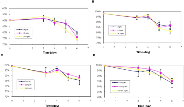
Four different growth factors were tested in single experiments (triplicate samples) for their ability to support cell growth and viability of a 293-F cell line in flat based Erlenmeyer flasks. CellRXs AE-IGF-I and dIGF-I have been benchmarked against commercially available hr-IGF-1 and insulin, in order to assess the efficacy of cell proliferation and level of enhancement of protein production. Figure 1 demonstrates that the growth did not differ significantly after the addition of growth factors among the cells, except for hrIGF-I (1 ng/ml), which almost generated a cell mass of 1 x 10⁶ cells/ml between day 4 and 5. CellRXs IGF-I varian seems to have better or comparable proliferation properties on 293-F cells than insulin and hr-IGF-I. It is important to note that a whole series of triplicate (hrIGF, 1ng/ml) was excluded because of insufficient material. A high standard deviation was found among the samples for insulin, probably due to the variation between the samples.
The cell viability remained high at around 90% after 96h of cultivation, for insulin it even lasted for 120h. The cells treated with hr-IGF-I had a lower viability, only 90% at the starting point, in contrast to the others experiments figure 2.
Nevertheless, it has not contributed to worse outcomes as viability values were at approximately the same levels of all samples, except for the last days when the viability of the hr-IGF-I deteriorate slightly more than the others. None of the IGF-I variants didn’t have any better effect on viability than insulin.
Figure 1. 293-F cell growth with different additives: (A) hrIGF-I, (B) AE-IGF-I, (C) dIGF-I and (D) Insulin in flat based Erlenmeyer flasks. n = 3, except hrIGF-I 1ng/ml (n = 2) because of insufficient material.
11
Figure 2. 293-F cell viability with different additives: (A) hrIGF-I, (B) AE-IGF-I, (C) dIGF-I and (D) Insulin in flat based Erlenmeyer flasks. n = 3, except hrIGF-I 1ng/ml (n = 2) because of insufficient material.
Antibody production
The average antibody production rate was calculated using ELISA data. The results show that antibody production resulting from addition of 10 000 ng/ml insulin can be compared with 25 ng/ml or 100 ng/ml of dIGF-I. Another comparison of similar expression levels is between insulin (100 ng/ml) and AE-IGF-I (5 ng/ml). This indicates that AE-IGF-I is a possible growth factor alternative instead of insulin because of the smaller quantities that promotes protein
production, in contrast to the large amounts of insulin that is required. The
antibody production of the cells with hr-IGF-I addition may have been affected as the only had a viability of 90% at the starting point of the experiment, in contrast to the other growth factors. It is important to note that data points obtained from ELISA for hrIGF-I, is based on n = 1-3 because of too low, or too high signals from the ELISA. The experiment should be repeated before any conclusions can be drawn. This doesn’t apply for the other experiments, which received values based on n = 2-3 for each data point figure 3.
12
Figure 3. Antibody production with different growth factor supplements: (A) hrIGF-I, (B) AE-IGF-I, (C) dIGF-I and (D) Insulin in flat based Erlenmeyer flasks. n = 1-3 for hrIGF-I and n = 2-3 for the rest.
Cell culture experiments in MTPs
It was not feasible to keep the cells alive due to evaporation and liquid loss from the microplate, despite that fresh media was added on every day (from day 3) so the total working volume was approximately 200 µl/well. Excessive evaporation was noted, mostly in the edges of the microplate (compare chart A with B) figure 4. The evaporation was widespread on day 4 and 5 in the wells, so it was difficult to estimate exactly how much media that be added. About 50-100 µl was added to each well. Despite addition of fresh media, the wells were almost empty on day 6, so no samples could be taken for determination of antibody concentration (except day 3, 4 and 5). It appears that liquid evaporation from wells is a limitation with this system. Evaporation in MTPs seems to be a common problem and is more pronounced in the outermost wells than in the centre of the plate, leading to variable osmolalities in different wells [9, 26-27]. Several experiments have been made, and all had problems with evaporation. Despite the failure of keeping the cells alive for a longer period, the cells managed to produce the protein of interest (data not shown). The high signals from the ELISA attempt may mean that a high amount of proteins were produced in the wells or that media has evaporated in the wells and proteins have been concentrated due to evaporation. The difference between the triplicate samples is too widespread to make any conclusions about the production level (data not shown). Howsoever, protein production in
microwells seems to be feasible if the problem with evaporation and liquid loss could be solved.
13
Figure 4. Evaporation rates from a 96 MTP with a working volume of 200 µl/well with a cell concentration of 1 x 10⁶ cells/ml in each well. Chart A shows wells with middle located positions, and B shows edge positions.
SDS-PAGE
The results in figure 5 shows that proteins and peptides could be found in all lanes loaded with samples. The cells stimulated with hrIGF-I, dIGF-I and AEIGF-I expressed the correct protein of expected size and secreted it. What was expected was a band of 25 kDa (light chain) and a band of 50 kDa (heavy chain), which together constitute a whole IgG of 150 kDa (25 +25 +50 +50). This can be ascertained by comparing the protein bands in lane 2, 3 and 4 with the protein bands (control) in lane 6. Lane 5 shows unidentified protein bands, perhaps
proteins from cell lysis (other proteins produced in the cells), or degradation of the proteins of interest so smaller protein fragments have migrated in the gel. It is difficult to judge if the correct protein has been produced because of the other proteins bands in the lane. However a light protein band at 50kDa is visible but it is hard to discern from the background. At 25 kDa it is even more difficult to determine this.
Figure 5. SDS-PAGE analysis of 293-F cell extracts. Lane 2-4 shows protein bands from 293-F cells treated with different growth factor supplements: (1) Protein standard, (2) hrIGF-I, (3) dIGF-I, (4) AE-IGF-I, (5) Proteins from MTP attempt (no growth factor added), (6) Purified reference IgG, (8) Protein standard.
A B cell based proliferation assay was set up as a functional test of the expressed proteins. IgG expressed with hrIGF-I, AE-IGF-I and dIGF-I added as growth factors was functional and not different from purified IgG in this assay, demonstrating that the added growth factors did not adversely affect the functionality of the produced antibody. Data not disclosed.
14
Conclusions
Drug development processes are highly expensive and time-consuming for
pharmaceutical companies, especially costly is clinical trials to bring new drugs to market. The boom in pharmaceutical development worry researchers that believe it will hamper development of new medicines and affect academia and consumers. The result of high development costs through higher drug prices is a burden that society will bear, and it will affect the consumers. Escalating costs will limit innovation [28-29]. Therefore it is crucial to create value and to eliminate as much waste as possible from an operation. A systematic scale-down of cultivation of mammalian cells expressing therapeutically important proteins (potential drug candidates) in microtiter format, could reduce both labour and material costs. At the same time, key process data could be obtained much quicker and eliminate waste and costs in early stage process development. This process can also easily be automated since companies have developed robots that can feed and stack plates automatically. Although evaporation is a limitation with this system, I believe it is possible to overcome by amending parameters such as shaking frequency, humidity and working volume. Duetz et al (2000) managed to solve the problem with excessive liquid evaporation by using a sandwich lid system on top of the microtiter plates [30]. It might be worth looking on this or a similar system to overcome the problem with evaporation. An optimal working volume in combination with a growth factor e. g. insulin or IGF-I could overcome the
problem with evaporation and decrease project time and cost.
It was expected that the dIGF-I and AE-IGF-I should have a better effect on cell proliferation and increase antibody production more than insulin, since previous work have shown that a variant of IGF-I (LongTM R3 IGF-I) demonstrated better performance than insulin in vitro [17, 31]. By monitoring cell viability and growth of 293-F cells, I’ve shown that the growth factors did not differ significantly from each other when it comes to cell proliferation in serum free conditions. The difference is that insulin is needed in much higher concentration to achieve the same results. Both dIGF-I and AE-IGF-I are potent alternatives to insulin when it comes to antibody production, since they have demonstrated equivalent or better performance. If the IGF-I variants can give equivalent or even better results than insulin and in smaller doses, it means that the material cost is minimized when using IGF-I instead. Current price of insulin and other growth factors and pricing of the IGF-I variants determines the profitability. This study should encourage further research into the potential of dIGF-I and AE-IGF-I as supplements to improve antibody yield at production scale and cell proliferation.
15
References
[1] Howanitz JH, Howanitz PJ (2001). Timeliness as a quality attribute and strategy. American Journal of Clinical Pathology, 166, 311-315.
[2] Hesse F, Wagner R (2000). Developments and improvements in the
manufacturing of human therapeutics with mammalian cell cultures. Trends in
Biotechnology, 18, 173-180.
[3] Wurm FM (2004). Production of recombinant protein therapeutics in cultivated mammalian cells. Nature Biotechnology, 22, 1393-1398.
[4] Kapust RB, Waugh DS (1999). Escherichia coli maltose-binding protein is uncommonly effective at promoting the solubility of polypeptides to which it is fused. Protein Science, 8, 1668-1674.
[5] Sørensen HP, Mortensen KK (2005). Soluble expression of recombinant proteins in the cytoplasm of Escherichia coli. Microbial cell factories, 4:1, 1-8. [6]Villaverde A, Carrió MM (2003). Protein aggregation in recombinant bacteria: biological role of inclusion bodies. Biotechnology Letters, 25, 1385-1395.
[7] Marston FA (1986) The purification of eukaryotic polypeptides synthesized in Escherichia coli. The Biochemical journal, 240, 1-12.
[8] Duetz WA, Witholt B (2004). Oxygen transfer by orbital shaking of square vessels and deepwell microtiter plates of various dimensions. Biochemical
Engineering Journal, 17, 181-185.
[9] Li F et al (2006). A systematic approach for scale-down model development and characterization of commercial cell culture processes. Biotechnology
Progress, 22, 696-703.
[10] Girard P et al (1999). Small scale bioreactor system for process development and optimization. Animal Cell Technology: Products from Cells, Cells as
Products, 323–327.
[11] Lye GJ et al (2003). Accelerated design of bioconversion processes using automated microscale processing techniques. Trends in Biotechnology, 21, 29-37. [12] Micheletti M, Lye GJ (2006) Microscale bioprocess optimization. Current
opinion in biotechnology, 17, 611-618.
[13] Doig SD, Lye GJ, Pickering SCR, JM Woodley (2002). The use of microscale processing technologies for quantification of biocatalytic Baeyer-Villiger oxidation kinetics. Biotechnology and Bioengineering, 80, 32-49.
[14] Harms P, Kostov Y, Rao G (2002). Bioprocess monitoring. Current Opinion
in Biotechnology, 13, 124-127.
[15] Barrett et al (2009). Microwell engineering characterization for mammalian cell ulture process development. Biotechnology and Bioengineering, 105, 260-275.
[16] Baganz F et al (2009). Fed-batch operation of an industrial cell culture process in shaken microwells. Biotechnology Letters, 32, 73-78.
[17] Chirkova L, Grosvenor S, Standfield S, Voorhamme D (2007). Enhanced CHO cell performance with a combination of CellPrime™ recombinant transferring and LONG®R3IGF-1. Bioprocessing journal, 6, 45-51. [18] Butler I et al (2004). An analogue of IGF-I. Bioprocess International. 2-7. [19] Adams TE, VC Epa, TPJ Garrett, CW Ward (2000). Structure and function of the type 1 insulin-like growth factor receptor. Cell Mol. Life Sci, 57, 1050-1093.
16
[20] Morris AE, Schmid J (2000). Effects of insulin and LongR3 on serum-free Chinese Hamster Ovary cell cultures expressing two recombinant proteins.
Biotechnology Progress, 16, 693-697.
[21] Alessi DR, Lawlor MA (2001). PKB/Akt: a key mediator of cell
proliferation, survival and insulin responses?. Journal of Cell Science, 114, 2903-2910.
[22] James DC et al (2006). Control of culture environment for improved polyethylenimine-mediated transient production of recombinant monoclonal antibodies by CHO cells. Biotechnology Progress, 22, 753-762.
[23] Voorhamme D, Yandell CA (2006). LONG™R3IGF-I as a more potent alternative to insulin in serum-free culture of HEK293 cells. Molecular
Biotechnology, 34, 201-204.
[24] Kahn RC, Saltiel RA (2001). Insulin signalling and the regulation of glucose and lipid metabolism. Nature, 414, 799-806.
[25] Graham FL et al (1977) Characteristics of a Human Cell Line Transformed by DNA from Human Adenovirus Type 5. Journal of General Virology, 36, 59-74.
[26] Deshpande RR, Heinzle E, Wittmann C (2004). Microplates with integrated oxygen sensing for medium optimization in animal cell culture. Cytotechnology,
46, 1-8.
[27] Duetz WA, Witholt B (2001). Effectiveness of orbital shaking for aeration of suspended bacterial cultures in square-deepwell microtiter plates. Biochemical
Engineering Journal, 7, 113-115.
[28] Collier R (2009). Rapidly rising clinical trial costs worry researchers.
Canadian Medical Association Journal, 180, 277-278.
[29] Collier R (2009). Drug development cost estimates hard to swallow.
Canadian Medical Association Journal, 180, 279-280.
[30] Duetz WA (2000). Methods for intense aeration, growth, storage, and replication of bacterial strains in microtiter plate. Applied and environmental
microbiology, 66, 2641-2646.
[31] Fung V, Thomas JN (1994). Comparison of LongTM R3 IGF-I with insulin in the support of cell growth and recombinant protein expression in CHO cells.