Author: Yaseen Al Hwamdeh Supervisor: Marianne Selim Örebro Universitet
School of Health and Medical Science
Biomedicine and Methods in Medical Diagnostics Degree Project, MC2006, 2nd Level, 15 credits VT 2015
Urinary Stone Diagnosis
Non-Contrast Computed Tomography versus
Intravenous Urography
ii
Summary:
Urinary stone is a health problem affecting 12-20% of population during life time, the risk of stone recurrence is 50%. Until recently, intravenous urography (IVU) was used as the traditional imaging procedures for urinary system stone diagnosis. IVU has lower accuracy in detecting stones, and other urinary system condition. Non contrast computed tomography (NCCT) became the golden standard for diagnosis urinary system stones. This study, reviews NCCT and IVU impact of diagnostic value, advantages and disadvantages, to address the dispensability of IVU at the present time. Computer-based searches of PubMed, AMED and CINAHL-Plus databases, combined with additional searches in radiology Journals and hand searches, were taken for studies in which patients have suspected urinary stone and undergone both NCCT and IVU. Inclusion of this review were based on criteria that was established to asses each study eligibility. The matched studies were taken as final result for searched. Ten studies were retrieved that direct compared both modalities in diagnosis urinary stone and meets the inclusion criteria. All reviewed studies reported higher sensitivity for NCCT than IVU with comparable specificity. IVU showed approximately half NCCT dose, and higher scanning time. IVU negative finding requires further imaging examinations, however NCCT provide an unexpected disease conditions save time and cost. NCCT is the last choice when other modalities failed to diagnose urinary stones. Based on this review NCCT is more accurate and informative diagnostic modality for urinary stones diagnosis. NCCT avoid contrast media usages, save time and are costly effective. The radiation dose is not higher with valuable diagnosis and the IVU is not an ideal modality to diagnosis renal stone.
Keywords: Urinary Calculi; Computed Tomography; Intravenous Urography.
Abbreviations:
CT: Computed Tomography
KUB: Kidney, Ureters and Bladder x-ray film IVU: Intravenous urography
NCCT: Non-contrast Computed Tomography MPR: Multi-planer reconstruction
3
Introduction
Urinary stone is considered as one of the most common urinary system disorders that affect kidney’s normal physiology. Urinary stone is affecting a wide range of population during life time, the risk of developing stone in adults is about 12 % in united states, and higher in the middle east up to 20% in Saudi Arabia .( 1 ) Anatomically urinary system consists of two beanshape- kidneys connected to bladder through ureters. The ureters connect the kidneys to the bladder from posterior base side. The bladder temporary store urine, then drain it outside the body by urethra. Urinary system function to filtrate out blood waste, maintain fluid balance, and regulation blood pressure. Urinary system dysfunctions result in homeostasis imbalance and abnormal urine drainage; eventually caused by urinary tract infection, tumours, or congenital abnormalities. Urinary system infection mostly results in stones formation, urine reflux, and urinary tract obstruction to variant degrees. (2)
Stones are abnormal irregular structures varies in their chemical composition, size and location. Stone formation is a slowly process that takes from month to several years. Usually stones size over 5 mm cannot pass throughout the body without physician intervention, therefore it might block or partially obstruct urine tract, thus, might cause uncomfortable conditions for patient and affect kidney function. Stones have many signs and symptoms depend on stone size and location include vague flank pain, abdominal pain, hematuria, difficulty and frequency of urinations, in addition to nausea and vomiting in some cases.(3) In relation to their chemical components, urinary stones divided into calcium stones, uric acid stones, struvite stones and cysteine stones. Calcium stones are the most common stone type, divided into calcium oxalate and calcium phosphate stone that form up to (60 - 90%) of kidney stones.(1)
Radiographic imaging basically depends on degree of tissue attenuation with x-ray. This process affected by tissue density, atomic number and x-ray photon energy. Radiopaque is the detectable tissue that appears clearly on x-ray images, on country radiolucent is not visualized on x-ray images. Calcium stones considered radiopaque stone however, pure uric acid stone is radiolucent stone due to its composition. (4)
Urinary Stones is considered one of the oldest problems that faced physicians since the Hippocrates time. (5) since then, laboratory test and imaging techniques are indispensible in use.
4 However, limitation of laboratories tests that provides incomplete scope of stones size and locations. Imaging procedures has introduced to provide informative image about urinary system variant anatomy, and physiological effects that include stones size and location.
Imaging modalities used for diagnostic urinary stones include Non-Contrast Computed Tomography (NCCT), Intravenous Urography (IVU). Many factors varies between these techniques including sensitivity, specificity, radiation dose, in addition to examination time, cost and availability in health care institution.
IVU introduced in 1923 and was the modality of choice for evaluating urinary system pathology including urinary stone. (6) IVU is conducted through injection of contrast media followed by series of x-ray images. IVU provides clear images to how execration process progress, tract blockage site and size and narrowing degree. IVU is able to detect any renal lesions that enhanced by Contrast media. Contrast media is chemical substances includes iodine, injected intravenously and excreted by kidneys, to provide enhanced-differentiation of urinary tract passage. The main disadvantage of contrast media is possibility of adverse reaction and toxicity. The incidence of acute adverse reaction is about 15% of patient after administration of contrast media. The reaction degree are mostly mild despite that, severe or life threatening reaction may occurs. Reaction sign include nausea, vomiting, however sever reaction may developed to bronchospasm or cardiac collapse in few seconds. Contrast toxicity affect renal function and may cause renal failure especially for those with high creatinine level or low kidney function. Reaction occurs due to contrast chemical composition, high viscosity, or high injection rate. Patient whom have unstable medically conditions or debilitated and cardiovascular disease should perform kidney function test, before attend to any examination used contrast media injection.(7)
NCCT provides thin slice axial images that visualize both urinary and extra-urinary within inside scanned abdomen, and can identify stones, new growth, and various abnormal pathologies with high resolution. NCCT involved into urinary system imaging modalities in 1995.(8) Since then NCCT has widely spread and became the modality of choice for detection of urinary system stones. With exception for few pure matrix stone NCCT can identify both radiopaque and radiolucent stones with accurate size measurements and high accuracy of up to 100 %.(9) in addition Multi planner reconstruction (MPR) is alternative technique that provides full scope of
5 inside tract details. MPR provides additional full-length images of whole urinary tract in multi-plans, and three dimensional reconstructions to correct finding and further details.
IVU is still considered one of first line imaging modality in diagnosis of urinary stones to date. The efficacy and superiority of NCCT over IVU in detection urinary stone had been demonstrated in many studies among last two decades.(8,12,13,16-19) Since then the studies focused on value of NCCT as gold standard providing informative diagnosis for urinary stone, and additional finding in absence of stone. Studies also focused on ability to reduce NCCT radiation dose, and the best scanning protocol with acceptable radiation dose and informative diagnosis. (10,11,14 ,15)
Aim of this study is to compare diagnostic value, advantages and disadvantages for both NCCT and IVU as a modality of choice for urinary stones diagnosis.
Methods:
Literature searches were taken for studies in which patients of suspected urinary stone that were undergone both non-contrast computed tomography and intravenous urography. Direct comparison was considered the factors that determine which best fit the aim of this study. Using the medical subject heading (MESH) term, PubMed, AMED and CINAHL-Plus. Computer -based searches were run to retrieve articles that contain the key words of “urinary calculi”, “computed tomography”, and “intravenous urography”. The searches were started with first keyword “urinary calculi” and the rest of keywords were added up consequently. The limits of English language and the year of publication from 1998 until 2014 were applied to databases.
Additional searched were conducted in case the database searches were overlooked some articles. Searches were conducted using same keywords in radiology Journals (Korean journal
radiology, American Society of Emergency Radiology, and of the American journal of Roentgenology). The references of all included articles were searched by hand additionally. The
dates were chosen to be the same as the previous database searches, where the title of any article indicated comparison of NCCT and IVU, the abstract were reviewed.
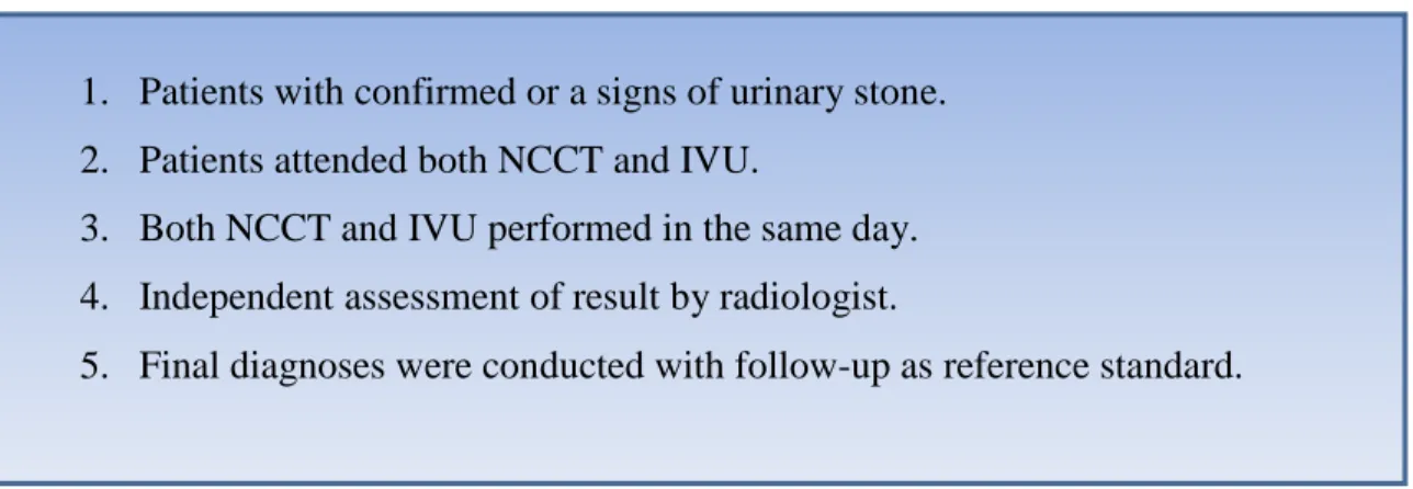
6 Inclusion of this review were based on criteria that was established in prior to asses each article eligibility (Figure 2). The matched articles, to the criteria used in this review, were taken as the final result of searches.
Search result
Keyword “urinary calculi” searched from PubMed database revealed 31581 articles. Once “urinary calculi” was combined with “computed tomography” searches showed 1559 articles. The final combination of “urinary calculi” “computed tomography“, and “intravenous urography” revealed 118 articles. English language and year of publication chosen from 1998 to the sixth week of 2015 were used as source to retrieve the article, collectively keywords and applied filters, showed 87 articles. All 87 articles reviewed screened by reading their abstract, 14 articles concluded that direct compared computed tomography and intravenous urography to diagnose urinary stone. Full text-reading of resulted in 6 articles that meet the inclusion criteria. (12,13 , 14 ,15, 16 , 17 ) The remaining articles were excluded. 6 studies was excluded because same patient did not attend for both NCCT and IVU. (18,19,20,21,22,23) The other two excluded because no clinical follow up was considered as reference to confirm the study result. (24,25)
CINAHL-Plus database searches collectively keywords “urinary calculi”, “computed tomography“, and “intravenous urography”, in addition to filters of english language and the limit for year of publications between 1998 to 2014. Searches have showed 11 articles. After screened all articles abstract, 2 articles were retrieved that compared computed tomography and intravenous urography to diagnose urinary stone and were excluded after full text reading
1. Patients with confirmed or a signs of urinary stone. 2. Patients attended both NCCT and IVU.
3. Both NCCT and IVU performed in the same day. 4. Independent assessment of result by radiologist.
5. Final diagnoses were conducted with follow-up as reference standard.
7 because one was a review,(26) and the other one did not meet inclusion criteria, because no clinical follow up were considered as reference to confirm the study result .(27)
AMED database searches with the same as the previous database searches did not revealed any study after combination of all keywords and filters. The Journal searches revealed two studies one was found in (Korean journal radiology), (28) and one in (American Society of Emergency
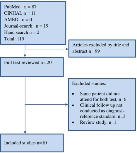
Radiology). (29) The hand search of obtained articles reference revealed 2 studies that were not found by the other searches (30,31). Ten studies were retrieved finally from all searches that meet inclusion criteria (Figure 3).
Comparison of included studies
Available data of included studies consist of (768) patients, (69%) were male. The prominent difference between studies was different modalities and imaging protocols that were used for both procedures, NCCT and IVU. The difference of NCCT scan was found in collimation, pitch
Full text reviewed n= 20 PubMed n = 87 CINHAL n = 11 AMED n = 0 Journal-search n = 19 Hand search n = 2 Total: 119
Articles excluded by title and abstract n= 99
Excluded studies:
Same patient did not attend for both test, n=6
Clinical follow up not conducted as diagnosis reference standard. n=3
Review study. n=1
Included studies n=10
8 and scanning area. Two Studies have used low dose CT scan protocol, however it had differences in the parameters used for dose reduction. The study by Liu (15) reduced slice number with increasing its width and adapted pitch, however the Meagher study (14) reduced the espouser factors, on the other hand the data were collected from four different hospitals, include five different machines and imaging protocol that were used. NCCT scanning area was varied, among ten studies eight performed scanning from top of the kidney to the pubic symphysis, however two studies used a different anatomical scanning area, one performed scan for whole abdomen pelvis, (30) and the other one from diaphragm to pubic symphysis. (31) IVU examinations also used different protocols, the differences founded in the number of images performed, imaging time after injection, excretion delay waiting time and imaging area covered. IVU exposure reported in two low dose studies; however patient dose and radiation risk were calculated with different method. (Table 1)
Methodology quality of included Studies
Even though strong criteria used to obtain high quality studies, the resulted studies hampered by poor reporting of methodology, results and personal patient details. Two studies missed reporting population gender and ages, (28,29) in addition the results of three Studies were not fully reported regarding the specificity and sensitivity. (14, 15, 31) There were poor reporting scanning factors for NCCT and IVU examination, exposure factor and number of actual total images not reported in 8 studies for IVU. In addition for NCCT two studies failed to report exposure factors, (12,29) and one more study had failed to report scanning parameter that used.(30) IVU additional film reported in three studies within 2, 6 and 24 hours for delayed contrast media excretion,(13,14 ,17) The other studies reported additional film without specific waiting time or number of films that performed, and one reported additional film for obstruction cases. (12) The Included studies performed CT scan examination before IVU within the same day, however the time between examinations varies among the studies. Five of the included studies performed IVU immediately after completion of NCCT, two within 6 hours and three studies within 24 hours.
9
Study Study type Population Sensitivity% Specificity % Examination time Examinatio
n Dose, cost
Using MPR
Additional finding
No. Mean Age NCCT IVU NCCT IVU NCCT IVU NCCT IVU
Miller(16) comparative accuracy 106 Male 38.8 Female 32.8 96 87 100 94 X X Cost NCCT 256$ IVU 240$
✓
Yilmaz(17) comparative accuracy97 41.2 94 52 97 94 5 Minutes ≥20 Minutes NCCT dose 3 times higher than IVU
✓
✓
✓
Sourtzis(31) comparative accuracy 53 40 Range 18-72 100 66.6 X X 40-50 Sec + 5 Minutes MPR X X✓
Niall(30) comparative accuracy 40 Median 50 Range 11-72 100 64 92 92 Average 4 Minutes 20-195Minutes Mean 63 NCCT 380$ IVU 180 $✓
✓
Longo(29) comparative accuracy 149 Adult 98 83 95 95 15-20 Minutes 30-35 Minutes 99 Minutes in delay X✓
Wang JH(12) comparative accuracy 82 50.7 Range 24-80 98.5 59.1 100 100 30 Minutes 108 Minutes X✓
✓
Jeong(28) comparative accuracy 30 30 Adult 96 61 100 89 40 -50 Seconds X X✓
Wang LJ(13) comparative accuracy 82 Range 22- 67 100 74.2 93.8 100 X X X✓
Meagher(14) comparative accuracy and dose 69 42.3 93 X X X 5 Minutes 80 Minutes NCCT 3.5mSv IVU 1.5 mSv Liu(15) comparative accuracy and dose 60 42 97 X 96 X Typical case 28 Seconds X IVU 1.33 mSv NCCT 2.8 mSv✓
✓
10 All results evaluated independently by physician who were blinded to other procedure results and clinical findings to avoid biasing results. The final diagnosis in all studies were confirmed with clinical follow-up as reference standard, by stone confirmation with spontaneous passage, diagnosis as other pathological finding, other investigation, or interventional recovery with extracorporeal shock wave lithotripsy (ESWL), Ureteroscopy, or surgical removal.
Result of included Studies
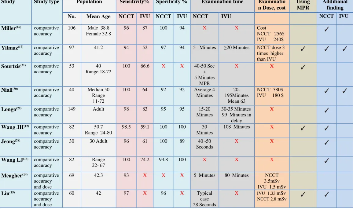
All study results were reported NCCT as higher sensitivity than IVU. NCCT and IVU sensitivity showed (93 -100%) and (52 - 87%) respectively. The lowest sensitivity value for NCCT showed 93% in Meagher study (14) and the highest for IVU 0.87% in Miller study (16). Three studies Sourtzis (31), Niall O (30) and Wang LJ (13) reported 100 % sensitivity for NCCT. However, five studies reported sensitivity below 67 % for IVU. These result explained superiority of NCCT over IVU. Both procedures specificity showed relative similarity in values through all studies. Specificity values for NCCT and IVU were ranged from 92% to 100% and from 89 % to 100% respectively. Three studies showed same specificity value for both procedures. (12, 29,30) The highest specificity value reported 100% three times for NCCT and two times for IVU. Three studies found NCCT had higher specificity than IVU by 6.2%, 3% and 11%,(16,17,28) However, one study found IVU higher than NCCT by 6.2%.(13) Specificity was not fully reported in three studies, (14,15,31) however the results showed that both NCCT and IVU had a comparable values of sensitivity and specificity. All indicated studies are mentioned in details in Table 1.
Dose, Time and reconstruction
The studies by Meagher (14) and Liu (15) showed that calculation of radiation-dose was different,
Liu study (15) estimated the effective dose equivalent (H
E), however the Meagher study (14) calculated the absorbed dose. IVU showed approximately half NCCT dose in low dose protocol studies. The lowest effective dose reported was 2.8 mSv for NCCT and 1.3 mSv for IVU in the
Liu study (15). The rest of the studies failed to report radiation dose except Yilmaz study (17) mentioned that NCCT dose was three times higher than IVU. In eight studies IVU showed significantly higher scanning time than the NCCT, among these studies NCCT time were ranged from 28 second to 30 minutes including the time for reconstruction, however IVU times ranged from 20 to 108 minutes. MPR were used as a part of NCCT protocol in four studies to confirm diagnosis and additional finding. (Table 1)
11
Discussion
Imaging modality for patient with suspected urinary stone depends on many factors; confirmation of stone and capability to provide alternative diagnosis whenever it is possible. The other factors including hazard of radiation dose, contrast adverse reaction, availability, examination cost and time. (32)
IVU has been the traditional imaging modality of choice for evaluation of patients suspected of having urinary system abnormality, rather than suspected of having stones, the higher percentage of using IVU as the standard procedure to anatomical and physiological delineate urinary system. NCCT limited by offering full scope of the morphological aspects only. Many urologists still have the idea that IVU is giving the exact pathway of the urinary system with the ability to see any radiopaque stones in this pathway. While the new multi-planner planning in the CT scan system can also help for the correct surgical intervention.
This subject is chosen to address the impact of still using IVU in diagnosing urinary stone for consideration when choosing the appropriateness of imaging modality especially in the third world country and Middle East, were urinary stones are very common health problem. In addition to increase attention to all aspects of IVU to clarify the differentiation with NCCT in diagnosing urinary stones, thus help to prevent misdiagnosis.
IVU has main advantages include evaluation of urinary function, the degree of ureteral obstruction, delay or absence of excretion, and anatomical localization of stones. Therefore, anomalies and tumour in the kidney collecting system and ureters are rather easy to identify. On the contrary, IVU disadvantages include low detection and poor distinguishing of radiolucent stone. In addition to the contrast media adverse reaction and possibility of vein rapture during injection. (33)
Based on the studies performed, NCCT provides the highest accuracy in detecting urinary stone in comparison to IVU. NCCT exploit benefits of cross sectional images and MPR option, NCCT is considered the golden standard imaging modality for detection of renal stone.(8,18) The study conducted by Miller et al (16) to compare between NCCT and IVU in detection renal stones has confirmed 75 patients to have a stone among 106 patients, 71%. NCCT showed 96% sensitivity and 100% specificity to detect renal stone, whereas IVU showed 87% and 94% for sensitivity
12 and specificity respectively. The rest of 29% had no stones diagnosed. Those patients had other illness such as renal or pelvic mass, prostatitis, musculoskeletal pain and or pyelonephritis. Miller concluded that NCCT was significantly better than IVU.
Wang JH et al (12) compared accuracy of IVU and NCCT, in this study follow up was used as reference standard for stone detection. Sixty six patients have confirmed to have urinary stone among 82 patents 80.5%. NCCT showed 98.5% sensitivity for detection stone. However, urinary
stones was detected in 39 patents out of 66, 59.1% using IVU. This has suggested that NCCT overcome the limitation of IVU in stones detection, include radiolucent stone, small size stone, and obstruction cases with weak contrast execration. Result of 31.7% of patient, whom have undergone IVU, were needed to be confirmed by other diagnostic modalities. Tumour identified in a patient using the MPR technique was not visualized by IVU, it was a lesion no involvement of renal collecting system.
The use of NCCT in addition to MPR provide details of ureters from the kidneys to the bladder, it provides additional finding including lesions outside collecting system hard to visualize by IVU. Moreover, it is able to differentiate classification out of urinary stones. NCCT images are also beneficial in some cases, in which, it shows abnormal tissue-masses that can be associated un-specifically to other diseases-conditions such as masses, haemorrhage, calcifications, gases, and fluid collections that estimated to 27%-47% of patients. (8,34)
Viweg et al (35) in study on NCCT value without correlation to IVU, out of 105 patients, NCCT identified urinary stones in 49 patients, 29 with other abdominal disease and 21 identified as healthy people. Author concludes that patients did not require any further imaging examination and similar examination cost in both techniques, and NCCT positive finding need no additional confirmation by other imaging modalities, on the contrary, IVU negative results that usually need to be confirmed by NCCT, in addition to accurate prospective about stone size in longitudinal and transverse axes .( 27,17)
IVU diagnostic value depends on using of contrast media, consequently IVU significantly gave more details about kidney execration-processes in relation to time, rather than NCCT. However, it is difficult to identify the tract or stone, when superimposition extensive feces or bowel gases over urinary system which affects final diagnosis. On contrary NCCT eliminate risk of adverse reaction and toxicity associated with contrast media and overcome bowel superimposition problem.
13 In respective to cost and time, NCCT might be a bit more expensive than IVU. However, NCCT is significantly faster than IVU. Examination time for NCCT does not exceed 30 min even in obstructed urinary track. There are potential delays of IVU-films that may require up to 24 hours following of excretion process. Eikefjord et al (27) analysed the cost effectiveness for both NCCT
and IVU for patient whom effected or susceptible urinary stone, author reported that NCCT has lower differential cost and an overriding alternative choice in order to diagnose urinary tract stones with no need to run further investigation.
When comparing two modalities which utilize ionization radiation, radiation dose must be considered. There is a risk consistently present with radiation exposure, and proportional to the exposure level. Special concern should be considered for children, pregnancy and young adult patients since the risk increased. NCCT reported to be higher radiation dose than IVU. Radiation dose basically depend on used protocol, however for both NCCT and IVU protocols differ among health care department and machine brand and specification. Many studies estimate NCCT effective dose. Denton et al (23) reported NCCT dose three times higher than IVU, effective dose for IVU calculation showed 1.5 mSv for three films protocol without delayed film and 4.7 mSv for NCCT in standard. Since the prominent limitation of NCCT is the high radiation dose. Low dose protocol was developed to reduce patient radiation exposure and with same accuracy. The effective dose reported to be 50-75 % lower earlier protocol with same accuracy level. (14) IVU effective dose lower than NCCT even with low dose protocol, however NCCT accuracy is higher than IVU and is not affected if the protocol is changed. For this review IVU showed approximately half NCCT dose in low dose protocol studies. The lowest effective dose reported was 2.8 mSv for NCCT and 1.3 mSv for IVU in Liu study (15).
Advanced technology influence the safety to patients by lowering x-rays dose which is safer to conduct the procedure. However in the past decades the radiation dose was higher due to lack of advance equipments, which were the obstacle facing the medical professionals. The possible risk fears regarding higher radiation dose are always guiding CT scan manufacturers for further progression of radiation dose reduction tools. MDCT has undergone technical development starting with early generation of 4, 16, and 64 slices, to the most recent models of 320 slice CT scan. These tools include automated tube current modulation by changing mAs, automated tube potential selection by changing kVp, and providing advanced three dimensional MPR. All of
14 these factors contribute to maximum radiation dose reduction. However, many health care providers did not take in the advantages of these capabilities because of lack of familiarity and training how these tools work. Also, those tools are built into scanner software with relatively simple interfaces.(36) Despite of published reports about superiority of NCCT over IVU, IVU is still being used today by numerous health care providers. In developing countries especially middle east and third world countries.(37) For example, middle Eastern Gulf States have the highest incidence of stone compared with developed country, due to adverse combination of dietary and environmental factors.(38) IVU examination are mostly used in local hospitals in the countryside compared to the major cities. However IVU has been replaced by NCCT long ago in Europe. According to European Association of Urology guidelines last editions, NCCT is recommended in confirmation of urinary stone diagnosis, this is due to its superiority to IVU and radiation risk can be reduced using low dose protocol.(39) Many reasons contribute that IVU is still in use. Economic reasons might be the main due to high cost of CT scan machine therefore examination cost, and decrease its choice by health care provider. In addition to lack of familiarity and training on CT scan advanced technology software such as three dimensional MPR, and physician familiarity with IVU.
The finding of this review is that across studies, NCCT has drawn the greatest attention and replaced IVU in diagnosis of renal stone. NCCT showed to be more effective to be used in detection of urinary stones, avoided contrast media usage, and shorter examination time. The cost of NCCT examination as compared with that of IVU varies among institutions but is comparable. However, radiation dose for both NCCT protocol is higher than IVU, particularly not complicated IVU. IVU varies in result with potentially, negative finding that need to be confirm by other diagnostic modality, however NCCT provide an accurate diagnosis, save the time and cost. NCCT is always the last choice when other modalities fail to diagnose urinary stones. Nowadays NCCT is the golden standard imaging modalities in diagnosis urinary stones with high accuracy. (8,16,17,40,41)
Conclusion:
Based on this review NCCT is better than IVU in detection of urinary stone, avoiding contrast media usages, save time and is cost effective. Developmental progression on imaging equipment
15 reduced the radiation dose. IVU replaced by NCCT in developed countries and still in use in third world countries. IVU is not ideal modality for diagnosis urinary stone, it is recommended to be completely replaced by NCCT with respect to increase the awareness, training and economic development to support and sustain quality health services.
References
1. López M, Hoppe B. History, epidemiology and regional diversities of urolithiasis. Pediatr Nephrol. 2010; 25(1):49-59.
2. Field MJ. The renal system : basic science and clinical conditions. 2nd ed. ed. Pollock CA, Harris DC, editors. Edinburgh ; New York: Edinburgh ; New York : Churchill Livingstone/Elsevier; 2010.
3. Portis AJ, Sundaram CP. Diagnosis and initial management of kidney stones. Am Fam Physician. 2001; 63(7):1329-38.
4. Wiederkehr MR, Moe OW. Uric Acid Nephrolithiasis: A Systemic Metabolic Disorder. Clin Rev Bone Miner Metab. 2011;9(3-4):207-17.
5. Dardioti V, Angelopoulos N, Hadjiconstantinou V. Renal diseases in the Hippocratic era. Am J Nephrol. 1997; 17(3-4):214-6.
6. Wrenn K. Emergency intravenous pyelography in the setting of possible renal colic: is it indicated? Ann Emerg Med. 1995; 26(3):304-7.
7. Thomsen HS, Morcos SK. Radiographic contrast media. BJU Int. 2000;86 Suppl 1:1-10. 8. Smith RC, Rosenfield AT, Choe KA, Essenmacher KR, Verga M, Glickman MG, et al. Acute
flank pain: comparison of non-contrast-enhanced CT and intravenous urography. Radiology. 1995; 194(3):789-94.
9. Mostafavi MR, Ernst RD, Saltzman B. Accurate determination of chemical composition of urinary calculi by spiral computerized tomography. J Urol. 1998;159(3):673-5.
10. Gurung J, Khan MF, Maataoui A, Herzog C, Bux R, Bratzke H, et al. Multislice CT of the pelvis: dose reduction with regard to image quality using 16-row CT. Eur Radiol. 2005;15(9):1898-905.
16
11. Moore CL, Daniels B, Ghita M, Gunabushanam G, Luty S, Molinaro AM, et al. Accuracy of reduced-dose computed tomography for ureteral stones in emergency department patients. Ann Emerg Med. 2015;65(2):189-98.e2.
12. Wang JH, Shen SH, Huang SS, Chang CY. Prospective comparison of unenhanced spiral computed tomography and intravenous urography in the evaluation of acute renal colic. J Chin Med Assoc. 2008;71(1):30-6.
13. Wang LJ, Ng CJ, Chen JC, Chiu TF, Wong YC. Diagnosis of acute flank pain caused by ureteral stones: value of combined direct and indirect signs on IVU and unenhanced helical CT. Eur Radiol. 2004;14(9):1634-40.
14. Meagher T, Sukumar VP, Collingwood J, Crawley T, Schofield D, Henson J, et al. Low dose computed tomography in suspected acute renal colic. Clin Radiol. 2001;56(11):873-6. 15. Liu W, Esler SJ, Kenny BJ, Goh RH, Rainbow AJ, Stevenson GW. Low-dose nonenhanced
helical CT of renal colic: assessment of ureteric stone detection and measurement of effective dose equivalent. Radiology. 2000;215(1):51-4.
16 . Miller OF, Rineer SK, Reichard SR, Buckley RG, Donovan MS, Graham IR, et al. Prospective comparison of unenhanced spiral computed tomography and intravenous urogram in the evaluation of acute flank pain. Urology. 1998;52(6):982-7.
17. Yilmaz S, Sindel T, Arslan G, Ozkaynak C, Karaali K, Kabaalioğlu A, et al. Renal colic: comparison of spiral CT, US and IVU in the detection of ureteral calculi. Eur Radiol. 1998;8(2):212-7.
18. Pfister SA, Deckart A, Laschke S, Dellas S, Otto U, Buitrago C, et al. Unenhanced helical computed tomography vs intravenous urography in patients with acute flank pain: accuracy and economic impact in a randomized prospective trial. Eur Radiol. 2003;13(11):2513-20. 19. Mendelson RM, Arnold-Reed DE, Kuan M, Wedderburn AW, Anderson JE, Sweetman G,
et al. Renal colic: a prospective evaluation of non-enhanced spiral CT versus intravenous pyelography. Australas Radiol. 2003;47(1):22-8.
20. Rekant EM, Gibert CL, Counselman FL. Emergency department time for evaluation of patients discharged with a diagnosis of renal colic: unenhanced helical computed tomography versus intravenous urography. J Emerg Med. 2001;21(4):371-4.
17 21. Thomson JM, Glocer J, Abbott C, Maling TM, Mark S. Computed tomography versus intravenous urography in diagnosis of acute flank pain from urolithiasis: a randomized study comparing imaging costs and radiation dose. Australas Radiol. 2001;45(3):291-7.
22. Homer JA, Davies-Payne DL, Peddinti BS. Randomized prospective comparison of non-contrast enhanced helical computed tomography and intravenous urography in the diagnosis of acute ureteric colic. Australas Radiol. 2001;45(3):285-90.
23. Denton ER, Mackenzie A, Greenwell T, Popert R, Rankin SC. Unenhanced helical CT for renal colic--is the radiation dose justifiable? Clin Radiol. 1999;54(7):444-7.
24. Ben Nakhi A, Gupta R, Al-Hunayan A, Muttikkal T, Chavan V, Mohammed A, et al. Comparative analysis and interobserver variation of unenhanced computed tomography and intravenous urography in the diagnosis of acute flank pain. Med Princ Pract. 2010;19(2):118-21.
25. Wong SK, Ng LG, Tan BS, Cheng CW, Chee CT, Chan LP, et al. Acute renal colic: value of unenhanced spiral computed tomography compared with intravenous urography. Ann Acad Med Singapore. 2001;30(6):568-72.
26. Jindal G, Ramchandani P. Acute flank pain secondary to urolithiasis: radiologic evaluation and alternate diagnoses. Radiol Clin North Am. 2007;45(3):395-410, vii.
27. Eikefjord E, Askildsen JE, Rørvik J. Cost-effectiveness analysis (CEA) of intravenous urography (IVU) and unenhanced multidetector computed tomography (MDCT) for initial investigation of suspected acute ureterolithiasis. Acta Radiol. 2008;49(2):222-9.
28. Ryu JA, Kim B, Jeon YH, Lee J, Lee JW, Jeon SS, et al. Unenhanced spiral CT in acute ureteral colic: a replacement for excretory urography? Korean J Radiol. 2001;2(1):14-20. 29. Longo J, Akbar SA, Schaff T, Jafri ZH, Jackson RE. A prospective comparative study of
non-contrast helical computed tomography and intravenous urogram for the assessment of renal colic. Emerg Radiol 2001;8(5):285e92.
30. Niall O, Russell J, MacGregor R, Duncan H, Mullins J. A comparison of noncontrast computerized tomography with excretory urography in the assessment of acute flank pain. J Urol. 1999;161(2):534-7.
18 31. Sourtzis S, Thibeau JF, Damry N, Raslan A, Vandendris M, Bellemans M. Radiologic investigation of renal colic: unenhanced helical CT compared with excretory urography. AJR Am J Roentgenol. 1999;172(6):1491-4.
32. Dalla Palma L, Pozzi-Mucelli R, Stacul F. Present-day imaging of patients with renal colic. Eur Radiol. 2001;11(1):4-17.
33 . Heidenreich A, Desgrandschamps F, Terrier F. Modern approach of diagnosis and management of acute flank pain: review of all imaging modalities. Eur Urol. 2002;41(4):351-62.
34. Dalrymple NC, Verga M, Anderson KR, Bove P, Covey AM, Rosenfield AT, et al. The value of unenhanced helical computerized tomography in the management of acute flank pain. J Urol. 1998;159(3):735-40.
35. Vieweg J, Teh C, Freed K, Leder RA, Smith RH, Nelson RH, et al. Unenhanced helical computerized tomography for the evaluation of patients with acute flank pain. J Urol. 1998;160(3 Pt 1):679-84.
36. Sabarudin A, Sun Z, Ng KH. A systematic review of radiation dose associated with different generations of multidetector CT coronary angiography. J Med Imaging Radiat Oncol. 2012;56(1):5-17.
37 . Hale Z, Hanna E, Miyake M, Rosser CJ. Imaging the urologic patient: the utility of intravenous pyelogram in the CT scan era. World J Urol. 2014;32(1):137-42.
38. Robertson WG. Stone formation in the Middle Eastern Gulf States: A review. Arab J Urol. 2012;10(3):265–72.
39 . Turk C, Knoll T, Petrik A, Sarica K, Skolarikos A, Straub M, et al. guidelines on urolithiasis. EAU. 2015.
40. Fowler KA, Locken JA, Duchesne JH, Williamson MR. US for detecting renal calculi with nonenhanced CT as a reference standard. Radiology. 2002; 222(1):109-13.
41 . Sheafor DH, Hertzberg BS, Freed KS, Carroll BA, Keogan MT, Paulson EK, et al. Nonenhanced helical CT and US in the emergency evaluation of patients with renal colic: prospective comparison. Radiology. 2000; 217(3):792-7.