ACTA UNIVERSITATIS
UPSALIENSIS
Digital Comprehensive Summaries of Uppsala Dissertations
from the Faculty of Science and Technology
1492
Can Bone Void Fillers Carry Load?
Behaviour of Calcium Phosphate Cements Under
Different Loading Scenarios
INGRID AJAXON
ISSN 1651-6214 ISBN 978-91-554-9865-8
Dissertation presented at Uppsala University to be publicly examined in Häggsalen, Ångströmlaboratoriet, Lägerhyddsvägen 1, Uppsala, Friday, 12 May 2017 at 09:15 for the degree of Doctor of Philosophy. The examination will be conducted in English. Faculty examiner: Prof. Uwe Gbureck (University of Wuerzburg).
Abstract
Ajaxon, I. 2017. Can Bone Void Fillers Carry Load? Behaviour of Calcium Phosphate Cements Under Different Loading Scenarios. Digital Comprehensive Summaries of Uppsala
Dissertations from the Faculty of Science and Technology 1492. 67 pp. Uppsala: Acta
Universitatis Upsaliensis. ISBN 978-91-554-9865-8.
Calcium phosphate cements (CPCs) are used as bone void fillers and as complements to hardware in fracture fixation. The aim of this thesis was to investigate the possibilities and limitations of the CPCs’ mechanical properties, and find out if these ceramic bone cements can carry application-specific loads, alone or as part of a construct. Recently developed experimental brushite and apatite cements were found to have a significantly higher strength in compression, tension and flexion compared to the commercially available CPCs chronOS™ Inject and Norian® SRS®. By using a high-resolution measurement technique the elastic moduli of the
CPCs were determined and found to be at least twice as high compared to earlier measurements, and closer to cortical bone than trabecular bone. Using the same method, Poisson's ratio for pure CPCs was determined for the first time. A non-destructive porosity measurement method for wet brushite cements was developed, and subsequently used to study the porosity increase during in vitro degradation. The compressive strength of the experimental brushite cement was still higher than that of trabecular bone after 25 weeks of degradation, showing that the cement can carry high loads over a time span sufficiently long for a fracture to heal. This thesis also presents the first ever fatigue results for acidic CPCs, and confirms the importance of testing the materials under cyclic loading as the cements may fail at stress levels much lower than the material’s quasi-static compressive strength. A decrease in fatigue life was found for brushite cements containing higher amounts of monetite. Increasing porosity and testing in a physiological buffer solution (PBS), rather than air, also decreased the fatigue life. However, the experimental brushite cement had a high probability of surviving loads found in the spine when tested in PBS, which has previously never been accomplished for acidic CPCs. In conclusion, available brushite cements may be able to carry the load alone in scenarios where the cortical shell is intact, the loading is mainly compressive, and the expected maximum stress is below 10 MPa. Under such circumstances this CPC may be the preferred choice over less biocompatible and non-degradable materials.
Keywords: Calcium phosphate, bone cement, brushite, apatite, monetite, porosity, solvent
exchange, degradation, compressive strength, diametral tensile strength, flexural strength, elastic modulus, Poisson’s ratio, fatigue
Ingrid Ajaxon, Department of Engineering Sciences, Applied Materials Sciences, Box 534, Uppsala University, SE-75121 Uppsala, Sweden.
© Ingrid Ajaxon 2017 ISSN 1651-6214 ISBN 978-91-554-9865-8
List of Papers
This thesis is based on the following papers, which are referred to in the text by their Roman numerals.
I Ajaxon, I., Persson, C. Mechanical properties of brushite calcium phosphate cements. In Shi, D. (Ed.). The world scientific
encyclope-dia of nanomedicine and bioengineering II: Bioimplants, regenera-tive medicine, and nano-cancer diagnosis and phototherapy – vol-ume 3: Design of bioactive materials for bone repair and regenera-tion. Singapore: World Scientific Pte Ltd, pp. 285-300. In press.
II Luo, J.*, Ajaxon, I.*, Ginebra, M.P., Engqvist, H., Persson, C. (2016) Compressive, diametral tensile and biaxial flexural strength of cutting-edge calcium phosphate cements. Journal of the
mechani-cal behavior of biomedimechani-cal materials, 60(C):617-627.
III Ajaxon, I.*, Acciaioli, A.*, Lionello, G., Ginebra, M.P., Öhman-Mägi, C., Baleani, M., Persson, C. Elastic properties and strain-to-crack-initiation of calcium phosphate bone cements: revelations of a high-resolution measurement technique. Submitted.
IV Ajaxon, I., Maazouz, Y., Ginebra, M.P., Öhman, C., Persson, C. (2015) Evaluation of a porosity measurement method for wet calci-um phosphate cements. Journal of Biomaterials Applications, 30(5):526-536.
V Ajaxon, I., Öhman, C., Persson, C. (2015) Long-term in vitro degra-dation of a high-strength brushite cement in water, PBS and serum solution. Biomed Research International, 2015:575079.
VI Ajaxon, I., Öhman Mägi, C., Persson, C. (2017) Compressive fatigue properties of an acidic calcium phosphate cement – effect of phase composition. Journal of Materials Science: Materials in Medicine, 28(3):41.
VII Ajaxon, I., Holmberg, A., Öhman-Mägi, C., Persson, C. Compres-sive fatigue properties of a high-strength degradable calcium phos-phate bone cement – influence of porosity and environment.
Manu-script.
Reprints were made with permission from the respective publishers.
* Authors contributed equally to this work and should be regarded as joint authors.
Author’s contributions
My contributions to the papers included in this thesis are:
Paper I Part of planning, all of literature collection, part of evaluation
and writing.
Paper II Part of planning, experimental work, evaluation and writing.
Paper III Part of planning, experimental work, and evaluation, major
part of writing.
Paper IV Major part of planning, experimental work, evaluation and
writing.
Paper V Major part of planning, experimental work, evaluation and
writing.
Paper VI Major part of planning, all experimental work, major part of evaluation and writing.
Paper VII Major part of planning, part of experimental work, major part
Contents
Introduction ... 11
Bone and bone replacement materials ... 13
Calcium phosphates in bone repair ... 19
Preparation and characterization methods ... 21
Cement preparation ... 21
Phase composition ... 22
Porosity ... 24
Mechanical assessment ... 26
Degradation ... 31
Summary of aims and objectives ... 33
Mechanical properties under quasi-static load ... 34
Compressive strength ... 34
Diametral tensile strength ... 40
Biaxial flexural strength ... 42
Elastic properties – Young's modulus and Poisson's ratio ... 43
Measuring the porosity of wet CPCs ... 46
In vitro degradation of brushite cements ... 48
Mechanical properties under cyclic load ... 52
Summary and Conclusions ... 60
Advances in method development ... 60
Advances in knowledge of mechanical properties ... 60
When can bone void fillers carry load? ... 61
Future perspectives ... 64
Swedish summary – Kan bencement bära last? ... 65
Acknowledgements ... 69
Abbreviations
α-TCP Alpha-tricalcium phosphate
β-TCP Beta-tricalcium phosphate
CPC Calcium phosphate cement
DCPA Dicalcium phosphate anhydrous, or Monetite
DCPD Dicalcium phosphate dihydrate, or Brushite
DIC Digital image correlation
FBS Foetal bovine serum
HA Hydroxyapatite
L/P-ratio Liquid to powder ratio
MCPM Monocalcium phosphate monohydrate
Micro-CT Micro computed tomography
MIP Mercury intrusion porosimetry
Nf Number of cycles to failure
OCP Octacalcium phosphate
PBS Phosphate buffered saline
PMMA Poly(metyl methacrylate)
RT Room temperature
SEM Scanning electron microscopy
σCS Compressive strength
σDTS Diametral tensile strength
σBFS Biaxial flexural strength
Smax Maximum compressive stress level (in fatigue)
SPP Disodium dihydrogen pyrophosphate
Introduction
Medical writings of ancient civilizations show that the earliest successful implants in humans were in the skeletal system1. Archaeological findings
reveal that spare parts, including both bone and teeth, were taken from ani-mal and human remains when needed. Additionally, different materials such as coral, shell, ivory, wood and metals were also used to replace missing bone and teeth. Over the centuries, the advancements in sterilization and surgical procedures, as well as the development of different synthetic mate-rials, have opened up a large flora of spare parts that can be used to replace and/or restore the function of traumatized or degenerated tissue, and – per-haps most importantly – to reduce pain, and thus improve the quality of life of the patients.
Today, there is a great need for synthetic materials aimed at treating dam-aged and diseased bone. One reason for this is that the population of the developed countries is steadily growing older, and with that the number of elderly affected by fragility fractures is increasing2,3. We also live more ac-tive lifestyles even at advanced ages, and demand well-functioning bodies throughout our lives.
A candidate bone replacement material is calcium phosphate cement (CPC), which is a synthetic biomaterial that over the last 15 years has gained a great deal of interest due to its similarity to the mineral phase of bone4.
CPCs are already used clinically as bone void fillers and as a complement to
hardware in fracture fixation5. Studies have shown benefits in terms of
im-proved stability and faster recovery when using CPCs in distal radius (wrist) fractures6, earlier weight bearing after tibial plateau (knee) and calcaneaus
(heel) fractures6,7, improved stability in femoral head and neck (hip) frac-tures6,8, successful repair of large cranial bone defects9,10, and less pain at
fracture sites11. Moreover, CPCs have been demonstrated to stabilize certain vertebral (spine) fractures12,13.
Currently there are several commercial calcium phosphate-based cements available14,15, but their clinical use is partly limited by poor mechanical
prop-erties, and, particularly, by a lack of knowledge of their behaviour during elastic deformation as well as their long term mechanical resistance. This is unfortunate, since CPCs have potential benefits compared to less biocompat-ible and non-resorbable bone replacement materials that are widely used today, e.g., metals and polymeric materials. Moreover, improvements in the mechanical properties have been achieved for newly developed CPCs16-18,
which may widen their use as bone replacement materials. The aim of this thesis was to study the behaviour of CPCs under different loading scenarios, especially the long-term properties, in order to evaluate the possible future use of these cements in load bearing applications. More specifically, the aim is trying to answer: i) What are the limitations and possibilities of these types of cements? ii) Can CPCs carry application-specific loads? Alone or as part of a construct?
The thesis consists of one book chapter and six scientific papers, each in-vestigating one or more of these questions, or providing methodology need-ed for the evaluation. This summary aims to put the studies into context and present an overall discussion of the results.
Bone and bone replacement materials
Bones have important tasks in the body: they provide structural support, protect vital organs, function as storage for salts (e.g. calcium and phospho-rous), and continuously supply the body with blood cells (produced by the bone marrow)19. Bone tissue is a composite material that contains a mineral
part consisting of carbonated hydroxyapatite (~40-70%), a protein matrix that contains collagen (~20-40%), water (~10%), and minor amounts of non-collagenous proteins20,21.
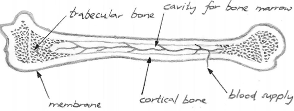
Figure 1. Cross-section of a long bone.
A bone generally consists of a compact outer layer (cortical or compact bone) with a much more porous interior (trabecular, cancellous or spongy bone)19, see Figure 1. These two tissues have very different mechanical
properties; cortical bone is stronger and stiffer than trabecular bone, see
Ta-ble 1. Depending on the anatomical site, and even within different regions of
the same site, the strength and stiffness of the osseous tissue can vary sub-stantially. The mechanical properties also vary among individuals, with age, sex, activity level, and due to different pathologies.21
Table 1. Mechanical properties of bone. Ultimate compres-sive strength [MPa] Ultimate tensile strength [MPa] Elastic modulus [GPa] Poisson’s ratio Cortical bone 95-23022,23 40-14021 9-2522,24 0.46-0.5825,26 Trabecular bone 1-30 27,28 1-1121 0.01-524,27,29,30 0.06-0.9525
Bone is a living organ that is constantly subjected to modifications and maintenance, a process called remodeling, i.e., removing old bone matrix and producing new. Mechanical loading of the bone tissue stimulates the remodeling process, but it is also triggered through other signaling pathways. The balance between bone resorption and renewal is critical, and during the first 20-30 years of our lives the balance is positive whereas an increase in
resorption is seen as we grow older.20 Bone remodeling can be disrupted by
different diseases, such as osteoporosis, which leads to a lower bone mineral
density, and a higher risk of fractures3. Women have a much higher risk of
suffering an osteoporotic fracture than men: in Sweden, women have a 46% and men a 22% risk of osteoporotic fracture after the age of 5031,32.
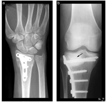
Even though bone has efficient mechanisms to heal by itself when frac-tured, some cases may require bone substituting materials, e.g., critical size defects (when the gap between the fractured surfaces is too big), some oste-oporotic fractures, or after removal of bone tumors11,33,34. Bone void fillers are commonly used as complements to metallic fracture-fixation hardware, such as those seen in Figure 2.
Figure 2. a) The author’s wrist after an adventurous day of snowboarding in the
Swedish mountains, showing fracture fixation using a metallic plate and screws. b) Tibial plateau fracture fixated with metallic hardware and augmented with a calcium phosphate cement (indicated with arrow). Reprinted from Larsson and Bauer6 with
permission.
An ideal material for bone repair and replacement would integrate well with bone, restore the mechanical properties of the defect and eventually be placed by new bone, such that the mechanical integrity of the bone is re-established as soon as possible. The gold standard for fracture repair and bone void filling is to use autologous tissue (bone tissue from the same pa-tient)6,35. However, there are several drawbacks with this method, including the need of multiple surgeries, donor-site morbidity, limitation in the amount of available tissue, and insufficient mechanical stability of the repair35,36. Another option is to use allografts (tissue from another patient of the same species) or xenografts (tissue from a different species), but these are also limited in supply, can be rejected by the patient's immune system, provide limited or no osteoinductive properties (stimulate new bone tissue formation) and there is a risk, even though it is extremely low, of disease transmis-sion35,36.
To circumvent the limitations with natural bone grafts, the focus has shifted towards synthetic bone graft substitutes. There are several different types of biomaterials used as bone replacement materials: encompassing all material classes – metals, polymers, ceramics and composites, both
non-resorbable and non-resorbable. Among ceramic biomaterials, calcium phosphates provide an interesting alternative due to their similarity to the mineral con-tent of human bone tissue. Moreover, calcium phosphates can be resorbable, preferably resorbing at the same rate that new bone is formed6;
osteoconduc-tive (serve as a scaffold or template for new bone to grow into)5,37; osteoin-ductive9,10,38; and can be prepared as an injectable material that hardens in vivo5. These properties depend on the specific calcium phosphate formula-tion; some common calcium phosphates used in bone repair will be de-scribed in the next section.
Calcium phosphates in bone repair
The first time a synthetic calcium phosphate was introduced in vivo was in 1920 when Albee, assisted by Morrison, attempted to prove bone regrowth and union of surgically created defects in rabbit bone, with successful re-sults39. At that time, the knowledge that bone consisted of calcium phosphate
had been around since the Uppsala scholar Johan Gottlieb Gahn had made the discovery in the late 18th century1.
CPCs are most commonly based on the orthophosphate ion (PO!!!). Some
common calcium orthophosphates are listed in Table 2. The lower the calci-um-to-phosphate (Ca/P) ratio, the more water-soluble the calcium ortho-phosphate is – something that affects the CPCs degradation behaviour40,41. Table 2. Some common calcium orthophosphates.
Ca/P Compound
Abbrevia-tion Chemical formula
0.5 Monocalcium phosphate monohydrate MCPM Ca(H2PO4)2 · H2O
1.0 Monetite (dicalcium phosphate anhydrous) DCPA CaHPO4
1.0 Brushite (dicalcium phosphate dihydrate) DCPD CaHPO4 · 2 H2O
1.33 Octacalcium phosphate OCP Ca8H2(PO4)6 ·5 H2O
1.5 Alpa-tricalcium phosphate α-TCP Ca3(PO4)2
1.5 Beta-tricalcium phosphate β-TCP Ca3(PO4)2
1.67 Hydroxyapatite HA Ca10(PO4)6(OH)2
Calcium phosphate-based biomaterials can be used as cements, putties, granules, and blocks42. Blocks can be cut into any desired shape, but it is difficult to get a good contact between implant and bone in irregularly shaped defects. Moreover, sintered calcium phosphate blocks are considered almost inert, thus lacking the advantages of a resorbable material. Calcium phosphate granules on the other hand, can be produced in different sizes and can be efficiently packed into defects with difficult geometries. However, granules have the disadvantage that they can escape the defect and may therefore need a surrounding membrane, or be embedded in a matrix, to re-main in place. Cements have considerable advantages compared to granules or sintered calcium phosphates: they can be injected directly into the fracture site, thus providing good cavity filling potential, they set at physiological temperature, and they can be resorbable. The injectability allow for minimal-ly invasive surgery, a technique entailing a shorter and easier surgical
proce-dure, as well as a lower infection risk and other complications associated with open surgery. The focus of this thesis is on such self-setting CPCs.
CPCs are obtained by mixing one or more of the calcium phosphate salts listed in Table 2 with an aqueous solution, that sets through a dissolution-precipitation reaction: the powder(s) dissolve in the solution, and then starts to nucleate and grow crystals. As more and more crystals grow, they become entangled, thus creating a mechanically rigid body. Between the entangled crystals there will be empty spaces. This intrinsic porosity occurs on the nano and micro scale and is created in the cement during setting. Besides, pores will be created in the cement when air is incorporated during mixing and moulding. CPCs can also be prepared pre-mixed, which means that the powder phase is mixed with a non-aqueous liquid and the setting reaction starts when the cement paste comes into contact with water43,44.
CPCs are commonly subdivided into two cement types that are distin-guished by the pH during setting. When the pH is higher than 4.2 the end product is a basic CPC, called apatite (carbonated apatite; calcium-deficient hydroxyapatite; hydroxyapatite, HA); and when the pH is lower than 4.2, the product is an acidic CPC, called brushite (dicalcium phosphate dihydrate, DCPD) or monetite (dicalcium phosphate anhydrous, DCPA). Brushite pre-cipitates faster than monetite, and is thus generally the main product when the pH is lower than 4.2. However, when the setting conditions are water-deficient monetite is formed instead. Moreover, higher temperatures promote the transformation of brushite into monetite.45,46 Most commercially
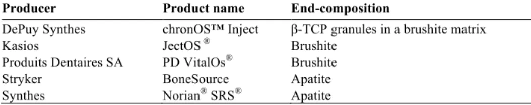
availa-ble CPC formulations have apatite as end-product, since more research effort has been put into basic CPCs compared to acidic ones. Some different com-mercial formulations of CPCs, including all available brushite cements, are presented in Table 3. Brushite and monetite have shown faster resorption rates compared to apatite cements5,47,48 and can have osteoinductive
ef-fects9,10,38, depending on the formulation of the acidic cement, which may be beneficial for faster bone regrowth. These properties are of course of high clinical relevance.
Table 3. Some commercially available CPCs. Detailed reviews of commercial CPCs
have been published elsewhere14,15.
Producer Product name End-composition
DePuy Synthes chronOS™ Inject β-TCP granules in a brushite matrix Kasios JectOS ® Brushite
Produits Dentaires SA PD VitalOs® Brushite
Stryker BoneSource Apatite Synthes Norian® SRS® Apatite
Ideally, a bone substitute material should have mechanical properties similar to the host bone. Some common techniques to assess these properties of CPCs are presented in the next chapter, together with details of how to pre-pare the cements.
Preparation and characterization methods
The CPCs used in this thesis were prepared and analyzed in a number of ways and the purpose of this chapter is to give an overview of the methods used, while detailed parameters and settings can be found in the respective papers.
Cement preparation
The main focus of this thesis was on experimental formulations of brushite cements developed in-house. Experimental formulations of apatite and monetite developed by collaborators, and commercially available brushite based and apatite based cements, were also evaluated for comparison.
Paper VI investigated an earlier generation of an experimental brushite
cement developed in-house. This cement consisted of a powder phase of as-received monocalcium phosphate monohydrate (MCPM) and beta-tricalcium phosphate (β-TCP). To prolong the setting time, disodium dihydrogen pyro-phosphate (SPP) was added to the powder mixture. Water was used as the liquid phase. This earlier formulation had a lower compressive strength
compared to an optimised brushite cement formulation16 that was studied in
Papers II, III, IV, V and VII, due to a different ratio between the β-TCP and
MCPM, no selection of particle sizes and a higher liquid-to powder (L/P) ratio.
An experimental high-strength brushite cement that, in previous in-house work16, had been optimised in terms of compressive strength, while
main-taining an acceptable injectability and setting time, was studied in Papers II,
III, IV, V and VII. The cement was prepared by mixing MCPM, sieved to
obtain particle sizes below 75 µm, with β-TCP, and SPP. The liquid phase used for this optimised brushite cement formulation was a citric acid solu-tion, which increased the solubility of the powders49. This formulation has been thoroughly studied by others in terms of setting time, injectability, mi-crostructure, porosity, compressive and diametral tensile strength, and phase composition16.
The powder phase of the experimental monetite cement studied in Paper
III was a mixture of MCPM and β-TCP, and the liquid phase was glycerol.
This cement was developed to improve the handling characteristics of the cement as mixing is no longer required in the operating theatre – the cement
can be delivered pre-mixed and the setting reaction starts when the glycerol is exchanged with body fluids43.
Experimental apatite cements were prepared from either a fine (Paper II) or a coarse (Papers III and IV) alpha-tricalcium phosphate (α-TCP) powder, provided by a collaborator. The preparation of the α-TCP powders has been described elsewhere17. Precipitated HA particles were added as a nucleation
agent in the α-TCP. The liquid phase was a sodium hydrogen phosphate solution, added as an accelerant17. The setting reaction is accelerated when a
fine α-TCP powder is used compared to a coarse α-TCP, which also leads to differences in the microstructure50. These cement formulations have been
thoroughly studied before by others in terms of setting time, compressive strength, porosity, pore size distribution, specific surface area and in vitro cell response17,18,51.
Two commercially available cements, chronOS™ Inject (brushite based) and Norian® SRS® (apatite based), were studied as reference materials in
Paper II. Both cements are indicated for use in bone voids in the extremities
and the pelvis but are not allowed for use in, e.g., the spine, for use alone in load-bearing applications or in the presence of an on-going infection. The
powder and liquid components of chronOS™ Inject and Norian® SRS® were
used as-received.
To form a cement paste, the powder and liquid phases of all cement types were either mixed by hand (Paper VI) or in a mechanical mixing device (Papers II, III, IV, V and VII). The latter was used to allow for a more effi-cient mixing of the two phases. The pastes were moulded in custom-made moulds for each specific mechanical test (described below). Specimens were left to harden in 100% humidity (chronOS™ Inject in Paper II and brushite cements in Paper VI), in deionized water (monetite in Paper III), or in phos-phate buffered saline (PBS, Papers II, III, IV, V and VII) at 37°C for 24 hours, 72 hours (monetite in Paper III) or 7 days (apatite prepared from coarse α-TCP powder in Papers III and IV) to allow for complete setting of the cements. The monetite cement in Paper III was subsequently dried in air for 24 hours and then autoclaved (sterilized).
Phase composition
Characterization of the crystalline structure and precise phase quantity of CPCs is of high importance to understand their resulting material properties. X-ray diffraction (XRD) and Rietveld refinement are easily employable tools for this purpose, and were therefore used in Papers II, III, IV, V, VI and VII.
A crystalline material possesses a long-range periodical arrangement of atoms (contrary to amorphous materials that have no such order) that creates atomic planes with a higher atomic density. When a monochromatic (single wavelength) X-ray beam is incident on the sample, these planes will
elas-tically scatter photons and create a pattern that is characteristic for each crys-talline material. By collecting the scattered beams over a range of angles an XRD pattern can be obtained, and by matching it with reference patterns, the crystalline structure of the sample can be determined.
Rietveld refinement is a technique that can be used to quantify the phase composition of the material52. It utilizes a theoretical approach to create an
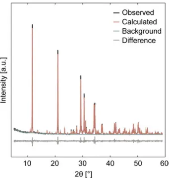
XRD pattern, based on crystal parameters retrieved from crystallographic databases, together with details of the X-ray diffractometer and the scan settings. The calculation process is iterated until the calculated pattern matches the measured XRD pattern. From this calculation the refined pa-rameters can be obtained. In Papers II, III, IV, V, VI and VII the quantitative phase composition was refined and was used to verify the crystalline compo-sition of the cement specimens and evaluate if any phase transformations occurred, as well as to connect it to the mechanical properties of the ce-ments. An example of an XRD measurement and a refined pattern can be seen in Figure 3.
Figure 3. Diffraction pattern of a brushite cement (optimised formulation) measured
by XRD (observed) and the corresponding Rietveld refinement (calculated).
Porosity
The porosity of CPCs has an important impact on several properties of the material, such as mechanical behaviour, degradation and bioactivity. As
already mentioned, an intrinsic nano- and microporosity is created in the CPCs during the setting reaction. The chemical formulation, particle sizes of the starting materials, different additives (such as accelerators and retard-ants) and L/P-ratio affect the porosity of the cement. However, pores are also created during preparation of the cement as air bubbles become entrapped during mixing of the cement paste and moulding of specimens.
There are several different methods to determine the porosity of CPCs,
e.g., helium pycnometry16,53-57, mercury intrusion porosimetry (MIP)17,18,53,58,
water evaporation53, solvent resaturation59, solvent exchange53, and
micro-computed tomographic imaging (micro-CT)60.
In helium pycnometry the skeletal density (density of the specimen ex-cluding pores) of the material is determined by pumping helium into a chamber with well-calibrated volume, and evaluating the pressure difference between the empty chamber and the same chamber filled with the specimen of interest (normally crushed into smaller pieces prior to analysis to enable a properly filled chamber). Helium pycnometry can only be performed on specimens that have been dried prior to analysis. The porosity can be calcu-lated from the skeletal density and the apparent density (density of the spec-imen including pores, determined by, e.g., a calliper or Archimedes’ princi-ple).
In water evaporation, it is assumed that all pores are filled with water and the volume of the pores is equal to the volume of the evaporated water. The porosity can be calculated from the mass differences before and after drying the specimen, together with the apparent volume.
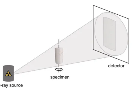
Micro-CT is a non-destructive method that utilises X-rays to image the object of interest in 2D, see Figure 4. A series of images are taken by rotat-ing the specimen, and the images can then be reconstructed to create a 3D model of the object. By separating the solid material from the background (including pores) the porosity can be determined. The advantage is that also closed pores can be taken into account, however, the sizes of the pores in-cluded in the analysis depend on the resolution of the scanner (typically ~5 µm). Therefore, in the case of CPCs, most pores are not taken into account, as they typically are in the size range of ten nanometers to a few microme-ters17,18,53. Nonetheless, micro-CT is a versatile tool that can be used to
Figure 4. Sketch showing the principle of micro-CT.
Several of the porosity measurement methods commonly used for CPCs are associated with drawbacks, such as high cost (e.g., helium pycnometry, MIP, micro-CT), use of toxic chemicals (e.g., MIP), lengthy analysis (e.g., MIP, micro-CT), that might lead to fewer specimens being analysed, compromis-ing statistical power. Micro-CT also has the above-mentioned disadvantage of having a limited resolution. Most importantly, most of the commonly used porosity measurement methods (with micro-CT being one exception) are destructive to a certain degree, preventing the specimens from being used in consecutive characterization: MIP contaminates the specimens with mercu-ry, and with helium pycnometry and water evaporation the specimen needs to be dried prior to analysis. Previously it has been shown that the mechani-cal properties of dry cements may be higher compared to their wet counter-part61-63, and the drying process might also lead to an unwanted phase
trans-formation53. Hence, a porosity measurement method for wet cements would
be very useful. Such a method would allow for the porosity and, e.g., me-chanical properties to be determined for the very same wet cement specimen, which is highly advantageous for the characterization of cements intended for direct injection in vivo.
Contrary to helium pycnometry and water evaporation, solvent exchange does not require drying of the specimen. In solvent exchange the apparent volume, 𝑉!, of the wet cement specimen is first determined (as previously described). The specimen is then immersed in a solvent (that should not alter the properties of the cement) and the mass of the specimen is recorded until complete exchange occurs (i.e., the change in mass is stable and the water in the pores are assumed to be completely exchanged with solvent). The porosi-ty, Φ, can be calculated from the following relation (Equation 1):
Φ = !!"#!!!"#$%&' !!!!!!!"#$%&'
!! , (1)
where 𝑚!"# is the mass of the wet specimen in air, 𝑚!"#$%&' is the mass of the specimen after complete exchange, 𝜌!!! and 𝜌!"#$%&' are the densities of
water and solvent, respectively. A solvent exchange method was developed for CPCs in Paper IV and was subsequently used in Papers V and VII to assess the porosity of the wet cements.
Mechanical assessment
The mechanical properties of CPCs are most commonly evaluated under a quasi-static load. Often the compressive strength is the only parameter used to evaluate the mechanical properties of the CPCs. The reason for this is the simplicity of the method compared to, e.g., tensile testing, and bending, es-pecially with respect to sample preparation and test set-up for these brittle materials.5,64 As already mentioned, more research effort has been devoted to
apatite cements as they have traditionally presented higher strengths than brushite cements. However, brushite cements have attracted more attention recently as they provide an interesting alternative for clinical applications
and as some new formulations have shown improved strengths16,65. A review
of many studies, collectively, was needed to understand the mechanical properties of brushite cements and determine knowledge gaps. Hence the aim of Paper I was to review the available literature on mechanical proper-ties of brushite cements. The aim of Paper II was to evaluate and compare the mechanical properties of the recently developed high-strength brushite cement and a fast-setting apatite cement, with two commercially available cements, under different loading scenarios, namely compressive, tensile and flexural.
Compressive strength
Quasi-static compressive strength, 𝜎!", of CPCs is evaluated by placing the specimen between two compression platens and applying a continuously increasing displacement until the specimen fails, see Figure 5a. Since there is no standard for ceramic bone cements, relevant parts of the testing
stand-ard for polymeric bone cements (e.g., poly(methyl methacrylate), PMMA)66
are commonly used for the testing, e.g., the dimensions of the specimens. The compressive strength is calculated from the load at failure, 𝐹!"#, and the cross-sectional area perpendicular to the loading direction, 𝐴 (Equation 2):
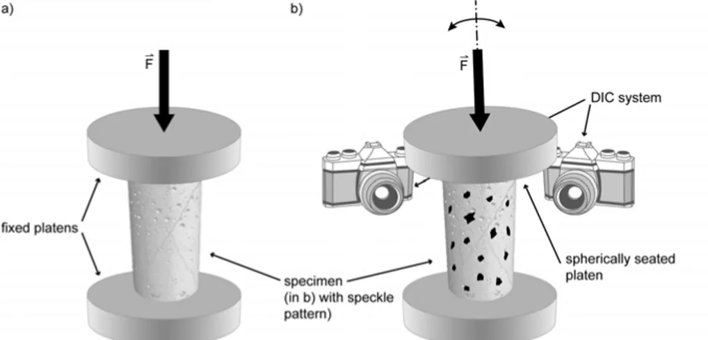
Compression tests of CPCs are sensitive to end artefacts, e.g., non-parallel end surfaces, where the reduced contact area leads to local stresses at the specimen ends. As an alternative, spherically seated compressions platens can be used to decrease the effects of the end-artefacts, see Figure 5b.
Figure 5. Experimental set-up of compressive strength tests using a) fixed
compres-sion platens and b) spherically seated platens. In b) the principles of digital image correlation (DIC) is also illustrated.
The compressive strength can be related to the porosity of the cement through the empirical relation (Equation 3)67:
𝜎!"= 𝜎!𝑒!!!, (3)
where 𝜎!" is the compressive strength, 𝜎! is the strength of a fully dense
cement (a cement with no porosity at all), Φ the porosity and 𝑞 is a
dimen-sionless constant. This equation has been widely used to couple the porosity to the strength of various cements before68,69.
Diametral tensile strength
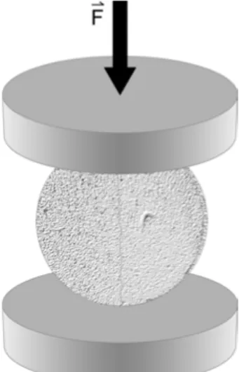
Tensile strength (sometimes referred to as direct tensile strength) is difficult to measure for brittle ceramics like CPCs, due to the difficulties in preparing specimens without surface flaws, and problems associated with fixing them on the testing machine. A method that has commonly been used instead is diametral tensile strength, 𝜎!"#, or indirect tension, also referred to as the Brazilian test, see Figure 6. In this method a circular disc is loaded in com-pression and it is assumed that tensile stresses perpendicular to the applied load break the specimen into two halves. The diametral tensile strength is calculated from the load at failure, 𝐹!"#, and the diameter, 𝑑, and height, ℎ, of the disc (Equation 4):
𝜎!"#=!!!"!!"#, (4) It has previously been shown that diametral tensile strength underestimates the strength of a brushite cement in comparison to (direct) tensile strength61. Though diametral tensile strength does not give the same stress distribution as a direct tensile strength test, it is a simple method that allows for compari-son of different cement formulations, and it has therefore been widely ap-plied for CPCs and is also employed for cements used within the dental70 and construction fields71.
Figure 6. Experimental set-up for diametral tensile strength test.
Biaxial flexural strength
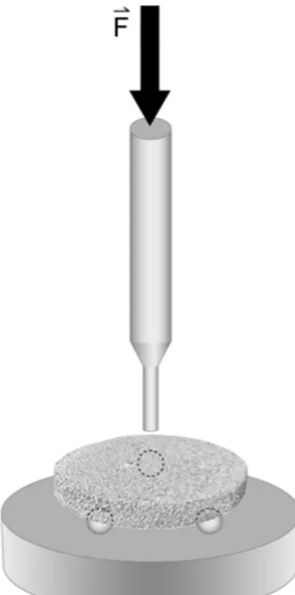
There are several methods to assess the flexural strength of CPCs, such as piston-on-3-ball (biaxial flexural strength, 𝜎!"#), 3- and 4-point bending. In piston-on-3-ball, a disc shaped specimen is supported on three balls and is centrally loaded to failure by a piston, see Figure 7. The biaxial flexural strength, is calculated from the load at failure, 𝐹!"#, and the thickness of the disc at its centre, 𝑑 (Equation 5):
𝜎!"# =!!.!"#$!!!"#!!!, 𝑋 = 1 + 𝜐 ln ! ! ! + !!! ! ! ! ! , (5) 𝑌 = 1 + 𝜐 1 + 𝑙𝑛 !! ! + 1 − 𝜐 !! !,
where 𝜈 is Poisson’s ratio, 𝐴 is the radius of the support circle, 𝐵 is the radi-us of the loaded area and 𝐶 is the sample radiradi-us.
Testing a disc shaped specimen (such as in piston-on-3-ball) is claimed to be less sensitive to surface flaws compared to 3- and 4-point bending (that utilises a bar shaped specimen), and piston-on-3-ball is thus a more readily used method for CPCs.
Figure 7. Experimental set-up for biaxial flexural strength test.
Elastic modulus and Poisson’s ratio
Literature values of elastic properties of CPCs are scarce. The elastic modu-lus of CPCs is commonly calculated from the linear part of the stress-strain curve obtained from compression testing, a method sometimes referred to as the platen-technique, or the method is not specified at all72,73. It is a simple method, but it may suffer from low accuracy, as the precision of the strain measurements relies on the resolution of the built-in position device measur-ing the displacement of the compression platens. The position device is gen-erally not sensitive enough to small deformations, such as those of brittle CPCs, as it usually is designed to measure displacement over a large length span (typically > 50 cm).
Contrary to the platen-technique, digital image correlation (DIC), see
Figure 5b, is a method that measures the deformation directly on the
speci-men surface. By applying a speckle pattern on the specispeci-men prior to testing and recording the deformation process by digital cameras, the movement of individual speckles can be tracked from the acquired images, and the strain can thus be calculated.74 This method has a higher accuracy compared to the platen-technique, since the resolution of the images is higher than the built-in position device of most materials testbuilt-ing machbuilt-ines. From these images, it
is also possible to calculate Poisson’s ratio, as the deformation both parallel and perpendicular to the loading direction can be determined. Values of Poisson’s ratio of CPCs are indeed also largely lacking in the literature (available only for some apatite cements75), and accurate values of elastic
modulus and Poisson’s ratio are crucial for, e.g., numerical models. Hence, the study in Paper III, using DIC to assess the elastic properties of CPCs, was highly motivated.
Fatigue
Quasi-static loading is an easy way to assess the mechanical properties of CPCs and to compare different cement types and formulations to each other. However, in vivo repeated loading can be expected, and quasi-static strength does not determine for how long the material will resist the mechanical load-ing scenario. To aid in the prediction of the CPCs behaviour when implanted into the body, the in vitro fatigue properties need to be studied. One way to characterize a material’s behaviour in fatigue, is by applying a cyclic load to an uncracked, smooth-surfaced specimen and counting the number of cycles to failure, Nf. Thus, by varying the maximum applied stress level, Smax, an
S-N curve can be created. The test usually has a predetermined number of
cy-cles after which the test will be stopped called the run-out limit. The influ-ence of environment on the fatigue properties can also be studied by per-forming the tests in a testing chamber with a circulating heated liquid bath connected to the dynamic materials testing system.
Very few publications exist on the fatigue behaviour of CPCs76-79,
where-as bone cements bwhere-ased on PMMA have been extensively studied80,81. A
like-ly reason for this is the time-consuming nature of fatigue testing and its as-sociated cost (the fatigue tests presented in this thesis corresponds to almost a year of continuous testing), and the brittleness of CPCs, leading to a scatter in results due to the variation of flaw sizes and amounts within the cements. Therefore, Papers VI and VII aimed at investigating the fatigue properties of brushite cements and study the effect of phase composition, porosity and liquid environment on the fatigue behaviour.
Degradation
The ideal, resorbable biomaterial would degrade at such a rate that resorp-tion of the material would be balanced by the formaresorp-tion of new bone tissue of the host6. The in vivo degradation rate depends on many different parame-ters, such as the type of CPC, the porosity of the implant, and the site of implantation41,58,82,83. At neutral pH, the in vivo degradation rate can be pre-dicted to be in the order brushite > monetite > octacalcium phosphate (OCP) > apatite (the first resorbing the fastest). However, the surface of a highly soluble CPC can be covered by a poorly soluble phase, therby reducing the
degradation rate82. Brushite is only metastable under physiological condi-tions and can be converted to monetite, OCP or apatite depending on the surrounding conditions.
Even though experiments performed in vitro will differ from those per-formed in vivo in terms of, e.g., chemical as well as biological environment, or fluid flow, in vitro testing of CPCs can still be a good starting point to predict the behaviour of the materials. A number of studies have investigated the in vitro degradation of brushite cements, showing that the degradation of the material occurs through dissolution, disintegration and/or conversion to a more stable phase5,54,82,84-89. The studies have been performed in water87,88,
PBS or similar54,84-86,89-92, or in foetal bovine serum (FBS)54,84,89, and the deg-radation has been studied in terms of, e.g., change in mechanical strength54,84,85,90-92, porosity54,84, and chemical composition of the ce-ments54,83,86-90,92,93.
There is a lack of data in the literature on how in vitro degradation affect important cement properties such as the compressive strength, phase compo-sition, porosity, and pore size distribution, especially for high-strength brushite cements over longer periods of time. The aforementioned publica-tions studied in vitro degradation of cements with strengths lower than 15 MPa, except for Cama et al.87 (the strength of their cement was ~28 MPa, i.e.
still much lower than the optimised brushite formulation studied in Paper V), which may be very different to the degradation of brushite cements with a higher strength, which has a lower porosity. Moreover, the time frame for bone fracture healing ranges from ~6 weeks up to several months or longer, depending on fracture site, age and health status of the injured patient20,94. Long-term studies are therefore important when one wants to evaluate the mechanical as well as chemical behaviour of CPCs and determine their limi-tations in terms of applications. However, most in vitro degradation studies of brushite cements found in the literature have been terminated after 2-4 weeks of incubation time, and the compressive strength, porosity and phase composition has never before been evaluated beyond 4 weeks of in vitro degradation54,84-88,90,91. Hence, the aim of Paper V was to investigate the
long-term in vitro degradation of a high-strength brushite cement in terms of compressive strength, porosity, pore size distribution and phase composition.
Summary of aims and objectives
This thesis aims to evaluate CPCs in different loading scenarios in order to determine their possible future use in load-bearing applications.
The first objectives of this work were to review the available literature on the mechanical properties of brushite cements (Paper I) and to evaluate and compare the compressive, tensile and flexural strengths of an experimental high-strength brushite cement developed in-house and a fast setting apatite cement developed at a collaborator with commercially available brushite and apatite based cements (Paper II).
Secondly, the objective was to determine the elastic modulus and Pois-son’s ratio of CPCs (a high-strength brushite cement, an apatite cement and a monetite cement were evaluated) using a high-resolution measurement tech-nique (Paper III). Accurate values of the elastic modulus and Poisson’s ratio are crucial for material development aimed at matching these properties and for computational modelling.
Thirdly, the objectives were to develop a non-destructive wet porosity measurement method (Paper IV), and to use this method to study the in vitro degradation of a high-strength brushite cement (Paper V). A long-term eval-uation of the porosity, pore size distribution, compressive strength, and phase composition was conducted in order to study how the cements behave within a time frame relevant for bone fracture healing.
Finally, the last objective was to study how two different formulations of brushite cements behave under dynamic loading. The fatigue properties were investigated in relation to the phase composition of the cement (Paper VI) and in relation to the porosity of the cement and the chemical surroundings (Paper VII).
Mechanical properties under quasi-static load
The different CPCs studied in this thesis were evaluated under quasi-static compression (Papers I-VII), diametral tensile strength (Paper II), and biaxial flexural strength (Paper II). The elastic modulus was evaluated under quasi-static compression using the platen-technique as well as DIC. The latter technique was also used to determine Poisson’s ratio (Paper III). This chap-ter presents the obtained results.
Compressive strength
The compressive strength varies greatly between different types of CPCs, due to differences in, e.g., chemical composition, particle size of the starting powders, entanglement of the crystals, porosity, pore size distribution, as well as sample preparation, storage and testing conditions. Apatite cements have traditionally been reported to be stronger than brushite cements in compression5,64. However, recent studies have produced brushite
formula-tions with strengths comparable to apatite cements16,65, whereas the strength of monetite cement is generally lower due to a higher porosity38,95.
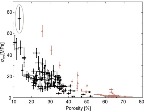
As previously mentioned, there is a strong relationship between the poros-ity of a CPC and its mechanical properties. This relationship was investigat-ed by evaluating the compressive strength and porosity of brushite cements presented in the literature (Paper I). The combined results from 16 scientific studies are summarized in Figure 8. A large variation in compressive strength was found, from approximately 1 to 74 MPa, and a clear relation to porosity can be discerned, with a reduction in strength as the porosity in-creases, according to Equation 3. Generally, using citric acid as liquid phase smaller particle sizes of reactant powders, decreasing the L/P-ratio, mixing the cement mechanically rather than by hand, and testing the cements when they have been dried result in brushite cements with improved strength45,65.
Figure 8. Relationship between compressive strength and porosity for brushite
ce-ments. Different markers indicate values from different studies. References can be found in Paper I. Strength tested on wet specimens is shown in black and dry spec-imens in colour. The circle indicates the optimised brushite formulation studied in
Papers II, III, IV, V and VII.
The main focus of this thesis is on an optimised brushite cement formulation found in the upper left corner of Figure 8, having a strength in compression of ~74 MPa and a porosity around 13%16. In Paper II, the compressive
strength of this cement was compared to that of an apatite cement and two
commercially available cements, chronOS™ Inject and Norian® SRS®
(hav-ing brushite and apatite as the main end-products of the set cements, respec-tively) (Paper II), see Figure 9.
The brushite cement was found to be almost 20 times stronger than chronOS™ Inject, which can partly be explained by the large difference in porosity (~11% and ~33% for brushite and chronOS™ Inject, respectively). Moreover, chronOS™ Inject has a high amount of filler particles (~55 wt% unreacted ß-TCP), which has previously been shown to be detrimental for the strength if present in too high amounts45.
The apatite cement was stronger than Norian® SRS®, although the differ-ence was not as pronounced compared to the brushite-based cements, likely due to more similar porosities (the apatite cement had a porosity ~41% and Norian® SRS® ~44%). The apatite cement contained mainly HA (~91 wt%
HA), whereas Norian® SRS® contained less HA (~85 wt%), and also some
unreacted calcite (~5 wt%) which may result in less efficient crystal entan-glement, and thus explain the lower strength.
All cements, except Norian® SRS®, had a significantly higher dry com-pressive strength compared to their wet counterparts. The difference in
strength between wet and dry CPCs has been shown before61-63, and has also
been observed for other self-setting cements, e.g., cements based on calcium sulphate dihydrate96. A likely explanation for this observation was proposed by Andrews97: breaking of cement structures happen by overcoming
fric-tional forces between interlocked crystals rater than breaking of individual crystals. By the introduction of water in the structure, the frictional force is reduced and thus the strength of the cement is decreased.
The wet strengths of brushite, apatite and Norian® SRS®are higher
com-pared to reported trabecular bone strengths (see Table 1) suggesting that the cements have the potential to be used in certain load-bearing applications.
Figure 9. Wet and dry strengths of two experimental formulations and two
commer-cial formulations of brushite and apatite cements. Groups with the same letter are not significantly different at a significance level of α = 0.05.
Wet brushite, monetite and apatite cements were also loaded in compression using both fixed and spherically seated compression platens (Paper III). No significant differences in strength were seen for monetite (fixed: ~12 MPa, spherically seated: ~13 MPa) and apatite cements (fixed: ~35 MPa, spheri-cally seated: ~39 MPa), whereas a significant increase was seen for brushite cements when spherically seated platens (~56 MPa) were used compared to
fixed platens (~34 MPa), see Figure 10. The lower strengths measured with the fixed platens for brushite cements are likely due to end-artefacts. Non-parallel ends result in a reduced contact area and an increase in local stress-es. Brushite cements are stiffer compared to apatite and monetite (see
Fig-ure 13), which will result in higher local stresses and therefore a greater
measured difference between fixed and spherically seated platens.
The compressive strength of the three cement types is within the range previously found for the same type of materials64,65,98. In comparison to the
compressive strength of bone, as already mentioned brushite and apatite cements have strengths in the upper range of trabecular bone, whereas monetite cements are in the middle of the range (see Table 1). The compres-sive strength of cortical bone exceeds all of the cements.
Figure 10. Wet compressive strengths for brushite, monetite and apatite cements
determined with fixed and spherically seated compression platens. Groups with the same letter are not significantly different at a significance level of α = 0.05.
Diametral tensile strength
Diametral tensile strength of brushite, apatite, chronOS™ Inject and Norian®
SRS® was investigated for both wet and dry specimens (Paper II), see
also seen for diametral tensile strength: both brushite and apatite cements were found to be stronger than the commercial brushite- and apatite-based formulations. Drying was seen to significantly affect the diametral tensile strength of chronOS™ Inject and the apatite cement. The same effect of friction reduction by the presence of water as previously discussed may ex-plain this difference between wet and dry strength. Interestingly, the strengths of brushite and Norian® SRS® was not significantly affected by drying. Microstructural features, e.g., amount of pores, shape and entangle-ment of the crystals, have an impact on the strength and may also influence the difference between wet and dry strength.
The diametral tensile strength of the brushite cement is considerably higher (about 10 times) compared to what has previously been found before for a brushite cement (~1.3 MPa for a moist cement)99, whereas the strength of the apatite cements is in the same range of earlier findings64. In
compari-son to the tensile strength of trabecular bone (see Table 1), the wet diametral tensile strength of the brushite cement resides at the higher end of the range, apatite is in the middle and the commercial cements are in the lower end of the range. Cortical bone is much stronger in tension compared to the studied CPCs.
Figure 11. Wet and dry diametral tensile strength of two experimental formulations
and two commercial formulations of brushite and apatite cements. Groups with the same letter are not significantly different at a significance level of α = 0.05.
Biaxial flexural strength
Piston-on-3-ball100 was used to assess the flexural strength of brushite and apatite cements, as well as chronOS™ Inject and Norian® SRS®, see Fig-ure 12. Again, the brushite cement was superior to the commercial
brushite-based formulation in flexural strength and the apatite cement had higher strength than Norian® SRS®. The strength of brushite and Norian® SRS® was not affected by drying, whereas drying significantly affected the strength of chronOS™ Inject and apatite.
The biaxial flexural strength of the brushite cement (~31 MPa) is higher
compared to previously reported values for brushite cement (~7 MPa)99. The
flexural strength of Norian® SRS® (wet strength ~11 MPa) is higher
com-pared to what has been found before for the same cement (wet strengths ~0.5-0.7 MPa)79. However, the latter values are for specimens tested in
tradi-tional 3- and 4-point bending, which tend to give lower strengths compared to biaxial flexural strength, since bending bars are more sensitive to surface flaws compared to the smaller discs99.
Figure 12. Wet and dry biaxial flexural strength of two experimental formulations
and two commercial formulations of brushite and apatite cements. Groups with the same letter are not significantly different at a significance level of α = 0.05.
Elastic properties – Young's modulus and Poisson's ratio
The wet elastic properties of brushite, monetite and apatite were studied using the platen-technique and DIC (Paper III). The types of platen used and the method to assess strains were seen to significantly affect measured val-ues of the elastic moduli of the cements, see Figure 13. DIC presented the highest elastic moduli for all three types of CPCs (brushite ~24 GPa, apatite ~14 GPa and monetite ~7 GPa), whereas the platen technique resulted in significantly lower values (roughly half of that of DIC). The elastic moduli from fixed and spherically seated platens were in the upper part of the range of previously published data for CPCs (0.4-8 GPa), including moduli values that were also obtained from the platen-technique or the method was not specified72,73,101. By using spherically seated platens, end-artefacts could be diminished and for brushite that was found to be the stiffest cement, higher elastic moduli were obtained.For these brittle CPCs it is difficult to measure the small strains the ce-ments may suffer from before failure using the platen-technique. In fact, based on the DIC results, the deformation is close to the detection limit of the built-in position device controlling the platen displacement (the uncer-tainty of the position devices were ±0.01 mm and the strain to when cracks appeared was ~0.02 mm). Strains determined by DIC, on the other hand, are inherently more accurate and precise, as previously discussed. Moreover, in DIC the strains are directly measured on the specimen surface, thus end arte-facts do not affect these measurements. Measurement errors related to the platen-technique have been demonstrated by others, e.g., for mechanical testing of calcium sulphate dihydrate cements96.
In comparison to bone, the elastic moduli obtained by DIC of the three studied CPCs are in the range of cortical bone, but higher compared to the moduli of trabecular bone (see Table 1).
Figure13. Elastic modulus for wet brushite, monetite and apatite cements
deter-mined using the platen-technique (fixed and spherically seated platens) and DIC. Groups with the same letter are not significantly different at a significance level of α = 0.05.
From DIC it is possible to determine the deformation both in the direction of loading and in the perpendicular direction, and therefore the wet Poisson’s ratio for the three types of cements could be determined: ~0.26 for brushite, ~0.24 for monetite, and ~0.21 for apatite. Values of Poisson’s ratio of CPCs are scarce in the literature, in fact for bulk cements, it has only been deter-mined before for a pre-mixed apatite/chitosan cement by an ultrasound through-transmission method75. Poisson’s ratios found herein for brushite,
monetite and apatite cements are within the range found in the study by Rajzer et al (0.19-0.26)75. Poisson’s ratio of the CPCs is in the range of
Measuring the porosity of wet CPCs
Solvent exchange is a porosity measurement method that does not include a drying step, and is commonly used for other types of cement, such as Port-land cements and concrete102, and, less commonly, brushite cements53. How-ever, when methanol was previously used as solvent for the brushite cements it affected the phase composition of the cement, likely due to an interaction between the small, more polar methanol molecule and brushite53. In Paper IV, isopropanol, having a larger, less polar molecule compared to methanol,
was evaluated as an alternative solvent in a wet porosity measurement meth-od for brushite and apatite cements. It proved to be an easy and non-destructive way to determine the porosity of wet brushite cements that had no effect on either the phase composition or the compressive strength. Moreover, the method only gave a slightly lower porosity (~1.5 percentage points) in comparison to water evaporation and helium pycnometry, hence it was used in subsequent studies (Papers V and VII).
For the apatite cements, the isopropanol exchange took much longer time compared to brushite cements; the mass loss reached steady-state within 24 hours for brushite compared to ~350 hours for apatite. This could be due to the smaller pores present in the apatite cements compared to brushite ce-ments that may retard the diffusion process. Immersing the specimens in isopropanol also affected the compressive strength of the apatite cements, whereas the strength of brushite cements was the same before and after im-mersion, see Figure 14. The higher strengths seen for the apatite cements after being immersed in isopropanol may be due to a continuation of the setting reaction; previously it has been shown that longer setting times can allow for a continued transformation of α-TCP to HA resulting in a higher strength50. Due to the long exchange time and the alteration of the strength,
Figure 14. Wet compressive strengths of a) apatite cements and b) brushite cements
after setting in PBS (control) or immersed in isopropanol and stored at room tem-perature (RT) until complete solvent exchange. Groups with the same letter are not significantly different at a significance level of α = 0.05.
In vitro degradation of brushite cements
For CPCs that are designed to resorb in vivo it is important to study the deg-radation of the cements to determine their limitations in terms of applica-tions. The compressive strength, porosity, chemical composition, and micro-structure of brushite cements were evaluated over 25 weeks of in vitro deg-radation (Paper V). The degdeg-radation study was performed in three different liquids: PBS and a serum solution (containing FBS) were chosen to simulate
in vivo conditions, while water was chosen to be able to compare to
previ-ously performed degradation studies and also to investigate if the in vitro degradation experiments could be simplified.
Initially, the brushite cements had an open porosity of ~13%, which in-creased by 0.3-0.8 percentage points per week, concomitant to a decrease in strength, see Figure 15. After 25 weeks of degradation the porosity had roughly doubled, and the strength halved, with variations depending on the soaking liquid. The lowest strength concomitant with the highest increase in porosity was noted for specimens immersed in PBS. The closed porosity determined by micro-CT decreased slightly over time and was comparable for specimens kept in the different liquids after 25 weeks of incubation. The increase in porosity is lower than what has previously been reported for a brushite cement84, but the studies differ in terms of, e.g., degradation proto-cols and, most importantly, the initial properties of the cement. A cement with large pores and a high surface area is likely to degrade faster compared to a cement with small pores and a small surface area. The brushite cement studied herein has a lower porosity compared to those previously evaluated.
Figure 15. a) Open porosity (determined by solvent exchange) and b) compressive
strength of brushite cements degraded in H2O, PBS and a serum solution over 25
weeks.
The brushite cement slowly dissolved in the three different degradation me-dia; a volume decrease was confirmed by both gravimetric analysis and mi-cro-CT. The degradation rate in terms of volume, or mass, was lower com-pared to other brushite cements (see Table 4), likely due to the intrinsically lower porosity of the investigated cement.
Table 4. Summary of in vitro degradation rates of brushite cements found in the
literature.
L/P ratio Liquid Time Degradation rate
[mass percentage points/week]
Reference 0.22 ml/g H2O 25 weeks 0.37 Paper V 1.33 ml/g H2O 16 days 7.1-8.9 88 0.22 ml/g PBS 25 weeks 0.35 Paper V 0.57 ml/g PBS 90 days 1.5 84 0.5 ml/g PBS 90 days 1.2-1.9 92 0.57 ml/g PBS 28 days 2.1-4.8 54 0.8 ml/g Ringers solution 28 days 2.8 85
0.5 ml/g PBS 21 days 1.8 91
1 g/g PBS 14 days 2.9-11.7 90
1 g/g PBS 14 days 0.5-8.7 86 0.22 ml/g Serum 25 weeks 0.22 Paper V
0.57 ml/g FBS 90 days 4.4 84
0.57 ml/g FBS 28 days 16.0 54
Micro-CT analysis revealed considerable changes in the microstructure of the surface from 10 weeks and onward, see Figure 16. It appeared as a flaky layer, see Figure 17, which XRD analysis revealed to consist of OCP, and was most prominent for specimens kept in PBS. OCP is known to be
meta-stable and will transform to apatite with time41. However, apatite was never observed during the time frame of the degradation study. In a recent in vitro study of a brushite cement, such a shell of OCP was already present after 5
days of degradation in PBS89. Bannerman et al. showed that the shell
pene-trated deeper and deeper, followed by dissolution of brushite, with increas-ing degradation time. Brushite has also been shown to transform to OCP in
vivo58, revealing a whisker-like morphology similar to what was found in
Paper V.
Figure 16. Micro-CT cross-sections of brushite cements after 0, 10 and 25 weeks of
degradation in three different liquids.
Figure 17. Scanning electron microscopy images showing the outer surface layer
after 25 weeks (this specific specimen had been kept in PBS).
Even though OCP is a more stable phase than brushite at physiological pH, the OCP layer that was formed during the in vitro degradation of the brushite cement in the present work did not affect the rate of increase in open porosi-ty, decrease in object volume and decrease in compressive strength. The addition of SPP is also likely to affect the degradation properties of the ce-ment. In fact, it has been shown clinically that CPCs containing pyrophos-phate ions can have a favourable resorption rate and stimulate bone for-mation, thus providing a good integration with the surrounding tissue in
![Table 1. Mechanical properties of bone. Ultimate compres-sive strength [MPa] Ultimate tensile strength [MPa] Elastic modulus [GPa] Poisson’s ratio Cortical bone 95-230 22,23 40-140 21 9-25 22,24 0.46-0.58 25,26 Trabecular bone 1-30 27,28 1-11 21](https://thumb-eu.123doks.com/thumbv2/5dokorg/5464033.141985/14.727.105.631.114.239/mechanical-properties-ultimate-strength-ultimate-strength-cortical-trabecular.webp)