Clin Oral Impl Res. 2019;30:833–848. wileyonlinelibrary.com/journal/clr
|
8331 | INTRODUCTION
It is currently accepted that treatment of peri‐implantitis regu‐ larly requires surgical intervention to get adequate access to the
contaminated implant surface (Klinge, Klinge, Bertl, & Stavropoulos, 2018; Renvert & Polyzois, 2018). Indeed, a variety of protocols, including mechanical or chemical means, or combinations thereof, aiming at implant surface decontamination have been proposed. Received: 1 January 2019
|
Revised: 6 April 2019|
Accepted: 18 June 2019DOI: 10.1111/clr.13499
R E V I E W A R T I C L E
Mechanical and biological complications after
implantoplasty—A systematic review
Andreas Stavropoulos
1,2| Kristina Bertl
1,3| Sera Eren
4| Klaus Gotfredsen
51Department of Periodontology, Faculty of
Odontology, University of Malmö, Malmö, Sweden
2Division of Conservative Dentistry
and Periodontology, University Clinic of Dentistry, Medical University of Vienna, Vienna, Austria
3Division of Oral Surgery, University Clinic
of Dentistry, Medical University of Vienna, Vienna, Austria
4Postgraduate Course
Periodontology, Medical University of Vienna, Vienna, Austria
5Department of Oral Rehabilitation, Faculty
of Health and Medical Sciences, School of Dentistry, University of Copenhagen, Copenhagen, Denmark
Correspondence
Andreas Stavropoulos, Department of Periodontology, Faculty of Odontology, University of Malmö, Carl Gustafs väg 34, 20506 Malmö, Sweden.
Email: andreas.stavropoulos@mau.se
Funding information
KOF/Calcin Foundation of the Danish Dental Association; Austrian Society of Implantology (ÖGI)
Abstract
Objectives: Implantoplasty, that is, the mechanical modification of the implant, in‐
cluding thread removal and surface smoothening, has been proposed during surgical peri‐implantitis treatment. Currently, there is no information about any potential me‐ chanical and/or biological complications after this approach. The aim of the current review was to systematically assess the literature to answer the focused question “Are there any mechanical and/or biological complications due to implantoplasty?”.
Materials and methods: A systematic literature search was performed in three da‐
tabases until 23/09/2018 to assess potential mechanical and/or biological compli‐ cations after implantoplasty. All laboratory, preclinical in vivo, and clinical studies involving implantoplasty were included, and any complication potentially related to implantoplasty was recorded and summarized.
Results: Out of 386 titles, 26 publications were included in the present review (six lab‐
oratory, two preclinical in vivo, and 18 clinical studies). Laboratory studies have shown that implantoplasty does not result in temperature increase, provided proper cooling is used, but leads in reduced implant strength in “standard” dimension implants; fur‐ ther, preclinical studies have shown titanium particle deposition in the surrounding tissues. Nevertheless, no clinical study has reported any remarkable complication due to implantoplasty; among 217‐291 implants subjected to implantoplasty, no implant fracture was reported during a follow‐up of 3–126 months, while only a single case of mucosal discoloration, likely due to titanium particle deposition, has been reported.
Conclusions: Based on all currently available, yet limited, preclinical in vivo and clini‐
cal evidence, implantoplasty seems not associated with any remarkable mechanical or biological complications on the short‐ to medium‐term.
K E Y W O R D S
complication, implant threads, implantoplasty, peri‐implantitis, systematic review
This is an open access article under the terms of the Creative Commons Attribution‐NonCommercial‐NoDerivs License, which permits use and distribution in any medium, provided the original work is properly cited, the use is non‐commercial and no modifications or adaptations are made.
Systematic reviews of studies on the various clinically applicable decontamination protocols, however, have shown that (a) complete implant surface decontamination cannot be achieved, neither by me‐ chanical nor chemical means alone, not even under laboratory (in vitro) settings, (b) in general, combinations of mechanical and chem‐ ical means appeared more effective, (c) there is large variation in the effectiveness of the various approaches, among other reasons, de‐ pending on the type of implant surface micro‐structure, and (d) the more structured the implant surface, the more difficult is it to de‐ contaminate (Louropoulou, Slot, & Van der Weijden, 2014; Ntrouka, Slot, Louropoulou, & Van der Weijden, 2011). In this context, pre‐ clinical in vivo studies have shown that differences in implant sur‐ face micro‐structure may indeed influence disease progression rate and the outcome of treatment in terms of extent/severity of residual peri‐implant inflammation. Specifically, peri‐implantitis progresses faster at rough implants compared with machined‐surface implants and there is a variation in progression rate among the various micro‐ structures (Albouy, Abrahamsson, Persson, & Berglundh, 2008; Berglundh, Gotfredsen, Zitzmann, Lang, & Lindhe, 2007; Carcuac et al., 2013), while there are remarkable differences in the size of the residual inflammatory infiltrate (i.e., from relatively small to quite large) and in the distance of the infiltrate to the bone (i.e., from rel‐ atively far away to almost in contact), among implants with similar surface roughness (i.e., moderately rough), but of different micro‐ structural design (Albouy, Abrahamsson, Persson, & Berglundh, 2011). In this latter study, the least residual inflammatory infiltrate— which was also located furthest from the bone (i.e., 1 mm)—was ob‐ served around implants with a machined surface.
One approach suggested to address effectively the above‐men‐ tioned concerns associated with rough implants affected by peri‐implan‐ titis, is implantoplasty, that is, the mechanical removal (grinding) of the implant threads and the rough implant surface, rendering thus a relatively “smooth” implant surface. Implantoplasty is performed at the aspects of the implant, where due to defect anatomy only a limited potential for
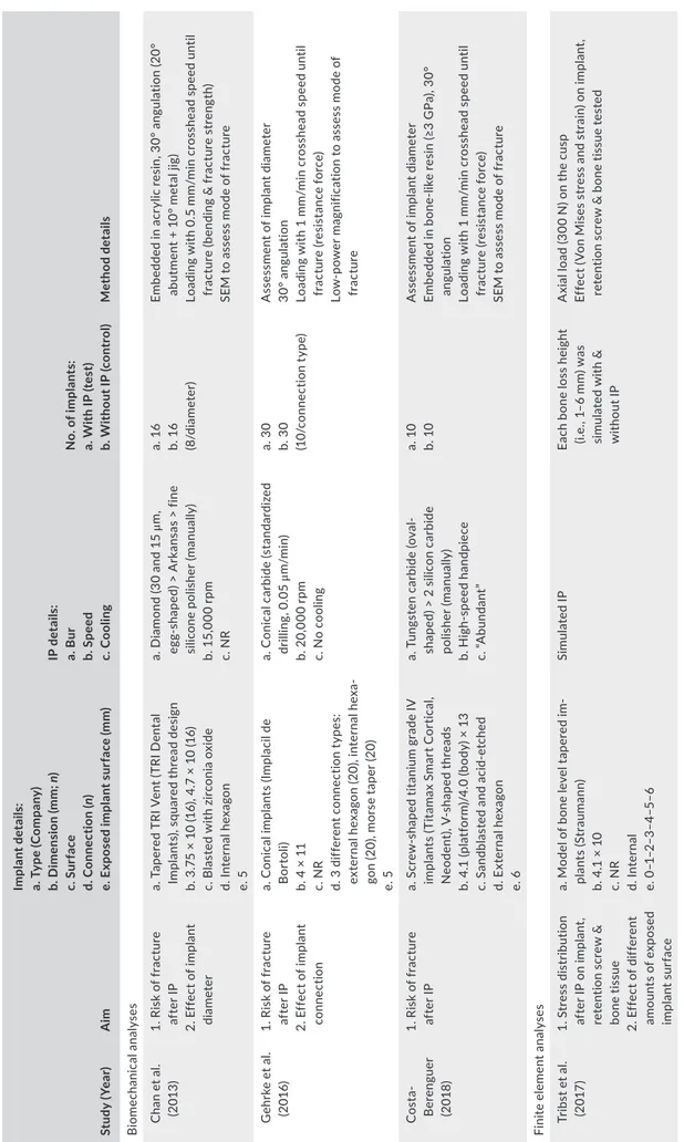
bone regeneration and/or re‐osseointegration after healing can be ex‐ pected, that is, the supra‐bony or dehiscenced aspects of the implant. Indeed, a few clinical studies have reported successful clinical and radio‐ graphic outcomes after surgical treatment of peri‐implantitis combined with implantoplasty (Matarasso, Iorio Siciliano, Aglietta, Andreuccetti, & Salvi, 2014; Pommer et al., 2016; Romeo et al., 2005; Romeo, Lops, Chiapasco, Ghisolfi, & Vogel, 2007; Schwarz, Hegewald, John, Sahm, & Becker, 2013; Schwarz, Sahm, Iglhaut, & Becker, 2011). Nevertheless, perforation of the implant body, destruction of the implant‐abutment connection, overheating of the implant during grinding causing thermal damage to the surrounding bone, or induction of mucosal staining and/ or increased risk for late inflammatory reactions due to titanium particle deposition, generated from the grinding procedure, appear as reason‐ able concerns. Further, reduction of the implant mass (implant diame‐ ter) at its coronal aspect, occasionally also involving the implant collar (Figure 1), may compromise implant strength and lead to an increased rate of late mechanical complications, for example, implant collar defor‐ mation, loosening of the supra‐structure, fixation‐screw fracture, and implant fracture; this may, in turn, lead to recurrent peri‐implant biolog‐ ical complications and/or require explantation. Currently, there is only limited information on this topic and there is no comprehensive system‐ atic appraisal of possible complications associated with implantoplasty.
Thus, the aim of the current review was to systematically assess the literature to answer the focused question “Are there any me‐ chanical and/or biological complications due to implantoplasty?”.
2 | MATERIAL AND METHODS
2.1 | Protocol and eligibility criteria
The present systematic review was performed according to the cri‐ teria of the Preferred Reporting Items for Systematic Reviews and Meta‐analyses (PRISMA; Appendix S1; Liberati et al., 2009; Moher, Liberati, Tetzlaff, & Altman, 2009). The following inclusion criteria
F I G U R E 1 (a and b) Clinical case from authors' clinic illustrating that implantoplasty often results in significant reduction in implant wall thickness, occasionally involving also the collar (white arrow and line)
were applied during literature search on original studies without publication year restriction: (a) English or German language; (b) labo‐ ratory (in vitro), preclinical in vivo, or clinical trials; (c) reporting on dental implants subjected to implantoplasty; (d) in case of preclinical in vivo and clinical trials, ≥1 month follow‐up after peri‐implantitis surgery; and (e) full text available.
2.2 | Information sources and literature search
Electronic search was performed on three sources (last search 23/09/2018; no date restriction used): MEDLINE (PubMed), Scopus (Ovid), and CENTRAL (Ovid). The database MEDLINE (PubMed) was searched with the following keywords: (periimplant* OR peri‐ implant*) AND (implantoplasty OR implant surface decontamination OR implant surface debridement OR implant surface modification OR implant surface detoxification OR implant threads). The asterisk (*) was used as a truncation symbol. For the other two databases, comparable terms were used, but modified to suit specific criteriaof the particular database. Additionally, a screening of the reference lists and a forward search via Science Citation Index of the included papers was performed. Grey literature was searched for in opengrey. eu and is reported under other sources.
2.3 | Data collection and extraction
Two authors (SE and KB) independently checked title, abstract, and finally full text on the predefined eligibility criteria. Abstracts with unclear methodology were included in full‐text assessment to avoid exclusion of potentially relevant articles. One author (SE) repeated the literature search. Kappa scores regarding agreement on the arti‐ cles to be included in the full‐text analysis and those finally chosen were calculated. In case of ambiguity, consensus through discussion was achieved together with a third author (AS). Further, two authors (SE and KB) extracted twice the following data (if available): author; year of publication; design and aim of the study; inclusion criteria; numbers of animals/patients; implant‐related details [i.e., number,
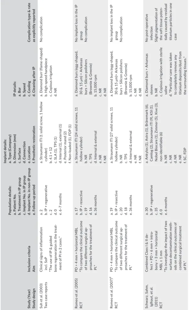
T A B LE 1 C ha ra ct er is tic s of th e 6 in cl ud ed la bo ra to ry p ub lic at io ns St ud y ( Yea r) A im Im pla nt de ta ils : a. T yp e ( C om pa ny ) b. D im en si on ( m m ; n ) c. S ur fa ce d. C on ne ct io n ( n) e. E xp os ed i m pl an t s ur fa ce ( m m ) IP d et ai ls : a. B ur b. S pee d c. C oo lin g N o. o f i m pl an ts : a. W ith I P ( te st ) b. W ith ou t I P ( co nt ro l) Me th od de ta ils B io m echa nic al a na ly se s C ha n e t a l. (2 013 ) 1. R is k o f f ra ct ur e af te r I P 2. E ff ec t o f i m pl an t di ame ter a. T ap er ed T RI V en t ( TR I D en ta l Imp la nt s) , s qua re d t hr ea d d es ig n b. 3 .7 5 × 10 (1 6) , 4 .7 × 1 0 (1 6) c. B la st ed w ith z irc on ia o xi de d. In te rn al h ex ag on e. 5 a. D ia m on d ( 30 a nd 1 5 μm , eg g‐ sh ap ed ) > A rk an sa s > fin e sili co ne p oli sh er (m an ua lly ) b. 1 5, 00 0 rp m c. N R a. 1 6 b. 1 6 (8 /d ia me ter ) Em be dd ed i n a cr yl ic r es in , 3 0° a ng ul at io n ( 20 ° ab ut m en t + 1 0° m et al j ig ) Lo ad in g w ith 0 .5 m m /m in c ro ss he ad s pe ed u nt il fr ac tu re ( be nd in g & f ra ct ur e s tr en gt h) SE M t o a ss es s m od e o f f ra ct ur e G eh rk e e t a l. (2 016 ) 1. R is k o f f ra ct ur e af te r I P 2. E ff ec t o f i m pl an t co nne ct io n a. C on ic al i m pl an ts ( Im pl ac il d e B or to li) b. 4 × 1 1 c. N R d. 3 d iff er en t c on ne ct io n t yp es : ex te rn al h ex ag on (2 0) , i nt er na l h ex a‐ go n ( 20) , m or se ta pe r ( 20) e. 5 a. C on ic al c ar bi de ( st an da rd ize d dr illi ng , 0 .05 μ m /min ) b. 2 0, 00 0 rp m c. N o c oo lin g a. 3 0 b. 3 0 (1 0/ co nne ct io n t yp e) A ss es sm en t o f i m pl an t d ia m et er 30 ° a ng ula tio n Lo ad in g w ith 1 m m /m in c ro ss he ad s pe ed u nt il fr ac tu re (r es is ta nc e f or ce ) Lo w ‐p ow er m ag ni fic at io n t o a ss es s m od e o f fr ac tu re C os ta‐ B er en gu er (2 018 ) 1. R is k o f f ra ct ur e af te r I P a. S cr ew ‐s ha pe d t ita ni um g ra de I V im pl an ts (T ita m ax S m ar t C or tic al , N eo den t), V ‐s ha pe d t hr ea ds b. 4 .1 ( pl at fo rm )/ 4. 0 ( bo dy ) × 1 3 c. S an db la st ed a nd a ci d‐ et ch ed d. E xt er na l h ex ag on e. 6 a. T ung st en c ar bi de (o val ‐ sh ap ed ) > 2 s ili co n c ar bi de po lish er (ma nu al ly ) b. Hi gh ‐s pe ed ha nd pi ec e c. “A bu nd an t” a. 10 b. 10 A ss es sm en t o f i m pl an t d ia m et er Em be dd ed in b on e‐ lik e re si n (≥ 3 G Pa ), 30 ° an gula tio n Lo ad in g w ith 1 m m /m in c ro ss he ad s pe ed u nt il fr ac tu re (r es is ta nc e f or ce ) SE M t o a ss es s m od e o f f ra ct ur e Fi ni te e lemen t a na ly se s Tr ib st e t a l. (2 017 ) 1. S tr ess di st rib ut io n af te r I P o n i m pl an t, re te nt io n s cr ew & bo ne ti ss ue 2. E ff ec t o f d iff er en t am ou nt s of e xp os ed im pla nt s ur fa ce a. M od el o f b on e l ev el t ap er ed i m ‐ pl an ts (S tr au m an n) b. 4 .1 × 1 0 c. N R d. Inte rn al e. 0 –1 –2 –3 –4 –5– 6 Sim ula te d I P Ea ch b on e l os s h ei gh t (i. e. , 1 –6 m m ) w as si m ul at ed w ith & w ith ou t IP A xi al lo ad (3 00 N ) o n th e cu sp Ef fe ct ( Vo n M is es s tr es s a nd s tr ai n) o n i m pl an t, re te nt io n s cr ew & b on e t is su e t es te d (C on tinues )
type (company/system), dimensions, surface, connection, jaw loca‐ tion, exposed implant surface, prosthetic restoration]; details related to implantoplasty (i.e., bur type, bur speed, use and type of cooling, cleaning procedure after implantoplasty); complications directly re‐ lated to implantoplasty, for example, perforation of the implant body, destruction of implant‐abutment connection, implant loss shortly after peri‐implantitis surgery due to overheating, and induction of mucosal staining due to titanium particle deposition; follow‐up pe‐ riod; late complications likely related to implantoplasty, for example, implant collar deformation, repeated loosening of the supra‐struc‐ ture, fixation‐screw fracture, implant fracture, and inflammation due to titanium particle deposition; and any other complication. If not specifically reported, data/values were calculated from graphs/ta‐ bles included in the publications, where deemed relevant.
2.4 | Synthesis of results
The results of the included studies were summarized and pooled whenever possible.
2.5 | Methodological and reporting
quality assessment
Due to the specific research question herein, aiming to summarize any reported complication after implantoplasty, irrespective of the aim of the individual studies, or the clinical outcome of the evaluated interventions, no study quality assessment was performed.
3 | RESULTS
3.1 | Study selection
The flow chart of the literature search is presented in Figure 2. Kappa scores regarding agreement on the articles to be included in the full‐ text analysis and those finally chosen were 0.91 and 0.95, respectively (p < 0.001). Out of a total of 391 records assessed, 26 publications were finally included: six laboratory (Chan et al., 2013; Costa‐Berenguer et al., 2018; Gehrke, Aramburú Júnior, Dedavid, & Shibli, 2016; Sharon, Shapira, Wilensky, Abu‐Hatoum, & Smidt, 2013; de Souza Júnior et al., 2016; Tribst, Piva, Shibli, Borges, & Tango, 2017), two preclini‐ cal in vivo (Schwarz, Mihatovic, Golubovic, Becker, & Sager, 2014; Schwarz, Sahm, Mihatovic, Golubovic, & Becker, 2011), and 18 clini‐ cal publications (Englezos, Cosyn, Koole, Jacquet, & De Bruyn, 2018; Geremias et al., 2017; Matarasso et al., 2014; Nart, de Tapia, Pujol, Pascual, & Valles, 2018; Pommer et al., 2016; Ramanauskaite, Becker, Juodzbalys, & Schwarz, 2018; Romeo et al., 2005; Sapata, de Souza, Sukekava, Villar, & Neto, 2016; Schwarz et al., 2013; Schwarz, John, & Becker, 2015; Schwarz, John, Mainusch, Sahm, & Becker, 2012; Schwarz, John, Sahm, & Becker, 2014; Schwarz, John, Schmucker, Sahm, & Becker, 2017; Schwarz, Sahm, & Becker, 2014; Schwarz, Sahm, Iglhaut, et al., 2011; Suh, Simon, Jeon, Choi, & Kim, 2003; Thierbach & Eger, 2013). The two preclinical in vivo publications (Schwarz, Mihatovic, et al., 2014; Schwarz, Sahm, Mihatovic, et al.,
St ud y ( Yea r) A im Im pla nt de ta ils : a. T yp e ( C om pa ny ) b. D im en si on ( m m ; n ) c. S ur fa ce d. C on ne ct io n ( n) e. E xp os ed i m pl an t s ur fa ce ( m m ) IP d et ai ls : a. B ur b. S pee d c. C oo lin g N o. o f i m pl an ts : a. W ith I P ( te st ) b. W ith ou t I P ( co nt ro l) Me th od de ta ils H ea t p ro du ct io n a na ly se s Sh ar on e t a l. (2 013 ) 1. H ea t p ro du ct io n du ring IP 2. E ff ec t o f b ur t yp e a. B io co m ( M IS I m pl an ts ) b. N R c. S an db la st ed a nd a ci d‐ et ch ed d. N R e. N R a. 4 d iff er en t b ur s: d ia m on d “p re m iu m li ne ” ( m ed iu m g ra in s) , di am on d ( m ed iu m g ra in s) , c ar ‐ bi de , s m oo th ( co nt ro l) b. 3 40 ,0 00 rp m c. 2 5 m l/ m in a. N R b. 0 A c an al w as p re pa re d in th e ap ic al p ar t o f e ac h im ‐ pl an t t hr ou gh it s ve rt ic al a xi s fo r t he e le ct ro de s IP p er fo rm ed fo r 6 0 s w ith s ta nd ar di ze d pr es su re (1 00 g ) de S ou za Jú ni or e t a l. (2 016 ) 1. H ea t p ro du ct io n du ring IP 2. E ff ec t o f b ur t yp e a. E as y Po ro us (C on ex ão S is te m as d e Pr ot és e) b. N R c. D ua l a ci d‐ et ch ed d. E xt er na l h ex ag on e. N R a. 3 d iff er en t b ur s ( cy lin dr ic al sh ap e) : d ia m on d, tu ng st en c ar ‐ bi de & m ult ila mina r b. 3 50 ,0 00 rp m c. 2 0 m l/ m in a. 3 6 b. 0 IP o n a s ta nd ar di ze d a re a o f 1 4 m m 2 w ith s ta nd ‐ ar di ze d p re ss ur e ( 10 0 g) Te m pe ra tu re a ss es sm en t a t t he i m pl an t & i n 1 m m di st an ce in the b one N ote : I P, imp la nt op la st y; N R , n ot re po rt ed ; S EM , sc an ni ng el ec tr on m ic ro sc op e. T A B LE 1 (Co nti nue d)
2011) and the clinical of Romeo et al. (2005) and Romeo et al. (2007) publications reported on different aspects basically of the same study population. Further, the publications of Schwarz et al. (2013), Schwarz et al. (2012), Schwarz et al. (2017) are follow‐ups of the study popula‐ tion presented in Schwarz, Sahm, Iglhaut, et al. (2011)), and some of the patients included in Schwarz, Sahm, Iglhaut, et al. (2011)) were also included in Ramanauskaite et al. (2018).
3.2 | Study characteristics, populations, and
interventions
Tables 1, 3, and 4 present study characteristics of the included labo‐ ratory, preclinical in vivo, and clinical publications, respectively.
3.2.1 | Laboratory studies
Three studies (Chan et al., 2013; Costa‐Berenguer et al., 2018; Gehrke et al., 2016) reported on implant strength to resist fracture after im‐ plantoplasty, based on altogether 56 implants of different diameters and connection types subjected to implantoplasty versus 56 intact
implants. One study (Tribst, et al.) assessed stress distribution after implantoplasty on the implant components and surrounding tissue in relation to the extent of exposed implant surface, by means of finite element analysis. Finally, two studies (Sharon et al., 2013; de Souza Júnior et al., 2016) measured heat production during implantoplasty at the implant and surrounding bone, using different types of burs.
3.2.2 | Preclinical in vivo studies
Two publications (Schwarz, Mihatovic, et al., 2014; Schwarz, Sahm, Mihatovic, et al., 2011) report on the same study including 48 non‐loaded implants installed in six beagle dogs, out of which 24 were subjected to implantoplasty. Assessment of complications included clinical observa‐ tions after surgery and histological analysis at the end of the study.
3.2.3 | Clinical studies
Eighteen clinical publications (six RCTs, five prospective case se‐ ries, four case reports, and three retrospective analyses) report on 217–291 implants subjected to implantoplasty and on 129 implants TA B L E 2 Results of the 6 included laboratory publications
Study (Year) Results after IP
Biomechanical analyses
Chan et al. (2013) 1. All 3.75‐mm implants fractured at implant body; IP reduced ss the bending strength by 17% from 614
to 511 N, but did not affect ss fracture strength (322 vs. 325 N for IP and controls, respectively). Cracks developed from the implant platform
2. For 4.7‐mm implants bending (803 N) and fracture (430 N) strength were not affected by IP; fractures occurred only at the abutment screw
Gehrke et al. (2016) 1. Mean final diameter: external hexagon (3.1 mm/22% reduction) > internal hexagon (3.2 mm/19% reduc‐
tion) > morse taper (3.3 mm/19% reduction)
2. Mean fracture strength was ss reduced: internal hexagon (496 N/40% reduction) > external hexagon (487 N/37% reduction) > morse taper (718 N/20% reduction)
3. Increased variation of the fracture strength after IP
Costa‐Berenguer (2018) 1. Minimal reduction of the implant’s inner body diameter (0.2 mm)
2. No ss differences in the resistance force between test & control group (896 and 880 N, respectively) 3. All test and 5 control specimens fractured at the implant body, 5 control specimens fractured at the abut‐
ment screw Finite element analyses
Tribst et al. (2017) 1. Von Mises stress on the implant: The stress increase on the implant body due to IP, ranged from 44% to
85%, but it was more or less independent of the extent—in height—of implant grinding
2. Von Mises stress on the retention screw: The stress increase on the retention screw due to IP ranged from 0% to 35% and in general stress increased with the extent—in height—of implant grinding 3. Bone micro‐strain within the bone tissue: Micro‐strain on peri‐implant bone tissue depended on the
extent of simulated bone loss but not on IP; micro‐strain was critical when the endo‐osseous portion of the implant was smaller than the exposed portion
Heat production analyses
Sharon et al. (2013) 1. Under proper cooling conditions, only minimal thermal changes (1.5–1.8°C), which represent no risk for
the surrounding soft and hard tissues, were recorded for all type of burs; there were no ss differences among different burs
de Souza Júnior et al. (2016) 1. Mean temperature increase at the implant: diamond (4.7°C) > multilaminar (2.5°C) > tungsten carbide
(1.1°C; ss differences among the groups; tungsten carbide smallest variation)
2. Mean temperature increase in the bone: diamond (1.4°C) > multilaminar (1.0°C) > tungsten carbide (0.9°C; no ss differences among groups; multilaminar smallest variation)
without implantoplasty, in 288–327 patients [variation in number of implants/patients is due to uncertainty regarding the number of patients in the study of Ramanauskaite et al., 2018 included also in Schwarz, Sahm, Iglhaut, et al. (2011)] and regarding the number of im‐ plants treated with implantoplasty in the study of Thierbach & Eger, 2013); the follow‐up ranged between 3 and 126 months, with 2/3 of the publications having a follow‐up ≤36 months. The type of burs used for implantoplasty is reported in 16 publications, while other details, for example, revolutions per minute, were only sparsely reported.
3.3 | Summary of results and reported
complications
Tables 2, 3, and 4 present study results and reported complications of the included laboratory, preclinical in vivo, and clinical publica‐ tions, respectively.
3.3.1 | Laboratory studies
Implantoplasty did not affect significantly implant strength and resistance to fracture of wide diameter implants (i.e., 4.7 mm di‐ ameter; Chan et al., 2013). In contrast, variable results were re‐ ported for narrow/regular diameter implants (i.e., 3.75–4.1 mm diameter); in one study, a minimal reduction (i.e., 0.2 mm) in the core diameter of the implant did not significantly affect implant strength (Costa‐Berenguer et al., 2018), while average strength re‐ duction of about 17%–40%—depending on implant platform/con‐ nection design—was observed in two other studies (Chan et al., 2013; Gehrke et al., 2016). In a finite element analysis (Tribst et al., 2017), implantoplasty resulted in a stress increase on the im‐ plant body of 44%–85%, more or less independent of the extent of simulated bone loss height, thus also of the extent—in height—of im‐ plant grinding; implantoplasty did not affect bone micro‐strain, which depended on the extent of simulated bone loss and was critical when the endo‐osseous portion of the implant was smaller than the exposed portion. Finally, when implantoplasty is performed under water irriga‐ tion, only a minimal increase (max. 1.8°C) in the surrounding tissue temperature was temporarily observed, irrespective of the type of bur used (Sharon et al., 2013; de Souza Júnior et al., 2016).
3.3.2 | Preclinical in vivo studies
No post‐operative complications after implantoplasty were reported (Schwarz, Mihatovic, et al., 2014; Schwarz, Sahm, Mihatovic, et al., 2011); a slight to moderate deposition of titanium particles in the ad‐ jacent tissues, associated with a localized inflammatory cell infiltrate, was observed histologically 12 weeks post‐operatively.
3.3.3 | Clinical studies
No implant loss or other severe complication directly attributed to implantoplasty was reported in any of the clinical studies. In two
T A B LE 3 C ha ra ct er is tic s a nd re su lts o f t he 2 in cl ud ed p re cl inic al p ub lic at io ns St ud y ( Yea r) Mo de l de ta ils : a. A ni m al ( ty pe , n o. ) b. N o. o f i m pl an ts w ith /w ith ou t I P c. F ol lo w ‐up p eri od (mo nt hs ) Im pla nt de ta ils : a. T yp e ( C om pa ny ) b. D im en si on ( m m ) IP d et ai ls : a. B ur b. C oo lin g C om pl ic at io n t yp e & r at e Sc hw ar z, S ah m , M ih at ov ic , e t a l. ( 20 11 ) a nd Sc hw ar z, M ih at ov ic , e t a l. ( 20 14 ) a a. D og (b ea gl e, 6 ) b. 2 4 ( at s up ra cr es ta l a sp ec ts ) / 24 c. 3 (s ub mer ge d/ no n‐ lo ade d) a. C am lo g® S cr ew ‐L ine Imp la nt , Pr om ot e® p lu s ( C am lo g B io te ch no lo gi es A G ) b. 3 .8 × 1 1 a. D ia m on d > A rk an sa s b. “ co pi ou s i rr ig at io n w ith sa lin e so lu tio n” 1. N o c om pl ic at io ns s uc h a s a lle rg ic r ea c‐ tio ns , s w elli ng s, a bs ce ss es , o r i nfe ct io ns w er e o bse rv ed 2. H is to lo gi ca l a na ly si s r ev ea le d a s lig ht t o m oder at e dep os iti on o f t ita ni um p ar tic le s in t he a dj ac en t t is su es , w hi ch w as a ss oc i‐ at ed w ith a l oc al ize d i nf la m m at or y c el l in fil tr ate A bb re vi at io n: IP , i m pl an to pl as ty . aD at a a re b as ed o n t he s am e a ni m al s.
T A B LE 4 C ha ra ct er is tic s a nd re su lts o f t he 1 8 in cl ud ed c linic al p ub lic at io ns St ud y ( Yea r) St udy d es ig n Inc lus io n cri te ria A im Po pul ati on d et ai ls : a. P at ie nt N o. b. T re at m en t i n I P g ro up c. I m pl an t N o. i n I P g ro up d. I m pl an t N o. i n n on ‐I P g ro up e. F ol lo w ‐u p p er io d Im pla nt de ta ils : a. T yp e ( C om pa ny ) b. D im en si on ( m m ) c. S ur fa ce d. C on ne ct io n e. L oc at io n f. P ro st he tic r es to ra tio n IP d et ai ls : a. B ur b. S pee d c. C oo lin g d. C le an in g a ft er I P C om pl ic at io n t yp e & r at e as e xp lic itl y r ep or te d Su h e t a l. ( 20 03 ) Tw o c as e r ep or ts C lin ic al s ig ns o f i nf la m m at io n inc l. S oP “T he u se o f I P & g ui de d bo ne re ge ne ra tio n i n t he tr ea t‐ m en t o f P I i n 2 ca se s.” a. 2 b. IP + re gen er at iv e c. 2 d. 0 e. 6 –7 m on th s a. S tr au m an n I TI ( 1 s ol id s cr ew , 1 h ol lo w cy lin der ) b. 4 .1 × 1 2 c. S LA (1 ), TP S (1 ) d. In te rn al (1 ), ex te rn al (1 ) e. P os te rio r m an d ( 2) f. N on ‐lo ad ed ( 1) , F D P ( 1) a. D ia m on d b ur r ( fla m e‐ sh ap ed ) b. Hi gh ‐s pe ed ha nd pi ec e c. C opi ous ir rig at io n d. N R N o c omp lic at io n Ro m eo e t a l. ( 20 05 ) RC T PD > 4 m m + h or iz on ta l M B L “T o c ompa re the c lin ic al ou tc om e of t w o d iff er en t s ur gi ca l a p‐ pr oa ch es f or t he t re at m en t o f PI .” a. 17 b. I P + re se ct iv e c. 1 9 d. 1 6 e. 3 6 m on th s a. S tr au m an n I TI ( 24 s ol id s cr ew s, 1 1 ho llo w c yl in der ) b. N R c. T PS d. In te rn al & e xt er na l e. N R f. N R a. D ia m on d b ur s ( eg g‐ sh ap ed , 30 & 1 5 μm ) > A rk an sa s bu rs > S ili co n p ol is he rs (B ro w ni e & G re eni e) b. 1 5, 00 0 rp m c. N R d. N R N o i m pl an t l os s i n t he I P gr ou p N o c omp lic at io n Ro m eo e t a l. ( 20 07 ) d RC T PD > 4 m m + h or iz on ta l M B L “T o c ompa re the c lin ic al ou tc om e of t w o d iff er en t s ur gi ca l a p‐ pr oa ch es f or t he t re at m en t o f PI .” a. 1 9 b. I P + re se ct iv e c. 2 0 d. 18 e. 3 6 m on th s a. S tr au m an n I TI ( 27 s ol id s cr ew s, 1 1 ho llo w c yl in der ) b. N R c. T PS d. In te rn al & e xt er na l e. N R f. N R a. D ia m on d b ur s ( eg g‐ sh ap ed , 30 & 1 5 μm )> A rk an sa s bu rs > S ili co n p ol is he rs (B ro w ni e & G re eni e) b. 1 5, 00 0 rp m c. N R d. N R N o i m pl an t l os s i n t he I P gr ou p N o c omp lic at io n Sc hw ar z, S ah m , Igl ha ut , e t a l. (2 011 ) RC T C om bi ne d C la ss I & I I d e‐ fe ct + P D > 6 m m + in tr a‐ bo ny > 3 m m + h or iz on ta l M B L ≥ 1 m m “T o i nv es tig at e t he i m pa ct o f t w o su rf ac e de co nt am in at io n me th ‐ od s o n t he c lin ic al o ut co m es o f co m bi ne d s ur gi ca l t re at m en t of P I.” a. 3 0 b. IP + re gen er at iv e c. 3 5 d. 0 e. 6 m on th s a. A nk yl os (1 ), A st ra (1 ), B rå ne m ar k (6 ), C am lo g ( 2) , S tr au m an n I TI ( 9) , K SI ( 1) , N ob el B io ca re ( 1) , Z im m er ( 5) , X iv e ( 3) , no n‐ iden tif ia bl e (6 ) b. N R c. N R d. N R e. N R f. S C , F D P a. D ia m on d bu rs > A rk an sa s st one s b. N R c. C op io us i rr ig at io n w ith s te ril e sa lin e d. “ Pa rt ic ul ar c ar e w as t ak en to c om pl et el y r em ov e a ny tit ani um d ep os its /d us t f ro m th e su rr ou nd in g tis su es .” No p os t‐ op er at iv e inf ec tio n Sl ig ht p ig men ta tio n o f th e s of t t is su e p ot en ‐ tia lly c au se d b y r es id ua l tit an iu m pa rt ic le s i n o ne cas e (C on tinues )
St ud y ( Yea r) St udy d es ig n Inc lus io n cri te ria A im Po pul ati on d et ai ls : a. P at ie nt N o. b. T re at m en t i n I P g ro up c. I m pl an t N o. i n I P g ro up d. I m pl an t N o. i n n on ‐I P g ro up e. F ol lo w ‐u p p er io d Im pla nt de ta ils : a. T yp e ( C om pa ny ) b. D im en si on ( m m ) c. S ur fa ce d. C on ne ct io n e. L oc at io n f. P ro st he tic r es to ra tio n IP d et ai ls : a. B ur b. S pee d c. C oo lin g d. C le an in g a ft er I P C om pl ic at io n t yp e & r at e as e xp lic itl y r ep or te d Sc hw ar z e t a l. (2 012 ) a RC T C om bi ne d C la ss I & I I d e‐ fe ct + P D > 6 m m + in tr a‐ bo ny > 3 m m + h or iz on ta l M B L ≥ 1 m m “T o i nv es tig at e t he i m pa ct o f t w o su rf ac e de co nt am in at io n me th ‐ od s o n t he c lin ic al o ut co m es o f co m bi ne d s ur gi ca l t re at m en t of P I.” a. 24 b. IP + re gen er at iv e c. 2 6 d. 0 e. 2 4 m on th s a. A nk yl os (1 ), A st ra (1 ), B rå ne m ar k (4 ), C am lo g ( 1) , S tr au m an n I TI ( 7) , K SI ( 1) , N ob el B io ca re ( 1) , Z im m er ( 4) , X iv e ( 2) , no n‐ iden tif ia bl e ( 2) b. N R c. N R d. N R e. N R f. S C , F D P a. D ia m on d bu rs > A rk an sa s st one s b. N R c. C op io us i rr ig at io n w ith s te ril e sa lin e d. “ Pa rt ic ul ar c ar e w as t ak en to c om pl et el y r em ov e a ny tit ani um d ep os its /d us t f ro m th e su rr ou nd in g tis su es .” N o f ur the r c omp lic a‐ tio ns a ft er th e 6‐ m on th fo llo w‐ up Sc hw ar z e t a l. (2 013 ) a RC T C om bi ne d C la ss I & I I d e‐ fe ct + P D > 6 m m + in tr a‐ bo ny > 3 m m + h or iz on ta l M B L ≥ 1 m m “T o i nv es tig at e t he i m pa ct o f t w o su rf ac e de co nt am in at io n me th ‐ od s o n t he c lin ic al o ut co m es o f co m bi ne d s ur gi ca l t re at m en t of P I.” a. 2 1 ( ad di tio na l t re at m en t w as r eq ui re d i n 4 o f t he m ) b. IP + re gen er at iv e c. 2 1 d. 0 e. 4 8 m on th s a. A st ra (1 ), B rå ne m ar k (4 ), C am lo g (1 ), St ra um an n IT I ( 6) , K SI (1 ), N ob el B io ca re ( 1) , Z im m er ( 3) , X iv e ( 2) , n on ‐ iden tif ia bl e ( 2) b. N R c. N R d. N R e. N R f. S C , F D P a. D ia m on d bu rs > A rk an sa s st one s b. N R c. C op io us i rr ig at io n w ith s te ril e sa lin e d. “ Pa rt ic ul ar c ar e w as t ak en to c om pl et el y r em ov e a ny tit ani um d ep os its /d us t f ro m th e su rr ou nd in g tis su es .” Tw o i m pl an t l os se s ( du e t o di se ase p ro gr es si on a nd no t d ue t o a n I P‐ re la te d co mp lic at io n) , o the rw ise no fu rt he r c omp lic a‐ tio ns a ft er t he 2 4‐ m on th fo llo w‐ up Th ie rb ac h a nd E ge r (2 013 ) Pr os pe ct iv e c as e se rie s PD > 5 m m + M B L > 3 m m “T o i nv es tig at e t w o d iff er en t ty pe s o f P I & a ss es s t he i nf lu ‐ enc e o f d iff er en t t re at men t m easu re s.” a. 2 8 b. IP + re gen er at iv e c. 17 c d. 3 3 e. 3 m on th s a. “S cr ew im pl an ts ” b. N R c. N R d. N R e. N R f. N R a. “S m oo th ed & p ol is he d” b. N R c. N R d. N R No p os t‐ op er at iv e inf ec tio n N o c omp lic at io n M at ar as so e t a l. (2 014 ) Pr os pe ct iv e c as e se rie s PD ≥ 5 m m + B oP + ≥2 m m M B L or e xp os ur e of ≥ 1 im pl an t t hr ea d “T o a ss es s t he c lin ic al a nd ra di og ra ph ic ou tc om es a pp ly ‐ in g a c om bi ne d r es ec tiv e a nd re gen er at iv e a pp ro ac h i n t he tr ea tm en t o f P I.” a. 11 b. IP + re gen er at iv e c. 11 d. 0 e. 12 m on th s a. S tr au ma nn TL b. 4 .1 ( 9) , 3 .3 ( 2) c. S LA d. Inte rn al e. N R f. S C ( 5) , f ul l‐a rc h r ec on st ru ct io ns ( 4) , fix ed d en ta l p ro st he se s (2 ) a. E gg ‐s ha pe d d ia m on d bu rs > A rk an sa s bu rs > S ili co n po lis he rs (B ro w ni e & G re eni e) b. 1 5, 00 0 rp m c. N R d. S al in e s ol ut io n N o m uc os al t at to o N o c omp lic at io n T A B LE 4 (Co nti nue d) (C on tinues )
St ud y ( Yea r) St udy d es ig n Inc lus io n cri te ria A im Po pul ati on d et ai ls : a. P at ie nt N o. b. T re at m en t i n I P g ro up c. I m pl an t N o. i n I P g ro up d. I m pl an t N o. i n n on ‐I P g ro up e. F ol lo w ‐u p p er io d Im pla nt de ta ils : a. T yp e ( C om pa ny ) b. D im en si on ( m m ) c. S ur fa ce d. C on ne ct io n e. L oc at io n f. P ro st he tic r es to ra tio n IP d et ai ls : a. B ur b. S pee d c. C oo lin g d. C le an in g a ft er I P C om pl ic at io n t yp e & r at e as e xp lic itl y r ep or te d Sc hw ar z, J oh n, e t a l. (2 014 ) Sin gl e c as e r ep or t C om bi ne d c la ss I & I I d e‐ fe ct + P D > 6 m m + in tr ab on y 3.7 m m “Cl in ic al ou tc om es o f a c omb ine d su rg ic al t he ra py f or a dv an ce d PI w ith c on co m ita nt s of t t is su e vo lu m e a ugm en ta tio n u si ng a co lla ge n m at rix .” a. 1 b. IP + re ge ne ra tiv e +S TA c. 1 d. 0 e. 3 6 m on th s a. N R b. N R c. N R d. N R e. P os te rio r m an d f. O ver den tu re a. D ia m on d bu rs > A rk an sa s st one s b. N R c. C op io us i rr ig at io n w ith s te ril e sa lin e d. N R No p os t‐ op er at iv e inf ec tio n No IP ‐r el at ed c om pl ic a‐ tio n, b ut d ue t o f ra ct ur e th e b ar a tt ac hm en t w as re pl ac ed b y a l oc at or sy st em 1 5 m on th s a ft er the ra py Sc hw ar z, S ah m , e t al . ( 20 14 ) Pr os pe ct iv e c as e se rie s C om bi ne d C la ss I & I I d e‐ fe ct + P D > 6 m m + in tr a‐ bo ny > 3 m m + h or iz on ta l M B L ≥ 1 m m “T o e va lu at e t he c lin ic al o ut co m e of a c om bi ne d s ur gi ca l t he ra py of a dv an ce d P I l es io ns w ith co nc om ita nt s of t t is su e v ol ume au gmen ta tio n. ” a. 10 b. IP + re ge ne ra tiv e +S TA c. 13 d. 0 e. 6 m on th s a. B rå ne m ar k (1 ), C am lo g (3 ), St ra um an n IT I ( 5) , Z im m er ( 1) , n on ‐id en tif ia bl e ( 3) b. N R c. N R d. N R e. N R f. S C , F D P a. D ia m on d bu rs > A rk an sa s st one s b. N R c. C op io us i rr ig at io n w ith s te ril e sa lin e d. N R No p os t‐ op er at iv e inf ec tio n N o c omp lic at io n Sc hw ar z e t a l. ( 20 15 ) Ret ros pe ct iv e an al ys is o f r ee nt ry cas es C om bi ne d C la ss I & I I d e‐ fe ct + P D > 6 m m + in tr a‐ bo ny > 3 m m + h or iz on ta l M B L ≥ 1 m m “R ep or t o n t he c lin ic al d ef ec t he al in g a ft er c om bi ne d s ur gi ca l re se ct iv e/ re gen er at iv e t her ap y of a dv an ce d PI .” a. 5 b. IP + re gen er at iv e c. 5 d. 0 e. 8– 78 m on th s a. N R b. N R c. N R d. N R e. A nt er io r/ po st er io r m ax (1 /1 ), an te ‐ rio r/ po st er io r m an d ( 2/ 1) f. S C , F D P a. D ia m on d bu rs > A rk an sa s st one s b. N R c. C op io us i rr ig at io n w ith s te ril e sa lin e d. N R No p os t‐ op er at iv e inf ec tio n N o c omp lic at io n Po m m er e t a l. (2 01 6) Ret ros pe ct iv e c as e se rie s PD ≥ 4 m m + B oP + c lin ic al s ig ns of i nf la m m at io n + M B L “T o a ss es s l on g‐ te rm s uc ce ss o f PI t re at m en t a nd t he e ff ec tiv e‐ ne ss o f v ar iou s t he ra peu tic inte rv ent io ns .” a. 14 2 b. I P + O FD c. 7 0 d. 72 e. U p t o 1 08 m on th s a. N R b. N R c. N R d. N R e. N R f. S C ( 14 2) a. D ia m on d d rills b. N R c. N R d. N R Te n i mp la nt lo sse s a m on g th e I P t re at ed i m pl an ts (d ue t o d is ea se p ro gr es ‐ si on a nd n ot d ue t o a n IP ‐r el ate d c om pl ic at io n) N o c omp lic at io n T A B LE 4 (Co nti nue d) (C on tinues )
St ud y ( Yea r) St udy d es ig n Inc lus io n cri te ria A im Po pul ati on d et ai ls : a. P at ie nt N o. b. T re at m en t i n I P g ro up c. I m pl an t N o. i n I P g ro up d. I m pl an t N o. i n n on ‐I P g ro up e. F ol lo w ‐u p p er io d Im pla nt de ta ils : a. T yp e ( C om pa ny ) b. D im en si on ( m m ) c. S ur fa ce d. C on ne ct io n e. L oc at io n f. P ro st he tic r es to ra tio n IP d et ai ls : a. B ur b. S pee d c. C oo lin g d. C le an in g a ft er I P C om pl ic at io n t yp e & r at e as e xp lic itl y r ep or te d Sa pa ta e t a l. (2 01 6) C ase re po rt PD ≥ 4 m m + B oP + M B L > 2 m m C as e de sc rip tio n a. 1 b. IP + O FD c. 2 d. 4 e. 2 4 m on th s a. N R b. N R c. R ough d. E xt er na l h ex ag on e. M ax (6 ) f. Fu ll‐ ar ch fi xe d pr os th es is b ef or e & ov er den tu re a ft er s ur ger y a. D ia m on d bu rs > A rk an sa s bu rs > S ili co n p ol is he rs b. 1 5, 00 0 rp m c. C on st an t c oo lin g w ith s te ril e sa lin e d. C H X No p os t‐ op er at iv e inf ec tio n N o c omp lic at io n G er em ia s e t a l. (2 017 ) C ase re po rt “h ig h le ve l o f b on e lo ss ” “T o a na ly ze t he p la nk to ni c gr ow th o f S tr ep to co cc us m ut an s on t he s ur fa ce s o f 3 i m pl an ts re tr ie ve d a ft er 3 d iff er en t P I tr ea tm en ts. ” a. 1 b. N R c. 1 d. 2 e. 4 m on th s a. N eo de nt ( 3) b. 4 .1 × 9 c. D ua l a ci d‐ et ch ed d. E xt er na l h ex ag on e. N R f. No n‐ lo ade d a. “ ac co rd in g t o t he S ch w ar z pr ot oc ol” b. N R c. N R d. N R No c om pl ic at io n u nt il pl an ne d ex pl an ta tio n af te r 4 m on th s Sc hw ar z e t a l. (2 017 ) a RC T C om bi ne d C la ss I & I I d e‐ fe ct + P D > 6 m m + in tr a‐ bo ny > 3 m m + h or iz on ta l M B L ≥ 1 m m “T o i nv es tig at e t he i m pa ct o f t w o su rf ac e de co nt am in at io n me th ‐ od s o n t he c lin ic al o ut co m es o f co m bi ne d s ur gi ca l t re at m en t of P I.” a. 1 5 b. IP + re gen er at iv e c. 1 5 d. 0 e. 8 4 m on th s a. A st ra (1 ), B rå ne m ar k (1 ), C am lo g (1 ), S tr au m an n I TI ( 5) , K SI ( 1) , N ob el B io ca re ( 1) , Z im m er ( 2) , X iv e ( 1) , n on ‐ iden tif ia bl e ( 2) b. N R c. N R d. N R e. N R f. S C , F D P a. D ia m on d bu rs > A rk an sa s st one s b. N R c. C op io us i rr ig at io n w ith s te ril e sa lin e d. “ Pa rt ic ul ar c ar e w as t ak en to c om pl et el y r em ov e a ny tit ani um d ep os its /d us t f ro m th e su rr ou nd in g tis su es .” N o m uc os al t at to o N o i mp la nt fr ac tu re N o f ur the r c omp lic at io ns af te r t he 4 8‐ m on th fo llo w‐ up En gl ez os e t a l. (2 018 ) Pr os pe ct iv e c as e se rie s PD ≥ 6 m m “O ut co m e o f r es ec tiv e s ur ge ry in cl . a pi ca lly p os iti on ed f la p co m bi ne d w ith o st eo pl as ty & IP .” a. 2 5 b. I P + re se ct iv e c. 4 0 d. 0 e. 4 8 m on th s a. N ob el B io ca re ( 17 ), S tr au m an n & A nk yl os (8 e ac h) , A st ra , Z im m er & IM Z (2 e ac h) , B io m et 3i ( 1) b. N R c. Mo der at el y r ou gh d. N R e. A nt er io r/ po st er io r m ax (8 /1 4) , a nt e‐ rio r/ po st er io r m an d ( 9/ 9) f. SC (8 ), FD P (1 3) , c om pl et e fix ed d en ‐ tu re s (1 3) , o ve rd en tu re s (6 ) a. D ia m on d bu rs > A rk an sa s st on es > b ro w n & g re en po lis hi ng ru bb er fla m e‐ sha pe d bu rs b. N R c. “U nd er ir rig at io n” d. C H X , s al in e s ol ut io n N o c omp lic at io n T A B LE 4 (Co nti nue d) (C on tinues )
St ud y ( Yea r) St udy d es ig n Inc lus io n cri te ria A im Po pul ati on d et ai ls : a. P at ie nt N o. b. T re at m en t i n I P g ro up c. I m pl an t N o. i n I P g ro up d. I m pl an t N o. i n n on ‐I P g ro up e. F ol lo w ‐u p p er io d Im pla nt de ta ils : a. T yp e ( C om pa ny ) b. D im en si on ( m m ) c. S ur fa ce d. C on ne ct io n e. L oc at io n f. P ro st he tic r es to ra tio n IP d et ai ls : a. B ur b. S pee d c. C oo lin g d. C le an in g a ft er I P C om pl ic at io n t yp e & r at e as e xp lic itl y r ep or te d N ar t e t a l. ( 20 18 ) Pr os pe ct iv e c as e se rie s 2‐ /3 ‐w al l i nf ra bo ny d e‐ fe ct s ≥ 3 m m o f de pt h + PD > 5 m m + B oP a nd / or S oP “T o a ss es s t he c lin ic al a nd ra di og ra ph ic o ut co m es o f t he re gen er at iv e t re at men t o f P I us in g a v an co m yc in & t ob ra m y‐ ci n im pr eg na te d al lo gr af t.” a. 13 b. IP + re gen er at iv e c. 17 d. 0 e. 12 m on th s a. N ob el B io ca re (6 ), A vi ne nt (5 ), K lo ck ne r ( 4) , Bi oh or iz on s ( 2) b. N R c. N R d. N R e. M an d (1 0) , m ax (7 ) f. S C ( 10 ), F D P ( 7) – s cr ew ‐r et ai ne d ( 13 ), cemen te d ( 4) a. D ia m on d b ur ( 40 ‐ & 1 5‐ μm gr it si ze )> A rk an sa s st on e b. N R c. N R d. S al in e s ol ut io n No p os t‐ op er at iv e inf ec tio n N o c omp lic at io n Ra m an au sk ai te e t a l. (2 018 ) b Ret ros pe ct iv e c as e se rie s M B L + B oP a nd /o r S oP “T o c ompa re the c lin ic al ou t‐ co m es fo llo w in g c omb ine d s ur ‐ gi ca l t re at m en t o f P I a t i ni tia lly gr af te d a nd n on ‐g ra ft ed i m pl an t si te s.” a. 3 9 b. IP + re gen er at iv e c. 57 d. 0 e. 6–1 26 ; a ve ra ge 4 2 m on th s a. S ol id s cr ew ( on e o r t w o p ar t) t ita ni um im pla nt b. N R c. N R d. N R e. M ax (2 6) , m an d (3 1) , a nt er io r ( 15 ), po st er io r ( 42 ) f. Fi xe d or re m ov ab le im pl an t‐ su pp or te d pros th es es a. D ia m on d bu rs > A rk an sa s st one s b. N R c. C op io us i rr ig at io n w ith s te ril e sa lin e d. N R N o c omp lic at io n A bb re vi at io ns : B oP , b le ed in g on p ro bi ng ; C H X , c hl or he xi di ne ; F D P, fi xe d de nt al p ro st he si s; IP , i m pl an to pl as ty ; m an d, m an di bl e; m ax , m ax ill a; M B L, m ar gi na l b on e lo ss ; N R , n ot re po rt ed ; O FD , o pe n fla p de br id em en t; PD , p ro bi ng p oc ke t d ep th ; P I, pe ri‐i m pl an tit is ; R C T, ra nd om ize d co nt ro lle d cl in ic al tr ia l; SC , s in gl e cr ow ns ; S LA , s an db la st ed a ci d‐ et ch ed ; S oP , s up pu ra tio n on p ro bi ng ; S TA , s of t t is su e au g‐ men ta tio n; T L, ti ss ue le ve l; T PS , t ita ni um pl as m a s pr ay . aFo llo w ‐u p o f S ch w ar z, S ah m , I gl ha ut , e t a l. ( 20 11 ). bSo m e p at ie nt s w er e p ar t o f S ch w ar z, S ah m , I gl ha ut , e t a l. ( 20 11 ). cU nc le ar w he th er a ll 1 7 i m pl an ts r ec ei ve d I P. d M ai nl y s am e p at ie nt s a s i n R om eo e t a l. ( 20 05 ). T A B LE 4 (Co nti nue d)
studies, a total of 12 implant losses during follow‐up—due to disease progression—were reported (Pommer et al., 2016; Schwarz et al., 2013). In one study (Schwarz, Mihatovic, et al., 2014), a fracture of a bar attachment 15 months post‐operatively was observed, but it did not appear related to implantoplasty. Finally, the only complication that was attributed to implantoplasty was a slight pigmentation of the soft tissue in a single patient due to titanium particle deposition from grinding (Schwarz, Sahm, Iglhaut, et al., 2011).
4 | DISCUSSION
The present systematic review focused on possible mechanical and/ or biological complications after implantoplasty. Based on all cur‐ rently available, yet limited, preclinical in vivo and clinical evidence, implantoplasty appears not associated with any remarkable mechan‐ ical or biological complications.
Due to the subtractive nature of implantoplasty, it is reason‐ able to consider the possibility of various mechanical complica‐ tions either during the procedure (e.g., perforation of the implant body or destruction of the implant‐abutment connection) or at a later stage (e.g., implant collar deformation and loosening of the supra‐structure, fixation‐screw fracture, implant fracture). While the complications during the procedure might be avoidable, if care is taken, the late complications might not be able to control, as they
are depended on the altered (weakened) mechanical properties of the implant. Indeed, the laboratory studies included in this review have indicated that narrow/standard—but not wide—diameter im‐ plants suffer from a variable, mostly significant, extent of weak‐ ening due to implantoplasty (Chan et al., 2013; Costa‐Berenguer et al., 2018; Gehrke et al., 2016). In this context, the information regarding the clinical performance of implants subjected to im‐ plantoplasty is based on relatively limited numbers, with short‐ to medium‐term follow‐up, which may be considered as limitation of the current review. In particular, in the available studies re‐ porting on about 200–300 implants subjected to implantoplasty, specific information regarding implant dimensions was available for only about 15 implants—from those only two were narrow (i.e., 3.3 mm) and followed for only 1 year—while the information available regarding the implant design, type of connection (e.g., external‐hex or morse taper), type of reconstruction (e.g., single crowns, overdenture, fixed dental prosthesis, number of implants replacing how many units, etc.), jaw region, and opposing denti‐ tion (e.g., teeth, dentures, or implant‐born reconstructions) was not always clear, if reported at all. Implant design and type of con‐ nection are important in terms of the mechanical properties after implantoplasty, since they define the remaining implant wall thick‐ ness and strain distribution, and consequently the bending and fracture strength of the implant‐fixation screw‐abutment system. This was clearly demonstrated in one of the included laboratory studies, where implants with the same macro‐geometry and size (4 mm in diameter), but different connection type, showed marked differences in bending strength reduction (i.e., ranging from 20% to 40%) after implantoplasty (Gehrke et al., 2016). Further, the strains exerted at the implant‐fixation screw‐abutment system depend—among other factors—on the region of the mouth, the type of reconstruction, and the opposing dentition. For example, lower forces are usually observed in the anterior regions of the mouth and when the opposing dentition is fixed partial dentures on teeth, comparing to posterior regions and when the oppos‐ ing dentition is implant‐borne reconstructions (Hämmerle et al., 1995; Vallittu & Könönen, 2000). Thus, a single posterior narrow implant, subjected to implantoplasty, would be at higher risk for mechanical complications compared with a 3‐unit bridge on two standard diameter implants in the anterior region. Nevertheless, despite that the available information is limited and incomplete, and secure conclusions may not be drawn, based on the facts that the implants in the included clinical studies (a) were of different brands/systems, thus representing different connection types, (b) were placed in various positions in the mouth, implying most likely use of not only wide diameter implants, (c) represented different type of reconstructions, including single crowns, (d) about half of them were followed for about 4 years, and (e) basically did not suffer from any remarkable mechanical complications, it is rea‐ sonable to claim that, in praxis, implantoplasty does not appear to significantly compromise the mechanical properties of implants, at least on the short‐ to medium‐term; nevertheless, mechanical complications cannot be definitely excluded (Figure 3).
F I G U R E 3 Clinical case from authors' clinic illustrating a fracture at the implant collar (white arrow), at a single implant in the premolar region of the maxilla, approximately 3 years after implantoplasty
Except from mechanical complications, it is reasonable to con‐ sider also the possibility of biological complications, either during the procedure (i.e., overheating of the implant during grinding caus‐ ing thermal damage to the surrounding bone) or at a later stage (i.e., induction of mucosal staining and/or increased risk for inflamma‐ tory reactions due to titanium particle deposition, generated from the grinding procedure). It is known that increase in temperature between 42 and 45°C results in reversible heat‐shock (Li, Chien, & Brånemark, 1999), but the threshold for irreversible thermal damage to the bone is 47°C for 1 min (Eriksson & Albrektsson, 1983). Indeed, studies assessing the impact of implant‐abutment grinding on tem‐ perature levels at the implant/bone interface have shown that, de‐ pending on the type of bur used and the contact time of the bur with the abutment, there is quite some variation in temperature increase, but under standard water cooling from the dental unit the tem‐ perature remains generally below the threshold of thermal damage (Brägger, Wermuth, & Török, 1995; Gross, Laufer, & Ormianar, 1995; Huh, Eckert, Ko, & Choi, 2009). During implantoplasty, however, the grinding is performed directly on the implant; thus, temperature in‐ creases at the implant/bone interface may be different (i.e., higher) than what observed in the studies involving abutment grinding. In the two laboratory studies identified herein, temperature increase at the implant/bone interface during implantoplasty under standard water cooling was momentarily < 2°C, irrespective the type of bur or duration of grinding (Sharon et al., 2013; de Souza Júnior et al., 2016), that is, well below the 47°C threshold. Thus, overheating of the neighboring bone tissue during implantoplasty appears easy to control by means of standard cooling and does not pose any concern in terms of osseous thermal damage. In contrast, deposition of tita‐ nium particles to the neighboring hard and soft tissues due to grind‐ ing is more difficult to control and variable amounts of such particles should be expected remaining in the tissues surrounding the implant after implantoplasty. Indeed, in one of the clinical studies included herein, a single case of slight mucosal pigmentation, attributed to ti‐ tanium particle deposition from the grinding, was observed (Schwarz, Sahm, Iglhaut, et al., 2011). Concerns have indeed been raised, based on results of in vitro studies, regarding the possible role of titanium particles in peri‐implant tissues in terms of initiation or aggravation of inflammatory processes (Noronha Oliveira et al., 2018). In the only preclinical in vivo study identified that involved implantoplasty (Schwarz, Mihatovic, et al., 2014; Schwarz, Sahm, Mihatovic, et al., 2011), presence of titanium particles in the surrounding soft tis‐ sues was associated with only a limited extent, low grade, chronic inflammatory response. Further, except from the above‐mentioned single case of tissue discoloration, no clinical study included in this review mentioned any other adverse event related to titanium parti‐ cle deposition, despite the variable type of burs used and most likely the variable amount of deposition. In this context, a very recent con‐ sensus report concluded that on the basis of the available evidence, although the possibility cannot unequivocally be excluded, it is not likely that titanium particles elicit adverse biological reactions in the peri‐implant tissues (Schliephake et al., 2018).
In perspective, the focus of this review was on possible mechan‐ ical and/or biological complications of implantoplasty; thus, no at‐ tempt was made to particularly assess the efficacy of the procedure in general, or depending on the way it is performed, for example, which type of burs is used to grind/smoothen the implant surface, or whether implantoplasty is used as single approach or combined with a resective or regenerative approach. Nevertheless, it has to be mentioned that based on the currently available—relatively weak— evidence, implantoplasty appears to yield positive clinical and radio‐ graphic results, that is, low bleeding rates, shallow probing pocket depths, increased clinical attachment levels, and increased or stable bone levels on the short‐ to medium‐term (Matarasso et al., 2014; Pommer et al., 2016; Romeo et al., 2005, 2007; Schwarz et al., 2013; Schwarz, Sahm, Iglhaut, et al., 2011).
In conclusion, based on an appraisal of all currently available, yet limited, preclinical in vivo and clinical evidence, implantoplasty seems not associated with any remarkable mechanical or biological complications on the short‐ to medium‐term. The effectiveness of implantoplasty for the management of peri‐implantitis has yet to be determined.
ACKNOWLEDGEMENTS
Partial funding for the review was provided from KOF/Calcin Foundation of the Danish Dental Association and from the Austrian Society of Implantology (ÖGI).
CONFLIC T OF INTEREST All authors declare no conflict of interest. AUTHOR CONTRIBUTION A.S. and K.B. conceived the ideas; S.E and K.B. collected the data; S.E. and K.B. analyzed the data; and A.S. K.B, and K.G. led the writing. ORCID
Andreas Stavropoulos https://orcid.org/0000‐0001‐8161‐3754
Kristina Bertl https://orcid.org/0000‐0002‐8279‐7943
REFERENCES
Albouy, J. P., Abrahamsson, I., Persson, L. G., & Berglundh, T. (2008). Spontaneous progression of peri‐implantitis at different types of im‐ plants. An experimental study in dogs. I: Clinical and radiographic observations. Clinical Oral Implants Research, 19, 997–1002. https :// doi.org/10.1111/j.1600‐0501.2008.01589.x
Albouy, J. P., Abrahamsson, I., Persson, L. G., & Berglundh, T. (2011). Implant surface characteristics influence the out‐ come of treatment of peri‐implantitis: An experimental study in dogs. Journal of Clinical Periodontology, 38, 58–64. https ://doi. org/10.1111/j.1600‐051X.2010.01631.x
Berglundh, T., Gotfredsen, K., Zitzmann, N. U., Lang, N. P., & Lindhe, J. (2007). Spontaneous progression of ligature induced peri‐implantitis at implants with different surface roughness: An experimental study in dogs. Clinical Oral Implants Research, 18, 655–661. https ://doi. org/10.1111/j.1600‐0501.2007.01397.x
Brägger, U., Wermuth, W., & Török, E. (1995). Heat generated during preparation of titanium implants of the ITI Dental Implant System: An in vitro study. Clinical Oral Implants Research, 6, 254–259. Carcuac, O., Abrahamsson, I., Albouy, J. P., Linder, E., Larsson, L., &
Berglundh, T. (2013). Experimental periodontitis and peri‐implanti‐ tis in dogs. Clinical Oral Implants Research, 24, 363–371. https ://doi. org/10.1111/clr.12067
Chan, H. L., Oh, W. S., Ong, H. S., Fu, J. H., Steigmann, M., Sierraalta, M., & Wang, H. L. (2013). Impact of implantoplasty on strength of the implant‐abutment complex. The International Journal of Oral & Maxillofacial Implants, 28, 1530–1535. 10.11607/jomi.3227.
Costa‐Berenguer, X., García‐García, M., Sánchez‐Torres, A., Sanz‐Alonso, M., Figueiredo, R., & Valmaseda‐Castellón, E. (2018). Effect of im‐ plantoplasty on fracture resistance and surface roughness of stan‐ dard diameter dental implants. Clinical Oral Implants Research, 29, 46–54. https ://doi.org/10.1111/clr.13037
de Souza Júnior, J. M., Oliveira de Souza, J. G., Pereira Neto, A. L., Iaculli, F., Piattelli, A., & Bianchini, M. A. (2016). Analysis of effectiveness of different rotational instruments in implantoplasty: An in vitro study. Implant Dentistry, 25(3), 341–347. https ://doi.org/10.1097/ID.00000 00000 000381
Englezos, E., Cosyn, J., Koole, S., Jacquet, W., & De Bruyn, H. (2018). Resective treatment of peri‐implantitis: Clinical and radiographic outcomes after 2 years. The International Journal of Periodontics and Restorative Dentistry, 38, 729–735. 10.11607/prd.3386.
Eriksson, A. R., & Albrektsson, T. (1983). Temperature threshold levels for heat‐induced bone tissue injury: A vital‐microscopic study in the rabbit. Journal of Prosthetic Dentistry, 50, 101–107. https ://doi. org/10.1016/0022‐3913(83)90174‐9
Gehrke, S. A., Aramburú Júnior, J. S., Dedavid, B. A., & Shibli, J. A. (2016). Analysis of implant strength after implantoplasty in three implant‐ abutment connection designs: An in vitro study. The International Journal of Oral & Maxillofacial Implants, 31, e65–e70. https ://doi. org/10.11607/ jomi.4399.
Geremias, T. C., Montero, J. F. D., Magini, R. S., Schuldt Filho, G., de Magalhães, E. B., & Bianchini, M. A. (2017). Biofilm analy‐ sis of retrieved dental implants after different peri‐implanti‐ tis treatments. Case Reports in Dentistry, 2017, 1–5. https ://doi. org/10.1155/2017/8562050
Gross, M., Laufer, B. Z., & Ormianar, Z. (1995). An investigation on heat transfer to the implant‐bone interface due to abutment preparation with high‐speed cutting instruments. The International Journal of Oral & Maxillofacial Implants, 10, 207–212. https ://doi.org/10.1097/00008 505‐19950 0440‐00027
Hämmerle, C. H., Wagner, D., Brägger, U., Lussi, A., Karayiannis, A., Joss, A., & Lang, N. P. (1995). Threshold of tactile sensitiv‐ ity perceived with dental endosseous implants and natu‐ ral teeth. Clinical Oral Implants Research, 6, 83–90. https ://doi. org/10.1034/j.1600‐0501.1995.060203.x
Huh, J. B., Eckert, S. E., Ko, S. M., & Choi, Y. G. (2009). Heat transfer to the implant‐bone interface during preparation of a zirconia/alumina abutment. The International Journal of Oral & Maxillofacial Implants, 24, 679–683.
Klinge, B., Klinge, A., Bertl, K., & Stavropoulos, A. (2018). Peri‐implant diseases. European Journal of Oral Sciences, 126(Suppl 1), 88–94. https ://doi.org/10.1111/eos.12529
Li, S., Chien, S., & Brånemark, P. I. (1999). Heat shock‐induced necro‐ sis and apoptosis in osteoblasts. Journal of Orthopaedic Research, 17, 891–899. https ://doi.org/10.1002/jor.11001 70614
Liberati, A., Altman, D. G., Tetzlaff, J., Mulrow, C., Gøtzsche, P. C., Ioannidis, J. P. A., … Moher, D. (2009). The PRISMA statement for reporting system‐ atic reviews and meta‐analyses of studies that evaluate health care in‐ terventions: Explanation and elaboration. Journal of Clinical Epidemiology, 62, e1–e34. https ://doi.org/10.1016/j.jclin epi.2009.06.006
Louropoulou, A., Slot, D. E., & Van der Weijden, F. (2014). The effects of mechanical instruments on contaminated titanium dental implant surfaces: A systematic review. Clinical Oral Implants Research, 25, 1149–1160. https ://doi.org/10.1111/clr.12224
Matarasso, S., Iorio Siciliano, V., Aglietta, M., Andreuccetti, G., & Salvi, G. E. (2014). Clinical and radiographic outcomes of a combined resec‐ tive and regenerative approach in the treatment of peri‐implantitis: A prospective case series. Clinical Oral Implants Research, 25, 761–767. https ://doi.org/10.1111/clr.12183
Moher, D., Liberati, A., Tetzlaff, J., & Altman, D. G. (2009). Preferred re‐ porting items for systematic reviews and meta‐analyses: The PRISMA statement. PLoS Medicine, 6, e1000097. https ://doi.org/10.1371/ journ al.pmed.1000097
Nart, J., de Tapia, B., Pujol, À., Pascual, A., & Valles, C. (2018). Vancomycin and tobramycin impregnated mineralized allograft for the surgi‐ cal regenerative treatment of peri‐implantitis: A 1‐year follow‐up case series. Clinical Oral Investigations, 22, 2199–2207. https ://doi. org/10.1007/s00784‐017‐2310‐0
Noronha Oliveira, M., Schunemann, W. V. H., Mathew, M. T., Henriques, B., Magini, R. S., Teughels, W., & Souza, J. C. M. (2018). Can deg‐ radation products released from dental implants affect peri‐im‐ plant tissues. Journal of Periodontal Research, 53, 1–11. https ://doi. org/10.1111/jre.12479
Ntrouka, V. I., Slot, D. E., Louropoulou, A., & Van der Weijden, F. (2011). The effect of chemotherapeutic agents on contaminated titanium surfaces: A systematic review. Clinical Oral Implants Research, 22, 681–690. https ://doi.org/10.1111/j.1600‐0501.2010.02037.x Pommer, B., Haas, R., Mailath‐Pokorny, G., Fürhauser, R., Watzek, G.,
Busenlechner, D., … Kloodt, C. (2016). Periimplantitis treatment: Long‐term comparison of laser decontamination and implantoplasty surgery. Implant Dentistry, 25, 646–649. https ://doi.org/10.1097/ ID.00000 00000 000461
Ramanauskaite, A., Becker, K., Juodzbalys, G., & Schwarz, F. (2018). Clinical outcomes following surgical treatment of peri‐implanti‐ tis at grafted and non‐grafted implant sites: A retrospective anal‐ ysis. International Journal of Implant Dentistry, 4, 27. https ://doi. org/10.1186/s40729‐018‐0135‐5
Renvert, S., & Polyzois, I. (2018). Treatment of pathologic peri‐implant pockets. Periodontology, 2000(76), 180–190. https ://doi.org/10.1111/ prd.12149
Romeo, E., Ghisolfi, M., Murgolo, N., Chiapasco, M., Lops, D., & Vogel, G. (2005). Therapy of peri‐implantitis with resective surgery. A 3‐year clinical trial on rough screw‐shaped oral implants. Part I: Clinical
outcome. Clinical Oral Implants Research, 16, 9–18. https ://doi.
org/10.1111/j.1600‐0501.2004.01084.x
Romeo, E., Lops, D., Chiapasco, M., Ghisolfi, M., & Vogel, G. (2007). Therapy of peri‐implantitis with resective surgery. A 3‐year clini‐ cal trial on rough screw‐shaped oral implants. Part II: Radiographic outcome. Clinical Oral Implants Research, 18, 179–187. https ://doi. org/10.1111/j.1600‐0501.2006.01318.x
Sapata, V. M., de Souza, A. B., Sukekava, F., Villar, C. C., & Neto, J. B. C. (2016). Multidisciplinary treatment for peri‐implantitis: A 24‐month follow‐up case report. Clinical Advances in Periodontics, 6, 76–82. https ://doi.org/10.1902/cap.2015.150035
Schliephake, H., Sicilia, A., Nawas, B. A., Donos, N., Gruber, R., Jepsen, S., … Sánchez Suárez, L. M. (2018). Drugs and diseases: Summary and consensus statements of group 1. The 5th EAO consensus conference 2018. Clinical Oral Implants Research, 29(Suppl 18), 93–99. https :// doi.org/10.1111/clr.13270 .