2020 · Ahmed M. et al.
This work is licensed under a Creative Commons Attribution License (CC BY 4.0). R e s e a r c h a r t i c l e
urn:lsid:zoobank.org:pub:B4DDC9C7-69F4-40D1-A424-27D04331D1F8
Revision of the genus Cobbionema Filipjev, 1922
(Nematoda, Chromadorida, Selachinematidae)
Mohammed AHMED
1,*, Sven BOSTRÖM
2& Oleksandr HOLOVACHOV
31,2,3 Department of Zoology, Swedish Museum of Natural History, Box 50007,
SE-104 05 Stockholm, Sweden.
* Corresponding author: mohammed.ahmed@nrm.se 2 Email: Sven.Bostrom@nrm.se
3 Email: Oleksandr.Holovachov@nrm.se
1urn:lsid:zoobank.org:author:C6B054C8-6794-445F-8483-177FB3853954 2urn:lsid:zoobank.org:author:528300CC-D0F0-4097-9631-6C5F75922799 3urn:lsid:zoobank.org:author:89D30ED8-CFD2-42EF-B962-30A13F97D203
Abstract. This paper reports on the genus Cobbionema Filipjev, 1922 in Sweden with the description
of four species and a revision of the genus. Cobbionema acrocerca Filipjev, 1922 is relatively small in size, with a tail that has a conical proximal and a digitate distal section. Cobbionema cylindrolaimoides Schuurmans Stekhoven, 1950 is similar to C. acrocerca in most characters except having a larger body size and heavily cuticularized mandibles. Cobbionema brevispicula sp. nov. is characterised by short spicules and a conoid tail. Cobbionema acuminata sp. nov. is characterised by a long two-part spicule, a conical tail and three (one mid dorsal and two ventrosublateral) sharply pointed tines in the anterior chamber of the stoma that are located more anterior than in all the other species. We also present a molecular phylogeny of the family based on the nearly full-length 18S and the D2-D3 expansion segment of the 28S rRNA genes. Maximum Likelihood and Bayesian trees inferred from both genes strongly support a clade that included Cobbionema, Demonema Cobb, 1894 and Halichoanolaimus de Man, 1888 and another clade with Gammanema Cobb, 1920 and Latronema Wieser, 1954 nested together. None of the trees supported the monophyly of the subfamilies Choniolaiminae and Selachinematinae.
Keywords. West coast of Sweden, marine nematodes, predatory, stoma, phylogeny.
Ahmed M., Boström S. & Holovachov O. 2020. Revision of the genus Cobbionema Filipjev, 1922 (Nematoda, Chromadorida, Selachinematidae). European Journal of Taxonomy 702: 1–34.
https://doi.org/10.5852/ejt.2020.702
Introduction
The family Selachinematidae Cobb, 1915 is a group of predatory marine nematodes characterised by, among other features, annulated or striated cuticle with transverse rows of dots and a two-chambered buccal cavity with a complex structure (Tchesunov 2014). The family has two subfamilies, Choniolaiminae Schuurmans Stekhoven & Adam, 1931 and Selachinematinae Cobb, 1915, which together hold the
13 genera described so far (Leduc 2013; Tchesunov 2014). Seven species of this family had previously been reported in Sweden (https://dyntaxa.se/Taxon/Info/2000413).
The genus Cobbionema was first erected by Filipjev (1922) to accommodate Cobbionema acrocerca Filipjev, 1922, which was described based on female specimens. The genus is classified within the subfamily Choniolaiminae for having well-developed anterior and posterior buccal chambers. Within the family Selachinematidae, Choniolaimus Ditlevsen, 1918 is one of the genera with the closest resemblance to Cobbionema (Gerlach 1964). The two can be distinguished on the basis of the presence of an anterior and a posterior pharyngeal bulb. In Choniolaimus, the pharynx has a distinct anterior and posterior bulb whereas in Cobbionema, only the anterior bulb is present. In terms of the stoma structure, Cobbionema closely resembles Halichoanolaimus de Man, 1888, but the two can be differentiated by the length of the cephalic sensilla as well as the shape of the spicules. The second species to be included in this genus was Cobbionema cylindrolaimoides which according to the author (Schuurmans Stekhoven 1950) differs from C. acrocerca in having a more cuticularized wall in the posterior chamber of the buccal cavity. Gerlach (1964) did not accept this distinction and therefore considered them possible synonyms, even though a detailed argument to support this action was lacking. The two species were, however, kept as separate species by Gerlach & Riemann (1973). Cobbionema capense was later described by Furstenberg & Heyns (1987), and this species was distinguished from C. acrocerca and C. cylindrolaimoides in having a moderately cuticularized posterior chamber of the buccal cavity as well as having teeth ridges. The description of C. trigamma by Leduc (2013) was the first species description within this genus to include male specimens. This species was separated from C. acrocerca based on features of the mandibles in the posterior buccal cavity and from C. capense based on the differences in the type and orientation of their buccal armament.
Here, we describe two new species of Cobbionema with remarks on the genus and its relationship with other genera within Selachinematidae. We also present an analysis of the phylogeny of the family based on nearly full-length of the 18S and the D2-D3 expansion segments of the 28S ribosomal RNA sequences, by combining publicly available data and newly generated sequences.
Material and methods
SamplingBottom sediment samples were collected in several locations in the southern part of the Skagerrak and in Gullmarn Fjord off the west coast of Sweden. All samples were collected with a bottom dredge (Waren’s dredge) or box corer and further sieved in the laboratory before fixation. Nematodes were extracted from samples using a decanting and sieving method (smallest mesh sizes: 45 µm or 70 µm). Freshwater was used during sieving to induce an osmotic shock in nematodes inducing their detachment from the substrate. Material retained on the sieves was immediately fixed in a 4% formaldehyde solution or in 95% ethanol. For light microscopy, specimens were transferred to pure glycerine using Seinhorst’s (1959) rapid method as modified by De Grisse (1969).
For SEM, specimens were post-fixed in 1% osmium tetroxide (OsO4) and transferred to pure acetone through an acetone/distilled water series. Specimens were critical point dried in liquid CO2, mounted on stubs, gold-plated under vacuum to a thickness of 200 Å in an Agar High Resolution Sputter Coater Model 20, and examined in a Hitachi S-4300 SEM at an accelerating voltage of 10 kV.
Permanent nematode mounts on glass slides were prepared using the paraffin wax ring method. All curved structures were measured along the curved median line. Measurements in all tables are presented in µm as mean and (range) where appropriate. Terminology follows Maggenti (2005). Abbreviations are according to Hunt & Palomares-Ruis (2012). Other terminologies specific to this revision are also defined on Fig. 1. Specimens are deposited in the invertebrate collection of the Department of Zoology, Swedish Museum of Natural History, Stockholm, Sweden (SMNH).
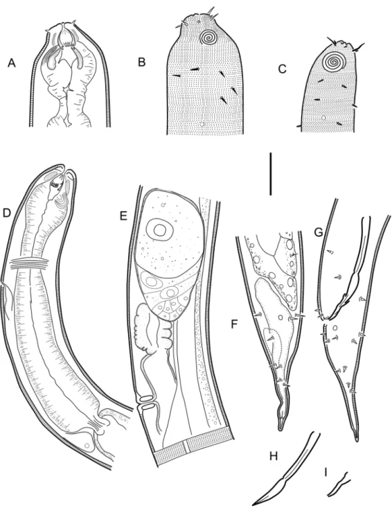
Fig. 1. Halichoanolaimus robustus (Bastian, 1865). Scanning electron micrographs. Abbreviations:
a = anterior row of pointed projections; b = posterior row of pointed projections; c = field of distinct denticles; d = the three tines (one middorsal, two ventrosublateral). Scale bar = 25 µm.
DNA extraction and amplification
DNA extraction was performed on Choniolaimus papillatus Ditlevsen, 1918, Cobbionema sp. (likely another undescribed species), Demonema rapax Cobb, 1894, Gammanema rapax (Ssaweljev, 1912), Gammanema rapax sensu Platt & Warwick 1988, Halichoanolaimus dolichurus Ssaweljev, 1912, H. norvegicus Allgén, 1940 and Latronema sp. (likely an undescribed species) using the Qiagen QiAmp DNA Micro kit (Qiagen, Sollentuna, Sweden). Individual nematodes (either freshly collected or stored in 95% ethanol at -20°C) were each placed in 1.5 ml microcentrifuge tubes containing 20 µl buffer ATL (Qiagen, Sweden) and stored at -20ºC until all samples were ready for extraction. During the extraction, 160 µl of buffer ATL was added to each sample. This was followed by the addition of 20 µl proteinase K, vortexing and incubation in an incubating microplate shaker (VWR, Spånga, Sweden) at 56ºC and 300 rpm overnight. The lysed samples were further processed to obtain pure DNA following the manufacturer’s instructions for genomic DNA extraction using the Qiagen QiAmp DNA Micro kit. Two regions of the rRNA gene, the nearly full-length of the 18S and the D2–D3 expansion segment of 28S, were amplified. The approximately 1800 bp region of the 18S rRNA gene was amplified as two overlapping fragments using the primer sets 988F–1912R for the first fragment and 1813F–2646R for the second fragment (Holterman et al. 2006). Polymerase chain reaction (PCR) for both fragments was performed in 25 µl reaction mix using Illustra Hot Start Mix RTG 0.2 ml reaction kit (GE Healthcare Life Sciences, Sweden). The reaction mix consisted of 1 µl (0.4 µM) of each primer, 2 µl template DNA and 21 µl nuclease-free water. The reaction conditions were 5 min at 95ºC; 5 cycles of (30 sec at 94ºC, 30 sec at 45ºC and 30 sec at 72ºC); 35 cycles of (30 sec at 94ºC, 30 sec at 54ºC and 30 sec at 72ºC); and a final extension for 5 min at 72ºC. The D2–D3 segment of the 28S rRNA gene was amplified using the primers D2Af and D3Br (Nunn 1992). PCR was performed in 25 µl reaction mix containing 1 µl (0.4 µM) of each primer, 2 µl template DNA and 21 µl nuclease-free water. The PCR conditions were 4 min at 94°C; 35 cycles of (94°C for 60 sec, 54°C for 90 sec and 72°C for 2 min); final extension for 10 min at 72°C. Enzymatic PCR clean-up was performed on the PCR product using Exonuclease I and Shrimp Alkaline Phosphatase (New England Biolabs, MA, USA). The purified PCR products were sent out to Macrogen Europe B.V. (Amsterdam, the Netherlands) for sequencing. Each amplicon was sequenced in both directions using the forward and reverse PCR primers.
The trace files of the individual sequences were visualized inside BioEdit (Hall 1999) and trimmed to high quality. The trimmed forward and reverse sequences were then assembled using Fragment Merger online tool (Bell & Kramvis 2013). The two fragments of the 18S rRNA gene were also assembled into contigs using the Fragment Merger online tool.
The nuclear ribosomal 18S and D2–D3 region of the 28S rRNA sequences were deposited in GenBank as follows. 18S rRNA accession numbers: MN786721–MN786722 (Choniolaimus papillatus), MN786723 (Cobbionema sp.), MN786724 (Demonema rapax), MN786725–MN786728, MN786730, MN786732 (Gammanema rapax), MN786729, MN786731 (Gammanema rapax sensu Platt & Warwick 1988), MN786733–MN786736 (Halichoanolaimus dolichurus), MN786737–MN786738 (Halichoanolaimus norvegicus), MN786739–MN786741 (Latronema sp.); D2-D3 of 28S rRNA accession numbers: MN786742–MN786745 (Choniolaimus papillatus), MN786746 (Cobbionema sp.), MN786747– MN786748 (Demonema rapax), MN786749–MN786752, MN786754, MN786756 (Gammanema rapax), MN786753, MN786755 (Gammanema rapax sensu Platt & Warwick 1988), MN786757– MN786760 (Halichoanolaimus dolichurus), MN786761–MN786763 (Halichoanolaimus norvegicus), MN786764–MN786766 (Latronema sp.).
Phylogenetic analysis
Alignments from Holovachov et al. (2017) for 18S rRNA gene and Holovachov et al. (2015) for D2D3 segment of the 28S rRNA gene were used as templates for alignment and annotation of the recently
published and newly generated sequences of Selachinematidae nematodes (see above). Secondary structure annotation was manually added to all non-annotated sequences using the JAVA-based editor 4SALE (Seibel et al. 2006), and all sequences were manually aligned to maximise apparent positional homology of nucleotides. Phylogenetic trees were built using RAxML ver. HPC2 (Stamatakis 2014) via the CIPRES portal (Miller et al. 2010) for the Maximum Likelihood inference of the partitioned dataset. The GTR nucleotide substitution model was used for non-paired sites, whereas the RNA7A (Higgs 2000) substitution model was used for paired sites. Bootstrap ML analysis was performed using the rapid bootstrapping option with 1000 iterations. For the Bayesian inference, PHASE 3 (Gowri-Shankar & Jow 2006) was used. The nucleotide substitution model REV GAMMA was used for non-paired sites and RNA16A paired-site substitution model for the paired sites (stems, hairpins) of the rRNA.
Sequence deposited in GenBank (Benson et al. 2018) under the accession number FJ040468 and identified as Synonchiella sp. belongs to Halichoanolaimus robustus (Bastian, 1865) de Man 1886, based on a reinvestigation of the images of the voucher specimen used for sequencing.
Results
Taxonomy
Phylum Nematoda Potts, 1932 Class Chromadorea Inglis, 1983 Order Chromadorida Chitwood, 1933
Family Selachinematidae Cobb 1915 Genus Cobbionema Filipjev, 1922
Type species
Cobbionema acrocerca Filipjev, 1922
Diagnosis
Adult characters
Body cylindrical, tapering slightly towards both extremities. Cuticle with fine transverse striation, with transverse rows of subcuticular dots, one row per annule. Lateral differentiation where punctations are irregularly spaced and further apart (cf. Bendiella Leduc, 2013) is absent. Lateral alae present in some species, shaped as single band of raised smooth cuticle. Somatic setae scattered along the body. Cephalic region narrower from the level of the base of the anterior chamber of the stoma. Inner labial sensilla indistinct. Outer labial sensilla small papilliform, located on the periphery of labial region. Cephalic sensilla setiform. Amphideal fovea lateral or ventrosublateral, large, sexually dimorphic in size, multispiral with circular outline. Anterior chamber of buccal cavity barrel-shaped or cylindrical in C. capense and C. trigamma, with 12 cuticularized rhabdions or six in C. trigamma, each with two pairs of large, pointed projections at posterior extremity. These projections are arranged in two horizontal rows, appearing from an en face view as two transverse circles of denticles separating the stoma into anterior and posterior chamber. Three (one middorsal and two ventrosublateral) prominent and sharply pointed tines (slender pointed projections, cf. Maggenti 2005) located a short distance in front of the anterior circle of denticles. Posterior buccal chamber cone-shaped, widest at (posterior or anterior) extremity (depending on the state of stomatal musculature), with strongly cuticularized Y-shaped mandibles; anterior branches of each mandible with denticles on its inner surface; posterior ‘stem’ of each mandible with strongly or weakly developed basal (submedian) knobs. The stoma structure of all studied specimens generally resembles that of Halichoanolaimus both in the arrangement of pointed projections in the anterior chamber of the stoma and the shape of the mandibles in the posterior chamber (Fig. 1). Anterior buccal chamber surrounded by strongly developed sphincter muscle. Pharynx muscularised,
with conspicuous anterior swelling surrounding the base of buccal cavity and widens posteriorly but not forming true posterior bulb. Cardia small, with cuticularized lumen. Tail straight or curved ventrally, conical in shape or with distinct conical proximal portion and a digitate (= subcylindrical) distal end. Caudal glands present, opening via a common spinneret, caudal gland cells/bodies at level with posterior part of intestine, anterior to cloaca or extending anterior to the posteriormost end of the intestine.
Male
Reproductive system diorchic, with outstretched anterior testis and reflexed posterior testis. Anterior and posterior testis can lie on either left or right side of intestine. Spicules paired and symmetrical, elongate, consisting of two sections separated by a constriction, nearly straight, with rounded manubrium, subcylindrical shaft, or short with bifid tips. Gubernaculum reduced only to two sublateral crura, arcuate conoid in shape, flanking distal part of spicules. Precloacal supplements setose or papilloid, midventral and range in number from one to five.
Female
Reproductive system didelphic, amphidelphic, ovary branches reflexed antidromously. Anterior ovary on left of intestine, posterior ovary on right side of intestine or vice versa. Vagina straight, without cuticularizations, surrounded by single sphincter muscle. Vulva midventral, a transverse slit. Rectum absent.
Remarks
Gerlach (1964) could not resolve the relationship of this genus with the other members of Selachinematidae due to the limited understanding of its buccal structure at that time. The descriptions of C. capense and C. trigamma have contributed useful insights into the nature of the buccal cavity of this genus. In spite of this, some differences in terminologies used to describe, possibly the same, features in the buccal cavity of these species need clarification. For example, pointed projections in C. trigamma and denticles in C. capense may refer to the same character; which could also be the same feature Schuurmans Stekhoven (1950) described as rows of denticular corpuscles. Also, the three chitinous hooks mentioned in the description of C. acrocerca could be the same as the teeth that were referred to in the description of C. capense. Moreover, an examination of the Swedish populations identified as C. acrocerca and C. cylindrolaimoides revealed that some of the characters described as unique to some species were actually found in others. For example, the Y-shaped rhabdions found in C. trigamma were also observed in all species of Cobbionema that were examined in this study and are very similar to the ones found in most species of Halichoanolaimus. In the descriptions of C. acrocerca and C. capense, a sphincter muscle around the anterior and part of the posterior chambers of the buccal cavity were described. This character has also been observed in all populations of C. acrocerca and C. cylindrolaimoides as well as the two new species described here, and depicted in Leduc (2013: fig. 10). This character has, however, not been observed in other genera of the family. There is therefore a possibility that this feature could be an apomorphic character which separates this genus from other genera within Selachinematidae.
Relationships
Cobbionema differs from all other genera of Selachinematidae in having a sphincter muscle around the anterior buccal chamber. Species of Cobbionema are similar to Halichoanolaimus in the rows of pointed projections at the intersection between the two buccal chambers as well as the three tines (one middorsal and two ventrosublateral) anterior to these two rows of pointed projections. In H. robustus, for example, the tail shape closely resembles that of some Cobbionema species. However, Cobbionema can be distinguished from Halichoanolaimus by the cephalic sensilla being setiform and not papilliform. Cobbionema can also be distinguished from Choniolaimus by the absence of terminal pharyngeal bulb
and shape of precloacal supplements. Table 1 summarizes some of the differentiating features between Cobbionema and other genera included in Selachinematidae.
Valid species
Cobbionema acrocerca Filipjev, 1922 Cobbionema acuminata sp. nov. Cobbionema brevispicula sp. nov.
Cobbionema capense Furstenberg & Heyns, 1987
Cobbionema cylindrolaimoides Schuurmans Stekhoven, 1950 Cobbionema trigamma Leduc, 2013
Cobbionema acrocerca Filipjev, 1922
Figs 2–3, Table 2
Diagnosis
Cobbionema acrocerca is particularly characterised by 0.59–0.86 mm long body; lateral alae present; body pores arranged on either sides of lateral alae; cephalic sensilla equal to 0.3–0.6 labial region diameters in length; amphideal fovea sexually dimorphic, with 4.25–5.25 turns in males and 3.25–3.75 turns in females; two rows of pointed projections and a third row with three prominent sharply pointed tines in the stoma, rows of projections close to one another longitudinally; spicules consisting of two sections, 50–58 µm long; single precloacal papilliform sensillum; tail with conoid proximal and digitate distal section.
Material examined
SWEDEN • 1 ♀; Skagerrak; 58º20′15.5″–15.2″ N, 10º40′06.9″–25.8″ E; 194–201 m depth; 10 Sep. 2012; “Inventering Bratten” leg.; soft bottom; SMNH-179216 • 1 ♂; Skagerrak; 58º19′15.6″–20.9″ N, 10º29′ 33.5″–34.0″ E; 352–374 m depth; 10 Sep. 2012; “Inventering Bratten” leg.; soft bottom; SMNH-179217 • 2 ♂♂; Skagerrak; 58º23′00.8″–22′00.8″ N, 10º20′28.8″–38.3″ E; 390–428 m depth; 10 Sep. 2012; “Inventering Bratten” leg.; soft bottom; SMNH-179218 • 2 ♂♂; Skagerrak; 58º22′17.8″–19.4″ N, 10º23′50.8″–24′03.2″ E; 351–387 m depth; 10 Sep. 2012; “Inventering Bratten” leg.; soft bottom; SMNH-179219 • 1 ♂; Skagerrak; 58º28′28.0″–31.1″ N, 10º33′19.1″–23.8″ E; 221–260 m depth; 11 Sep. 2012; “Inventering Bratten” leg.; soft bottom; SMNH-179220 • 3 ♂♂; Skagerrak; 58º28′12.1″–19.5″ N, 10º37′01.1″–07.1″ E; 180–216 m depth; 11 Sep. 2012; “Inventering Bratten” leg.; soft bottom; SMNH-179221, SMNH179222 • 1 ♂, 2 ♀♀; Skagerrak; 58º34′21.3″–16.6″ N, 10º38′11.2″–29.4″ E; 139– 153 m depth; 12 Sep. 2012; “Inventering Bratten” leg.; soft bottom; SMNH-179223 • 1 ♀; Skagerrak; 58º27′36.7″–43.3″ N, 10º32′52.0″–59.4″ E; 232–240 m depth; 12 Sep. 2012; “Inventering Bratten” leg.; soft bottom; SMNH-179224.
Description
Adult characters
Body cylindrical, tapering slightly towards both extremities. Cuticle with transverse rows of dots, no lateral differentiation. Lateral alae present, single band of smooth cuticle (Fig. 3F) starting at level with basal pharyngeal bulb and ending anterior to anus/cloaca. Body pores present, starting at level with anterior part of pharynx and extending to tail region, located sublaterally on either dorsal or ventral side of lateral alae, irregularly spaced. Somatic setae scattered along the body. Labial region bluntly-rounded, not set-off. Cephalic region of males and females narrower from the level of the base of the anterior chamber of the stoma. Six equal lips surrounding mouth opening. Inner labial sensilla indistinct. Outer labial sensilla small papilliform, located on the periphery of labial region. Cephalic sensilla setiform, equal to 0.3–0.6 labial region diameters in length, located at the same level as outer labial sensilla.
Amphideal fovea ventrosublateral, large, sexually dimorphic in size, multispiral with 4.25–5.25 turns in males and 3.25–3.75 turns in females, with circular outline, equal to about 0.5–0.6 of the corresponding body diameter in males and 0.3 in females. Five to seven pairs of cervical setae present, located posterior to amphid in subventral, ventrosublateral, subdorsal and dorsosublateral positions (cervical setae can be distinguished from somatic sensilla in having larger size and regular arrangement in pairs between amphid and nerve ring level). Buccal cavity voluminous, divided into anterior and posterior chambers. Anterior chamber of buccal cavity barrel-shaped, with 12 cuticularized rhabdions, each with two pairs of large, pointed projections at its posterior extremity. These projections are arranged in two horizontal rows, appearing from an en face view as two circles of denticles separating the stoma into anterior and posterior chamber. Three (one middorsal and two ventrosublateral) prominent and sharply pointed tines located a short distance in front of anterior circle of denticles. Posterior buccal chamber cone-shaped, widest at (posterior or anterior) extremity (depending on the state of stomatal musculature), with three strongly cuticularized Y-shaped mandibles; anterior branches of each mandible rather short and acute; posterior “stem” of each mandible enlarged but without basal (submedian) knobs (Fig. 3A–B). Anterior buccal chamber surrounded by strongly developed sphincter muscle. Each mandible is also supported by a strong longitudinal and tangential musculature, altogether appearing as a muscular bulb. Pharynx muscularised, with developed glandular tissue throughout its entire length; with conspicuous
Characters Leduc, 2013Bendiella Choanolaimusde Man, 1880 Ditlevsen, 1918Choniolaimus Filipjev, 1922Cobbionema GammanemaCobb, 1920 Halichoanolaimusde Man, 1886 KosswigonemaGerlach, 1964 Wieser, 1954Latronema CheironchusCobb, 1917 DemonemaCobb, 1894 PseudocheironchusLeduc, 2013 SynonchiellaCobb, 1933 SynonchiumCobb, 1920
Cuticular
ornamentations rows of dotsTransverse rows of dotsTransverse rows of dotsTransverse rows of dotsTransverse rows of dotsTransverse Transverse rows of dots Transverse rows of dots
Transverse rows of dots and longitudinal ridges Transverse
rows of dots rows of dotsTransverse Transverse rows of dots rows of dotsTransverse rows of dotsTransverse
Lateral
differentiation Dots wider apart Dots wider apart Absent Absent or with single ala Absent Absent Absent Absent Absent Absent Absent Absent Absent
Inner labial
sensilla Papilliform Papilliform Papilliform Papilliform Papilliform/ Setiform Papilliform Papilliform Setiform Papilliform Papilliform Papilliform Papilliform Papilliform
Outer labial
sensilla Papilliform Papilliform Setiform Papilliform/ Setiform Setiform Papilliform Setiform Setiform Papilliform Papilliform Papilliform Papilliform Papilliform
Cephalic
sensilla Papilliform Papilliform Setiform Setiform Setiform Papilliform Setiform Setiform Setiform Papilliform Papilliform Setiform Papilliform
Sphincter muscle around
stoma Absent Absent Absent Present Absent Absent Absent Absent Absent Absent Absent Absent Absent
Anterior pharyngeal
bulb Absent Present Present Present Absent Absent Present Absent Present Present Present Absent Absent
Posterior pharyngeal
bulb Absent Absent Present Absent Absent Absent Absent Absent Present Absent Present Absent Absent
Precloacal
supplements Absent Papilloid Cup-shaped Papilloid or setose Cup-shaped Setose Cup-shaped Setose Cup-shaped Cup-shaped Cup-shaped Cup-shaped Absent
Tail shape Elongated Short conical, almost
rounded Conical
Conical proximally, subcylindrical
distally
Conical Conical proximally, subcylindrical
distally Conical Conical
Bluntly conical Conical proximally, subcylindrical distally Rounded Conical, distally cylindrical with clavate terminus Short conical, digitate Table 1 (continued on the next page). Comparison between all genera of Selachinematidae Cobb, 1915
anterior swelling surrounding the base of buccal cavity; pharynx widens posteriorly but not forming true posterior bulb. Cardia small, with cuticularized lumen. Secretory-excretory system present, renette body located at level with pharyngo-intestinal junction, secretory-excretory pore located just posterior to nerve ring. Tail straight or ventrally curved, distinctly divided into conoid proximal and digitate distal sections. Caudal glands present, opening via a common spinneret, caudal gland cells/bodies may extend anterior to the posteriormost end of intestine.
Male
Reproductive system diorchic, with outstretched anterior testis and reflexed posterior testis, always on opposite sides of the intestine. Anterior testis on either left (n = 6) or right (n = 1) side of intestine, posterior testis on right (n = 6) or left (n = 1) side of intestine. Spicules paired and symmetrical, elongate, consisting of two sections, subcylindrical proximal section and conoid/fusiform distal section (Fig. 2H), equal to 1.9–2.8 anal body diameters in length. Gubernaculum reduced to sublateral crura, arcuate conoid in shape, with central bend (Figs 2I, 3E), flanking distal part of spicules. Single midventral precloacal papilliform sensillum located 6 µm from the cloacal opening. Further papilliform sensilla arranged in pairs subventrally and subdorsally anterior to cloaca and along tail.
Characters Leduc, 2013Bendiella Choanolaimusde Man, 1880 Ditlevsen, 1918Choniolaimus Filipjev, 1922Cobbionema GammanemaCobb, 1920 Halichoanolaimusde Man, 1886 KosswigonemaGerlach, 1964 Wieser, 1954Latronema CheironchusCobb, 1917 DemonemaCobb, 1894 PseudocheironchusLeduc, 2013 SynonchiellaCobb, 1933 SynonchiumCobb, 1920
Cuticular
ornamentations rows of dotsTransverse rows of dotsTransverse rows of dotsTransverse rows of dotsTransverse rows of dotsTransverse Transverse rows of dots Transverse rows of dots
Transverse rows of dots and longitudinal ridges Transverse
rows of dots rows of dotsTransverse Transverse rows of dots rows of dotsTransverse rows of dotsTransverse
Lateral
differentiation Dots wider apart Dots wider apart Absent Absent or with single ala Absent Absent Absent Absent Absent Absent Absent Absent Absent
Inner labial
sensilla Papilliform Papilliform Papilliform Papilliform Papilliform/ Setiform Papilliform Papilliform Setiform Papilliform Papilliform Papilliform Papilliform Papilliform
Outer labial
sensilla Papilliform Papilliform Setiform Papilliform/ Setiform Setiform Papilliform Setiform Setiform Papilliform Papilliform Papilliform Papilliform Papilliform
Cephalic
sensilla Papilliform Papilliform Setiform Setiform Setiform Papilliform Setiform Setiform Setiform Papilliform Papilliform Setiform Papilliform
Sphincter muscle around
stoma Absent Absent Absent Present Absent Absent Absent Absent Absent Absent Absent Absent Absent
Anterior pharyngeal
bulb Absent Present Present Present Absent Absent Present Absent Present Present Present Absent Absent
Posterior pharyngeal
bulb Absent Absent Present Absent Absent Absent Absent Absent Present Absent Present Absent Absent
Precloacal
supplements Absent Papilloid Cup-shaped Papilloid or setose Cup-shaped Setose Cup-shaped Setose Cup-shaped Cup-shaped Cup-shaped Cup-shaped Absent
Tail shape Elongated Short conical, almost
rounded Conical
Conical proximally, subcylindrical
distally
Conical Conical proximally, subcylindrical
distally Conical Conical
Bluntly conical Conical proximally, subcylindrical distally Rounded Conical, distally cylindrical with clavate terminus Short conical, digitate Table 1 (continued). Comparison between all genera of Selachinematidae Cobb, 1915 based on key
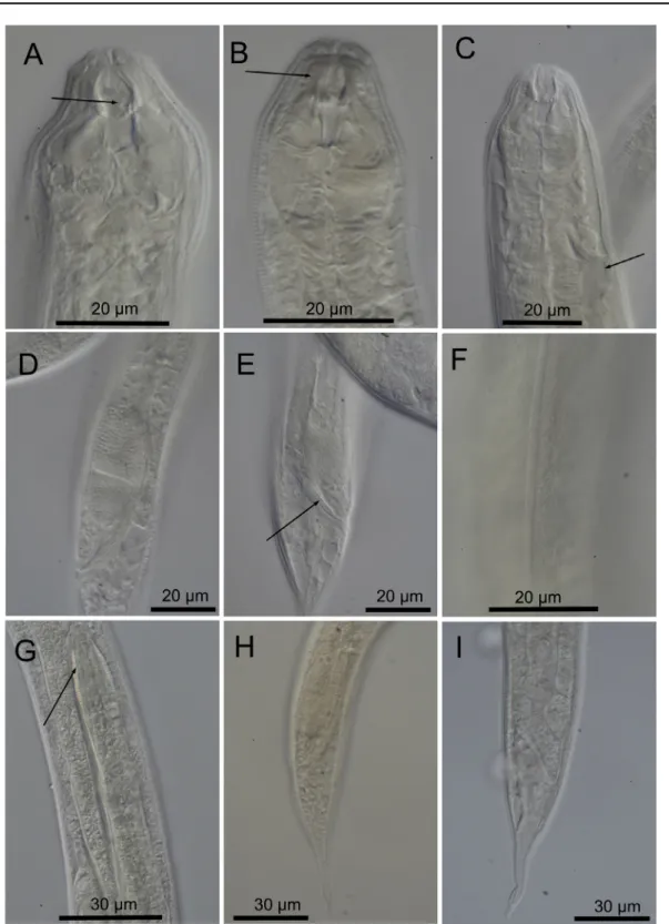
Fig. 2. Cobbionema acrocerca Filipjev, 1922. A. Female head region (ventral side to the right)
(SMNH-179223). B. Surface view of female head (ventral side to the right) (SMNH-(SMNH-179223). C. Surface view of male head (ventral side to the left) (SMNH-179217). D. Anterior part of male (ventral side to the left) (SMNH-179217). E. Female mid-body region showing anterior branch of the reproductive system and lateral ala 179216). F. Female caudal region 179216). G. Male caudal region (SMNH-179217). H. Spicule. I. Crura. Scale bar = 20 µm.
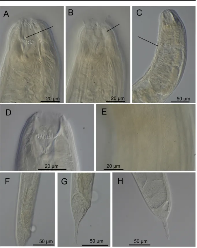
Fig. 3. Cobbionema acrocerca Filipjev, 1922. Light micrographs. A. Female stoma showing stoma
armament, arrow pointing to the left ventrosublateral tine (SMNH-179217). B. Anterior part of a female with arrow pointing to the sphincter muscle surrounding the anterior stoma chamber (SMNH-179217).
C. Anterior part of a female with arrow showing the ampulla of the secretory-excretory system
(SMNH-179218). D. Caudal region a male showing spicule. E. Caudal region of a male showing the crura (arrow) (SMNH-179218). F. Cuticle showing lateral ala (SMNH-179223). G. Nematode prey inside a female (arrow) (SMNH-179216). H. Male caudal region (SMNH-179217). I. Female caudal region (SMNH-179223).
Female
Reproductive system didelphic, amphidelphic, ovary branches reflexed antidromously. Anterior ovary on either left (n = 2) or right (n = 2) side of intestine, posterior ovary on either right (n = 2) or left (n = 2) side of intestine. Oviducts with large cylindrical cells. Uterus thin-walled, filled with round spermatozoa in fertilized females. Vagina straight, without cuticularization, surrounded by single sphincter muscle. Vulva midventral, a transverse slit. Intra-uterine egg not seen. Rectum absent.
Differential diagnosis
The original description of Cobbionema acrocerca by Filipjev (1922) was based on several female specimens of which only one was measured. The Swedish population examined here resembles the type specimen in terms of body size and a number of body measurements (Table 2), having unremarkable mandibles in the posterior stoma and proximal part of the tail being conical in shape. Cobbionema acrocerca differs from C. cylindrolaimoides in having a smaller body (588–860 µm vs 1360– 1755 µm in C. cylindrolaimoides), smaller spicules (50–58 µm vs 89–98 µm in C. cylindrolaimoides), weakly cuticularized mandibles (vs strongly cuticularised), and different tail shape in females (with conoid vs dome-shaped proximal section).
Cobbionema acrocerca differs from all other species of Cobbionema, except C. cylindrolaimodes, in the morphology of the cuticle (lateral alae present in C. acrocerca vs absent in all except C. cylindrolaimoides). Cobbionema acrocerca can also be distinguished from holotype male of C. trigamma in having smaller body size (592–666 µm vs 1305 µm) and smaller spicules. In comparison with paratypes of C. trigamma, however, C. acrocerca only differs from C. trigamma in the morphology of the cuticle (lateral alae present in C. acrocerca vs absent in C. trigamma).
Cobbionema acrocerca can be distinguished from C. capense in the relative positions of cephalic and outer labial sensilla (outer labial sensilla more posterior to cephalic sensilla in C. capense vs cephalic sensilla at the same level as outer labial sensilla) as well as the cuticle morphology (lateral alae present in C. acrocerca vs absent in C. capense).
Biology
Gut content included undigested remains (spicules and gubernacula) or partially digested entire bodies of a variety of nematode species (Fig. 3G), including Chromadoridae Filipjev, 1917.
Cobbionema cylindrolaimoides Schuurmans Stekhoven, 1950
Figs 4–5, Table 2
Diagnosis
Cobbionema cylindrolaimoides is characterised by 1.36–1.76 mm long body; lateral alae present; body pores arranged on either sides of lateral alae; cephalic sensilla, equal to 0.3–0.5 labial region diameters in length; amphideal fovea sexually dimorphic, with 3.5–4.25 turns in males and 3.25–3.5 turns in females; two rows of pointed projections and three prominent sharply pointed tines in the stoma, rows of projections close to one another longitudinally; spicules consisting of two sections, 89–98 µm long; single midventral precloacal papilliform sensillum; tail with conoid or dome shaped proximal and digitate distal section.
Material examined
SWEDEN • 3 ♀♀; Skagerrak; 58º22′20″ N, 11°09′26″ E; 25–50 m depth; 9 Aug. 2011; O. Holovachov leg.; muddy sand; SMNH-179211 • 3 ♀♀, 6 ♂♂; Skagerrak; 58º19′15.6″–20.9″ N, 10º29′33.5″–34.0″ E; 30 m depth; 11 Aug. 2011; O. Holovachov leg.; soft mud; SMNH-179212, SMNH-179213 • 1 ♀, 3 ♂♂; Skagerrak; 58º19′15.6″–20.9″ N, 10º29′33.5″–34.0″ E; 30 m depth; 11 Aug. 2011; O. Holovachov leg.; soft mud; SMNH-179214, SMNH-179215.
Description
Adult characters
Body cylindrical, tapering slightly towards both extremities. Cuticle with transverse rows of dots, without lateral differentiation. Lateral alae present, single band of smooth cuticle starting at level with basal pharyngeal bulb and ending anterior to anus/cloaca. Body pores present, starting at level with anterior part of pharynx and extending to tail region, located on either side of the lateral alae, irregularly spaced. Somatic setae scattered along the body. Labial region bluntly rounded, not set off. Depending on which muscles are constricted, cephalic region can be cylindrical or with a broad and shallow constriction at level with amphidial fovea. Six equal lips surrounding mouth opening. Inner labial sensilla indistinct. Outer labial sensilla small papilliform, located on the periphery of labial region. Cephalic sensilla setiform, equal to 0.3–0.5 labial region diameters in length, located at the same level as outer labial sensilla. Amphideal fovea ventrosublateral, large, sexually dimorphic in size, multispiral with 3.5–4.25 turns in males and 3.25–3.5 turns in females, with circular outline, equal to about 0.4– 0.5 of the corresponding body diameter in male and 0.2–0.3 in female. Two to three pairs of cervical setae present, located posterior to amphid in ventrosublateral and dorsosublateral positions. Buccal cavity voluminous, divided into anterior and posterior chambers, partially everted in some specimens. Anterior chamber of buccal cavity barrel-shaped, with 12 cuticularized rhabdions, each with two pairs of large, pointed projections at posterior extremity. These projections are arranged in two horizontal rows, appearing from an en face view as two circles of denticles separating the stoma into anterior and posterior chamber. Three (one middorsal and two ventrosublateral) prominent and sharply pointed, almost setiform tines located a short distance in front of anterior circle of denticles. Posterior buccal chamber cone-shaped, widest at (posterior or anterior) extremity, or cylindrical (depending on the state of stomatal musculature), with three strongly cuticularized Y-shaped mandibles; anterior branches of each mandible broad triangular, with a field of distinct denticles; posterior ‘stem’ of each mandible with strongly developed basal (submedian) knobs (Figs 4A, 5A). Anterior buccal chamber surrounded by strongly developed sphincter muscle. Each mandible is also supported by a strong longitudinal and tangential musculature, altogether appearing as a muscular bulb. Pharynx muscularised, with developed glandular tissue throughout its entire length; with conspicuous anterior swelling surrounding the base of buccal cavity; pharynx widens posteriorly but not forming true posterior bulb. Cardia small, with cuticularized lumen. Secretory-excretory system present, renette body located at level with pharyngo-intestinal junction, secretory-excretory pore located at level with nerve ring. Tail straight, or curved either dorsally or ventrally, distinctly divided into conoid (in males and some females) or dome-shaped (in females) proximal and digitate distal sections. Caudal glands present, opening via a common spinneret, caudal gland cells/bodies incaudal.
Male
Reproductive system diorchic, with outstretched anterior testis and reflexed posterior testis, always on opposite sides of the intestine. Anterior testis on right (n = 4) side of intestine, posterior testis on left side (n = 4) of intestine. Spicules paired and symmetrical, elongate, seemingly consisting of two sections, subcylindrical proximal section and conoid/fusiform distal section, equal to 2.2–2.4 anal body diameters in length. Gubernaculum reduced to sublateral crura, subcylindrical to conoid in shape, with proximal swelling in some specimens, flanking distal part of spicules (Fig. 4I). Single midventral precloacal papilliform sensillum located 15 µm from the cloacal opening (Fig. 4G). Further papilliform sensilla arranged in pairs subventrally and subdorsally anterior to cloaca and along tail.
Female
Reproductive system didelphic, amphidelphic, ovary branches reflexed antidromously. Anterior ovary on either right (n = 3) or left (n = 4) side of intestine, posterior ovary on either right (n = 5) or left (n = 2) side of intestine. Sperm present in the uterus. Vulva located at around midbody, a transverse slit.
Table 2
(continued on the next page)
. Morphometrics of Cobbionema acr ocer ca Filipjev , 1922, Cobbionema cylindr olaimoides Schuurmans Stekhoven, 1950, Cobbionema br evispicula sp. nov . and Cobbionema acuminata sp. nov . Character C. acr ocer ca C. cylindr olaimoides C. br evispicula sp. nov . C. acuminata sp. nov . ♂♂ ♀♀ ♀ ♂♂ ♀♀ ♀ Holotype ♂ Paratype ♂ Additional ♂ Additional ♀ Holotype ♂ Paratype ♂ Additional ♂ n 4 4 Filipjev (1922) 4 4 Schuurmans Stekhoven (1950) – – – – – – – L 623 (592–666) 713 (588–826) 860 141 1 (1360–1476) 1707 (1615–1755) 1456 1128 1083 1173 1370 1366 1183 1094 a 19 (16–22) 17.4 (14–20) 16 23 (21–26) 15 (14–17) 17 18.3 22.6 25.1 19.7 30.0 31.5 25.9 b 4.8 (4.5–5) 5.8 (5.3–7.0) 4.3 6.3 (6.2–6.4) 6.5 (6.1–6.9) 7 5.8 6.0 6.1 6.4 6.7 6.0 6.8 c 12 (1 1–12) 11 (9–13) 21 18 (16–20) 20 (18–22) 13.7 15.3 16.9 12.6 19.3 19.2 18.8 c’ 2.3 (2.0–2.8) 2.5 (1.8–3.6) 2.0 2 (1.8–2.0) 1.7 (1.2–2.0) 2.5 2.1 2.1 2.3 2.5 2.3 2.0 V (%) – 50 (48–52) 55 – 46 (42–51) 45 – – – 55 – – –
Labial region diameter
13 (10–17) 15 (14–16) 19 28 (22–35) 27 (21–34) 32 25 29 28 35 15 15 15
Body diameter at level of amphid
20 (17–26) 22 (20–24) 25 39 (36–43) 53 (44–60) 31 34 34 41 23 20 21
Outer labial sensilla length
? ? 3 ? 4.9 0.7 0.7 ? 0.7 0.7 ? 1.4
Cephalic sensilla length
6.8 (5.4–8.1) 6.1 (4.4–6.7) 8 9 (80–10) 11 (10–12) 8 10 14 12 12 ? 10
Position of cephalic setae bases from anterior end
? ? ? ? 5 5 3 4 2 2 2
Amphid from anterior end
7.3 (4.9–14.0) 7.2 (6.7–8.1) 11 (8–12) 14 (1 1–17) 9 9 7 10 5 6 6 Amphid width 11 (8–12) 7 (5–8) 5 16 (14–18) 11 (13–16) 14 15 16 12 14 12 12
Distance from anterior end to base of anterior stoma
9 (8–9) 11 (7–12) 14 (14–15) 28 (26–30) 15 14 16 19 14 13 12
Table 2 (continued). Morphometrics of Cobbionema acr ocer ca Filipjev , 1922, Cobbionema cylindr olaimoides Schuurmans Stekhoven, 1950, Cobbionema br evispicula sp. nov . and Cobbionema acuminata sp. nov . Character C. acr ocer ca C. cylindr olaimoides C. br evispicula sp. nov . C. acuminata sp. nov . ♂♂ ♀♀ ♀ ♂♂ ♀♀ ♀ Holotype ♂ Paratype ♂ Additional ♂ Additional ♀ Holotype ♂ Paratype ♂ Additional ♂ W idth of posterior stoma chamber 4.5 (3–5) 6 (4–8) 7 (6–9) 15 (1 1–18) ? 13 21 21 11 9 17 Length of mandibles 14 (14–16) ? 12 (1 1–14) 19 (16–21) 12 12 12 12 10 10 10
Nerve ring from anterior end
45 (41–48) ? 80 87 99 (87–106) 92 ? 75 75 87 81 79 65
Excretory pore from anterior end
56 (50–62) 58 (42–72) 104 (97–1 10) 118 (99–143) 92 89 89 97 109 106 78
Pharyngeal region length
129 (1 18–138) 126 (85–155) 200 225 (218–238) 264 (236–285) 212 194 180 192 213 203 197 161
Maximum body diameter
33 (27–38) 42 (29–50) 53 61 (57–67) 115 (102– 125) 62 48 47 70 46 38 42
Spicule length along arc
55 (50–58) – 93 (89–98) 29 32 30 – 64 65 55 Crura length 14 (13–16) – 31 (28–33) 9 8 – 24 17 18
Anal body diameter
24 (20–26) 29 (18–36) 19 41 (39–44) 51 (42–64) 60 33 33 33 48 29 27 29 Tail length 54 (49–58) 65 (63–68) 39 78 (73–84) 83 (76–91) 82 71 70 108 71 62 58 Amphid width / cbd (%) 51 (47–59) 30 (26–33) 17 41 (36–47) 28 (24–32) 27 44 43 47 29 61 62 57
Nerve ring from ant. end / pharyngeal region length (%)
36 (35–38) ? 40 37 (37–38) ? 42 39 41 40 40 40 Excr . pore
from ant. end / pharyngeal region length (%)
44 (43–45) 46 (39–51) 46 (43–49) 45 (41–50) 48 49 46 46 54 54 48
Cephalic sensilla length/lip region diameter
51 (47–56) 40 (32–47) 33 (26–43) 42 (29–54) 33.3 35.7 48.8 33.3 81.0 ? 63.6
Fig. 4. Cobbionema cylindrolaimoides Schuurmans Stekhoven, 1950. A. Female head region (ventral
side to the left) 179211). B. Surface view of female head (ventral side to the left) (SMNH-197211). C. Surface view of male head (ventral side to the right) (SMNH-179212). D. Anterior part of male (ventral side to the right) (SMNH-179212). E. Female mid body region showing anterior branch of the reproductive system and an egg 179211). F. Dorsoventral view of spicules 179213). G. Lateral view of spicules 179214). H. Spicule. I. Crura. J. Female tail (SMNH-179211). Scale bar = 20 µm.
Fig. 5. Cobbionema cylindrolaimoides Schuurmans Stekhoven, 1950. Light micrographs. A. Female
stoma showing stoma armament, arrow pointing to the right ventrosublateral tine (SMNH-179211).
B. Anterior part of a female showing sphincter muscle surrounding the anterior stoma chamber (arrow)
(SMNH-179212). C. Anterior part of a female with arrow showing the ampulla of the secretory-excretory system (SMNH-179211). D. Male stoma showing buccal armament (SMNH-179213). E. Cuticle showing lateral ala 179211). F. Caudal region of a male showing spicule and crura (SMNH-179214). G. Male tail (SMNH-(SMNH-179214). H. Female tail (SMNH-179212).
Differential diagnosis
Swedish specimens of C. cylindrolaimoides resemble the only female specimen used by Schuurmans Stekhoven (1950) for the original description in body size and most body measurements (Table 2), presence of strongly cuticularized mandibles in the posterior stoma section and sigmoid-conoid shape of the tail. Gerlach (1964) synonymised it with C. acrocerca (and followed by Bezerra et al 2020) but in Gerlach & Riemann (1974) they are treated as separate species. As discussed above, C. cylindrolaimoides differs from C. acrocerca in larger body (1360–1755 µm vs 588–860 µm in C. acrocerca), longer spicules (89–98 µm vs 50–58 µm in C. acrocerca) and mandibles with strongly developed basal knobs (vs mandibles without discernible basal knobs) and shape of female tail.
Based on the updated morphological diagnosis, C. cylindrolaimoides most closely resembles the holotype male of C. trigamma in having relatively large body size (1360–1476 µm vs 1305 µm in C. trigamma) and long spicules (89–98 µm vs 114 µm in C. trigamma). The only clear morphological difference between C. cylindrolaimoides and C. trigamma is in the morphology of the cuticle (lateral alae present in C. cylindrolaimoides vs absent in C. trigamma). Paratypes of C. trigamma differ from C. cylindrolaimoides in much smaller body size (1360–1755 µm vs 566–600 µm in both male and female C. trigamma).
Furthermore, C. cylindrolaimoides differs from C. capense, known from a single female, in body size (1456–1755 µm in females vs 688 µm in C. capense), cuticle morphology (lateral alae present in C. cylindrolaimoides vs absent in C. capense) and different morphology of the stoma (see Table 3).
Biology
Gut content included undigested remains (spicules and gubernacula) or even partially digested entire bodies of a variety of nematode species, including Cyatholaimidae Filipjev, 1918 and Axonolaimidae Filipjev, 1918.
Cobbionema brevispicula sp. nov.
urn:lsid:zoobank.org:act:E6F3CD60-DCE1-4F96-BB28-410199A02705 Figs 6–7, Table 2
Diagnosis
Cobbionema brevispicula sp. nov. is characterised by 1.08–1.37 mm long body; lateral alae absent; body pores present; cephalic sensilla equal to 0.3–0.5 labial region diameters in length; amphideal fovea sexually dimorphic, with 3.5–3.75 turns; two rows of pointed projections and a third row with three prominent sharply pointed tines in the stoma, rows of projections close to one another longitudinally; spicules elongate, nearly straight, with rounded manubrium, subcylindrical shaft and bifid tips, 29–32 µm long. Three or five midventral precloacal papilliform sensilla; tail curved ventrally and conoid in shape.
Etymology
The species name refers to relatively short spicule in the species, compared to other congeners.
Material examined Holotype
SWEDEN • ♂; Skagerrak; 58º22′14″ N, 11º05′00″ E; 30–70 m depth; 9 Aug. 2014; O. Holovachov leg.; gravel, mud and algae; SMNH Type-9219.
Paratypes
Table 3.
Comparison between all six known species of the genus
Cobbionema
Filipjev
, 1922 based on original descriptions and studied slides.
Characters C. trigamma Leduc, 2013 C. capense Furstenber g & Heyns, 1987 C. cylindrolaimoides Schuurmans Stekhoven, 1950 C. acrocerca Filipjev , 1922 C. brevispicula sp. nov . C. acuminata sp. nov . L male (µm) 600–1305 NA 1360–1476 592–666 1083–1 173 1094–1366 L female (µm) 566 688 1456–1755 588–860 1370 NA
Amphideal fovea in males
multispiral; 4.25 turns, 0.3–0.5 cbd NA multispiral; 3.5–4.25 turns, 0.4–0.5 cbd multispiral; 4.25–5.25 turns, 0.5–0.6 cbd multispiral; 3.5–3.75 turns, 0.4–0.5 cbd multispiral; 3.75 turns, 0.6 cbd
Amphideal fovea in females multispiral; 4.25 turns, 0.3 cbd wide
multispiral; 6.8 µm wide multispiral; 3.25–3.5, 0.2–0.3 cbd multispiral; 3.25–3.75 turns, 0.3 cbd multispiral; 3.5–3.75 turns, 0.3 cbd NA
Sphincter muscle around stoma not described but visible in photographs
present present present present present Buccal armature 24 pointed projections two groups of teeth and four groups of denticles two rows of pointed projections; three regularly arranged tines
two rows of pointed
projections; three regularly
arranged tines
two rows of pointed projections; three regularly arranged tines
two rows of pointed
projections; three regularly
arranged tines
Rhabdions in posterior part of stoma three pairs fused posteriorly
described as rugae
three cuticularized Y–shaped mandibles three cuticularized Y–shaped mandibles three cuticularized Y–shaped mandibles three cuticularized Y–shaped mandibles
Lateral alae absent not described present present absent absent
Lateral body pores
present not described present present present absent Spicule length µm 57–1 14 NA 89–98 50–58 29–32 55–65 µm
Mid-ventral precloacol supplement
one precloacal
seta
NA
one precloacal papilla
one precloacal papilla
three or five precloacal
papillae
one precloacal papilla
Anus in female
described as absent but present in drawings
present
absent
absent
absent
NA
Size variability within species
significant not described not significant not significant not significant not significant Tail shape Conoid proximal and digitat e distal sections
Conoid proximal and
subcylindrical distal sections
Conoid or dome–shaped
proximal and digitate distal
sections
Conoid proximal and digitate distal sections
Curved ventral, conoid
in shape
Additional non-type material
SWEDEN • 1 ♀; Skagerrak; 58º17′32″ N, 11º11′24″ E; 45–55 m depth; 10 Sep. 2012; O. Holovachov leg.; coarse sediment with algae; SMNH-182041.
Description
Adult characters
Body cylindrical, tapering slightly towards both extremities. Cuticle with transverse rows of dots, without lateral differentiation. Lateral alae in the shape of a smooth band of cuticle absent. Body pores present (fewer and less obvious in males), starting at level with anterior part of pharynx and extending to tail region, located along either side of the lateral alae, irregularly spaced. Somatic setae scattered along the body. Labial region bluntly-rounded, not set-off. Cephalic region of one male and a female demarcated by a broad and shallow constriction at level with amphid base (Fig. 6A–B); cephalic region of other two males subcylindrical (Fig. 6C–D); this is likely determined by the level of constriction of circum-stomatal sphincter. Six equal lips surrounding mouth opening. Inner labial sensilla indistinct. Outer labial sensilla small papilliform, located on the periphery of labial region. Cephalic sensilla setiform, equal to 0.3–0.5 labial region diameters in length, located at the same level as outer labial sensilla. Amphideal fovea ventrosublateral, large, sexually dimorphic in size, multispiral with 3.5–3.75 turns, with circular outline, equal to about 0.4–0.5 of the corresponding body diameter in male and 0.3 in female. Three to four pairs of cervical setae present, located posterior to amphid in ventrosublateral (1–2 pairs) and dorsosublateral (2 pairs) positions. Buccal cavity voluminous, divided into anterior and posterior chambers. Anterior chamber of buccal cavity barrel-shaped, with 12 cuticularized rhabdions, each with two pairs of large, pointed projections at posterior extremity. These projections are arranged in two horizontal rows, appearing from an en face view as two circles of denticles separating stoma into anterior and posterior chamber. Three (one middorsal and two ventrosublateral) prominent and hollow tines located a short distance in front of anterior circle of denticles. Posterior buccal chamber cone-shaped or cylindrical, widest at (posterior or anterior) extremity (depending on the state of stomatal musculature), with three strongly cuticularized Y-shaped mandibles; anterior branches of each mandible with a field of distinct denticles/knobs on its inner surface; posterior ‘stem’ of each mandible with strongly developed basal (submedian) knobs. Anterior buccal chamber surrounded by strongly developed sphincter muscle. Each mandible is also supported by a strong longitudinal and tangential musculature, forming a muscular anterior pharyngeal bulb. Pharynx muscularised, with developed glandular tissue throughout its entire length; the anterior pharyngeal bulb surrounds the base of buccal cavity; pharynx widens posteriorly but not forming true posterior bulb. Cardia small, with cuticularized lumen. Secretory-excretory system present, renette body located at level with pharyngo-intestinal junction, secretory-excretory pore located at level with nerve ring. Tail curved ventrally, conoid in shape. Caudal glands present, opening via a common spinneret, caudal gland cells/bodies incaudal in the majority of examined specimens, at level with posterior part of intestine in one male.
Male
Reproductive system diorchic, with outstretched anterior testis and reflexed posterior testis, always on opposite sides of the intestine. Anterior testis on either left (holotype) or right (paratype) side of intestine, posterior testis on either right (holotype) or left (paratype) side of intestine. Spicules paired and symmetrical, elongate, nearly straight, with rounded manubrium, subcylindrical shaft and bifid distal tips, equal to 0.9–1.0 anal body diameters in length (Figs 6H, 7F). Gubernaculum reduced to sublateral crura, conoid in shape, flanking distal part of spicules. Three or five midventral precloacal papilliform sensilla located in a row, starting 20–22 µm in front of cloacal opening. Further papilliform sensilla arranged as follows: one subventral pair located half-way between posteriormost midventral papilliform sensillum and cloacal opening; several subventral and several subdorsal pairs along the tail in a variable manner (Fig. 6F–G).
Female
Reproductive system didelphic, amphidelphic, ovary branches reflexed antidromously. Anterior ovary on right side of intestine, posterior ovary on left side of intestine. Other details of female reproductive system morphology cannot be observed due to suboptimal preservation of the specimen. Vulva located
Fig. 6. Cobbionema brevispicula sp. nov. A. Female head region (ventral side to the right)
(SMNH-182041). B. Surface view of female head (ventral side to the right) (SMNH-(SMNH-182041). C. Surface view of the male holotype head (ventral side to the right) (SMNH Type-9219). D. Anterior part of male holotype (ventral side to the right) (SMNH Type-9219). E. Female caudal region (SMNH-182041). F–G. Male holotype caudal region (SMNH Type-9219). H. Spicule. I. Crura. Scale bar = 20 µm.
Fig. 7. Cobbionema brevispicula sp. nov. Light micrographs. A. Stoma of male holotype showing
stoma armament, arrow pointing to the left ventrosublateral tine (SMNH Type-9219). B. Anterior part of male holotype showing sphincter muscle surrounding the anterior end of posterior stoma chamber (arrow) (SMNH Type-9219). C. Anterior part of a female; arrow pointing to a mandible with heavily cuticularized basal knob (SMNH-182041). D. Cuticle showing no lateral ala (SMNH Type-9219).
E. Male tail (SMNH 9219). F. Caudal region of male holotype showing spicule (SMNH
just posterior to midbody. Intra-uterine egg not seen. Rectum reduced, a string of undifferentiated tissue stretching from posterior blunt end of intestine towards ventral body side is visible.
Differential diagnosis
Both males and females of C. brevispicula sp. nov. can be distinguished from all previously described species of Cobbionema by their conical tail (vs conoid proximal portion and digitate distal section). Males of C. brevispicula sp. nov. are also different from all other previously described species in the unique shape and size of their spicules (nearly straight, with bifid tips and measuring 0.9–1.0 anal body diameters long vs two-sectioned spicules consisting of subcylindrical proximal part and a conoid/ fusiform distal section and measuring 1.9–2.8 anal body diameters long).
Biology
Gut content included undigested remains (spicules and gubernacula) of a variety of nematode species, including a possible Cyatholaimidae.
Cobbionema acuminata sp. nov.
urn:lsid:zoobank.org:act:AF9C8B50-B488-4425-A883-8A1ACAB423E7 Figs 8–9, Table 2
Diagnosis
Cobbionema acuminata sp. nov. is characterised by 1.09–1.37 mm long body; lateral alae and body pores absent; cephalic sensilla equal to 0.6–0.8 labial region diameters in length; amphideal fovea large, with 3.75 turns; two rows of pointed projections and three prominent sharply pointed tines in the stoma, rows of projections wider apart longitudinally; spicules consisting of two sections, 55–65 µm long; single midventral precloacal papilliform sensillum; tail curved ventrally, conical in shape.
Etymology
The species name refers to the presence of acute, sharply pointed tines in the stoma.
Material examined Holotype
SWEDEN • ♂; Skagerrak, Hållö island; 58º20.35′ N, 11º12.70′ E; 19 Jul. 2014; 14–17 m depth; O. Holovachov leg.; sand and shells; SMNH Type-9221.
Paratype
SWEDEN • 1 ♂; same collection data as for holotype; SMNH Type-9222.
Additional non-type material
SWEDEN • 1 ♂; Skagerrak, Bonden island; 58º12.64′ N, 11º18.95′ E; 10 Nov. 2012; 15–22 m depth; O. Holovachov leg.; sand and shells; SMNH-182043.
Description
Adult characters
Body cylindrical, tapering slightly towards both extremities. Cuticle with transverse rows of dots, without lateral differentiation. Lateral alae absent. Body pores absent. Somatic setae scattered along the body. Labial region bluntly-rounded, not set-off, but cephalic region demarcated by a broad and shallow constriction at level with amphidial fovea. Six equal lips surrounding mouth opening. Inner labial sensilla indistinct. Outer labial sensilla small papilliform, located on the periphery of labial region.
Cephalic sensilla setiform, equal to 0.6–0.8 labial region diameters in length, located at the same level as outer labial sensilla. Amphideal fovea ventrosublateral, large, multispiral with 3.75 turns, with circular outline, equal to about 0.6 of the corresponding body diameter. Four pairs of cervical setae present, located just posterior to amphid in ventrosublateral (at the base of each amphid) and dorsosublateral positions (Fig. 8B). Buccal cavity voluminous, divided into anterior and posterior chambers. Anterior chamber of buccal cavity barrel-shaped, with 12 cuticularized rhabdions, each with two pairs of large, pointed projections at posterior extremity. These projections are arranged in two horizontal rows, appearing from an en face view as two circles of denticles separating stoma into anterior and posterior chamber. Three (one middorsal and two ventrosublateral) prominent and sharply pointed tines (with hollow conoid base) located a short distance in front of anterior circle of denticles. Posterior buccal chamber cone-shaped, widest at (posterior or anterior) extremity (depending on the state of stomatal musculature), with strongly cuticularized Y-shaped mandibles; anterior branches of each mandible with a field of minute denticles/knobs on its inner surface; posterior ‘stem’ of each mandible with strongly developed basal (submedian) knobs. Anterior buccal chamber surrounded by strongly developed sphincter muscle. Each mandible is also supported by a strong longitudinal and tangential musculature, altogether appearing as a muscular bulb. Pharynx weakly muscularised, with strongly developed glandular tissue throughout its entire length; with conspicuous anterior swelling surrounding the base of buccal cavity; pharynx widens posteriorly but not forming true posterior bulb. Cardia small, with cuticularized lumen. Secretory-excretory system present, renette body located at level with pharyngo-intestinal junction, secretory-excretory pore located short distance posterior to nerve ring. Tail curved ventrally, conical in shape. Caudal glands present, opening via a common spinneret, caudal gland cells/ bodies at level with posterior part of intestine, anterior to cloaca.
Male
Reproductive system diorchic, with outstretched anterior testis and reflexed posterior testis. Anterior testis on right side of intestine, posterior testis on left side of intestine. Spicules paired and symmetrical, elongate, seemingly consisting of two sections separated by a constriction, subcylindrical/fusiform proximal section and conoid distal section, equal to 1.9–2.4 anal body diameters in length. Gubernaculum reduced only to sublateral crura, arcuate conoid in shape, flanking distal part of spicules. Single midventral precloacal papilliform sensillum located 17 µm in front of cloacal opening (Fig. 8D). Further six papilliform sensilla arranged as follows: one subventral pair located half-way between midventral papilliform sensillum and cloacal opening; one subventral pair short distance posterior to cloalcal opening and one ventro-sublateral pair at anterior third of tail length. One pair of dorsosublateral setae located along middle third of tail.
Female
Not found.
Differential diagnosis
Cobbionema acuminata sp. nov., known from three males, differs from all previously described species in arrangement of the stoma armaments (rows of pointed projections and tines widely spaced longitudinally vs rows of projections and tines closer longitudinally in all other species, the three sharply pointed tines more erect and prominent vs pointed posteriorly and shorter in other species). Cobbionema acuminata sp. nov. is also unique in its lack of body pores (present in all other species except for C. capense where no mention was made of the presence or absence of body pores in the description). Cobbionema acuminata sp. nov. differs from males of C. acrocerca and C. cylindrolaimoides in its tail shape (conical in C. acuminata sp. nov. vs conoid proximal and digitate distal sections in C. acrocerca and C. cylindrolaimoides) and lacking lateral alae (present in C. acrocerca and C. cylindrolaimoides). Cobbionema acuminata sp. nov. also differs from C. acrocerca in most body measurements (see Table 2). Cobbionema acuminata sp. nov. differs from holotype and paratype males of C. trigamma
Fig. 8. Cobbionema acuminata sp. nov. A. Male paratype head region (ventral side to the right) (SMNH
Type-9222). B. Surface view of male holotype head (ventral side to the right) (SMNH Type-9221).
C. Anterior part of male holotype (ventral side to the right) (SMNH Type-9221). D. Caudal region of
in its body diameter (25.9–31.5 vs 16 and 17 in holotype and paratype of C. trigamma), number of pointed projections in the stoma (48 vs 24 in C. trigamma) and tail shape (conical vs conoid proximal and digitate distal sections in C. trigamma). Because C. capense is known only from a single female specimen, its comparison with C. acuminata sp. nov., for which no female specimens were found, cannot be based on a character with known sexual dimorphisms such as body size. The two can be distinguished based on their tail shape (conical vs conoid proximal and subcylindrical distal sections in C. capense). Cobbionema acuminata sp. nov. resembles males of C. brevispicula sp. nov. in body length (1094–1366 µm vs 1083–1173 µm). The two, however, can be distinguished by their spicule shape and
Fig. 9. Cobbionema acuminata sp. nov. Light micrographs. A. Stoma of male holotype (SMNH
Type-9221) showing stoma armament, arrow pointing to the right ventrosublateral tine. B. Anterior part of a male showing sphincter muscle surrounding the anterior stoma chamber (arrow). C. Stoma of male holotype (SMNH Type-9221) showing the arrangement of the pointed projections and the left ventrosublateral tine. D–E. Caudal region of male paratype (SMNH Type-9222) showing the crura (arrow) and spicule. F. Male tail, holotype (SMNH Type-9221).
length (two-sectioned spicules consisting of subcylindrical proximal part and a conoid/fusiform distal section and measuring 1.9–2.4 anal body diameters long vs nearly straight, with bifid tips and measuring 0.9–1.0 anal body diameters long).
Molecular phylogeny
Both the 18S and 28S rRNA genes datasets were analysed using Maximum Likelihood (RAxML) and Bayesian inference methods (mcmcphase in PHASE 3). Analysis of the 18S rRNA dataset produced similar, but not identical tree topologies for the Maximum Likelihood and Bayesian inference methods (see Figs 10–11). The main topological differences between the two approaches concerned the relationship between Choniolaimus papillatus and the ((Halichoanolaimus + Demonema Cobb, 1894) + Cobbionema) clade and the relationship between Choanolaimus psammophilus de Man, 1880 and Synonchium sp. Similarly, the 28S rRNA dataset produced tree topologies that were broadly comparable for the two inference methods (see Figs 12–13). The main difference between the tree topologies involves the position of Pseudocheironchus ingluviosus Leduc, 2013, the position of Cobbionema within the (Bendiella + Cobbionema + Demonema + Halichoanolaimus) clade and the relationships
Fig. 10. Maximum Likelihood phylogenetic tree of Selachinematidae Cobb, 1915 inferred from 18S
rRNA data, using alignments based on secondary structure models using GTR GAMMA for unpaired sites and RNA7A for paired sites.
within the Latronema Wieser, 1954 clade. All four trees indicate a close relationship between Latronema and Gammanema Cobb, 1920 through strong branch support (bootstrap support: 94–100 and posterior probability: 1). Cobbionema and Halichoanolaimus were nested within the same clade as was expected based on their morphological similarities. Demonema was also embedded within the same clade.
Discussion
This study presents the first record of the genus Cobbionema in Sweden. Other new records for the family Selachinematidae include Choniolaimus crassus Vitiello, 1971, C. panicus Gerlach, 1956, Halichoanolaimus norvegicus, Gammanema rapax and Synonchiella riemanni Warwick, 1970 (https://dyntaxa.se/Taxon/Info/2000413). Including the two new species of Cobbionema to the list of new records increases it to nine, making an overall total of 16 species for the Swedish fauna. The entire family list now comprises four species of Halichoanolaimus, four species of Choniolaimus, two species of Synonchiella Cobb, 1933, one species of Gammanema, one species Latronema and four species of Cobbionema (Table 4).
Fig. 11. Bayesian phylogenetic tree of Selachinematidae Cobb, 1915 inferred from 18S rRNA data,
using alignments based on secondary structure models using REV GAMMA for unpaired sites and RNA16A for paired sites.
Morphologically, Cobbionema is unique among the other genera of the family in the spicule shape in males, the sphincter muscle around the anterior stoma chamber and in having anterior not posterior pharyngeal bulb. The first ever mention of the sphincter muscle around the anterior part of the stoma was made in the original description of the type species, C. acrocerca, by Filipjev (1922). In the description of C. cylindrolaimoides, although no reference was made on the presence of such sphincter muscle, the similarities in the stoma armament of Cobbionema and that of Halichoanolaimus was mentioned. Furstenberg & Heyns (1987) provided a detailed description of the stoma armament of C. capense, the third member of the genus, and which as the aforementioned two, was described without males. The description of C. trigamma, which included males for the first time, also described in more detail the structure of the stoma. Based on the description of C. trigamma and all four species examined in this study, the gamma-shaped rhabdions in the posterior chamber of the buccal cavity used to distinguish C. trigamma from the other species of Cobbionema is most likely a shared character for the genus. Furthermore, the total number of pointed projections in the stoma could be more than previously reported (at least 48 in all species examined in this study). Aside from the substantial size difference between C. acrocerca and C. cylindrolaimoides, and perhaps the way that this manifests into difference in mandibular cuticularization and spicule size, they share almost all of the useful species diagnosis features
Fig. 12. Maximum Likelihood phylogenetic tree of Selachinematidae Cobb, 1915 inferred from 28S
rRNA data, using alignments based on secondary structure models using GTR GAMMA for unpaired sites and RNA7A for paired sites.