DOCT OR AL DISSERT A TION IN ODONT OL OG Y MIC HELE S T OCC HER O MALMÖ UNIVERSIT ON INFLUEN CE OF AN UNDERSIZED IMPL ANT SITE ON IMPL ANT S TABLIT Y AND OSSEOINTEGR A TION
MICHELE STOCCHERO
ON INFLUENCE OF AN
UNDERSIZED IMPLANT
SITE ON IMPLANT STABILITY
AND OSSEOINTEGRATION
O N I N F L U E N C E O F A N U N D E R S I Z E D I M P L A N T S I T E O N I M P L A N T S T A B I L I T Y A N D O S S E O I N T E G R A T I O N
Malmö University
Faculty of Odontology Doctoral Dissertation 2018
© Michele Stocchero 2018 ISBN 978-91-7104-969-8 (print) ISBN 978-91-7104-970-4 (pdf)
MICHELE STOCCHERO
ON INFLUENCE OF AN
UNDERSIZED IMPLANT
SITE ON IMPLANT STABLITY
AND OSSEOINTEGRATION
Malmö University, 2018
Faculty of Odontology
Department of Oral and Maxillofacial Surgery
and Oral Medicine
Malmö, Sweden
This publication is available in electronic format at: https://muep.mau.se/handle/2043/26189
To my father and mother, and to my future wife, Marta
This thesis is number 53 in a series of investigations on implants, hard tissues and the locomotor apparatus originating from the Department of Biomaterials, University of Gothenburg, the Department of Prosthodontics/ Material Sciences and Department of Oral & Maxillofacial Surgery and Oral Medicine, Malmö University.
1. Anders R Eriksson DDS, 1984. Heat-induced Bone Tissue Injury. An in vivo investigation of heat tolerance of bone tissue and temperature rise in the drilling of cortical bone.
Thesis defended 21.2.1984. External examiner: Docent K-G. Thorngren. 2. Magnus Jacobsson MD, 1985. On Bone Behaviour after Irradiation. Thesis defended 29.4.1985. External examiner: Docent A. Nathanson. 3. Fredric Buch MD, 1985. On Electrical Stimulation of Bone Tissue. Thesis defended 28.5.1985. External examiner: Docent T. Ejsing-Jörgensen. 4. Peter Kälebo MD, 1987. On Experimental Bone Regeneration in Titanium Implants. A quantitative microradiographic and histologic investigation using the Bone Harvest Chamber.
Thesis defended 1.10.1987. External examiner: Docent N. Egund.
5. Lars Carlsson MD, 1989. On the Development of a new Concept for Orthopaedic Implant Fixation.
Thesis defended 2.12.1989. External examiner: Docent L-Å Broström. 6. Tord Röstlund MD, 1990. On the Development of a New Arthroplasty. Thesis defended 19.1.1990. External examiner: Docent Å. Carlsson. 7. Carina Johansson Res Tech, 1991. On Tissue Reaction to Metal Implants. Thesis defended 12.4.1991. External examiner: Professor K. Nilner. 8. Lars Sennerby DDS, 1991. On the Bone Tissue Response to Titanium Implants.
Thesis defended 24.9.1991. External examiner: Dr J.E. Davies.
9. Per Morberg MD, 1991. On Bone Tissue Reactions to Acrylic Cement. Thesis defended 19.12.1991. External examiner: Docent K. Obrant. 10. Ulla Myhr PT, 1994. On factors of Importance for Sitting in Children with Cerebral Palsy.
11. Magnus Gottlander MD, 1994. On Hard Tissue Reactions to Hydroxyapatite-Coated Titanium Implants.
Thesis defended 25.11.1994. External examiner: Docent P. Aspenberg. 12. Edward Ebramzadeh MScEng, 1995. On Factors Affecting Long-Term Outcome of Total Hip Replacements.
Thesis defended 6.2.1995. External examiner: Docent L. Linder.
13. Patricia Campbell BA, 1995. On Aseptic Loosening in Total Hip Replacement: the Role of UHMWPE Wear Particles.
Thesis defended 7.2.1995. External examiner: Professor D. Howie.
14. Ann Wennerberg, DDS, 1996. On Surface Roughness and Implant Incorporation.
Thesis defended 19.4.1996. External examiner: Professor PO. Glantz. 15. Neil Meredith BDS MSc FDS RCSm, 1997. On the Clinical Measurement of Implant Stability Osseointegration.
Thesis defended 3.6.1997. External examiner: Professor J. Brunski. 16. Lars Rasmusson DDS, 1998. On Implant Integration in Membrane-Induced and Grafter Bone.
Thesis defended 4.12.1998. External examiner: Professor R. Haanaes. 17. Thay Q Lee MSc, 1999. On the Biomechanics of the Patellfemoral Joint and Patellar Resurfacing in Total Knee Arthroplasty.
Thesis defended 19.4.1999. External examiner: Docent G. Nemeth. 18. Anna Karin Lundgren DDS, 1999. On Factors Influencing Guided Regeneration and Augmentation of Intramembraneous Bone.
Thesis defended 7.5.1999. External examiner: Professor B. Klinge.
19. Carl-Johan Ivanoff DDS, 1999. On Surgical and Implant Related Factors Influencing Integration and Function of Titanium Implants. Experimental and Clinical Aspects.
Thesis defended 12.5.1999. External examiner: Professor B. Rosenquist. 20. Bertil Friberg DDS MDS, 1999. On Bone Quality and Implant Stability Measurements.
Thesis defended 12.11.1999. External examiner: Docent P. Åstrand. 21. Åse Allansdotter Johansson MD, 1999. On Implant Integration in Irradiated Bone. An Experimental Study of the Effects of Hyperbaric Oxygeneration and Delayed Implant Placement.
22. Börje Svensson FFS, 2000. On Costochondral Grafts Replacing Mandibular Condyles in Juvenile Chronic Arthritis. A Clinical, Histologic and Experimental Study.
Thesis defended 22.5.2000. External examiner: Professor Ch. Lindqvist. 23. Warren Macdonald BEng, MPhil, 2000. On Component Integration on Total Hip Arthroplasties: Pre-Clinical Evaluations.
Thesis defended 1.9.2000. External examiner: Dr A.J.C. Lee
24. Magne Røkkum MD, 2001. On Late Complications with HA Coated Hip Arthroplasties.
Thesis defended 12.10.2001. External examiner: Professor P. Benum. 25. Carin Hallgren Höstner DDS, 2001. On the Bone Response to Different Implant Textures. A 3D analysis of roughness, wavelength and surface pattern of experimental implants.
Thesis defended 19.11.2001. External examiner: Professor S. Lundgren. 26. Young-Taeg Sul DDS, 2002. On the Bone Response to Oxidised Titanium Implants: The role of microporous structure and chemical composition of the surface oxide in enhanced
osseointegration.
Thesis defended 7.6.2002. External examiner: Professor J.E. Ellingsen. 27. Victoria Franke Stenport DDS, 2002. On Growth Factors and Titanium Implant Integration in Bone.
Thesis defended 11.6.2002. External examiner: Associate Professor E. Solheim.
28. Mikael Sundfeldt MD, 2002. On the Aetiology of Aseptic Loosening in Joint Arthroplasties and Routes to Improved cemented Fixation.
Thesis defended 14.6.2002. External examiner: Professor N. Dahlén. 29. Christer Slotte CCS, 2003. On Surgical Techniques to Increase Bone Density and Volume. Studies in Rat and Rabbit.
Thesis defended 13.6.2003. External examiner: Professor C.H.F. Hämmerle. 30. Anna Arvidsson MSc, 2003. On Surface Mediated Interactions Related to Chemomechanical Caries Removal. Effects on surrounding tissues and materials.
Thesis defended 28.11.2003. External examiner: Professor P. Tengvall. 31. Pia Bolind DDS, 2004. On 606 retrieved oral and craniofacial implants. An analysis of consequently received human specimens.
32. Patricia Miranda Burgos DDS, 2006. On the influence of micro- and macroscopic surface modifications on bone integration of titanium implants. Thesis defended 1.9.2006. External examiner: Professor A. Piattelli. 33. Jonas P. Becktor DDS, 2006. On factors influencing the outcome of various techniques using endosseous implants for reconstruction of the atrophic edentulous and partially dentate maxilla.
Thesis defended 17.11.2006. External examiner: Professor K.F. Moos. 34. Anna Göransson DDS, 2006. On Possibly Bioactive CP Titanium Surfaces.
Thesis defended 8.12.2006. External examiner: Professor B. Melsen. 35. Andreas Thor DDS, 2006. On plateletrich plasma in reconstructive dental implant surgery.
Thesis defended 8.12.2006. External examiner: Professor E.M. Pinholt. 36. Luiz Meirelles DDS MSc, 2007. On Nano Size Structures for Enhanced Early Bone Formation.
Thesis defended 13.6.2007. External examiner: Professor Lyndon F. Cooper. 37. Pär-Olov Östman DDS, 2007. On various protocols for direct loading of implant-supported fixed prostheses.
Thesis defended 21.12.2007. External examiner: Professor B. Klinge. 38. Kerstin Fischer DDS, 2008. On immediate/early loading of implant supported prostheses in the maxilla.
Thesis defended 8.2.2008. External examiner: Professor K. Arvidsson Fyrberg.
39. Alf Eliasson 2008. On the role of number of fixtures, surgical technique and timing of loading.
Thesis defended 23.5.2008. External examiner: Professor K. Arvidsson Fyrberg.
40. Victoria Fröjd DDS, 2010. On Ca2+ incorporation and nanoporosity of titanium surfaces and the effect on implant performance.
Thesis defended 26.11.2010. External examiner: Professor J.E. Ellingsen. 41. Lory Melin Svanborg DDS, 2011. On the importance of nanometer structures for implant incorporation in bone tissue.
Thesis defended 01.06.2011. External examiner: Associate professor C. Dahlin.
42. Byung-Soo Kang MSc, 2011. On the bone tissue response to surface chemistry modifications of titanium implants.
Thesis defended 30.09.2011. External examiner: Professor J. Pan.
43. Kostas Bougas DDS, 2012. On the influence of biochemical coating on implant bone incorporation.
Thesis defended 12.12.2012. External examiner: Professor T. Berglundh. 44. Arne Mordenfeld DDS, 2013. On tissue reaction to and adsorption of bone substitutes.
Thesis defended 29.5.2013. External examiner: Professor C. Dahlin. 45. Ramesh Chowdhary DDS, 2014. On efficacy of implant thread design for bone stimulation.
Thesis defended 21.05.2014. External examiner: Professor Flemming Isidor. 46. Anders Halldin MSc, 2015. On a biomechanical approach to analysis of stability and load bearing capacity of oral implants.
Thesis defended 28.05.2015. External examiner: Professor J. Brunski. 47. Francesca Cecchinato MSc, 2015. On magnesium-modified titanium coatings and magnesium alloys for oral and orthopaedic applications: in vitro investigation.
Thesis defended 20.11.2015. External examiner: Professor C. Stanford. 48. Jonas Anderud DDS, 2016. On guided bone regeneration using ceramic membranes.
Thesis defended 27.05.2016. External examiner: Professor S. Lundgren 49. Silvia Galli DDS, 2016. On magnesium-containing implants for bone applications.
Thesis defended 08.12.2016. External examiner: Professor J.E. Ellingsen. 50. Bruno Chrcanovic DDS MSc, 2017. On Failure of Oral Implants. Thesis defended 08.06.2017. External examiner: Associate Professor B. Friberg.
51. Pär Johansson DDS, 2017. On hydroxyapatite modified PEEK implants for bone applications.
Thesis defended 15.12.2017. External examiner: Professor L. Rasmusson. 52. Ali Alenezi DDS MSc, 2018. On enhancement of bone formation using local drug delivery systems.
53. Michele Stocchero DDS, 2018. On influence of an undersized implant site on implant stability and osseointegration
Thesis to be defended 14.12.2018. External examiner: Professor S. Lundgren.
TABLE OF CONTENTS
THESIS AT A GLANCE ... 15
ABSTRACT ... 16
LIST OF PAPERS ... 19
Contribution by the respondent ...20
ABBREVIATIONS ... 21
INTRODUCTION ... 23
OSSEOINTEGRATION AS A BIOMECHANICAL PROCESS .... 25
Primary and Secondary stability ...25
Insertion torque ...27
Resonance Frequency Analysis ...29
Removal Torque ...30
Static and dynamic peri-implant bone strain ...30
OSSEOINTEGRATION AS A BIOLOGIC PROCESS ... 33
Bone-Implant Interface ...33
Peri-implant bone healing ...34
Bone modeling/remodeling ...35
Basic Multicellular Unit ...36
SURGICAL ASPECTS OF OSSEOINTEGRATION ... 40
Surgical principles for osseointegration ...40
Osteonecrosis and bone overheating ...41
Bone quality assessment ...45
Implant design ...46
Undersized drilling techniques ...48
Alternative surgical techniques ...50
AIMS ... 55
MATERIALS AND METHODS ... 56
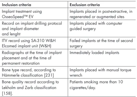
Data collection (systematic review) ...56
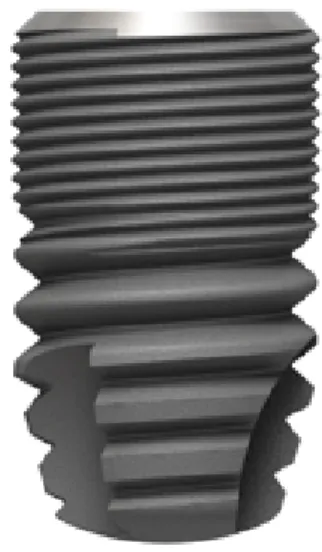
Implant system and drilling protocol ...58
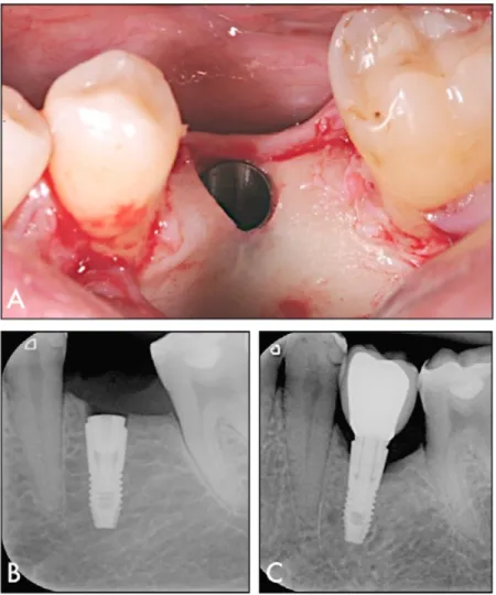
Patient selection and clinical treatment ...59
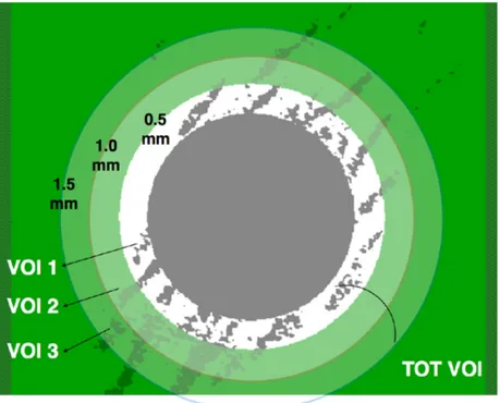
In vivo methods ...62 Stability assessment ...63 Micro-computed tomography ...64 Histology ...64 In silico methods ...66 Statistics ...70 RESULTS ... 73 Systematic review ...73 Clinical analysis ...76 Biologic analysis ...80 Biomechanical analysis ...83 In silico analysis ...84 DISCUSSION ... 88
Undersized drilling in trabecular bone ...88
Undersized drilling in cortical bone ...91
Intracortical bone remodeling around undersized implant sites ...95
The influence of intra-cortical remodeling spaces on material properties ...99
Clinical significance and a new definition of undersized drilling ...102
Clinical considerations on the Insertion Torque ...105
Limitations of the studies ...107
CONCLUSIONS AND FUTURE PERSPECTIVES...109
POPULÄRTVETENSKAPLIG SAMMANFATTNING ...112 AKNOWLEDGEMENTS ...115 REFERENCES ...120 PAPERS I–IV ...147
THESIS A
T A GL
AN
CE
THESIS A
T A GL
AN
CE
St ud y A im Il lu st ra ti on K ey f in d in g s (I ) B io m ec ha nic al, B io lo gic , a nd C lin ic al O ut com es of U nde rs iz ed Im pl ant Sur gi ca l Pr epa ra tion: A S ys te m at ic R ev ie w To co m pi le t he cu rr en t ev id en ce o n bi om ech an ical , b io lo gi c, an d cl in ical o ut co m es o f un dersi zed sur gi ca l pr epa ra tion pr ot oc ol s i n den tal im pl an t su rg ery . A n u nd ersi zed d ril lin g p ro to co l i s ef fect iv e i n IT V in lo w -de ns ity b one . Bi ol og ic re spons e i n long -te rm h ea lin g af te r u nd ersi ze d im pl an t pl ac em ent is com pa ra bl e t o tha t i n the nonunde rs iz ed s ur gi ca l d rillin g pr ot oc ol. Clin ic al ev id en ce, al bei t s par se, in di cat e t hat unde rpr epa ra tion i n l ow -d en sit y b on e i s a saf e pr oced ur e. (II) C lin ic al C on sid era tio ns of A da pte d D ril lin g Pr ot oc ol by Bone Q ua lit y Pe rc ept ion . To ev al uat e I TV an d M BL o f an im pl an t s ys tem af te r a cl in ic al ly pe rc ei ve d bon e qu al ity –a da pt ed dr illin g. Ex ce ss iv e I TV in de ns e bone ca n ca us e ne ga tiv e m ar gi na l bon e r es pons es . A pr es ur gi ca l ra di ogr aphi c a ss es sm ent and the pe rc ept ion of bo ne qu al ity ar e n eces sar y to sel ect an o pt im al dr illin g pr ot oc ol and t o m ini m iz e s ur gi ca l tra um a. (III) In flu en ce o f d iff ere nt dri lli ng p re pa ra tio n on cor tic al bone : A bi om ec ha ni ca l, hi st ol ogi ca l, and m ic ro -C T st udy o n sh ee p. To in ve sti ga te th e e xte nt o f c orti ca l b on e re m ode ling be tw ee n 2 dif fe re nt d rillin g pr ot oco ls by m ea ns of h isto m orp ho m etri c, µC T, and bi om ech an ical an al ys es . Im pl an ts in se rt ed int o unde rs iz ed sit es ha ve an in creased b io m ech an ical p erf orm an ce, b ut pr ov oke d m aj or re m ode ling of the cor tic al bone du rin g t he earl y h eal in g p eri od co m pared to nonunde rs iz ed pr epa ra tions . A fte r 10 w ee ks , no di fferen ce w as o bserv ed . (I V ) In si lic o m ul ti-sc al e a na ly sis of the ef fe ct of res or pt io n ca vi tie s on pe ri-im pl an t b on e af ter un dersi zed d ril lin g t ech ni qu e. To ev al uat e t he m ech an ical p ro per ties an d l oad di st rib ut ion of pe ri-im pl an t co rt ical b on e m od el w ith an d w ith ou t r es or pt io n ca vi tie s. Co rti ca l b on e w ith res or pt io n ca vi tie s s how ed im pai red m ec ha ni ca l pr op er tie s. S tre ss/ st rai n di st rib ut io n su gg est s t hat th is bo ne m od el is m or e pr one to m ic roda m ag e, thus de la yi ng the heal in g pr oces s.ABSTRACT
The use of dental implants for the rehabilitation of edentulous areas is an established treatment, showing high success rates. Primary stability is one of the pre-requisites for osseointegration, and it is ensured by the mechanical interlocking at the bone to implant interface. Current procedures have changed from the original protocols, towards a reduction of treatment time. Nowadays, the achievement of a great magnitude of primary stability is demanded in clinical practice, since there is a trend to load the implant immediately or in the early stages after implant insertion. Aiming on this, several modifications have been introduced, such as more aggressive implant design, modified surfaces and novel surgical techniques. Undersized drilling preparation is one of the most commonly adopted protocols during the implant surgery. This technique creates an osteotomy that is consistently smaller than the implant diameter, so that a tight interfacial contact and compression is created. Clinically this is perceived with an increase of the insertion torque value (ITV).
Albeit commonly performed, several aspects of undersized drilling are still not well investigated. It was hypothesized that a great magnitude of compression at the implant insertion would generate tissue damage and may trigger a negative bone response during the healing time. This could lead to an impairment of bone material properties, a decrease of stability and marginal bone loss. Based on a clinical need, the general aim of this thesis was a more consistent understanding of the effects of an undersized drilling osteotomy.
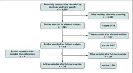
Study I aimed to provide the current evidence based on the literature on biomechanical, biologic and clinical outcomes. An electronic and a manual search were undertaken including in vitro,
animal, and clinical studies in which an undersized drilling protocol was compared with a non-undersized drilling protocol. 29 studies met the inclusion criteria, including 14 biomechanical, 7 biologic, 6 biologic and biomechanical, and 2 clinical. A meta-analysis was not performed. Several studies showed that implants inserted with an undersized drilling approach reached a significantly higher ITV than conventional drilling in low-density substrates, while this effect is less evident in denser substrates. Similar long-term bone-to-implant contact (BIC) was achieved between bone-to-implants inserted with undersized and non-undersized protocols. Results in the short term were inconclusive. Clinical studies did not show negative outcomes for undersized drilling in low-density bone, although clinical evidence was sparse.
In study II, clinical outcomes were evaluated with a retrospective design, which included 87 patients treated with 188 implants inserted with an adapted drilling protocol according to the surgeon’s perception of bone quality. ITV and Marginal bone loss (MBL) between implant placement and permanent restoration was calculated. ITV differed significantly based on mandible/maxilla, bone quality, implant diameter, and drilling protocol. Median MBL was 0.05 mm (0.00; 0.24). A significant difference was found between the mandible and maxilla and between drilling protocols. In particular, significantly higher MBL was found in the undersized drilling protocol. Multiple regression models were built to test the effect of independent variables on the outcomes. ITV was influenced by bone quality and implant diameter. MBL was influenced by bone quality, implant diameter, ITV, and the interaction between bone quality and ITV. It was estimated that MBL was greater with increased bone density and ITV.
Study III aimed to evaluate in vivo the extent of cortical bone remodeling and the bone integration of implants placed after different drilling protocols. Forty-eight implants were inserted into the sheep mandible following two drilling protocols: undersized preparation and non‐undersized preparation. Healing time was set at 5 and 10 weeks. Removal torque (RTQ) was measured and the peri-implant
bone was scanned using a micro-computed tomography (μ‐CT). Bone
volume density (BV/TV) was calculated in pre‐determined hollow cylinders. Total BIC and newly‐formed BIC (newBIC) and Bone Area Fraction Occupancy (BAFO) was measured. Results showed that, at
5 weeks of healing, significantly greater RTQ value was present for the undersized group, while non-undersized group presented greater values of BV/TV, newBIC and BAFO. No differences were noted at 10 weeks.
The purpose of study IV was to assess bone material properties and to predict the strain/stress distribution on cortical bone using a multi-scale in silico model. Two types of micro-multi-scale bone structures were assessed: cortical bone models with resorption cavities and without resorption cavities, following undersized and non-undersized drilling protocols respectively. In a macro-scale model, oblique load of 100N was simulated. Maximum principal stress/strain, and shear stress/ strain were calculated. Bone with resorption cavities presented anisotropic material properties. Compared to bone without cavities, greater maximum values of Maximum principal stress/strain was calculated, both in macro- and micro-scale models. These values were located at the implant neck area and in the proximity of cavities respectively. Greater values of shear stress/strain were found in the test along the mandibular longitudinal plane.
In summary, this thesis suggested that undersized drilling technique can cause negative effects in the cortical bone. The literature indicated that undersized drilling technique is effective in increasing the ITV in low-density bone. However, ITV is mostly influenced by bone quality, rather than drilling protocol. Furthermore, high ITV can induce greater bone resorption in dense bone. Cortical bone has the capability to maintain high levels of rotational stability at undersized sites in the early phases of healing, despite the great amount of micro-damage. From a biologic point of view, this procedure causes a reduced apposition of newly formed bone at the interface and it initiates an intense bone resorption activity in the surrounding tissue. This creates a temporary porosity into cortical bone, reducing the volume of peri-implant mineralized tissue. Intra-cortical resorption cavities caused an impairment of material properties and compromised mechanical behavior. This bone model is more prone to micro-damage and to a delayed healing process. Therefore, avoiding early loading protocols is recommended. Future clinical studies should focus on the longer-term outcome of undersized drilling, since the current clinical evidence is insufficient.
LIST OF PAPERS
The dissertation is based on four papers, which will be referred in the main text by their Roman numerals. The papers are appended at the end of the thesis.
I. Biomechanical, Biologic, and Clinical Outcomes of Undersized
Implant Surgical Preparation: A Systematic Review.
Stocchero M, Toia M, Cecchinato D, Becktor JP, Coelho PG, Jimbo R.
The International Journal of Oral & Maxillofacial Implants. 2016;31(6):1247–63.
II. Clinical Considerations of Adapted Drilling Protocol by Bone Quality Perception.
Toia M, Stocchero M, Cecchinato F, Corrá E, Jimbo R, Cecchinato D.
The International Journal of Oral & Maxillofacial Implants. 2017;32(6):1288–95.
III. Influence of different drilling preparation on cortical bone: A biomechanical, histological, and micro-CT study on sheep. Stocchero M, Toia M, Jinno Y, Cecchinato F, Becktor JP, Naito Y, Halldin A, Jimbo R.
IV. In silico multi-scale analysis of the effect of resorption cavities on peri-implant bone after undersized drilling technique. Stocchero M, Jinno Y, Toia M, Jimbo R, Lee C, Yamaguchi S, Imazato S, Becktor JP.
(Submitted)
Reprint permissions have been granted from:
Paper I: Quintessence Publishing Company Inc, License Number 4427150671453
Paper II Quintessence Publishing Company Inc, License number 4427150611044
Paper III: John Wiley and Sons, License number: 4462351433318
Contribution by the respondent
The respondent performed most of the work from planning, experimental work (with the exception of the histological preparation, the histological imaging and the in silico modeling) and he performed the analysis of data. The respondent was also the main contributor to writing the manuscripts.
ABBREVIATIONS
2D Two-dimensional
3D Three-dimensional
BAFO Bone Area Fraction Occupancy
BIC Bone to Implant Contact
BMP Bone Morphogenetic Protein
BMU Basic Multicellular Unit
BV/TV Bone Volume Density
CBCT Cone Beam Computed Tomography
EM Elastic Modulus
PR Poisson’s Ratio
FEA Finite Element Analysis
ITQ Insertion Torque
ITV Insertion Torque Value
kV Kilovolt
MBL Marginal Bone Loss
mm Millimetre
MPStrain Maximum Principal Strain
MPStress Maximum Principal Stress
newBIC Newly Formed Bone to Implant Contact
OPG Osteoprotegerin
RANK Receptor Activator Of Nuclear Factor
Kappa-Β
RANKL Receptor Activator Of Nuclear Factor
RFA Resonance Frequency Analysis
ROI Region of Interest
rpm Revolutions per Minute
RTQ Removal Torque
s Second
Tcutting Cutting Torque
Tfriction Friction Torque
TGF-β Transforming Growth Factor Beta
totBIC Total Bone to Implant Contact
VOI Volume of Interest
µA Microampere
µCT or
Micro-CT Micro-Computed Tomography
INTRODUCTION
Dental implants are medical devices used for the rehabilitation of complete [1], partial [2], or single [3] edentulous areas. Thanks to the discoveries conducted from the 1960s in the laboratory headed by Prof. P.I. Brånemark, modern dental implantology is a scientifically solid branch of dentistry, which stands on the concept of osseointegration. This term describes a direct contact between living bone and load bearing implant observed from light microscopy [4,5]. This definition involves two key concepts, one belonging to biologic aspects and one to mechanical: the histological interface between a living tissue and an inorganic surface and the load-carrying function of implants.
Unquestionably, implant rehabilitation is nowadays a common procedure, based on an established predictability and supported by high success rate on long-term clinical data [6]. Nevertheless, failures are still encountered [7].
It is true that the use of dental implants is currently adopted even in challenging situations, that were previously avoided if not contraindicated [8]. Technological and procedural innovations have been continuously introduced, from new implant designs to new clinical procedures. Despite the indisputably superior performance of current treatments, many clinical routines have changed over the years. A vast part of the theoretical background is based on the early protocols and may appear out of date; the research field itself may not face the rapid evolution of implant therapy. Basically, current trends aim to a simplified and time-reduced treatment often without the support of evidence from the literature. Clinical opinion-leaders and implant manufacturers have been encouraging immediate or
early implant loading, before reaching a complete osseointegration. Implant surgical routines have changed considerably over time. Modifications have been made, intended to improve implant stability, together with more aggressive implant designs. However the biologic interactions between bone and implant are rarely investigated with novel procedures. It is likely that several unexpected implant failures may be explained because of the lack of observation of the basic concepts of osseointegration.
Before analyzing the influence of the surgical protocol on peri-implant bone, main biological and biomechanical aspects of osseointegration will be reviewed.
OSSEOINTEGRATION AS A
BIOMECHANICAL PROCESS
Primary and Secondary stability
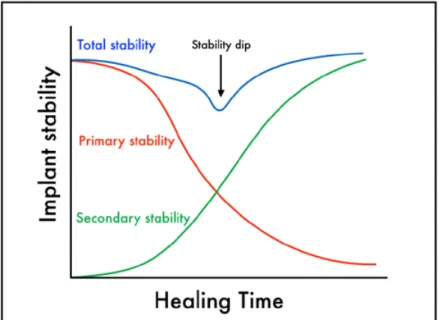
Implant stability has been regarded as one of the most important factors for a successful osseointegration. It should be maintained from the time of insertion and throughout the healing period [9]. On the contrary, implant mobility is a visible sign of failure, mostly caused by fibrous encapsulation [10]. The initial mechanical interlocking between the implant and bone is known as primary stability. This is provided mostly by the engagement with cortical bone [11]. On the other hand, bone formation on the implant surface provides the secondary stability. This requires the biological process of de novo bone formation [12]. Implant stability is commonly subjected to modifications with time (Figure 1)[13], according to the type of contact between the implant and the surrounding bone tissue [14]. After the initial peak of stability, due to mechanical anchorage, the primary fixation decreases until the so-called stability dip. In this period, the shift from the mechanical to the biological stability occurs. The initial drop of stability is caused by osteoclastic activity [15] together with visco-elastic relaxation phenomena of pre-stressed bone [16]. The following increase of stability is guaranteed by the apposition of new bone supported by osteoblasts along with the remodeling process.
The concept of implant stability can be measured as implant micro-motion, that is the displacement of an implant relative to the surrounding bone. This can be influenced by several factors, such as the mechanical engagement between the threads and the bone,
the geometry of the implant, the magnitude of biologic attachment and physical properties of the surrounding bone tissue (i.e. elastic modulus) [17]. A minimal degree of micro-motion seems not to be necessarily followed by fibrous encapsulation, and it was recognized that implants with limited amount of micro-motion can actually integrate [18]. However the degree of such mobility is debatable. It was hypothesized that a threshold of tolerated mobility lies between 50 and 150 µm [19,20]. Moreover, it was observed that a low range of micro-motion at the interfacial tissues may even have a positive biologic effect, such as stimulation of interfacial bone formation [21,22]. Thus, one of the main objectives during clinical procedures is to maintain the level of micro-motion below the dangerous threshold, even when immediate or early loading protocols are applied. Thus, one of the primary focuses during implant surgery is the achievement of a good and appropriate level of primary stability.
Figure 1. A representation of the turnover from primary to secondary
stability of dental implants as described by Raghavendra et al. [14]. The total stability typically shows a lowest point (stability dip), due to decrease of the mechanical engagement before the onset of the biological fixation. Modified from Suzuki et al. [13].
The importance of assessing implant micro-motion reflects a major problem in implant dentistry: implant stability cannot be directly measured. However, there exist some methods for the clinical practice or experimental quantification of the grade of stability (primary and/ or secondary). The currently most used ones are: Insertion Torque, Resonance Frequency Analysis and Removal Torque evaluations.
Insertion torque
Insertion torque value (ITV) is the most common parameter used in the clinical practice to measure implants stability, due to the easy calculation at the time of surgery. Insertion torque (ITQ) is a measure of the rotational resistance during implant installment [23] and it is measured in Ncm. Many clinicians rely on this value as a good predictor for osseointegration [24]. Specifically, high ITV is often seen as desirable [25]. However, this statement is controversial, since good results in terms of clinical outcomes have been reported for implants inserted both at extremely high [26] and extremely low [27] ITV. Rotational mobility per se, at the time of insertion, does not necessarily lead to an impaired osseointegration of non-loaded implants [28].
A clinical common opinion claims that the increase of ITV can act as a stimulus for a reduced-time osseointegration. A systematic review stated that a high ITV is a prerequisite for immediate or early loading [29]. Hence, many researchers and clinicians have been discussing the threshold values for these procedures. Ottoni et al. [30] declared that an ITV above 32 Ncm is necessary to achieve osseointegration for immediate load on single implants. According to Aparicio et al. [31], an ITV in the range of 30 to 50 Ncm provides adequate stability. Lower values were recommended by Norton et al. [32], since 25 Ncm would seem more than sufficient to achieve successful immediately loaded single-tooth implants. These torque values have been proposed not just to prevent micro-motion, but also to withstand the forces of prosthetic procedures related to the type of implant connection and functional loading. Based on the implant-abutment interface design, different levels of tightening torque are recommended, from 15 Ncm to 30 Ncm or even more [33]. Thus, ITV should be greater than the torque values recommended for the immediate fixation of the abutment [34]. Otherwise the final force impressed to the screw would disrupt the initial implant interlocking,
28
along the prosthetic connection is debatable and not a focus of the present work.
De facto, any statement that suggests the ideal clinical protocol based on the ITV may not apply to every clinical situation, since ITV is not an absolute parameter for primary stability. Although some researchers reported that high ITV is positively correlated with primary stability [35,36], other studies had observed that such an association might not be present in all implant systems [37,38]. On the contrary, an in vitro study demonstrated that low levels of ITV might be associated with decreased micro-motion [39] when trimmed threaded implants are used.
From a theoretical point of view, ITQ is the sum of two distinct
processes: torque generated by bone cutting (cutting torque, Tcutting)
and torque generated by bone friction (friction torque, Tfriction). From these formulas it is clear that when interpreting the mere
ITV, several factors have to be considered. For example, Tfriction is
proportional to the implant radius raised to the power of 2. As a consequence, by simply switching to an implant with larger diameter, the ITV is significantly enhanced as evidenced by several authors in low-density bone [40,41]. In addition, ITV is largely affected by the area of contact with bone, specifically to the cortical layer [42]. This means that the thickness of the cortical bone plays a major role in the stress distribution at implant insertion. Another aspect
to be considered is whether ITV is generated mostly by Tcutting or by
Tfriction. If the implant cutting efficiency is high, there is more debris
production, but less bone deformation and mechanical damage. In Insertion Torque ≈ 𝑇𝑇!"##$%&+ 𝑇𝑇!"#$%#&'
More specifically: 𝑇𝑇!"##$%&= 𝐶𝐶×𝑝𝑝!×𝑟𝑟×𝑘𝑘! 𝑇𝑇!"#$%#&'= 𝑃𝑃×𝐴𝐴×µμ×𝑟𝑟 where: C: constant p: thread pitch r: radius
kc: specific cutting force P: critical pressure on the bone 𝐴𝐴 = 2𝜋𝜋𝜋𝜋×ℎ𝑒𝑒𝑒𝑒𝑒𝑒ℎ𝑡𝑡: area of contact µ: coefficient of friction.
contrast, if ITQ is mostly affected by friction, the implant impresses an increased compression with bone deformation. Consequently, the same ITV may have completely different bone effects based on implant geometry.
Hence, ITV provides site- and implant-specific information that cannot be used for a direct comparison with different implants, neither inter-individually or even in the same patient.
Resonance Frequency Analysis
A non-invasive method for measuring implant stability involves the use of resonance frequency analysis (RFA) of the implant–bone complex. Due to a transducer, which is directly tightened to the implant, RFA measures the stiffness and deflection of the implant into the bone [43]. One of the main advantages of the RFA is the reproducibility of this measure over time. In this manner the clinician could check the variation of the implant stability during the healing phase [44].
The frequency obtained is then translated into a value, which is called Implant Stability Quotient (ISQ), ranging from 0 to 100. This scale indicates that the higher is the value, the greater is the implant stiffness. Clinical guidelines suggest an ISQ>65 for successful osseointegration. An ISQ<50 may indicate an unstable implant [11]. However, the prognostic value of RFA has been questioned and some studies have shown that these measurements are more accurate in predicting implants that were at risk of failure after the establishment of osseointegration [45,46]. The basic assumption of over-time RFA measurements is that, since implant and the surrounding bone act as single unit, modifications of stiffness represent a modification in the bone-to-implant interface [11]. However, ISQ values have to be carefully interpreted, since they do not entirely rely on the quality of osseointegration. Besides from the stiffness of the implant-bone interface, the stiffness of the bone itself and the stiffness of the implant components can influence ISQ [12]. Moreover, other factors related to implant geometry strongly influence the ISQ values, such as implant length, diameter, macro- and micro-design, placement position and type of abutment [47,11]. Therefore, the clinical relevance of the RFA is comparing the stability in the follow-up of each individual implant, but, similarly to ITV, caution should be taken when comparing ISQ from different implants [48].
Removal Torque
Removal Torque (RTQ) test determines the critical torque value for disrupting the bone-to-implant interlocking by unscrewing the implant [49]. This destructive method is mainly used in research that does not involve humans. Analogously to ITQ, RTQ is affected by implant geometry and bone quality. In in vivo studies, RTQ is affected both by primary and secondary stability [24]. In fact, it is the result of initial interlocking between the implant and the bone (primary stability) and the osseointegration process after healing, remodeling, and bone apposition (secondary stability). Therefore, due to the controlled settings of the experiments, usually RTQ values can estimate the quality of osseointegration in terms of bone-to-implant contact [11]. However, it has to be remarked that residual strain in the peri-implant bone could influence the results by increasing the RTQ [50].
Static and dynamic peri-implant bone strain
One of the regulatory systems for bone physiology is mechanical stimulus. Bone has the capability to adapt its mass and structure according to the strains induced by loads, through modeling and remodeling processes [51]. This adaptive mechanism is regulated by the magnitude, frequency, rate and duration of the strain [52-54].
According to Frost’s theory on bone modeling/remodeling regulation, a certain degree of deformation is required for the initiation of the process and the maintenance of bone mass [55]. There is a threshold of physiological strains in which bone is triggered in a steady state of normal remodeling. In case of lower strains, bone resorption is initiated, while higher strains may lead to increased bone mass. On the other hand, peak load levels may cause bone micro-damage and stress fracture (Figure 2)[56]. Strains can be either of a static or a dynamic nature.
During implant installation with a certain ITV, static strains are generated in the surrounding bone. The magnitude of such strains depends on several factors, such as the implant geometry, status of the host bed, and preparation of the osteotomy site, which is of particular interest of the present thesis. When the diameter of the osteotomy is narrower than the diameter of the implant, implant stability is secured via press-fit phenomena. The concept of press-fit, which is
different from screw-fixation, is taken from orthopedic surgery. This stability is generated by the radial contact stresses, which develop frictional shear resistance to relative motion at the bone-implant interface [57,58].
The magnitude of strains that bone can tolerate depends primarily on bone quality [59]. If the induced static strain lies within the yield limit, the mechanical engagement between the bone and the implant is linearly related to the strain [60,61]. When the magnitude of the strain is higher, plastic deformation and numerous microcracks are expected, thus altering the pristine bone mechanical properties
[62,16]. Even if short cracks, i.e. less than 100 μm, can be caused
by drilling procedures [63], longer microcracks are generated during implant insertion [64]. From previous studies, it was hypothesized that bone damage created by static strain can trigger the modeling/ remodeling process [65]. According to a revision of the Frost theory on bone remodeling, bone is controlled by an inhibitory signal triggered by mechanical stimuli [66]. The remodeling process is restrained when load lies within the physiologic threshold. On the contrary, remodeling is increased when load is extremely low or excessively high, due to tissue damage [67]. In an in vivo study on rabbits by Halldin et al. [50], induced static strains were maintained 24 days after insertion. Interestingly, if the induced bone strains are beyond the ultimate cortical yield limit, there was no negative bone response in a rabbit tibia model [16]. On the contrary higher RTQ and a tendency of higher BIC was observed after 3 and 13 days.
In addition, bone has the capability to withstand static strains thanks to relaxation phenomena [68,69]. This means that when a deformation is impressed constantly, the measured force decreases over time. This can be explained by the visco-elastic properties of bone complex structural composition [70]. A decrease of intra-bony forces over time is further expected due to the removal of the pre-stressed bone by remodeling.
Dynamic strains that typically are generated in the peri-implant bone are due to masticatory functions and para-functions [71]. These forces involve a repeated pattern of cyclical impacts on the implant that are transferred to the surrounding bone [72]. In bone physiology, dynamic strains are the driving force behind bone modeling and remodeling [73]. A theory elaborated by Qin et al. [54], affirms
that bone mass maintenance is defined by the relation between the magnitude of stress and strain and loading cycle numbers per day. According to this theory, low stress/strain require a higher number of cycles to maintain the bone, while fewer cycles would lead to bone mass resorption. On the contrary, it is generally accepted that excessive masticatory loads on peri-implant bone would lead to marginal bone loss and eventually loss of osseointegration [74,75]. It was observed that dynamic overload induces typically to crater-shaped bone defects [76]. This pattern of bone loss may be explained by the excessive microdamage occurring at the interfacial bone, which could trigger a relevant bone remodeling. This process may not keep pace with accumulating damages, thereby predisposing to bone loss [77]. The investigation of implant load transferring is often performed with the help of Finite Element Analysis (FEA) models. Thanks to these computer simulations, stress and strain patterns can be calculated to explore the mechanism of peri-implant bone behavior [77,78].
Figure 2. Graphical illustration of the bone modeling/remodeling
regulation according to Frost’s theory. The strain magnitude predicts bone behavior, resulting in an increase or decrease in bone mass (vertical axis). In the left part, strains are low and bone remodeling is activated to decrease bone mass due to disuse. Above this threshold, a low-intensity remodeling activity is sustained for the preservation of bone mass. At higher strains modeling is triggered with an increase in bone mass. Pathological overload zone is defined when strains are greater than the repair capability. Modified from Su et al. [56].
OSSEOINTEGRATION AS A
BIOLOGIC PROCESS
Bone-Implant Interface
Histological and electron microscopical observations of osseointegrated implants in humans described the bone anchored implants to be provided with well-organized lamellar structures. The tight interface between the cells and the implant surface is not interposed by organized collagenous and fibroblastic matrix, but by an acellular layer. Histologic and electron microscopical observations described a layer, approximately 500 nm thick, composed by amorphous material, enriched in proteoglycans and glycoproteins, which separates the implant surface from the vital tissue [79,80]. A stable peri-implant tissue includes the presence of different types of cells and structures, which could be observed in normal conditions by light microscopy: osteoblasts, osteoclasts, multinucleate giant cells, monocytes, vessels, collagenous matrix and mineralized matrix [81]. Differently from the ideal scenario, a failure of osseointegration is described when soft connective tissue is interposed, i.e fibrous encapsulation. In this condition, the anchorage is not stable and the implant will be lost. A third situation can be considered when the bone in contact with the implant is partially necrotic. According to the early opinions, this anchorage is less reliable in bearing the occlusal load than a full osseointegration with vital bone. However it was stated that this interface might function as a temporary stabilization of the implant even for several years, before this is replaced or resorbed [4].
Light microscopy investigations of bone-to-implant interfaces have assessed the relative presence of mineralized tissue in contact with the implant surface, rather than fibrous tissue. This outcome is generally regarded as Bone-to-Implant Contact (BIC). The majority of the analyses involved the use of various animal models or, in some cases, human retrieved samples [82]. On the basis of the current literature, it can be said that there is no clear cut-off value of what we can consider as “osseointegration” [81]. Extremely different results can be obtained based on the type of model, type of bone, time of healing, type of implant, etc. Yet, a BIC of 100% is indeed to be regarded as unrealistic, while it is more reasonable to consider 60% as a result of good integration [83]. The rate of contact in the interface between mineralized bone and implant surface is subjected to continuous biological transformation and may change with time. More recent interpretations look at osseointegration as a dynamic process in a phase of equilibrium; if negative actors interfere, we could observe a break-down of the status [84].
Peri-implant bone healing
Once the osteotomy is prepared and the implant is placed into its final position, a cascade of biologic events are in involved in the bone formation adjacent to the implant. This process begins with the clot formation: blood cells, including erythrocytes, neutrophils and macrophages are interconnected through a network of fibrin [85]. This is followed by an inflammatory phase, in which blood and inflammatory cells, including neutrophils and then monocytes are recruited to the peri-implant tissues. Together with platelet activation, cytokines and growth and differentiation factors are released. At this time, angiogenesis and bone formation are stimulated. Thus, mesenchymal cells are recruited in the proximity to the titanium surface and they start to differentiate into osteogenic cells [85,86]. These cells deposit new bone matrix in the vicinity of the implant site. In such a manner, de novo bone formation may occur directly on the surface of the implant (contact osteogenesis), or either on the surface of the pre-existing bone approximating to the implant (distance osteogenesis) [87].
The unique interplay between implant geometry and site preparation in terms of gaps and interlocking areas induces distinct
bone-healing phenomena. According to several animal studies, two major healing patterns have been described [85, 60]. If void spaces are created between the implant surface and the osteotomy walls, an intra-membranous like healing scenario can be expected [88,89]. These bone-chamber structures are filled with blood clot and will rapidly evolve in woven bone formation [85,90,91]. Thus, when a large contact free surface is left, we can observe a high magnitude of de novo bone formation. Depending on the implant micro-topography, the new bone apposition may start in the center of the chamber for smooth surfaces, or directly at the implant interface for modified surfaces [90,92]. When new bone apposition occurs directly on the implant surface it has been defined as contact osteogenesis [87]. Immature bone structures are then replaced by lamellar bone in later stages [85].
A different healing pattern can be observed when the implant is inserted in intimate contact with the bone walls, called interfacial remodeling [89]. The tight interlocking between the fixture and the bone causes press-fit phenomena, with micro-cracks and diffuse damage formation [93]. Necrotic bone can be observed in the location of greater compression. This bone can be encountered as small spotted areas [94], or as wider regions [95]. The damaged bone is then removed with intense osteoclastic activity and eventually replaced with new bone, thanks to the time-consuming remodeling process. This so-called distance osteogenesis will result in a bone formation approximating the implant surface [87].
These two distinct healing processes are typically observed along the surface of the same site, for a standard screw-shape implant installed in a conventional osteotomy. Bone chambers will form between the threads, while interfacial remodeling will take place in the areas of primary engagement with the pristine bone, typìcally at the implant thread tips [85]. This favorable combination will provide both the biological advantages of de novo bone formation with the mechanical advantages of primary interlocking, which in turn provides stability to the implant.
Bone modeling/remodeling
The bone surrounding an implant is involved with complex physiologic mechanisms from the time of insertion and throughout the functioning period. Bone is a dynamic tissue that undergoes to
a continuous modification of its composition. As seen previously, bone has the adaptive capability to modify its mass and structure depending on the external stimuli. Thanks to the bone modeling process, bone mass can be altered. Based on the type of load, new bone formation (increase of bone mass) or resorption (decrease of bone mass) can occur.
On the other hand, the skeletal system has the capability to renovate itself with the lifelong process of bone remodeling. Thanks to this process a discrete volume of old bone is removed and then replaced by newly formed tissue, which would mineralize subsequently into new bone [96]. Remodeling has three main functions: balancing the body mineral content, adapting the bone structure to mechanical environment and repairing to bone micro-damage [97]. Thus, two kinds of remodeling can be distinguished [98]. In physiological conditions, a sustained not-site specific remodeling process takes place. Thanks to this mechanism, bone tissue is cyclically renovated with a turnover rate per year of 2-10% for cortical bone and 20-30% for trabecular bone [99,100]. A second type of bone turnover is targeted remodeling, which directs the action of the process to specific sites. This is the case of bone repair in case of bone injury. It has been established that targeted remodeling is triggered by micro-damage [67,101]. More specifically, the initiating factor has been pointed out to be the death of osteocytes [102]. Osteocyte apoptosis is induced by compressive stresses and strains and it is commonly a result of bone micro-damage [103,104]. It was observed that microcracks may impair canalicular fluid flow interconnecting adjacent osteocytes and this can lead to cell apoptosis [105]. As a matter of fact, pro-apoptotic molecules were identified in the proximity of microcracks [106]. Therefore, remodeling activities are regulated by strain as well, but not simultaneously to damage. In the presence of a relevant injury (i.e. surgical trauma), damage-induced remodeling would come first over the attempt of strain normalization [107,108].
Basic Multicellular Unit
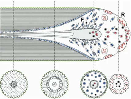
The remodeling process requires the coordinated action of catabolic and anabolic actions, i.e. bone resorption by osteoclasts and new bone apposition by osteoblasts, respectively. The entire process is orchestrated in specialized structures known as Basic Multicellular Units (BMUs) (Figure 3). An active BMU consists of number of
osteoclasts in the front, referred as the cutting cone, and a number of lining osteoblasts behind it, forming the closing cone. The structure is provided of blood supply and associated connective tissue [110]. BMUs are active both in cortical and cancellous bone: in the former they excavate tunnels, in the latter they are in contact with the endosteal surface of bone trabeculae. BMUs can proceed with a rate of 0.15-0.30 mm/day in rabbits [111] or 25-30 µm/day longitudinally, and 6-7 µm/day radially in humans [112]. It has been calculated that a BMU can travel roughly for 4 mm through the cortex with a life span of 200 days [112].
Dying osteocytes promote cells activation and guide the movement of BMUs through the cortex [113]. The direction of motion is oriented by the principal stress trajectory, but also toward micro‐ damage, depending on its proximity [114]. In cortical bone BMUs the tunneling resorption occurs in linear planes forming remodeling spaces, or remodeling cavities, or resorption cavities, which constitutes the Haversian canal formation for secondary osteons [66].
The remodeling takes place with temporal and spatial distinct phases: • Activation phase: the detection of the initiating signal for
remodeling is the first step. It is believed that the starting stimulus is influenced by the mechanical environment, including strain, stress, shear, pressure, liquid flow and biochemical agents [115]. The activation is a continuing process that occurs in the cutting cone of an active BMU. An osteocyte is at the center of this mechanism thanks to its mechanosensory properties, and it is responsible of the initiation of osteoclasts recruitment via the RANK/RANKL signaling [116]. A major role in this phase is also played by bone lining cells, which is a sub-popuation of osteoblasts. They fold together forming a canopy in proximity of osteoclasts and in contact with the bone surface. It is believed that they modulate the resorption activity by stimulating osteoclast differentiation and initiating the resorption phase by digesting non-mineralized bone matrix [117].
• Resorption phase: osteoclasts dissolve the mineralized bone in the front of the BMU forming resorption lacunae (Howship’s lacunae). The bone resorption mechanism includes a mineral dissolution and enzymatic degradation of the organic matrix [115].
• Reversal phase: this is the period of transition (10 days) between the osteoclastic and osteoblastic activity. Osteoclast formation and function is inhibited by cytokines, such as OPG [118]. Old osteoclasts that completed bone excavation ultimately undergo apoptosis [107]. The bone lining cells enter the resorption cavity and clean its bottom from bone matrix remnants, a fundamental phase for the subsequent deposition of the cement line for osteoblasts attachment [115].
Figure 3. A representation of a Basic Multicellular Unit (BMU) and
sequential events in the remodeling process of cortical bone (moving to the right). On the right, the cutting cone (R) is at the front of this structure and it is composed by osteoclasts. This moves through the bone cortex removing bone longitudinally and centrifugally. The bone formation (F) is promoted by osteoblasts. Four cross-sections of the gradual formation of secondary osteons are shown below. Cells from the osteoclast lines are in red, from the osteoblastic line in blue. The reversal cement line (green) is formed at the peripheral limit of the osteon. The reversal line represents the point at which cell function reverses from osteoclastic resorption to formation of bone by osteoblasts. Figure reprinted from Roberts et al. [109].
• Formation phase: the bone formation is promoted by osteoblasts. This stage takes longer than the resorption. In fact this is the longest phase of the remodeling process [115]. Osteoblasts recruitment and differentiation is promoted by molecules stored in the bone matrix and liberated during the bone resorption,
such as TGF-β [119]. The bone formation process is regulated
by bone morphogenetic proteins (BMPs) [120]. Osteoblasts proliferation corresponds to a secretion of the proteins of the osteoid tissue. After an average time of 15 days during which the tissue incurs to several modifications, the extracellular matrix undergoes a mineralization process [121].
• Termination phase: most of the cells gradually slow down their activity and become quiescent lining cells, while some osteoblasts remain trapped in the matrix and differentiate into osteocytes. The exact mechanism of cessation of the remodeling process is actually still unknown in large parts. A role seems to be played by newly formed osteocytes close to the resorption cavities, which may create inhibitory factors for further bone degradation.
The total time of active remodeling in human is around 17 weeks after placement of an implant [122].
SURGICAL ASPECTS OF
OSSEOINTEGRATION
Surgical principles for osseointegration
In the beginning of the osseointegration era, a fundamental study
by Albrektsson et al. [4] listed a number of pre-requisites for a
successful implant treatment based on 15-year clinical experience. Implant surgery has been regarded as one of the key-factors, together with implant material, implant design, implant surface, status of the bone site and loading conditions. In that review, a “delicate surgical technique” was advocated. The authors suggested the preparation with a minimized surgical trauma by continuous irrigation of cooling agents, adequate drill geometry to implant shape and adequate drill speed. Cortical bone preparation with counter-sink drills and tapping was also considered as mandatory before implant installation (Figure 4). The same author, twenty years later, emphasized again the importance of the surgeon and prosthodontist skills in the clinical success [123]. The author refers to a retrospective analysis on the outcome of almost 1000 implants placed in a single treatment facility during a certain year, and it was noted that one single operator was responsible of 40% of the failures. Another study focused on the outcome of implant rehabilitation using a single implant type from different surgeons and prosthodontist [124]. Again, it was found that there was a correlation between the outcome and the operator responsible for the treatment. In a recent multifactorial analysis on clinical factors associated to early implant failures, 9582 implants and 3448 implant operations were assessed [125]. According to the results, the “surgeon” factor showed the highest risk of early implant
failure. A similar observation was made in an analysis of 10.096 consecutively placed oral implants at a Malmö Clinic; one surgeon saw 12.2% failures which was statistically more than his peers did [126].
Another influential research team compiled a clinical guideline with integral surgical principles for successful implant rehabilitation. It was suggested that a proper surgical protocol plays an essential role, including the selection of the patient and the appropriate implant type (shape, length and diameter) [127]. It was advocated that a low-trauma surgical procedure should avoid unnecessary tissue damage during surgery and possible contamination of the implant site [128]. The basic principles to follow during an optimal surgery included: the respect of appropriate hygienic principles, copious cooling with refrigerated saline solution, the use of sharp drills, drilling procedures with intermittent movements without applying excessive pressure. A precise adaptation of the flap margins allowing a primary wound closure was considered as well [127]. Flapless surgery is an alternative approach for implant surgery. According to some reports, this technique can reduce the surgical time and patient discomfort [129]. Moreover, since the periosteal membrane is not elevated and the blood supply is not disrupted, it is claimed that bone trauma could be minimized in this manner [130]. Besides, from a clinical point of view, a meta-analysis stated that flapless and conventional flap approaches showed comparable radiographic marginal bone loss [131].
Osteonecrosis and bone overheating
Regardless of any precautions for a minimally invasive technique, implant surgery is a traumatic event for the alveolar ridge. From histological observations, a zone of necrotic bone at the interface with the implant is always to be expected immediately after implant
surgery [4] . As stated before, bone tissue provides a primary rigid
fixation of the implant during the initial phase of healing. For a long-term bone-to-implant structural interface, such tissue has to be replaced with vital bone in the remodeling process [122]. It was stated that the thinner this layer, the faster is the repair and thus the establishment of vital tissues at the implant interface [4] .
Osteonecrosis is primarily due to the disruption of blood vessels and bone over-heating. During implant site drilling procedures, the frictional heating of the surrounding bone is an unavoidable phenomenon. It has been demonstrated that the increase of the bone temperature over 47° for one minute can prevent the formation of new bone in contact with the implant [132]. It was observed that the extent of the necrotic zone around the drilling site is proportional to the amount of heat generated [133]. Osteonecrosis is a process that takes several weeks for repair. Histologically, necrosis is characterized by osteocyte cellular reduction, which is followed by osteoclastic resorption. The subsequent events follow the model of the remodeling process, with new bone apposition and secondary osteon formation. However, if the damaging stimulus is excessive, an abnormal osteoclastic activity can lead to irreversible damage to the bone structural composition [134]. This massive resorption of peri-implant bone can impair the osseointegration process [135]. Based on the severity of the necrosis encountered in orthopedic surgery, four grades were described [136]:
Figure 4. The original implant surgical protocol illustrated by Albrektsson
et al. (4]. A series of drills (a-d) had to be used, including a cortical enlargment with counter-sink (e) and final tapping of the recipient site (f), before implant installation (g). Modified from Albrektsson et al. (4].
Grade 0 No damage; no devascularization
Grade 1 Insulted tissue is eliminated by the subsequent
drilling; devascularization is observed Grade 2
Insulted tissue is not eliminated by subsequent drilling; devascularization and heat damage can be observed
Grade 3 The entire cross-section of bone, including
periostium is necrotic. The process is irreversible There are several factors associated with the generation of heating during the drilling procedures on cortical bone (Table 1).
Table 1. Influential parameters affecting bone heating during drilling
procedures. Modified from Augustin et al. [137].
Parameters of the drill Parameters of drilling Other parameters
Drill design Drilling speed Bone cortical thickness
Drill diameter Drilling force Guided surgery
Drill wearing Cooling
Drilling depth
Drill design is one of the most influential parameters on the generation of heat. Drill consists of three different parts: shank, body and cutting edge. The drill body is provided with flutes, which are limited by the outer portions of the body, called lands. The drill diameter is measured in the largest portion of the drills across the top of the lands. The clearance is the space provided between the land and the substrate walls. The non-cutting portion of the land, called flank, determines the clearance angle by which the material is cleared. Drills could be provided of relief angle, which is the surface adjacent to the cutting edge [137]. An in vitro study on three implant drill designs observed that those which were not provided with relief angles and with reduced clearance angles showed the highest increase of temperature [138]. Twist drills can be provided by different number of flutes. These can be helical or straight. It was observed that three fluted drills, compared with two-fluted drills showed higher cutting efficiency reduction of heat generation [139]. However these findings are in partial disagreement with another study, which observed that
two-fluted drills provided lower heat generation [140]. Regarding the drill diameter, larger drills produce significantly greater amount of heat. On the other hand larger drills have larger flutes, thus providing a better elimination of bone chips and debris [141]. Drill wear is another aspect that was often investigated. The repeated use and sterilization cause a reduction of cutting efficiency and therefore an increase of heat production [138]. The material of the drill, in conjunction with its design, can influence the wear amount and the cutting efficiency [142]. The drilling speed is a matter of debate. According to some research, increasing both the speed and the load allowed more efficient cutting with no significant increase of temperature. [143, 144]. On the other hand, in vivo experiments demonstrated that high drilling speed increases the duration and the degree of excessive temperatures and it was associated with an increase in the zone of necrosis, and subsequently more pronounced bone resorption [145]. However, one crucial aspect concerning the heat generation during drilling procedures is the force impressed to the drill. Higher forces deliver more energy to the tissue, thus increasing the friction and the temperature [137]. There is a general agreement on the importance of cooling during the drilling procedures. Double irrigation, seems to provide a better heat decrease compared with external and internal cooling [146]. The temperature of the cooling agent is relevant too [147]. As one could expect, the higher the drilling depth, the greater amount of heat is generated. However, the quality of bone remains another factor of importance. One could expect that in the deeper part of the osteotomy there is cancellous bone, while in the coronal part there is the cortical layer. The hardness of the latter is related to an increased friction, thus a greater increase of temperature [148]. The duration of the cortical bone drilling depends on the thickness of the layer. According to previous works, the thickness of rabbit cortical bone is 1.5 mm with average drilling duration of 5s, dog cortical bone is 3.5 mm with a duration of 15s. Human femur cortical thickness is 6-6.5 mm and the drilling takes 18 s [149,132]. The use of a surgical guide is another clinical aspect that can influence bone temperature. One negative factor of computer-guided surgery is that this technique is more susceptible of hazardous bone over-heating, increasing bone temperatures [150-152] and reduced cell viability [153].
Bone quality assessment
Optimal implant surgery should be preceded by an appropriate treatment plan, in which the surgeon has to consider several aspects. Once the ideal prosthetic-driven implant site is selected, bone quality and quantity have to be carefully evaluated in a pre-surgical assessment [154]. Bone quality has been regarded as one of the most influential factors affecting implant survival rates [10] and it has been proposed as a discriminating aspect for the determination of the length of the healing period [155]. Bone quality can be defined by mineral density, micro-architecture, and trabecular thickness [156,157]. A conventional and widely-reported bone classification for clinical implant dentistry was proposed by Lekholm & Zarb and it is based on the relative composition of cortical and trabecular bone [158]. The assessment of the bone quality is established by pre-clinical radiographic analysis in association with the tactile perception during surgery.
According to such classification, which is still broadly used, four types of bone were distinguished (Figure 5):
• Quality 1: Almost entire jaw is comprised of homogenous compact bone
• Quality 2: A thick layer of compact bone surrounds a core of dense trabecular bone
• Quality 3: A thin layer of cortical bone surrounds a core of dense trabecular bone
• Quality 4: A thin layer of cortical bone surrounds a core of low-density trabecular bone. Traditionally this bone was regarded as poor quality
However, this classification has some drawbacks. Clinical perceptions can hardly distinguish one bone quality from the next one, while it is easier to distinguish between “hard” and “soft” bone tissue [156]. The concept of good and poor quality, as well, is nowadays obsolete. The initial difficulties of reaching improved outcome in the low-density bone, is now improved, thanks to the technological advances in implant dentistry. In addition, recent research showed that when an osteotomy is prepared, trabecular bone heals faster than cortical bone [145]. This can be explained because trabecular bone possesses
![Figure 4. The original implant surgical protocol illustrated by Albrektsson et al. (4]](https://thumb-eu.123doks.com/thumbv2/5dokorg/3955028.76632/44.680.104.550.93.323/figure-original-implant-surgical-protocol-illustrated-albrektsson-et.webp)