DISSERTATION
ANDROGEN SIGNALING IN THE PLACENTA
Submitted by Ellane Rachael Cleys
Department of Biomedical Sciences
In partial fulfillment of the requirements For the Degree of Doctor of Philosophy
Colorado State University Fort Collins, Colorado
Summer 2014
Doctoral Committee:
Advisor: Gerrit Bouma Co-Advisor: Colin Clay Stuart Tobet
Copyright by Ellane Rachael Cleys 2014 All Rights Reserved
ABSTRACT
ANDROGEN SIGNALING IN THE PLACENTA
Placental estrogen signaling is known to regulate placental trophoblast function and differentiation. However, the role of placental androgen signaling has never been investigated, despite the rise of maternal serum androgens throughout gestation. Recent findings have shown increased maternal serum androgen in patients with the placental induced disorder preeclampsia. Preeclampsia, a maternal hypertension and proteinuria condition instigated by insufficient trophoblast differentiation and invasion into maternal spiral arteries, is also associated with increased placental expression of androgen receptor and an increased risk of incidence in patients with polymorphisms in androgen receptor that decrease androgen signaling. These findings suggest a crucial role for placental androgen signaling. Moreover, research investigating androgen’s role in cancer progression has shown that many androgen responsive genes regulate cell proliferation, differentiation to invasive phenotypes, and tissue vascularization, all processes necessary for normal placental development. Androgen signaling in tumor tissues is further regulated by androgen receptor complexes with histone lysine demethylases. These complexes are recruited to androgen response elements in DNA and dynamically regulate histone tail modifications for transcription initiation. This led us to the overall hypothesis that (1) androgen signaling in trophoblast cells is important for placental development, and (2) androgen receptor complexes with histone lysine demethylases in the placenta to regulate vascularization, growth and invasion factors in trophoblast cells. To test this hypothesis, we utilized a prenatal androgenization ewe model as well as human first trimester placental samples and immortalized
human trophoblast cell lines. Using the prenatal androgenized ewe model, we report for the first time expression of histone lysine demethylases in the placenta. Furthermore, we showed androgen receptor complexes with histone lysine demethylases and is recruited to an androgen response elements in the 5’untranslated flanking sequence of vascular endothelial growth factor in the sheep placenta. We also report that histone lysine demethylase are present in human first trimester syncytiotrophoblast and complex with androgen receptor in immortalized trophoblasts. Additionally, we demonstrated that androgen receptor complexes with histone lysine demethylases are also present in choriocarcinoma ACH-3P and BeWo cells. Dihydrotestosterone treatment in these cells led to down-regulation of androgen responsive genes, specifically
KDM3A and MMP2. Inhibition of androgen receptor through flutamide treatment altered mRNA
levels for genes regulating vascularization, including HIF1α, PPARα, and PPAR
y
. Hypoxia also decreased CYP19 levels, however, further investigation is needed to confirm dihydrotestosterone and flutamide effect on protein expression in trophoblast cells. These data suggest that histone lysine demethylases complex with androgen receptor to regulate androgen responsive genes, including those directing placental vascularization and development. However, further experiments are needed to confirm the necessity of histone lysine demethylases for targeted androgen signaling in trophoblast cells and to determine if androgen directly regulates trophoblast differentiation and invasion. These findings suggest androgen signaling may play a critical role in placental development.ACKNOWLEDGMENTS
I would like to thank my primary adviser, Dr. Gerrit Bouma, for generously taking me into his lab and for his support and guidance for all of my experiments, research, publications and career choices. I have truly enjoyed working with you and greatly appreciate your endless enthusiasm and support. I would also like to thank my co-adviser and committee members Dr. Colin Clay, Dr. Stuart Tobet, and Dr. Santiago Di Pietro for their encouragement, assistance, and intellectual contribution to my research and professional development. I greatly appreciate your inspiration and your drive to help me become a successful researcher and graduate student. A special thanks to Dr. Russ Anthony and Dr. Thomas Hansen for their generous mentorship and guidance throughout my graduate training as well. Your understanding and guidance has helped me develop professionally and assisted in shaping and directing my career goals. Thank you also to Dr. Quintin Winger and Dr. Jason Bruemmer for your input and time in editing publications and perfecting experiment designs. Additionally, I sincerely appreciate the endless support and guidance from the Animal Reproduction and Biotechnology Laboratory faculty, staff, and fellow graduate students. Working at the Animal Reproduction and Biotechnology Laboratory has been a wonderful experience and has granted me the opportunity to work with the most brilliant and supportive researchers and reproductive physiologists.
Thank you all so very much for contributing to my graduate research and dissertation. I hope you find the following dissertation a fair reflection of your inspiration and the generous contributions you have made to me over the past years.
DEDICATION
In dedication to my amazing family, for your endless support, encouragement and love. Thank you so much for everything Mom, Dad, and Jake.
TABLE OF CONTENTS ABSTRACT ... ii ACKNOWLEDMENTS ... iv DEDICATION ...v LIST OF TABLES ... ix LIST OF FIGURES ...x INTRODUCTION ...1
CHAPTER I: LITERATURE REVIEW ...4
PLACENTAL DEVELOPMENT IN THE HUMAN ... 5
PLACENTAL DEVELOPMENT IN THE SHEEP ... 8
MATRIX METALLOPROTEINASES IN THE PLACENTA ... 10
STEROIDOGENESIS AND HORMONE SIGNALING ... 12
PLACENTAL STEROIDOGENESIS AND RECEPTOR EXPRESSION ... 16
STEROIDOGENESIS IN THE MATERNAL-PLACENTA-FETAL UNIT ... 19
PLACENTAL ESTROGEN SIGNALING AND FUNCTION ... 22
PLACENTAL ANDROGEN SIGNALING AND FUNCTION ... 25
LOSS OF SEX HORMONE SIGNALING IN THE PLACENTA ... 27
ANDROGEN SIGNALING IN CANCER ... 28
ANDROGEN RECEPTOR INTERACTIONS WITH HISTONE LYSINE DEMETHYLASES ... 32
PLACENTAL DEVELOPMENTAL PROGRAMING ... 34
Epigenetics and Methylation ... 34
Imprinting in the Placenta ... 39
Sheep Models for Abnormal Placental and Developmental Programing ... 41
PRELIMINARY DATA AND CONCLUSIONS ... 45
JUSTIFICATION ... 47
Hypothesis ... 48
Aim 1 ... 48
Aim 2 ... 48
CHAPTER II: ANDROGEN RECEPTOR AND HISTONE LYSINE DEMETHYLASES IN OVINE PLACENTA ...53
SUMMARY ... 53
INTRODUCTION ... 54
MATERIALS AND METHODS ... 56
First Trimester Human Placenta Samples ... 58
DNA Isolation and ELISA of Global DNA Methylation ... 58
Protein Isolation and Western Blot ... 59
Immunohistolocalization ... 60
Placentome RNA Isolation and Real Time PCR ... 61
Coimmunoprecipitation of AR with KDM1A and KDM4D from Placentomes ... 63
Chromatin Immunoprecipitation with AR and KDM1A from Placentomes ... 64
RESULTS ... 66
Placentome Morphology and Fetal Growth ... 66
Global DNA Methylation in Placentomes ... 67
Changes in Epigenetic Factors and Steroid Hormone Receptors ... 67
Localization of AR, KDM1A and KDM4D in GD90 Placentomes ... 68
mRNA Levels of Androgen Responsive Genes in Placentomes ... 68
AR Complexes with Histone Demethylases and Binds to VEGFA ... 69
Immunolocalization of AR, KDM1A, and KDM4D in First Trimester Human Placenta ... 69
DISCUSSION ... 70
REFERENCES ... 92
CHAPTER III: ANDROGEN RECEPTOR INTERACTS WITH HISTONE LYSINE DEMETHYLASES AND REGULATES TRANSCRIPTION IN CHORIOCARCINOMA CELLS ...99
SUMMARY ... 99
INTRODUCTION ... 100
MATERIALS AND METHODS ... 103
Cell Culture Treatment with DHT, Flutamide, and Hypoxia ... 103
Cell Pellet RNA Isolation and Real Time PCR ... 105
Protein Isolation and Western Blot ... 106
Coimmunoprecipitation of AR with KDM1A ... 107
RESULTS ... 108
AR and KDM1A Protein Expression and Interaction in Choriocarcinoma Cells ... 108
Gene Expression Levels in Choriocarcinoma Cells ... 108
AR and KDMs in Telomerase Immortalized Swan71 Cells ... 110
DISCUSSION ... 111
REFERENCES ... 130
CHAPTER IV: DISCUSSION AND CONCLUSIONS ...141
REFERENCES ...146
APPENDIX ...174
APPENDIX I: MIRNA PROFILE IN SERUM EXOSOMES DURING THE FIRST HALF OF PREGNANCY AND MID-GESTATION IN SHEEP ...175
SUMMARY ... 175
RESULTS ... 178
Exosomes in Serum of Ewes ... 179
miRNAs in Exosomes Isolated from Maternal Circulation of Non-Pregnant (GD0), GD30, and GD90 Ewes ... 179
miRNAs in Exosomes Isolated from Umbilical Arterial and Venous Serum at GD90 ... 181
miRNA Levels in Cotyledon and Caruncle Tissue from GD90 Placentomes ... 181
DISCUSSION ... 182
MATERIALS AND METHODS ... 186
Animal Care and Serum Collection... 186
Serum Exosome Collection and miRNA Isolation ... 187
Placentome Collection and miRNA Isolation ... 188
Real Time PCR Analysis ... 188
Western Blot Analysis ... 190
REFERENCES ... 203
APPENDIX II: SUPPLEMENTARY FIGURES ...206
LIST OF TABLES
TABLE 1.1. Trophoblast immunolocalization of sex hormone receptors ...52 TABLE 2.1. List of antibodies and their dilutions used for Western blot or IHC protocols ...76 TABLE 2.2. List of primer sequences used for PCR of ChIP samples spanning an ARE and non-ARE region in the -5000bp 5' flanking sequence of VEGFA ...77 SUPPLEMENTAL TABLE 2.1. List of primer sequences used for real time PCR of sheep
placentomes ...86 TABLE 3.1. List of primer sequences used for real time PCR ...117 TABLE 3.2. List of antibodies and their dilutions used for Western blot protein detection ...118 APPENDIX TABLE I.I. Pathways associated with differentially expressed miRNAs, their
associated p-value, and number of molecules in pathway ...197 APPENDIX TABLE I.II. List of p-values associated with differentially expressed
miRNAs ...199 APPENDIX TABLE I.III. List of forward primers used in real time PCR ...200
LIST OF FIGURES
FIGURE 1.1. Structure of the primate placenta ...50
FIGURE 1.2. Steroid hormone synthesis ...51
FIGURE 2.1. Placentome morphology and fetal weight ...78
FIGURE 2.2. Global methylation in ovine GD90 placentomes ...79
FIGURE 2.3. Differential levels of epigenetic regulators with TP treatment ...80
FIGURE 2.4. Real time PCR and representative Western blot depicting placentome mRNA and protein levels in control and TP treated ewes ...81
FIGURE 2.5. Serial images of immunolocalized AR, KDM1A, and KDM4D in type A placentomes from control ewes at GD90 ...82
FIGURE 2.6. Real time PCR results of androgen responsive genes known to regulate trophoblast differentiation and proliferation ...83
FIGURE 2.7. Interaction of KDMs with AR ...84
FIGURE 2.8. Immunolocalization of AR, KDM1A, and KDM4D in first trimester human placenta samples ...85
SUPPLEMENTAL FIGURE 2.1. Real time PCR results for other epigenetic regulators ...87
SUPPLEMENTAL FIGURE 2.2. Representative Western blot blocking peptide controls ...88
SUPPLEMENTAL FIGURE 2.3. Real time PCR results for other androgen responsive genes known to regulate trophoblast differentiation and proliferation ...89
SUPPLEMENTAL FIGURE 2.4. ESR1 and ESR2 placentome mRNA and protein levels in control and TP treated ewes ...90
SUPPLEMENTAL FIGURE 2.5. Immunolocalization of ESR1 and ESR2 in type A placentomes from control ewes at GD90 ...91
FIGURE 3.1. Detection and interaction of AR and KDM1A in choriocarcinoma cells ...119
FIGURE 3.2. Changes in relative mRNA levels of androgen responsive genes with DHT treatment in ACH-3P cells ...120
FIGURE 3.3. Relative changes in mRNA in ACH-3P cells for genes regulating trophoblast differentiation with DHT treatment ...122
FIGURE 3.4. Changes in relative mRNA levels of androgen responsive genes with DHT treatment in BeWo cells ...123
FIGURE 3.5. Changes in relative mRNA levels with DHT treatment, hypoxia, and AR inhibition
48 hours after treatment in ACH-3P cells ...125
FIGURE 3.6. Changes in relative mRNA associated with DHT treatment and AR inhibition in BeWo cells ...127
FIGURE 3.7. Expression of AR and KDMs in Swan71 Cells ...128
SUPPLEMENTAL FIGURE 3.1. ACH-3P cells at 48 hours post-DHT treatment ...129
APPENDIX FIGURE I.I. Western blot of serum exosomal and placentome protein ...192
APPENDIX FIGURE I.II. Maternal serum exosomal miRNAs that decrease by GD90 (P<0.05) ...193
APPENDIX FIGURE I.III. Maternal serum exosomal miRNAs that increase by GD90 (P<0.05) ...194
APPENDIX FIGURE I.IV. Serum exosomal miRNAs that are differenentially expressed in umbilical vein and artery at GD90 ...195
APPENDIX FIGURE I.V. miRNAs differentially expressed in placentome tissue of fetal (cotyledon) and maternal (caruncle) origin at GD90 (P<0.05) ...196
APPENDIX II: Supplementary Figures ...206
APPENDIX FIGURE II.I. CLASSIFICATION OF OVINE PLACENTOMES COLLECTED AT GD90 FROM CONTROL AND PRENATAL ANDROGENIZED EWES ... 206
APPENDIX FIGURE II.II. END-POINT PCR FOR AR AND KDMS IN GD16/17 SHEEP CONCEPTUS .. 207
APPENDIX FIGURE II.III. IMMUNOLOCALIZATION OF AR, KDM1A, AND KDM4D IN GESTATIONAL DAY 17 SHEEP CONCEPTUS ... 208
APPENDIX FIGURE II.IV. CHANGES IN GLOBAL DNA METHYLATION IN TESTOSTERONE PROPIONATE TREATED OTR19 CELLS ... 209
APPENDIX FIGURE II.V. CHANGES IN RELATIVE MRNA IN OTR19 CELLS 48 HOURS AFTER TESTOSTERONE PROPIONATE TREATMENT ... 210
APPENDIX FIGURE II.VI. MATERNAL SERUM TESTOSTERONE LEVELS THROUGHOUT GESTATION IN THE EWE FED MODERATE AND HIGH NUTIENT DIET ... 211
INTRODUCTION
The placenta functions as a crucial and transitory organ for the support of the developing fetus, providing an intricate network of trophoblast-lined villi interdigitated in maternal decidua and blood for nutrient and gas exchange, endocrine signaling, and fetal waste disposal. During the first trimester, trophoblast cells from the embryo rapidly proliferate and differentiate for placental development, or placentation, to occur. Trophoblast function is regulated by a vast array of transcription factors and endocrine signaling. Specifically, the hypoxic environment of the uterus regulates estrogen production from trophoblast cells, promoting cytotrophoblast differentiation to syncytiotrophoblast and invasive extravillous trophoblasts in the human placenta for continued estrogen production and uterine spiral artery remodeling, respectively. In the sheep placenta, hormone-secreting binucleate trophoblasts fuse with maternal uterine epithelium for formation of a syncytium that functions in endocrine signaling, including production and release of sex hormones into maternal circulation.
While estrogen signaling, in particular estriol, estrone and estradiol, is known to aid in trophoblast proliferation and differentiation, other factors are necessary for proper placentation to occur. Vascular endothelial growth factor (VEGF) aids in trophoblast differentiation and is necessary for placental angiogenesis. Matrix metalloproteinases (MMPs) secreted from trophoblasts break down uterine extracellular matrix for placental growth and maternal spiral artery remodeling. Growth factors such as insulin like growth factors (IGFs), epidermal growth factor (EGF), and transforming growth factors (TGFs) are also expressed in the placenta for continued proliferation and turnover of trophoblasts throughout gestation. Interestingly, many of
the proteins known to regulate angiogenesis, trophoblast function and differentiation are androgen responsive genes, including VEGFA, MMPs, IGFs, EGF, and TGFs.
Although androgens are known to regulate a vast array of genes that direct processes necessary for normal placentation (i.e. vascularization, growth and differentiation factors), heretofore no one has investigated androgens’ role in placental development. This is of particular interest as estrogen has been shown to regulate trophoblast differentiation and invasion during the first trimester for maternal spiral artery remodel. Additionally, increased maternal serum androgens, increased placental androgen receptor (AR), and decreased placental aromatase activity have been reported in patients with preeclampsia. Preeclampsia is a placental-derived disorder where insufficient trophoblast differentiation and invasion leads to maternal hypertension, fetal growth restriction, and can result in fetal and/or maternal death. As AR protein has been immunolocalized in human trophoblast cells, these findings suggest that placental androgen signaling may have a critical function in regulating placentation through stimulating angiogenesis or trophoblast differentiation.
However, to identify the role of androgen signaling in the placenta, it is also necessary to investigate the role of histone lysine demethylases (KDMs). KDMs have been shown to form complexes with AR to initiate transcription of androgen responsive genes in cancer cells that might otherwise be epigenetically silenced. Additionally, several KDMs have been found to be androgen responsive, leading to a positive feedback on androgen signaling through KDMs in cancer cells. Although KDM presence and function has been primarily investigated in cancer, trophoblast function similarly to cancer in that they undergo an epithelial to mesenchymal
transition, express angiogenic factors to establish vascularization, and can differentiate to invade surrounding tissue. It is therefore of interest to determine if KDMs are present in the placenta and if they are capable of complexing with AR to regulate placental androgen signaling.
Identification of placental androgens’ function, as well as demonstrating AR interactions with KDMs in the placenta, will aid in clarifying the complex mechanisms driving placentation. In addition, by elucidating the mechanisms of placental androgen signaling, a better understanding is achieved as to how placental disorders occur, such as preeclampsia. This could potentially lead to new opportunities for early diagnosis.
CHAPTER I: LITERATURE REVIEW
Mammalian prenatal development is supported by a multifunctional and transitory organ, the placenta (Gude et al., 2004). The placenta functions not only for nutrient transport to the fetus, but also for gas exchange, removal of waste, and endocrine functions (Knobil and Neill 1998). Through playing such a multifunctional and necessary role in neonatal development, any aberrant placentation can lead to severe complications during pregnancy (Gude et al., 2004) as well as during postnatal development in humans and agricultural animals (Resnik 2002; Wu et al., 2006). In agricultural animals, insufficient placental function can lead to low birth weights, reduced efficiency, and decreased lean muscle tissue (Wu et al., 2006). In humans, dysfunctional placentation can lead to fetal intrauterine growth restriction (IUGR), preeclampsia (PE), and adult onset of diseases such as hypertension, diabetes, and coronary heart disease (Eriksson et al., 1999; Jaquet et al., 2000; Anderson 2007). In addition to causing fetal mortality and morbidity, abnormal placentation can lead to maternal mortality, hypertension, and stroke (Redman and Sargent 2003).
One such placental induced disorder in humans is PE, which is a serious pregnancy complication characterized by maternal hypertension, proteinuria, that is typically presented with IUGR (Redman 1990; 1991) and can only be treated by delivery of the placenta (Ilekis et al., 2007). PE develops when there is insufficient trophoblast invasion and remodeling of maternal spiral arteries during the first trimester (Redman 1990; 1991; Roberts et al., 1993), possibly as a consequence of an overactive maternal immune response to placentation (Redman et al., 1999; Moffett and Hiby 2007). Many risk factors for PE have been identified, including maternal
obesity, African or Native American descent, smoking, and male fetal sex (James 1995;2008;2013). PE occurs in approximately 5-7% of pregnancies worldwide (Roberts and Cooper 2001; Redman and Sargent 2005), causing 15-20% of maternal deaths in developed countries (Roberts et al., 1993; Roberts and Redman 1993; Sibai et al., 2005). IUGR without the presentation of PE occurs in approximately 8% of pregnancies worldwide (Resnik 2002). Despite the prevalence and severity of these placental disorders, little is understood about their etiology, likely due to the multifactorial development of onset (Kaufmann et al., 2003; Ilekis et al., 2007). To better understand abnormal placental development, in the hopes of developing better diagnostics and treatment, further research into pathways regulating normal placental development is necessary.
Placental Development in the Human
The fertilized human oocyte develops into a 58-cell blastocyst by 96 hours post fertilization and enters the endometrial cavity after three days post fertilization (Knobil and Neill 1998). The developing blastocyst appositions or aligns next to the uterine endometrium and forms the junctional zone where placental cells begin to invade maternal uterine decidua approximately one week post fertilization (Knobil and Neill 1998; Kaufmann et al., 2003). The cells surrounding the outside of the blastocyst are referred to as the trophectoderm (Kaufmann et al, 2003). The trophectoderm is composed of trophoblast cells, which differentiate into cytotrophoblast cells, functioning as the proliferative bipotential progenitor cells of the placenta (Enders 1968; Kaufmann et al., 2003) (Figure 1.1). The cytotrophoblasts can further differentiate to a continuous, multinuclear layer of syncytiotrophoblast where continued cell fusion form a syncytium that will be in direct contact with maternal blood (Brosens et al., 1967; Pijnenborg et
al., 1981; Benirschkle and Kaufmann 2000; Hirano et al., 2002; Guibourdenche et al., 2009). Proliferative cytotrophoblast cell columns attach to and interdigitated the junctional zone, forming anchoring villi (Enders 1968).
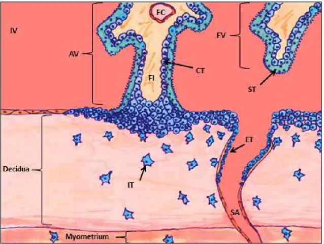
The invasive extravillous trophoblasts (also referred to as interstitial extravillous cytotrophoblasts) differentiate from the cytotrophoblast at anchoring villi and invade into the maternal decidua, invading up to the first third of the myometrium by the third trimester (Brosens 1988; Blankenship, et al., 1993; Kam et al., 1999; Kaufmann et al., 2003). To aid in the invasion processes, extravillous cytotrophoblast express an array of genes, including matrix metalloproteinases 2 (MMP2) and MMP9, to break down the extracellular matrix, in particular during the first trimester when placental vascularization is primarily established (Bass et al., 1994; Cross et al., 1994). With differentiation into an invasive extravillous phenotype, there is also a decrease in expression of transforming growth factor beta-3 (TGF-β3) and tissue inhibitor of metalloproteinases-2 (TIMP2) (Lee et al., 2010).
When the extravillous cytotrophoblast invade the maternal decidua, they position themselves next to the maternal spiral arteries and form plugs during the first trimester of pregnancy (Hustin et al., 1987; Moll et al., 1988; Nanaev et al, 1995; Burton et al., 1999; Guibourdenche et al., 2009). Endovascular extravillous cytotrophoblasts differentiate from extravillous trophoblasts to invade maternal spiral artery walls and lumen (Blankenship et al., 1993; Kaufmann et al., 2003). By 10-12 weeks of pregnancy, the plugs are lost and endovascular extravillous trophoblasts replace the epithelium, leading to dilation of the arteries, increased blood flow, and reduced flow resistance to the placenta by the second trimester (Brosens et al., 1967; Pijnenborg et al., 1981;
Benirschkle and Kaufmann 2000; Hirano et al., 2002; Guibourdenche et al., 2009). Endovascular extravillous cytotrophoblast invasion and re-modeling of the spiral arteries is highest in the middle of the placental bed, forming a discoid region where extensive nutrient and gas exchange occurs (Brosens 1988; Blankenship, et al., 1993; Kam et al., 1999; Kaufmann et al., 2003).
Rapid proliferation of the placenta occurs early during pregnancy (Korgun et al., 2006; Kar et al., 2007). At about 21 days post-conception, fetal-placental vasculogenesis initiates and circulation is established by 32 days post-conception (Demir et al., 1989; Zygmunt et al., 2003; Kaufmann et al., 2004; Torry et al., 2004; Demir et al., 2004, 2007; Arroyo and Winn 2008; Burton et al., 2009; van Oppenraaij et al., 2009). Placental vascularization is in part established by villous cytotrophoblast cells expressing vascular endothelial growth factor (VEGF) and other angiogenic factors during the first trimester (Hildebrandt et al., 2001).
Although the trophoblasts continue proliferation and cell turn-over throughout pregnancy (King and Blankenship, 1993; Kaufmann and Castellucci, 1997), there remains three layers of fetal tissue (trophoblast syncytium, fetal interstitium, and fetal endothelium) (Leiser and Kaufmann 1994), that separate fetal portal blood from maternal arterial blood (Leiser and Kaufmann 1994). This direct bathing of syncytium in maternal blood gives humans a hemochorial placenta (Enders 1965; Boyd and Hamilton 1970). Additionally, as the endovascular extravillous trophoblast invasion occurs in one concentrated area, this forms a discoid region where the majority of nutrient and gas exchange occurs. Therefore, the human hemochorial placenta is also a discoidal placenta (Boud and Hamilton 1970; Leiser and Kaufmann 1994), functioning to maintain
fetal-placental growth with a highly invasive extravillous and endovascular trophoblast invasion and arterial remodeling (Kaufmann et al., 2003).
Placental Development in the Sheep
In contrast to the human placenta, endovascular invasion by trophoblast cells does not occur within the sheep placenta (Kaufmann et al., 2003; Spencer et al., 2004; Carter 2007). Instead, the maternal endothelium and connective tissue is left intact (Leiser and Kaufmann 1994). Although the method of implantation is different in the ewe compared to the human, similarities remain in the process of apposition, trophoblast differentiation, and trophoblast function.
In the ewe, the morula enters the uterus at day 4, developing into a blastocyst by day 6 (Spencer et al., 2004). By day 8, the blastocyst hatches from the zona pellucida and is located in the ipsilateral uterine horn (Rowson and Moor 1966; Spencer et al., 2004). It undergoes a period of rapid cell proliferation and elongation, moving to the contralateral horn by day 13 if a singleton is present (Rowson and Moor 1966). During this period of elongation, and in contrast to the human, the extraembryonic membranes (chorion and yolk sac) form prior to implantation (Renfree 1982; Guillomot et al., 1993; Carson et al., 2000; Spencer et al., 2004). Rapid elongation of the ovine conceptus continues until day 16, when it adheres to the uterine epithelium (Spencer et al., 2004). The ovine conceptus then increases in size approximately threefold from gestational day 20 to 30 (Spencer et al., 2004; Grazul-Bilska et al., 2011).
Attachment and implantation begins at embryonic day 16, when mononuclear trophoblast cells from the trophectoderm adhere to the endometrial luminal epithelium (Spencer et al., 2004).
Between days 14 and 16, when the ovine conceptus is rapidly elongating and beginning to adhere to the endometrial luminal epithelium, binucleate trophoblast cells begin to differentiate from the mononuclear trophoblasts through consecutive nuclear divisions without cytokinesis (Spencer et al., 2004). Mononuclear trophoblasts, comparable to the invasive extravillous trophoblast in humans, also express MMP2 and MMP9, likely for improved branching of placental villi (Riley et al., 2000). Binucleate trophoblast cells do not adhere to the endometrial luminal epithelium, but they can fuse with epithelial cells and form a syncytium or hybrid symplasm that functions similarly to the human syncytium for the synthesis and secretion of hormones (Wooding 1992; Hoffman and Wooding 1993; Spencer et al., 2004). As giant binucleate cells fuse with endometrial luminal epithelium, they form trinucleate cells that further develop into syncytial plaques as more binucleate cells migrate and fuse (Wooding 1984; Spencer et al., 2004). Also similar to the human syncytium, continued cell turn-over and binucleate cell migration and fusion maintains the syncytium in the sheep placenta (Wooding 1984; Riley et al., 2000; Spencer et al., 2004). By gestational day 22, the entire trophectoderm has adhered to the endometrial luminal epithelium at placentome sites, described below (Boshier 1969; Guillomot et al., 1981; Spencer et al., 2004).
In ruminant animals, including sheep, areas along the endometrial luminal epithelium develop plaques or caruncles, which form progressively at gestational day 14 with depression of the luminal epithelium and crypt formation (Guillomot et al., 1981; Wimsatt 1950; Spencer et al., 2004). These specialized caruncle regions are the maternal side of the developing placenta at attachment sights, called placentomes (Wimsatt 1950; Spenser et al., 2004). Placentomes are composed of both a maternal portion (caruncle) and a fetal portion (cotyledon) (Wimsatt 1950;
Spencer et al., 2004). Syncytial plaques develop specifically within the placentome, covering the surface of caruncles for nutrient and gas exchange by fusion with endometrial luminal epithelium (Cross et al., 1994; Spencer et al., 2004). Placentomes are not present in the human placenta, but they do maintain a similar structure and function compared to the discoid region of invasion and nutrient exchange in the human placenta (Leiser and Kaufmann 1994). Inter-cotyledonary regions between placentomes are characterized by smooth chorion where nutrient and gas exchange can still occur (Bjorkman 1969; Leiser and Kaufmann 1994; Spencer et al., 2004).
As the sheep placenta has limited invasion of the maternal tissue (Leiser and Kaufmann 1994; Kaufmann et al., 2003; Spenser et al., 2004; Carter 2007), there are essentially six layers of tissue separating fetal from maternal blood in areas outside of the placentomes: maternal endothelium, connective tissue, maternal epithelium, fetal trophoblast, fetal connective tissue, and fetal endothelium (Leiser and Kaufmann, 1994). This type of placenta in sheep is called synepitheliochorial placenta (Wooding 1992; Leiser and Kaufmann 1994; Carter 2007), which is capable of transferring nutrients and gas across more tissue layers than are present in the human placenta (Cross et al., 1994).
Matrix Metalloproteinases in the Placenta
Matrix metalloproteinases (MMPs) play an important role in tissue remodeling for repair, cell migration, vascularization, and placentation. In particular, extravillous cytotrophoblast cells secrete MMPs to aid trophoblast differentiation and invasion into maternal decidua, and to establish placental vascularization (Huppertz et al., 1998; Solberg et al., 2003; Isaka et al., 2003; Munaut et al., 2003; Renaud et al., 2014). There are 28 known members in the MMP family and
they all function as calcium-dependent zinc endopeptidases that cleave internal peptide bonds in polypeptides and proteins (Bode and Maskos 2003). After translation, MMP pro-protein (pro-MMP) is processed in the endoplasmic reticulum, where pro-peptide is removed to allow access to the catalytic domain. The catalytic domain of MMPs contains zinc ion (Zn2+) that will interact with proteins and cleave peptide bonds, primarily through protein interactions at a glutamate residue within the MMP to initiate subsequent substrate hydrolysis. Once the pro-peptide is removed, active MMPs are secreted into the extracellular space or can be bound to the cell membrane. Substrate binding domain of MMPs functions for specificity of targeting proteins for degradation. Target proteins for MMPs are primarily extracellular matrix proteins, including collagen (types 1 through 10), gelatin, aggrecan, fibronenctin, elastin, and laminin (Bode and Maskos 2003; Manzetti et al., 2003). However, each MMP has specificity and affinity for specific extracellular matrix proteins (i.e. MMP9 targets gelatin and collagen type 4 and 5) (Manzetti et al., 2003). By this mechanism, MMPs can degrade extracellular matrix proteins, enabling cell migration or proliferation, vascularization, or other tissue remodeling processes (Munaut et al., 2003; Renaud et al., 2014).
During placentation, invading extravillous trophoblasts are known to secret MMPs to cleave extracellular matrix proteins in the decidua, aiding in the process of cell migration into maternal decidua and establishment of placental vascularization (Huppertz et al., 1998; Munaut et al., 2003; Solberg et al., 2003; Renaud et al., 2014). However, to control the function of MMPs, cells also secrete tissue inhibitors of metalloproteinases (TIMPs). TIMPs function by binding MMPs, usually at a 1:1 ration, and block MMP activity by inserting into the active site of the catalytic domain (Snoek-van Beurden and Von den Hof 2005; Stephenson et al., 2005). There are four
known TIMPs and they play a significant role in regulating tissue remodeling and cell invasion (Snoek-van Beurden and Von den Hof 2005). When extravillous trophoblasts of the placenta invade maternal decidua during the first trimester, decidual stromal cells secrete TIMPs, possibly as a mechanism to limit placental invasion (Hurskainen et al., 1996; Huppertz et al., 1998). Furthermore, research suggests that hormone secretion from trophoblasts may function in paracrine or autocrine signaling for regulation of placental MMP expression (Lee et al., 2003; Liao et al., 2003, Limaye et al., 2008; Comstock et al., 2008)
Steroidogenesis and Hormone Signaling
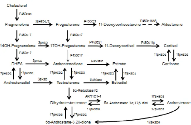
While placenta of all species functions for the transport of nutrients and gas for support of the developing fetal-placenta unit (Gude et al., 2004), it also has important endocrine functions necessary for fetal development and maternal signaling (Knobil and Neill 1998; Guibourdenche et al., 2009). Steroid hormone synthesis (steroidogenesis) involves the conversion of cholesterol to various steroid hormones through enzymatic processing (Miller and Auchus 2010). Free cholesterol in the cell cytoplasm, obtained either from endocytosis or de novo synthesis, is carried to the outer mitochondrial membrane by proteins with a StAR-related lipid transfer domain (START), such as StarD4 and StarD5 (Miller and Auchus 2010). StAR, the first-described member of the START protein family, plays the crucial role in facilitating the movement of cholesterol from the outer mitochondrial membrane to the inner mitochondrial membrane (Miller and Auchus 2010). Sterol response element binding proteins (SREBPs) and 3-hydroxy-3-methylglutaryl co-enzyme A reductase regulate the biosynthesis of cholesterol (Miller and Auchus, 2010). The entire process of steroidogenesis is show in Figure 1.2, with the first and rate limiting step being P450scc (CYP11A1 or cholesterol side chain cleavage enzyme) conversion of cholesterol to pregnenolone (Miller and Auchus 2010). This occurs in the inner mitochondrial membrane (Miller and Auchus 2010). Other
steroidogenic enzymes that function in steroidogenesis belong to two family groups: cytochrome P450 enzymes (P450) or hydroxysteroid dehydrogenases (HSDs) (Miller and Auchus 2010).
Cellular regulation of steroidogenesis occurs with posttranslational modifications of P450 and HSD steroidogenic enzymes (Miller and Auchus 2010). The end result is the cell-specific production of sex steroid hormones (androgens, progesterone, and estrogens), mineralocorticoids, or glucocorticoids (Miller and Auchus 2010). The production of specific steroids is determined by the origin of the endocrine tissue or cell type as specific steroidogenic enzymes may only be expressed in a given tissue or cell, such as in the adrenal gland or ovary (Miller and Auchus 2010). The steroid hormones, once secreted by the cell, can act in a paracrine or endocrine manner (Petraglia et al., 1996). For a cell to respond to steroid hormones, however, it must express their receptors (Mangelsdorf et al., 1995). For instance, tissue specific expression of estrogen receptors 1/alpha (ESR1), estrogen receptor 2/beta (ESR2), and androgen receptor (AR) can be seen in multiple cell types and tissues, including the male and female gonads and the placenta (Leung and Steele 1992).
Estrogens encompasses a family of sex steroid hormones that have an 18 carbon structure and can refer to both naturally occurring and synthetic hormones. All estrogens bind to estrogen receptors to initiate a cellular response. The estrogen family includes estrone (E1), estradiol (E2),
estriol (E3), and 16-hydroxy estrone. The most common biologically active estrogen is estradiol.
Similarly, androgens are a class of 19 carbon sex steroid hormones that includes the adrenal androgens, dehydroepiandrosterone (DHEA) and DHEA-S, as well as androstenedione, androstenediol, testosterone, and dihydrotestosterone (DHT). All androgens signal via binding to
the androgen receptor (AR), although, similar to estrogens, there is varying affinity of family members to the receptor (Senger 2003; Hu et al., 2010; Miller and Auchus 2010).
Steroid hormone receptors possess several domains for functional signaling, including a ligand binding domain and a DNA binding domain (Mangelsdorf et al., 1995). The traditional AR DNA binding domain or response element (ARE) is AGAACAnnnTGTTCT (Mangelsdorf and Evans 1995; Mangelsdorf et al., 1995; Comstock et al., 2008) and the traditional ESR1 DNA binding domain (ERE) is GGTCAnnnTGACC (Mangelsdorf et al., 1995). Binding of a sex hormone ligand to its receptor typically occurs within the cytoplasm or within the nucleus as steroid hormones are membrane permeable (Mangelsdorf et al., 1995). The ligand-bound receptor may then dimerize and bind directly to DNA where the DNA-response element or half-site is present (Mangelsdorf et al., 1995). Binding can also occur at other DNA sequences that are similar in sequence or shape to the traditional DNA binding domain (Comstock et al., 2008). The ligand-bound receptor further recruits other transcription cofactors to initiate transcription (Mangelsdorf et al., 1995). As this pathway of steroid hormone signaling initiates gene transcription, the effects of steroid signaling are not typically an immediate cellular response (Cooper and Hausman 2000).
However, studies have shown that steroid hormones also are capable of initiating rapid cellular responses, such as increased intracellular calcium (Berridge and Taylor 1988) that would precede gene transcription (Cooper and Hausman 2000). This suggests that steroid hormones are capable of binding cell membrane receptors to initiate a rapid cellular response via secondary messengers (Cooper and Hausman 2000). Indeed, despite traditional signaling via direct binding to DNA or
through transcription factor binding, steroid hormones appear to also signal through specific membrane receptors (Prossnitz et al., 2008; Thomas 2008). For instance, progesterone and estrogen signal through cell membrane receptors mPRα and PGMRC1, and GPR30 (GPER), respectively, possibly to initiate a different cellular response than the conventional nuclear steroid hormone receptor pathway (Prossnitz et al., 2008; Thomas 2008).
Receptor expression within a cell can also be regulated by tissue specific methylation within the promoter region, such as occurs in AR (Kinoshita et al., 2000; Jarrard et al., 1998), ESR1 (Fürst et al., 2012), and ESR2 transcription (Xue, et al., 2007). Methylation within the promoter region typically blocks gene transcription (Zhang and Meaney 2010), adding a layer of complexity to the system that ensures appropriate cellular signaling and response to steroid hormones. Additionally, tissue specific response regions may be present within the promoter region to regulate mRNA transcription, such as the placental specific promoter region for aromatase (CYP19) (Vanselow et al., 1999).
While steroid hormone signaling regulates transcription, circulating and cytoplasmic binding proteins can bind specific hormones to decrease their ability to signal while increasing their half-life (Anderson 2008). For instance, sex hormone-binding globulin (SHBG) is a glycoprotein that binds sex steroid hormones, in particular testosterone and estradiol (Bardin et al., 1981; Hammond 1990; Petra 1991). Circulating SHBG is further capable of regulating hormone signaling by transporting hormones directly to cells or tissue to initiate cellular signaling (Bordin and Petra 1980; Stanczky et al., 1986; Larriva-Sahd et al., 1991; Hryb et al., 1990).
Mature peptide hormones, in comparison, are composed of less than 50 amino acids and their signaling pathway is different from steroid hormones as they only signal via cell membrane receptors (Cooper and Hausman 2000). After a peptide hormone binds its membrane receptor, secondary messengers, such as calcium, rapidly amplify the signal of ligand binding, typically via phosphorylation of cytoplasmic proteins (Cooper and Hausman 2000). Ultimately, this leads to binding of transcriptional cofactors or repressors for regulation of gene expression (Cooper and Hausman 2000). In both situations, the products of either peptide or steroid hormone signaling can initiate gene transcription, resulting in protein products that have the potential to further regulated the transcription of other genes, referred to as secondary response genes (Cooper and Hausman 2000).
Placental Steroidogenesis and Receptor Expression
During the first trimester, at approximately the seventh week of pregnancy, sufficient syncytiotrophoblast have differentiated from cytotrophoblasts to become the primary hormone producing cells in the human placenta (Guibourdenche et al., 2009). The syncytiotrophoblast is in direct contact with maternal blood at approximately eleven weeks of gestation; it secretes hormones directly into maternal circulation (Guibourdenche et al., 2009). Syncytiotrophoblast secrete progesterone (Guibourdenche et al., 2009), human chorionic gonadotropin (hCG), placental lactogen (hPL), and placental growth hormone (hGH-v) (Jameson and Hollenberg 1993; Guibourdenche et al., 2009). While PL and GH function for mobilization of maternal nutrients for fetal-placental growth and support (Handwerger and Freemark 2000), progesterone and hCG function for uterine quiescence and pregnancy maintenance (Stoffer et al., 1977; Spencer and Bazer 2002).
As the syncytiotrophoblast is responsible for the production of progesterone in the human placenta, research has investigated the mechanisms of steroidogenesis and sex hormone signaling in this multinucleated cell layer. The steroidogenic enzymes present in the human syncytiotrophoblast include P450scc (pregnenolone production from cholesterol), 3β-HSD (progesterone and androstenedione production), 17β-HSD (testosterone production from androstenedione), and P450arom (or CYP19, estrogen production from testosterone) (Fournet-Dulguerov et al, 1987; Payne and Hales 2004; Tuckey et al., 2004; Guibourdenche et al., 2009; Sathishkumar et al., 2012). The syncytiotrophoblast in humans does not express P450 17α-hydroxylase-17:20; therefore, androgen production from pregnenolone and progesterone does not occur in the human placenta (Payne and Hales 2004; Tuckey et al., 2004; Guibourdenche et al., 2009). This, however, does not appear to be the case for ruminants, as both the goat and sheep placenta express P450c17 (Ma et al., 1999; Weng et al., 2005).
The syncytiotrophoblast in the human placenta are still capable of producing testosterone despite the lack of P450-17α (Payne and Hales 2004; Tuckey et al., 2004; Guibourdenche et al., 2009). To produce testosterone, syncytiotrophoblast endocytose maternal cholesterol via binding to lipoprotein receptor, VLDL receptor, or B1 scavenger receptor on the syncytiotrophoblast cell membrane (Guibourdenche et al., 2009). Free cytosolic cholesterol is available after lysosomal degradation of LDLs occurs (Guibourdenche et al., 2009). In the syncytiotrophoblast, the free cholesterol is selectively transported to the outer and inner mitochondrial membrane by sterol carrier protein-2 and metastatic lymph node 64 proteins, respectively (Guibourdenche et al., 2009). Within the syncytiotrophoblast’s mitochondria, cholesterol can be converted into
pregnenolone through the action of P450scc, which is further converted into progesterone via enzymatic action of 3β-HSD (Guibourdenche et al., 2009).
As androgens cannot be directly produced from pregnenolone or progesterone in the human placenta, an extracellular precursor is necessary for syncytiotrophoblast androgen and estrogen biosynthesis (Guibourdenche et al., 2009). The precursor dehydroepiandrosterone sulfate (DHEA-s) is produced by fetal and maternal adrenals and reaches the placenta from fetal and maternal circulation, respectively (Calvin et al., 1963; Guibourdenche et al., 2009). Once DHEA-s iDHEA-s taken up by DHEA-syncytiotrophoblaDHEA-st, it iDHEA-s hydrolyzed by DHEA-sterol DHEA-steroid DHEA-sulfataDHEA-seDHEA-s into 16α-hydroxy DHEA-s (Guibourdenche et al., 2009). 3β-HSD then metabolizes the hydrolyzed s-DHA into androstenedione, which can be further converted into testosterone by 17β-HSD (Guibourdenche et al., 2009). Placental CYP19 in the endoplasmic reticulum aromatizes androstenedione and testosterone in the syncytiotrophoblast for the production of C18-estrone and estradiol, respectively (Ryan 1959; Thompson and Siiteri 1974a,b).
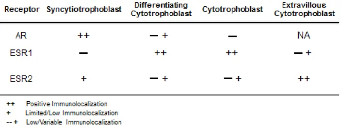
While the syncytiotrophoblast is steroidogenic and produces sex steroid hormones, it also appears to be regulated by sex-steroid hormones (discussed in more detail below). To respond to paracrine and endocrine hormone signaling, trophoblasts must express sex steroid hormone receptors (reviewed in Table 1.1). Interestingly, ESR1 does not appear to be expressed in differentiated syncytiotrophoblast (Kumar et al., 2009), but is expressed in cytotrophoblasts, differentiating cytotrophoblasts, and extravillous cytotrophoblasts (Bukovsky et al., 2003a,b; Schiessl et al., 2006; Kumar et al., 2009). AR, in contrast, is immunolocalized in differentiated syncytiotrophoblast and has variable immunoreactivity in differentiating cytotrophoblasts
(Iwamura et al., 1994; Hsu et al., 2009). ESR2 has relatively low levels of nuclear immunolocalization in cytotrophoblasts and differentiating cytotrophoblasts, but can be found in the cytoplasm of the syncytiotrophoblast and extravillous cytotrophoblasts (Bukovsky et al., 2003a,b; Schiessl et al., 2006; Kumar et al., 2009). The localization of AR, ESR1, and ESR2 has not yet been determined within the sheep placenta.
Expression of sex hormone receptors and aromatase also appears to be regulated by fetal sex and pathological conditions during pregnancy. For instance, higher aromatase is present in placenta from male fetuses compared with placenta from females in normal pregnancy, while preeclampsia increases aromatase expression in placenta from female fetuses and decreases aromatase in the placenta from male fetuses (Sathishkumar et al., 2012). Additionally, ESR1 expression can also be lost in cytotrophoblast cells, possibly contributing to the reduced placental invasion noted in preeclampsia (Bukovsky et al., 2003a,b).
Syncytiotrophoblast regulation of sex hormone signaling is further complicated by the expression of sex hormone-binding globulin (SHBG) within the syncytium (Larrea et al., 1993). SHBG preferentially binds testosterone and estradiol to potentially limit immediate hormone signaling or extend sex-hormone half-life (Bardin et al., 1981; Hammond 1990; Petra 1991).
Steroidogenesis in the Maternal-Placenta-Fetal Unit
For placental steroidogenesis, maternal cholesterol is first taken up by trophoblast cells via LDL receptor-mediated endocytosis or SR-B1-mediated uptake (Guibourdenche et al., 2009; Hu et al., 2010). Once internalized within the placental cells, it can be stored as esters in lipid droplets,
incorporated as free cholesterol into cellular membranes, or, depending on the placental cell, it can be utilized for steroidogenesis. In the placenta, syncytiotrophoblast (human), binucleate cells (sheep), and giant trophoblasts and spongiotrophoblasts (mouse) are steroidogenic cells, but they lack the ability to adequately synthesize cholesterol from acetate via de novo synthesis. Therefore, a maternal source of cholesterol is required and, during pregnancy, higher levels of circulating cholesterol can be found in the maternal blood (Malassiné et al., 2003; Tuckey et al., 2004; Guibourdenche et al., 2009). Cholesterol that is taken up via LDL receptor-mediated endocytosis is processed in an early endosome, where decreased pH allows for the receptor to disassociate and be recycled back to the plasma membrane. The now free cytoplasmic cholesterol (including that from SR-B1 mediated uptake) is trafficked to the mitochondria in syncytiotrophoblast (human), binucleate cells (sheep), and giant trophoblasts and spongiotrophoblasts (mouse), likely following microtubule trafficking. Cholesterol is first loaded onto the outer mitochondrial membrane and then transferred to the inner mitochondrial membrane via StAR. At the inner mitochondrial membrane, side chain cleavage occurs via P450scc enzymatic activity to produce pregnenolone. Pregnenolone (in the above listed cell types) is further converted to progesterone via enzymatic activity of 3β-HSD (Guibourdenche et al., 2009; Hu et al., 2010). At this point, the progesterone is primarily released back into maternal circulation. This is of particular importance in human and sheep, where placental production of progesterone maintains pregnancy. Interestingly, the human placenta is more steroidogenic than placentas of other mammals in that it produces much greater quantities of progesterone per unit volume (Malassine et al., 2003; Senger 2003; Miller and Auchus 2010).
In the human placenta, progesterone does not undergo further processing to form estrogens or androgens, as discussed above. Instead, the human placenta required maternal and fetal contributions of dehydroepiandrosterone sulfate (DHEA-S) (Calvin et al., 1963; Mastorakos and Ilias 2003; Payne and Hales 2004; Tuckey et al., 2004; Guibourdenche et al., 2009). In contrast, the sheep and rat placenta express 17α-hydroxylase and are capable of producing androgens; this would occur in the binucleate cells (sheep and rat) and in the spongiotrophoblasts (rat). The mouse placenta is also capable of converting progesterone into androstenedione, but lacks aromatase for the production of estrogens (Rembiesa et al., 1971; Arensburg et al., 1999; Ma et al., 1999; Mastorakos and Ilias 2003). In both the human and sheep placenta, DHEA can be further processed by 3β-HSD to form androstenedione. Androstenedione is further converted to testosterone via 17β-HSD while aromatase activity (which is present in the sheep and human placenta) can produce estrone and estradiol from these precursors, respectively (Bousquet et al., 1984; Strauss et al., 1996; Wooding et al., 1996; Guibourdenche et al., 2009: Hu et al., 2010; Mondragón et al., 2012).
In humans, sheep, and rodents, the fetal gonads cannot make progesterone, estrone, estradiol, or estriol. This is because the fetus lacks the enzymes 3β-HSD and aromatase, both of which are present in the placenta (Ryan 1959; Thompson and Siiteri 1974a,b; Arensburg et al., 1999; Guibourdenche et al., 2009). However, if the fetus is a male, there will be production of testosterone and dihydrotestosterone (DHT). When this occurs, maternal LDL cholesterol is taken up by the fetal gonadal tissue via LDL receptor mediated endocytosis or SR-B1 mediated selective uptake. Once inside the cell, cholesterol will be processed as describe above. When sexual differentiation of the male testes begins to occur (at the start of the second trimester in
humans or at day 30 of gestation in sheep), the differentiating Leydig cells will convert free cholesterol into pregnenolone. Conversely, circulating fetal pregnenolone of placental origin may also be taken up for steroidogenesis. Pregnenolone in Leydig cells undergoes enzymatic processing to produce testosterone and DHT, both of which are necessary for completion of male sexual differentiation (Gehani et al., 1998; Senger 2003; Hu et al., 2010). However, fetal derived testosterone will travel through fetal circulation, back to the placenta, where aromatase activity will convert it to estradiol (Guibourdenche et al., 2009). This agrees with human studies that have shown that fetal sex does not alter maternal serum or umbilical cord blood levels of testosterone and dihydrotestosterone (Dawood and Sexena 1977; Rodeck et al., 1985).
Additionally, the maternal gonad maintains steroidogenesis throughout pregnancy, though to varying degrees in human, sheep, and rodent. To begin with, circulating LDL is also taken up by ovarian cells (granulosa, theca, small or large luteal cells) and processed as described above. In luteal cells of the sheep, human, and rat ovary, pregnenolone is converted to progesterone via 3β-HSD. The amount of progesterone produced by the corpus luteum, and the amount of luteal tissue, is dependent on the stage of pregnancy. For instance, in early sheep and human pregnancy, luteal production of progesterone is high and will maintain pregnancy. However, by the second trimester, luteal production of progesterone is supplement by the placental production (Nelson et al., 1992; Quirke et al., 2001; Senger 2003; Miller and Auchus 2010).
Placental Estrogen Signaling and Function
The major estrogens produced by the human placenta are estrone, estriol and estradiol, which begin to increase during the third to thirteenth weeks of gestation (Tulchinsky and Hobel 1953;
Siiteri and MacDonald 1963; 1966; Loriaux et al., 1972). These estrogens appears to play a very crucial and complex role during placental development in humans. Depending on the stage of gestation and hypoxic conditions, placental estrogen signals for varying cellular functions and physiological effects (Albrecht et al., 2006). Interestingly, oxygen concentration regulates aromatase activity in the placenta (Thompson and Siiteri 1974a,b; Goto and Fishman 1977; Zachariah and Juchau 1977), which can lead to impaired estrogen biosynthesis in hypoxic conditions (Aw et al., 1985; Rodesch et al., 1992). This might contribute to estradiol’s varying functions between the first and second trimester, as a more hypoxic environment is present with restricted blood flow to the developing placenta during the first trimester when extensive differentiation of extravillous cytotrophoblasts and remodeling of the uterine spiral arteries occurs (Kaufmann et al., 2003). Additionally, this might lead to greater prominence of androgen signaling in syncytiotrophoblast in the first trimester as androgens up-regulates hypoxia inducible factors (HIFs) and vascular endothelial growth factor (VEGF) activity in a hypoxic environment (Shabisgh et al., 1999; Mabjeesh et al., 2003b; Cheng et al., 2004; Lissbrant et al., 2004; Boddy et al., 2005; Zhu and Kyprianou 2008).
As early as 11 weeks of gestation, HIF-1α activity is blocked by 2-methoxyestradiol (2-ME), a metabolite of estradiol that is present in the human placenta (Berg et al., 1983; Mabjeesh et al., 2003a; Ricker et al., 2004; Becker et al., 2008; Kanasaki et al., 2008). Under the hypoxic conditions that are present during the first trimester, 2-ME induces cytotrophoblast differentiation into an invasive phenotype (Lee et al., 2010). Cytotrophoblast differentiation also occurs in vitro as treatment of term human cytotrophoblasts with 17β-estradiol leads to differentiation into a syncytium of functionally mature syncytiotrophoblast capable of secreting
human chorionic gonadotropin (hCG) (Rama et al., 2004). Therefore, estrogen influences placental growth in the first trimester (Abdul-Karim et al., 1971) through increased syncytiotrophoblast differentiation and, thereby, increased synthesis of progesterone (Solomon 1994). It is widely accepted that the hypoxic environment during the first trimester leads to the trophoblast invasion (Zhou et al., 1993; Graham et al., 2000; Norwitz et al., 2001; Kadyrov et al., 2003; Bischof and Irminger-Finer 2005; Hayashi et al., 2005; Lyall 2006; Cohen and Bischof 2007; Lunghi et al., 2007; Robins et al., 2007; Rosario et al., 2008), likely through estradiol signaling such as with 2-ME (Lee et al., 2010).
At the start of the second trimester in primate pregnancy, there is a surge in estrogens that blocks the differentiation of cytotrophoblast cells into extravillous trophoblasts (Albrecht et al., 2006), likely facilitated through down-regulation of VEGF (Bonagura et al., 2008). This shift in estrogen function in the placenta during the second trimester, when estrogen levels surge, may be regulated by the increase in placental blood flow and oxygen at this time (Albrecht et al., 2006). However, once maternal spiral artery invasion has occurred and the high-flow, low-resistance blood flow is established to the placenta in the second trimester, high circulating estrogen levels are not required to maintain the uteroplacental blood flow that is established (Aberdeen et al., 2010), primarily because the invasion and establishment of the uteroplacental blood flow occurs in the first trimester with differentiation of the extravillous and endovascular cytotrophoblasts (Kaufmann et al., 2003).
Interestingly, reduced aromatase activity in the term placenta has been reported in preeclamptic patients, significantly reducing placental 17β-estradiol, estrone, and 2-methoxy-estradiol (Hertig
et al., 2010). This suggests that aberrant estrogen signaling during the first trimester in the preeclamptic placenta may contribute to the reduced placental invasion and pregnancy-induced hypertension presented in these patients (Redman and Sargent, 2003).
Placental Androgen Signaling and Function
Despite the research investigating the critical roles of estrogen in trophoblast cell function, there has been very limited research investigating the function of androgen signaling within the placenta. The limited research on androgen’s role in placental development has primarily focused on its role in preeclampsia (PE). The earliest report from Thoumsin et al. showed that there is decreased aromatase activity in preeclamptic placentas compared to the placentas from hypertensive patients (1982). Women with PE have increase serum testosterone concentrations compared to normotensive pregnancies (Atamer et al., 2004; Ghorashi and Sheikhvatan 2008; Hsu et al., 2009), regardless of fetal gender (Lorzadeh and Kazemirad 2012). Preeclamptic patients in the third trimester have higher serum total and free testosterone levels when compared to normotensive pregnancies, although serum concentrations of DHEA-S, androstenedione, and SHBG are not different (Acromite et al., 1999; Serin et al., 2001; Salamalekis et al., 2006). An increase in androstenedione, testosterone, and free testosterone has also been reported at 17 and 33 weeks of gestation in women who eventually developed preeclampsia, while DHEAS was elevated at week 17 only (Carlsen et al., 2005). Increased serum total and free testosterone has also been reported in pregnancies with placental induced hypertension at 37 weeks of gestation (Gerulewicz-Vannini et al., 2006).
Increased placental expression of androgen receptor (AR) is also reported in the syncytiotrophoblast and stromal cells of preeclamptic placentas compared to placentas from normotensive pregnancies (Hsu et al., 2009; Sathishkumar et al., 2012). While increased serum testosterone (Atamer et al., 2004; Ghorashi and Sheikhvatan 2008; Hsu et al., 2009; Lorzadeh and Kazemirad 2012) and increase placental AR (Hsu et al., 2009; Sathishkumar et al., 2012) would suggest that androgen signaling contributes to or is a result of the pathogenesis of PE, decreased androgen signaling may also contribute to placental insufficiency (Lim et al., 2011). For instance, women are at an increased risk of developing PE if they have a polymorphism of greater than 16 GGC repeats in the transcriptional activation domain of the AR gene, leading to decreased AR function and expression (Lim et al., 2011).
Interestingly, by six weeks after delivery, serum testosterone and free testosterone levels from preeclamptic patients reaches circulating concentrations similar to those of normotensive, control patients (Serin et al., 2001), strongly suggesting a placental source for circulating androgen. It has been proposed that serum testosterone levels could be used as a marker for predicting late onset preeclampsia (Carlsen et al., 2005; Lorzadeh and Kazemirad 2012); however, a conflicting study has shown that serum testosterone and SHBG are not significantly different during the first and second trimester in patients that later developed preeclampsia compared to matched controls (Tuutti et al., 2011). Despite this report, these data suggest that androgen signaling may be very important for placental development and function, and that any abnormality in androgen signaling may contribute to pregnancy complications like PE. As placental androgen may play crucial roles in regulating trophoblast function and differentiation, similar to placental estrogen, future studies on androgen’s role in trophoblast function and signaling are imperative.
Loss of Sex Hormone Signaling in the Placenta
Rodent knock out models of ESR1 and AR have been shown to be non-lethal (reviewed by Couse and Korach 1999; Yeh et al., 2002). ESR1 knock-out mice are infertile given the multifactorial functions of estrogen in developmental and reproductive physiology (reviewed by Couse and Korach 1999). In AR knock-out mice, only males are infertile while female mice have reduced litter sizes (Yeh et al 2002). Additionally, human cases of estrogen resistance have been reported, showing that functional loss of ESR1 is viable for fetal development (Smith et al 1994). In spite of these findings, the functional loss of ESR1 may be in part rescued with ESR2 function in some tissues (reviewed by Couse and Korach 1999). Additionally, infants born with placental aromatase deficiency (Harada et al., 1992a,b; Conte et al., 1994; Deladoëy et al., 1999) survived prenatal development (Shozu et al., 1991 Morishima et al., 1995; Carani et al., 1997), suggesting normal or at least compensatory placental development and function had occurred.
Human fetuses with androgen insensitivity also survive fetal development (Hughes and Evans 1987; Bangsboll et al., 1992; Brown 1995; Quigley et al., 1995; reviewed by Ahmed et al., 2000), suggesting that lost or perturbed sex hormone signaling may not be detrimental to placental development, despite findings of increased maternal serum androgens in compromised pregnancies (Atamer et al., 2004; Ghorashi and Sheikhvatan 2008; Hsu et al., 2009; Lorzadeh and Kazemirad 2012). However, other human case studies have suggested that functional loss of AR is correlated with multiple miscarriages (Wilson et al., 1974; Decaestecker et al., 2008). Heightened androgen signaling is also associated with recurrent spontaneous abortions and preterm labor (Karvela et al., 2008; Karjalainen et al., 2012). Given the conflicting reports on the
necessity of placental androgen signaling, further research is needed to clarify its role in normal placental development and trophoblast function.
Androgen Signaling in Cancer
To understand possible roles of androgen signaling in the placenta, insights can be obtained from studies on androgen signaling in cancer as cancer cells share many similarities to trophoblasts (Ferretti et al., 2007). For instance, cytotrophoblast cells undergo rapid cell divisions and are capable of maintaining an undifferentiated state; these features are also present in cancer (Ferretti et al., 2007). Additionally, cytotrophoblast can invade surrounding tissue, avoiding an immune response similar to cancer progression (Ferretti et al., 2007). Most importantly, trophoblasts are capable of recruiting maternal spiral arteries and remodeling them for increased vascularization of the placenta (Kaufmann et al., 2003). While cancer does not necessarily remodel arteries, tumors are capable of initiating vascularization (Ferretti et al., 2007).
Cancer cells and trophoblast cells also undergo an epithelial-mesenchymal transition (EMT). Mesenchymal cells are maintained in an undifferentiated state during gastrulation in the developing fetus (Vicovac and Aplin 1996). While mesenchymal cells are pluripotent cells capable of migration, polar epithelial cells can dedifferentiate to form mesenchymal cells in the EMT process. EMT is a normal process during gastrulation (or tissue development) in multicellular organisms. For EMT to occur, polar epithelial cells must undergo multiple changes, including loss of polarity, loss of specific cellular junctions (such as gap junctions that often separate epithelial cells and a basement membrane from underlying tissue), cytoskeletal remodeling, loss of E-cadherin expression, and redistribution of organelles. In addition, there are
molecular changes that induce epithelial cells to regain a pluripotent, mesenchymal state. Studies have shown that multiple signaling pathways are involved, but usually Wnt and Ras signaling, in cooperation with transforming growth factor beta (TGFβ) and mitogen activated protein kinase (MAPK), are necessary for EMT to occur. Once epithelial cells have returned to the de-differentiated, pluripotent state of mesenchymal cells, they are capable of migrating and proliferating via PI3K signaling, and have increased resistance to apoptosis (Theiry 2003; Kalluri and Weinberg 2009).
While EMT occurs primarily during gastrulation, it is also occurs in adult stages of multicellular organisms, likely as a mechanism to respond to physiological stress and allow for specific tissue repair mechanisms throughout the life of the organism. However, when molecular regulation of EMT goes awry, there can be uncontrolled proliferation of undifferentiated mesenchymal cells. These cells are capable of migrating to other tissue and cause disease states such as tissue fibrosis or cancer (Thiery 2003).
In addition to continuous embryonic and adult EMT, the trophoblasts in the placenta also undergoes EMT during development. The polar trophoblast cells surrounding the blastocyst undergo EMT to form cytotrophoblasts. Cytotrophoblasts, similar to mesenchymal cells, are considered pluripotent as they function as a placental stem cell, capable of migrating and differentiating into invasive extravillous cytotrophoblasts, endovascular extravillous cytotrophoblasts, and syncytiotrophoblast,. Early reports had suggested that the differentiation of cytotrophoblasts was also an EMT, although this is now debated. However, in contrasts to EMT, polar syncytiotrophoblast, which functions as an epithelial layer of the placenta, cannot revert