1
MPO measurements in DSS-treated mice
David Ahl
Bachelor’s thesis in Biomedicine Division of Integrative Physiology Department of Medical Cell Biology Uppsala University
2
Abstract
Inflammatory bowel disease is a group of pathological conditions where the intestine is
chronically inflamed. Recent studies indicate that probiotics ameliorate DSS-induced colitis in rats. To further investigate this, our laboratory has studied the effects of two Lactobacillus
reuteri strains in C57 Bl/6 mice treated with 3% DSS. The severity of disease was assessed
through both clinical observations (disease activity index) and histological evaluation. Using these methods, preliminary results suggest that probiotic protection can be demonstrated. To further reinforce these results, additional methods for evalution of disease severity are needed. This thesis focuses on the use of the ELISA technique for measurements of myeloperoxidase (MPO) in colon samples collected from mice in the probiotic study. MPO is an enzyme found abundantly in neutrophils, and measure of this enzyme is frequently used for approximation of neutrophil presence. MPO level is therefore used as an indicator of inflammatory status and thus, in the case of inflammatory bowel disease, severity of disease. The results indicate that MPO levels are decreased in mice with DSS-induced colitis when treated with probiotics, but that a larger sample group is needed for definite proof. Furthermore, this study investigates the effect of an additional freeze-thaw cycle on colon samples before ELISA measurements, and shows that no effect on MPO levels can be demonstrated.
3
Contents
Introduction ... 5
The large intestine ... 5
Inflammatory bowel disease (IBD) ... 5
Colonic microbiota ... 6
Probiotic protection from DSS-induced colitis in mice ... 6
Myeloperoxidase as a pro-inflammatory marker ... 6
Aim ... 7
Methods ... 7
Experimental animals ... 7
Homogenization of samples ... 8
Investigation of the effect of a repeated freeze-thaw cycle on MPO levels ... 8
Enzyme-linked immunosorbent assay (ELISA) ... 8
Results ... 11
DAI in the new mice group ... 11
ELISA measurements of MPO ... 11
Regression analysis ... 13
Mean group values ... 14
Discussion ... 16
ELISA measurements of MPO ... 16
Effect of a repeated freeze-thaw cycle on MPO levels ... 16
4
Abbreviations
DAI Disease Activity Index DSS Dextran Sulfate Sodium IBD Inflammatory Bowel Disease MPO myeloperoxidase
5
Introduction
The large intestine
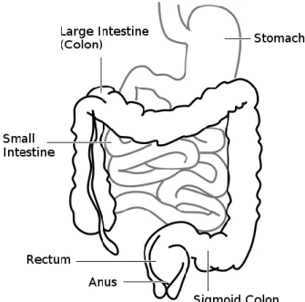
The large intestine consists of the cecum and the colon and is about 1.5 meters long (Fig. 1). Its main purposes are to absorb water and vitamins and to process feces towards the rectum. The colon wall is composed of three layers; the muscularis, the submucosa, and the epithelial-covered mucosa (towards lumen).
Inflammatory bowel disease (IBD)
Ulcerative colitis and Crohn’s disease are the most common pathological conditions in the IBD group. They are idiopathic and both manifest as chronic inflammation of the
gastrointestinal tract but differ in localization and nature of the inflammation. Ulcerative colitis affects only the colon and rectum and is limited to the mucosa while Crohn’s disease can affect any part of the gastrointestinal tract and be transmural. Also, the distribution of inflamed areas in Crohn’s disease may be segmental whereas ulcerative colitis is continuous. While fatalities are rare, IBD can be severely handicapping with symptoms such as abdominal pain, fever, vomiting, diarrhea, rectal bleeding and weight loss (6). The etiology for IBD is still unclear, but evidence exists that both genetic and environmental factors are involved in development of the disease. There are indications that the interaction of the mucosal immune system with the commensal microbiota of the gastrointestinal tract is somehow dysfunctional in patients that develop IBD (7). There is currently no curative therapy for IBD, and treatment primarily consists of antibiotic- and anti-inflammatory drugs that can control or dampen flares of the disease (6). In severe cases, surgical removal of inflamed parts of the gastrointestinal tract may be required.
6
Colonic microbiota
The large intestine is inhabited by 500 – 1,000 different species of bacteria that normally do not cause disease, called the commensal flora (11). The mucosal immune system must remain inactive when confronted with these massive amounts of microbes but at the same time be able to activate when pathogens are presented to it. It is speculated that the
relationship between the immune system and the commensal flora is somehow disturbed in patients that develop IBD (4).
Probiotic protection from DSS-induced colitis in mice
This project is based on ongoing work in our laboratory on probiotic protection from DSS-induced colitis in mice. The protective properties of two Lactobacillus reuteri strains, ATCC PTA 4659 (human) and R2LC (rat), in mice treated with 3% DSS (TdB Consultancy,
Uppsala, Sweden) in drinking water is investigated. The exact mechanism of DSS is unclear, but it is known that it damages the colonic mucosa and causes an inflammation similar to ulcerative colitis (10). It is therefore used for inducing a murine model of the disease. Male C57 Bl/6 mice (Taconic M&B, Ry, Denmark) were divided in five groups; 8d DSS (treatment with DSS for 8 days), 2d R2LC + DSS (2 day treatment with R2LC, five day break, then DSS for 8 days), 15d R2LC + DSS (treatment with R2LC for 15 days and DSS the last 8 days), 15d 4659 + DSS (treatment with ATCC PTA 4659 for 15 days and DSS for the last 8 days), and 15d saline + DSS (treatment with saline solution for 15 days and DSS the last 8 days). To evaluate the severity of colitis in the animals, a method of scoring clinical symptoms of the disease called disease activity index (DAI) was used. DAI is a scoring system widely used for assessment of murine colitis and is based on three clinical parameters; weight loss, stool consistency, and fecal blood content (Table 1) (9). In addition to DAI, more methods of evaluating disease severity are needed. This project will investigate the possibilities of disease evaluation using the ELISA technique for myeloperoxidase (MPO) measurements.
Myeloperoxidase as a pro-inflammatory marker
MPO is an enzyme found in high content in neutrophils, where it is used for converting hydrogen peroxide (H2O2) to hypochlorous acid (HOCl). HOCl is a reactive oxygen species that chronically damages biomolecules, making it cytotoxic both to pathogenic cells such as bacteria and normal tissue cells. A high intracellular concentration of antioxidants protects MPO-containing cells from damage induced by HOCl as well as other reactive oxygen species. Neutrophils use HOCl, and other bactericidal agents, as a weapon against pathogens that elicits an inflammatory response (2). The fact that neutrophils contain substantially more MPO than any other cell type (12) suggests that MPO could be a good measure of neutrophil presence, something that has been shown by Bradley et al. (3) and others. Neutrophils quickly migrate from the blood stream to sites of inflammation due to chemotaxic signaling by tissue
7
resident macrophages and other cells, often in response to a bacterial infection. Therefore, the number of leukocytes in a tissue can be used as an indication of the severity of inflammation. For this project, MPO levels are measured and used as an approximation of leukocyte count and therefore degree of inflammation.
Aim
This project will utilize the ELISA technique for MPO measurements to investigate if
probiotic protection from DSS-induced colitis can be demonstrated in samples collected in
this study.
For reasons described in the methods section of this work some samples will have gone through two freeze-thaw cycles before being analyzed with ELISA. Therefore, in this project we will also investigate if a repeated freeze-thaw cycle affects MPO levels.
Since this technique has never been utilized in our laboratory before, there will be a certain degree of method development involved in sample preparation and basic ELISA-technique.
Methods
Experimental animals
In addition to the mice used earlier in this study, seven more male C57 Bl/6 mice (Taconic M&B, Ry, Denmark) were used for this project with purpose of serving as control samples in the ELISA. They were also used for evaluation of the effect of an additional freeze-thaw cycle on MPO levels in samples. The mice were divided into two groups; 8d DSS (n = 4) and control (n = 3). Mice in the 8d DSS-group were treated with 3% DSS for 8 days, administered via drinking water. Control group mice were left untreated. A daily DAI-score was calculated as described above (Table 1). Fecal blood content was measured using the Hemoccult test kit (Beckman Coulter, Fullerton, CA, USA) in all cases where gross bleeding was not present.
Table 1. Scoring of disease activity index.
Score Weight loss Stool consistency Fecal blood content
0 None Normal Normal
1 1-5%
2 5-10% Loose Occult
3 10-20%
4 >20% Diarrhea Gross bleeding
The mice were killed on day 8 by cervical dislocation, then immediately opened up and the superior part of colon descendens removed for analysis.
8
Homogenization of samples
Samples were prepared in a glass homogenizer in a lysis buffer containing 200 mM NaCl, 5 mM EDTA, 10 mM tris, 10% glycerin, 1 mM PMSF, 1 µg/ml leupeptin and 28 µg/ml aprotinin (pH 7.4), according to instructions for ELISA-kit HK210 (Hycult Biotech, Uden, The Netherlands). Initially each sample was weighed and added together with 50 µl lysis buffer for the homogenising procedure. When the sample was fully suspended in solution, lysis buffer was used to rinse down any remains on the sides of the homogenizing tube, to an end volume of 20 µl lysis buffer per mg tissue. The homogenate was then transferred to a 1.5 ml eppendorf tube for two 15-minute centrifugations at 1500g and 4°C. The aliquot was snap-frozen in liquid nitrogen. All homogenized samples were stored at -70°C until running of the ELISA.
Investigation of the effect of a repeated freeze-thaw cycle on MPO levels
Colon samples collected in this study were snap-frozen in liquid nitrogen and then kept at -70°C until thawed and homogenized, and then snap-frozen in liquid nitrogen again. To investigate if this additional freeze-thaw cycle affects MPO levels, samples from the seven new mice used for this project were snap-frozen either before or after the homogenization procedure. Two samples from the DSS-treated group and two samples from the control group were homogenized immediately after extraction from the mice and the solutions then snap-frozen in liquid nitrogen. The remaining three samples were directly snap-snap-frozen after any remaining feces being removed and the tissue rinsed with de-ionized water. Twelve samples from the earlier mice and the additional four new directly snap-frozen samples were thawed from -70°C, cleaned and rinsed, homogenized, and then snap-frozen in liquid nitrogen as solutions.
Enzyme-linked immunosorbent assay (ELISA)
The ELISA/EIA-technique was first described by Peter Perlmann and Eva Engvall at Stockholm University in Sweden (5), and Anton Schuurs and Bauke van Weemen in The Netherlands (13) independently in 1971. The technique has since then been developed in numerous ways and has become a widely used (8) technique for antibody and antigen detection in research and development as well as in diagnostic medicine. The properties of being easy to quantify, versatile and simple, and having a high sensitivity (1) has accounted for much of the technique’s success. For this project the ELISA-kit HK210 from Hycult Biotech was used. HK210 is based on the sandwich method of ELISA. This kit uses 96-well microtiter plates coated with capture antibody specific to an epitope on mouse MPO and a tracer antibody specific to another epitope on the MPO molecule which is added after the samples. This combination of antibodies captures MPO molecules on the plate in between them, and with a series of washes and additions of detection agents, the MPO-level is
9
quantified through a correlating color change (Fig. 2) that can be exactly measured in a spectrophotometer (Fig. 3). For this project, the Tecan Safire2 plate reader (Tecan Nordic AB, Mölndal, Sweden) was used.
The addition and subsequent incubation of each reactant is followed by a thorough washing procedure using washing buffer included with the HK210-kit. The washing procedure is important in order to keep only the reactants that are bound to the MPO or molecules connected to it. This ensures that the absorbance value reflects the MPO level in the original sample.
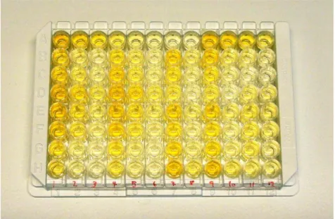
Figure 3. Working principle for sandwich-ELISA HK210. Wells of a 96-well microtiter plate are coated with capture antibody with specificity for mouse MPO. Homogenized samples are added to the wells where it binds to the capture antibody. A biotinylated tracer antibody is then added which binds to a different epitope on the MPO molecule. In the next step the enzyme streptavidin-peroxidase binds to biotin on the tracer antibody. Finally, tetramethylbenzidine (TMB) is added which acts as a substrate for streptavidin-peroxidase. TMB forms a colored product when allowed to react with streptavidin-peroxidase, the strength of the color change depending on how much MPO the original sample contains. The reaction is ended with stop solution being added and the absorption for each well can then be measured in a spectrophotometer at 450 nm. Adapted from product information & manual for ELISA-kit HK210, Hycult Biotech.
Figure 2. 96-well ELISA microtiter plate after addition of stop solution. A colorization corresponding to MPO level can be seen after finishing of the ELISA protocol. Exact absorbance measurements are performed at 450 nm in a spectrophotometer.
10
To be able to interpret the data obtained from an absorbance measurement, reference absorbance measurements for known concentrations are needed. This was obtained by using a series of standards. The standard solution is provided with the kit and contains a known concentration of MPO that is diluted in a serial manner so that the absorbance data can be plotted to generate a standard curve. This curve was used to convert absorbance measurements for the samples into concentration values. In the HK210-kit, the standard curve ranges from 0 to 250 ng/ml. Sample concentrations outside this range are not usable since they will not be on the standard curve. For this reason a suitable dilution of each sample is required to keep them in the 0 to 250 ng/ml range. All dilutions were made with dilution buffer provided with the HK210 kit. Since this analysis has not been carried out previously in our laboratory the dilution factors for the samples were not immediately obvious, and a trial-and-error approach was necessary. A rough estimate of a dilution factor of 1:200 for DSS-treated mice was obtained from Johanna Henriksnäs at VISIONAR Preclinical AB, Uppsala, Sweden. With this as a starting point, dilution factors for samples from each group were estimated with the DAI score from each mouse used as a guideline. Samples from the 15d R2LC + DSS-group and 15d 4659 + DSS-group were assayed in 1:5, 1:25, and 1:100 dilution while samples from the 8d DSS, 2d R2LC + DSS, and 15d saline + DSS-groups were assayed in 1:20 and 1:200 dilutions. Samples from the control group were only diluted 1:5. All samples including standards were analyzed on the ELISA plate in duplicates in accordance to kit instructions to generate a mean absorbance value for each sample. The dilution of samples and duplicates of each dilution results in each sample occupying between two to six wells on the 96-well ELISA plate, limiting the number of samples that can be analyzed. For this project, a total of 19 samples were used; two 8d DSS from the earlier mice, two 8d DSS from the new mice (one re-frozen, one directly homogenized), one from 2d R2LC + DSS, two from 15d saline + DSS, three controls from the new mice (one re-frozen, two directly homogenized), two extra controls taken from untreated mice used in another study at our laboratory (both re-frozen), three 15d R2LC + DSS, and four 15d 4659 + DSS. These samples were chosen to best represent each group with respect to the mean DAI.
11
Results
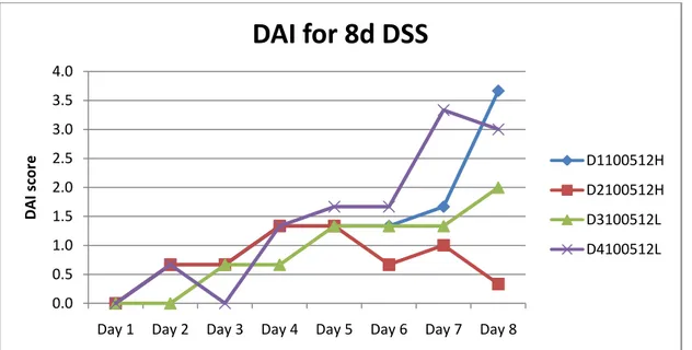
DAI in the new mice group
The new mice were divided into two groups; 8d DSS and untreated control. DAI scores were calculated daily by assessment of clinical symptoms (weight loss, stool consistency, and fecal blood content) for the 8d DSS-group.
Figure 4. Disease activity index in the four animals from the new group with colitis induced by 3% DSS.
Three of the 8d DSS-mice showed clinical symptoms of colitis on day 8 while one mouse showed very few clinical symptoms for reasons unknown to us (Fig. 4). The two mice with the highest DAI score were used for MPO analysis with ELISA. Colon tissue from D1100521H was snap-frozen in liquid nitrogen before being homogenized while tissue from D4100521L was homogenized immediately after being removed from the mouse and then frozen. DAI for the untreated group was considered to be zero since earlier studies have never shown any clinical symptoms in untreated animals. From this group, colon tissue from one mouse was snap-frozen in liquid nitrogen before being homogenized while tissue from the two other mice was immediately homogenized.
ELISA measurements of MPO
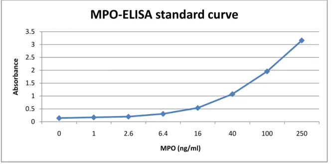
Most of the sample dilutions turned out to be within the range of the standard curve. The standard curve is almost linear in the 0 to 40 ng/ml range (Fig. 5). Since all of our samples had one or more dilution with a concentration within this range, the two upper absorbance values of the standard curve was omitted in order to get a more accurate trend line to calculate sample concentration values from (Fig. 6). The highest dilution for the 15d R2LC + DSS and 15d 4659 + DSS was 1:100 and 1:200 for the 8d DSS, 2d R2LC + DSS, and 15d saline +
0.0 0.5 1.0 1.5 2.0 2.5 3.0 3.5 4.0
Day 1 Day 2 Day 3 Day 4 Day 5 Day 6 Day 7 Day 8
D A I sc o re
DAI for 8d DSS
D1100512H D2100512H D3100512L D4100512L12
DSS. In these dilutions, all of the samples were within the 0 to 40 ng/ml range and for this reason we decided to use them for the calculations of concentration. There was one sample in the 8d DSS group and one in the 15d 4659 + DSS group that was unrealisticly low in the highest dilutions. Therefore, samples from the 1:20 and 1:25 dilutions respectivly were used in these cases.
Figure 5. Original standard curve.
Figure 6. Standard curve with the two highest absorbance values omitted for better trend line fit.
0 0.5 1 1.5 2 2.5 3 3.5 0 1 2.6 6.4 16 40 100 250 A b sor b an ce MPO (ng/ml)
MPO-ELISA standard curve
y = 0.0234x + 0.1456 R² = 0.9992 0 0.2 0.4 0.6 0.8 1 1.2 0 5 10 15 20 25 30 35 40 45 A b sor b an ce MPO (ng/ml)
13
Sample concentration values were calculated with Magellan TM – Data Analysis Software (Tecan Trading AG, Switzerland) using the modified standard curve described above (Table 2).
Table 2. Sample concentrations calculated from standard curve. DAI scores obtained from the earlier mice (15d R2LC + DSS, 15d 4659 + DSS, 8d DSS, 2d R2LC + DSS, 15d saline + DSS) and new data (8d DSS, Ctrl). “H” and “L” at the end of 8d DSS and Ctrl sample names depict “directly snap-frozen” and “directly homogenized”, respectively. “H”-samples have gone through a second freeze-thaw cycle.
Sample Group DAI MPO (ng/ml) 100220b 15d R2LC + DSS 1.3 210 100220d 15d R2LC + DSS 0.7 479 100220f 15d R2LC + DSS 0 1727 100222b 15d 4659 + DSS 1.3 955 100222c 15d 4659 + DSS 1.3 483 100222d 15d 4659 + DSS 1.3 118 100222e 15d 4659 + DSS 2 123 D1100512H 8d DSS 3.7 2192 D4100512L 8d DSS 3 768 100218A 8d DSS 2.7 408 100218C 8d DSS 3.7 2033 100218D 2d R2LC + DSS 4 680 100222G 15d saline + DSS 3.7 1243 100222H 15d saline + DSS 3.7 2929 K1100423H Ctrl 0 49 K2100512H Ctrl 0 85 K3100512L Ctrl 0 72 K4100512L Ctrl 0 58 R1100427H Ctrl 0 40
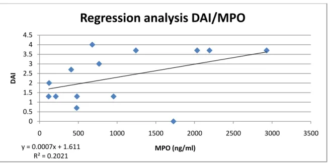
Regression analysis
In order to visualize the relationship between DAI score and MPO levels, a regression analysis was made (Fig. 7).
Figure 7. Regression analysis showing the relationship between DAI score and MPO levels.
y = 0.0007x + 1.611 R² = 0.2021 0 0.5 1 1.5 2 2.5 3 3.5 4 4.5 0 500 1000 1500 2000 2500 3000 3500 DAI MPO (ng/ml)
14
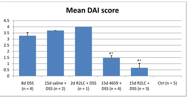
Mean group values
Because of the low correlation coefficient, mean group values for DAI (Table 3) (Fig. 8,10) and MPO (Table 4) (Fig. 9,10) were calculated.
Table 3. Mean group DAI values. DAI scores obtained from the earlier mice (15d R2LC + DSS, 15d 4659 + DSS, 8d DSS, 2d R2LC + DSS, 15d saline + DSS) and new data (8d DSS, Ctrl). n = no. of mice.
Group n Mean DAI SEM
8d DSS 4 3.3 0.3 15d saline + DSS 2 3.7 0.0 2d R2LC + DSS 1 4.0 15d 4659 + DSS 4 1.5 0.2 15d R2LC + DSS 3 0.7 0.4 Ctrl 5 0.0
Figure 8. Group DAI score. DAI scores obtained from the earlier mice (15d R2LC + DSS, 15d 4659 + DSS, 8d DSS, 2d R2LC + DSS, 15d saline + DSS) and new data (8d DSS, Ctrl). Values are means ± SE; n = no. of mice. *P < 0.01 relative to 15d saline + DSS; +P < 0.01 relative to 8d DSS. 0 0.5 1 1.5 2 2.5 3 3.5 4 4.5 8d DSS (n = 4) 15d saline + DSS (n = 2) 2d R2LC + DSS (n = 1) 15d 4659 + DSS (n = 4) 15d R2LC + DSS (n = 3) Ctrl (n = 5)
Mean DAI score
*+
15
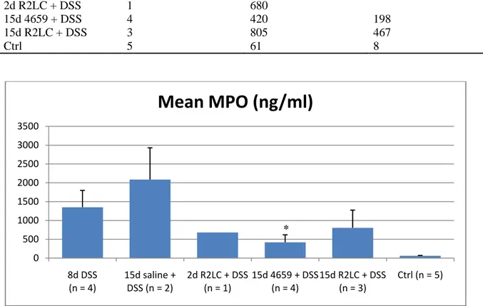
Table 2. Mean group MPO values. n = no. of mice.
Group n Mean MPO SEM
8d DSS 4 1350 447 15d saline + DSS 2 2086 843 2d R2LC + DSS 1 680 15d 4659 + DSS 4 420 198 15d R2LC + DSS 3 805 467 Ctrl 5 61 8
Figure 9. Group MPO values. Values are means ± SE; n = no. of mice. *P < 0.05 relative to 15d saline + DSS.
15d 4659 + DSS shows significance (P < 0.05) against 15d saline + DSS. Because of the similarities in treatment of the 8d DSS and 15d saline + DSS groups, MPO values from these groups were pooled and tested for significance against the other groups. Significance against this combined group was also shown only for 15d 4659 + DSS.
Figure 10. Group MPO values and group DAI scores. DAI scores obtained from the earlier mice (15d R2LC + DSS, 15d 4659 + DSS, 8d DSS, 2d R2LC + DSS, 15d saline + DSS) and new data (8d DSS, Ctrl).Values are means; n = no. of mice.
0 500 1000 1500 2000 2500 3000 3500 8d DSS (n = 4) 15d saline + DSS (n = 2) 2d R2LC + DSS (n = 1) 15d 4659 + DSS (n = 4) 15d R2LC + DSS (n = 3) Ctrl (n = 5)
Mean MPO (ng/ml)
0 0.5 1 1.5 2 2.5 3 3.5 4 4.5 0 500 1000 1500 2000 2500 8d DSS (n = 4) 15d saline + DSS (n = 2) 2d R2LC + DSS (n = 1) 15d 4659 + DSS (n = 4) 15d R2LC + DSS (n = 3) Ctrl (n = 5) DAI M P O (n g/m l)Mean MPO / mean DAI
Mean MPO Mean DAI *
16
Discussion
ELISA measurements of MPO
In this project, I have investigated if MPO measurements using ELISA technique is a suitable method for assessment of disease severity in a murine model of ulcerative colitis. DAI is a well established method of assessing disease severity, and one could therefore expect the severity of inflammation and thus MPO levels to correlate to DAI to a certain degree. The regression analysis of the MPO measurements shows a low correlation coefficient with DAI. This can largely be contributed to a few samples with very different DAI-MPO level relations in combination with a too small data set. One sample (100220f) from the 15d R2LC + DSS group and the sample from the 2d R2LC + DSS group (100218D) are mainly responsible for the low correlation coefficient. Removal of these from the data set would drastically increase R2. Hopefully, analysis of the remaining samples will increase data set size without adding too many near-outliers and thus increase the amount of variability of MPO that can be explained by DAI. In future studies, a measurement of protein concentration in sample solutions will be conducted. This will reveal any discrepancies in the homogenization process of the samples that might explain the large variation of MPO levels in some groups.
Because of the poor correlation between individual DAI score and MPO level, the mean DAI and MPO of each group were calculated. For DAI, the numbers show that the mean values for the groups treated with probiotics are significantly lower (P < 0.01) than both 8d DSS and 15d saline + DSS. This is true also for the mean MPO level of the 15d 4659 + DSS group which shows significance (P < 0.05) against 15d saline + DSS as well as against the
combined group of 8d DSS and 15d saline + DSS. Combining these groups is reasonable
considering that the only difference between them is a 15 day treatment with saline solution. These data indicate that the measure of MPO levels with ELISA in samples collected from mice with DSS-induced colitis can be used as a technique for assessing disease severity, but that a greater number of samples has to be used in order to get statistical evidence.
Effect of a repeated freeze-thaw cycle on MPO levels
The experimental protocol of this study results in samples from the earlier mice undergoing two freeze-thaw cycles. To investigate if this affects MPO levels, a new group of mice was used where the colon samples were treated with either one or two freeze-thaw cycles. The sample in the new 8d DSS group that underwent two freeze-thaw cycles shows a higher MPO level than the sample that only underwent one. It is considered unlikely that an additional freeze-thaw cycle would increase MPO levels, and therefore the difference of the 8d DSS samples is probably due to some other factor than freezing. One reasonable explaination could be that the higher MPO level is just reflecting the higher DAI of these samples compaired to their directly frozen counterparts. Another possibility is that the concentration of dissolved tissue in the sample solutions after homogenization is not equal for
17
all samples. As stated earlier, this will be investigated in future studies by measuring protein concentrations in the sample solutions. Neither in the control samples can any correlation be seen between MPO level and number of freeze-thaw cycles.
This indicates that two freeze-thaw cycles do not affect MPO levels more than one cycle does.
18
References
1. Crowther JR. Overview of ELISA in relation to other disciplines. Methods Mol Biol
516: 1-8, 2009.
2. Baynes JW and Dominiczak MH. In: Medical Biochemistry: Elsevier Mosby, 2005, p.
505.
3. Bradley P, Priebat D, Christensen R, and Rothstein G. Measurement of cutaneous
inflammation: estimation of neutrophil content with an enzyme marker. J Invest
Dermatol 78: 206-209, 1982.
4. De Jager P, Franchimont D, Waliszewska A, Bitton A, Cohen A, Langelier D, Belaiche J, Vermeire S, Farwell L, Goris A, Libioulle C, Jani N, Dassopoulos T, Bromfield G, Dubois B, Cho J, Brant S, Duerr R, Yang H, Rotter J, Silverberg M, Steinhart A, Daly M, Podolsky D, Louis E, Hafler D, Rioux J, Consortium QIG, and Consortium NIG. The role of the Toll receptor pathway in susceptibility to
inflammatory bowel diseases. Genes Immun 8: 387-397, 2007.
5. Engvall E and Perlmann P. Enzyme-linked immunosorbent assay (ELISA).
Quantitative assay of immunoglobulin G. Immunochemistry 8: 871-874, 1971. 6. Hendrickson B, Gokhale R, and Cho J. Clinical aspects and pathophysiology of
inflammatory bowel disease. Clin Microbiol Rev 15: 79-94, 2002.
7. Kaser A, Zeissig S, and Blumberg R. Inflammatory bowel disease. Annu Rev Immunol 28: 573-621, 2010.
8. Lequin R. Enzyme immunoassay (EIA)/enzyme-linked immunosorbent assay (ELISA). Clin Chem 51: 2415-2418, 2005.
9. Murthy S, Cooper H, Shim H, Shah R, Ibrahim S, and Sedergran D. Treatment of
dextran sulfate sodium-induced murine colitis by intracolonic cyclosporin. Dig Dis Sci 38: 1722-1734, 1993.
10. Okayasu I, Hatakeyama S, Yamada M, Ohkusa T, Inagaki Y, and Nakaya R. A
novel method in the induction of reliable experimental acute and chronic ulcerative colitis in mice. Gastroenterology 98: 694-702, 1990.
11. Sears C. A dynamic partnership: celebrating our gut flora. Anaerobe 11: 247-251,
2005.
12. Trush M, Egner P, and Kensler T. Myeloperoxidase as a biomarker of skin irritation
and inflammation. Food Chem Toxicol 32: 143-147, 1994.
13. Van Weemen B and Schuurs A. Immunoassay using antigen-enzyme conjugates. FEBS Lett 15: 232-236, 1971.