Linköping University Post Print
The effects of Dickkopf-1 antibody on
metaphyseal bone and implant fixation under
different loading conditions
Fredrik Agholme, Hanna Isaksson, Stuart Kuhstoss and Per Aspenberg
N.B.: When citing this work, cite the original article.
Original Publication:
Fredrik Agholme, Hanna Isaksson, Stuart Kuhstoss and Per Aspenberg, The effects of Dickkopf-1 antibody on metaphyseal bone and implant fixation under different loading conditions, 2011, BONE, (48), 5, 988-996.
http://dx.doi.org/10.1016/j.bone.2011.02.008
Copyright: Elsevier Science B.V., Amsterdam.
http://www.elsevier.com/
Postprint available at: Linköping University Electronic Press
The effects of Dickkopf-1 antibody on
metaphyseal bone and implant fixation
under different loading conditions
Fredrik Agholme
1*, Hanna Isaksson
2, Stuart Kuhstoss
3, Per Aspenberg
11
Orthopaedics, Department of Clinical and Experimental Medicine, Faculty of
Medicine, Linköping University, SE-58183, Linköping, Sweden
2
Department of Applied Physics, University of Eastern Finland, FI-70211,
Kuopio, Finland.
3
Lilly Research Laboratories, Lilly Corporate Center, 46285 Indianapolis, IN,
USA
* Corresponding Author
Fredrik Agholme
Experimental Orthopaedics/KEF Linköping University Medical Faculty SE-581 85 Linköping Sweden
Phone #: +4613-224116 Email: fredrik.agholme@liu.se
Authors’ emails:
Fredrik Agholme: fredrik.agholme@liu.se Hanna Isaksson: hanna.isaksson@uef.fi Stuart Kuhstoss: kuhstoss_stuart@lilly.com Per Aspenberg: per.aspenberg@liu.se
Abstract
The secreted protein Dickkopf-1 (Dkk1) is an antagonist of canonical Wnt signaling,
expressed during fracture healing. It is unknown if it is involved in the mechanical control of bone maintenance. We investigated the response to administration of a Dkk1 neutralizing antibody (Dkk1-ab) in metaphyseal bone under different loading conditions, with or without trauma. In this three part experiment, 120 rats had a screw or bone chamber inserted either unilaterally or bilaterally in the proximal tibia. Mechanical (pull-out) testing, µCT and histology were used for evaluation. The animals were injected with either 10 mg/kg Dkk1-ab or saline every 14 days for 14, 28, or 42 days. Antibody treatment increased bone formation around the screws and improved their fixation. After 28 days, the pull-out force was increased by over 100%. In cancellous bone, the bone volume fraction was increased by 50%. In some animals, one hind limb was paralyzed with Botullinum toxin A (Botox) to create a
mechanically unloaded environment. This did not increase the response to antibody treatment with regard to screw fixation, but in cancellous bone, the bone volume fraction increased by 233 %. Thus, the response in unloaded, untraumatized bone was proportionally larger, suggesting that Dkk1 may be up-regulated in unloaded bone. There was also an increase in thickness of the metaphyseal cortex. In bone chambers, the antibody treatment increased the bone volume fraction. The results suggest that antibodies blocking Dkk1 might be used to stimulate bone formation especially during implant fixation, fracture repair, or bone disuse. It also seems that Dkk1 is up-regulated both after metaphyseal trauma and after unloading, and that Dkk1 is involved in mechano-transduction.
Introduction
Most fractures occur in osteoporotic cancellous bone in metaphyseal regions, such as the hip, spine or forearm. Screws, pins and plates may be inserted to stabilize these fractures. In cancellous bone, the response to the trauma of inserting a screw is similar to metaphyseal fracture repair: both are examples of trauma-induced membranous bone formation. This new bone formation is important for the strength of screw fixation, especially when the initial fixation is weak. Consequently, the regeneration of bone after trauma can be estimated by measuring the mechanical fixation of screws and the formation of new bone around them. The early fixation of total joint replacements also depends on a fracture-type response in
metaphyseal bone. Increased bone formation at early stages in the healing or incorporation process might provide a better long-term prognosis due to the correlation between improved early fixation and reduced risk of late loosening [1].
Dickkopf-1 (Dkk1) is a secreted glycoprotein. It is a potent Wnt antagonist [2], vital for head and limb development [2-3]. Dkk1 binds to low-density lipoprotein related proteins 4, 5 and 6 (LRP4/5/6) [4-7]. The exact mode of action is unclear, but it appears that Dkk1 directly competes with Wnt-ligand in binding to LRP6 [8] thus antagonizing Wnt signalling [9] and increasing β-catenin degradation. This β-catenin accumulation favors mesenchymal stem cell commitment for an osteogenic fate [10]. By modulating Wnt signaling, it is possible to achieve an anabolic effect in bone. Skeletal trauma causes a local increase in Wnt signaling [11] and it has been shown that the pathways involved in bone Wnt signaling are necessary for bone healing [11-12] and for bone formation in general [13-15]. Fracture repair can be influenced by blocking Dkk1 in mice [16] and a decrease in Dkk1 gene expression leads to an increase in bone mass and strength [15, 17].
Sclerostin, another bone specific antagonist to Wnt signaling, has been shown to be an important mediator of the response and adaptation of bone to mechanical loading [18]. It has also been shown that Dkk1 expression is influenced by mechanical load [19], although not to the same degree as sclerostin. To further investigate this, we compared the response to
antibodies blocking Dkk1 in traumatized and untraumatized bone, under both loaded and unloaded conditions.
This study tested three hypotheses. The first was that systemic administration of a Dkk1 neutralizing antibody increases the healing response to trauma, thereby improving the fixation of an implanted screw. The second hypothesis was that the same antibody promotes bone generation in general, leading to increased density in untraumatized bone and increased bone formation in a titanium chamber. The third was that these responses are dependent on
Materials and Methods
Experimental overview
The study consists of three sets of in vivo experiments. All studies involve insertion of an implant into the proximal tibia of one or both legs. A total of 120 male, 10 week old Sprague Dawley rats (Taconic, Lille Skensved, Denmark) with a mean weight of 360 ± 30 grams were used.
In the first experiment (screw fixation), we studied the response to metaphyseal trauma by inserting a stainless steel screw in the right proximal tibia of 40 rats. The fixation was
evaluated by mechanical pull-out testing after either 14 or 28 days (N = 4 x 10). In the second experiment, we studied the capacity for bone regeneration by inserting a titanium chamber (bone conduction chamber, BCC) at the same location in 40 rats. The bone regeneration was evaluated using histology after 28 days or µCT after 42 days (N = 4 x 10). In the third experiment, we studied the role of mechanical loading, by injecting Botullinum toxin A (Botox, Allergan, Irvine, CA, USA) to paralyze the muscles in one of the hind limbs in 40 animals. Screws were inserted bilaterally in the proximal tibiae for 28 days. These were either of steel, for mechanical testing (N = 2 x 12), or of PMMA for morphometry measurements by µCT (N = 2 x 8).
After surgery, all rats were randomly assigned to either antibody or saline injections. Animals were euthanized using carbon dioxide after 14, 28 or 42 days and both tibias were harvested. From the animals with a titanium chamber, evaluated with µCT, the L5 vertebra was also collected.
The rats were given free access to food and water during the experiment, and were housed three per cage at 21◦C in a room with 12 hours light and 12 hours dark cycle. The study was approved by the Regional Ethics Committee for Animal Experiments and institutional guidelines for care and treatment of laboratory animals were followed.
Implants
Stainless steel screws were used for pull-out testing. The screw heads were designed to enable mounting in a materials testing machine. The threaded part of the screw was 2.8 mm long and 1.6 mm in diameter. To avoid metal artifacts during µCT measurements, screws of the same size were made out of polymethylmethacrylate (PMMA). Bone formation adjacent to PMMA in similar models appears histologically similar to that seen around stainless steel [20]. We have previously used PMMA screws to study bone formation in the proximity of an implant [21]. To study new bone formation, we used the bone chamber [22]. It consists of a titanium screw with a cylindricalinterior space 2 mm wide and 7.5 mm long. The chamber is empty at insertion, and its bone content after a certain time will reflect the capability for new bone formation by membranous ossification.
Dkk1-Antibody
A chimeric mouse/rat Dkk1 neutralizing antibody (Dkk1-ab) was provided by Lilly (Lilly Research Laboratories, Indianapolis, USA). This antibody consists of mouse variable domains fused to rat IgG1 kappa constant domains. Antibody solution, 10 mg/kg, or corresponding volume of saline was given by subcutaneous injection every 14 days, starting the day after surgery. The operators and evaluators were blinded for treatment during the course of the experiment.
Botox injection
Animals allocated for unloading were anesthetized with isoflurane and given Botox
intramuscularly using an insulin-syringe in the calf (Three injections of 1 U) and quadriceps femoris muscles (Two injections of 1 U). The hind limb for treatment (left or right) was randomly chosen. Two days after the injection, all animals presented an obvious limp and did not bear weight on the injected limb. This condition persisted for the duration of the
experiment. The animals underwent implantation surgery on the third day after the Botox injection.
Surgical procedure
The surgical procedures were identical to those explained in earlier work [21-22]. The rats were anesthetized with isoflurane and operated on under aseptic conditions. Briefly, a 1.4 mm insertion hole was hand drilled in the cancellous bone, approximately 3 mm distal to the tibial physis. A steel or PMMA screw was inserted in the hole and gently screwed in place. Animals that received a bone chamber were subjected to the same procedure, but a 3.2 mmdiameter drill was used to enlarge the hole created inthe medial cortex. The chamber was then screwed into the holeuntil its cap rested on the periosteal bone surface.In case of bilateral implants the same procedure was repeated on the other limb. The animals were fully weight bearing
immediately after awakening from anaesthesia. Rats received 0.007 mg of buprenorphine as post operative analgesic every 12 hours for 48 hours.
Mechanical evaluation
All analyses were performed by investigators who were blinded for antibody treatment. Harvested bones were kept moist by saline irrigation and all bones were tested within one hour after harvesting. Steel screws were tested for pull-out strength in a computerized
materials testing machine (100 R; DDL Inc. Eden Prairie, MN, USA), at a cross-head speed of 0.1 mm/s. These type of measurements have been described in detail earlier [23]. The
machine recorded the peak force and the energy uptake until the force had dropped to 90% of maximum. It also calculated the stiffness from the slope of the force/distance curve at a point chosen manually by the investigator. The peak pull-out force was considered the primary variable.
To determine if any observed effects were due to a change in the healing response, or due to a general effect on the skeleton, the fixation of incorporated screws was compared with screws inserted at the end of the experiment. A screw was inserted post mortem in the untraumatized contra-lateral tibia, similarly as during the surgical procedure. This screw was tested for pull-out strength immediately. The same screw was used for all samples. The screw was cleaned between each animal.
Histomorphometry
Histomorphometry was conducted on the bone chamber contents 28 days after insertion, all analyses were performed by investigators who were blinded for treatment. After the tibia was harvested, the tissue contents were removed from inside the chamber, decalcified, and
preparedfor histology. Sections parallel to the longaxis of the chamber were stained with hematoxylin and eosin.
The evaluation was done bymanual point counting within an area of interest from the bottom of the chamber, i.e. the ingrowth end, towards the frontier of the advancingnew bone
formation. Only the central third ofthe specimen was evaluated, i.e. bone close to the titanium walls was excluded. We used an overlay grid to count points covering new living bone and
total points. On average 160 points were counted per specimen. The bone ingrowth distance was measured by dividing the area covered by newbone with the width of the sample. These measurements were conducted using computer software (Cell-D, Olympus, Germany) on one image of the best section from each specimen [22]. All specimens were reassessed after one month to determine the standarderror of the measurement, which was 4% for bone volume fraction.
MicroCT
For the second bone chamber experiment, 42 days after insertion, the chamber contents, the proximal tibia (untraumatized side) and the L5 vertebra were scanned. For the Botox
experiments, both tibias and the bone surrounding the PMMA screw were scanned. The µCT scanner acquired topographic images of the bone with an isotropic voxel size of 15µm
(Skyscan 1172, v. 1.5, Skyscan, Aarteselar Belgium) at the energy settings of 100 kV and 100 µA, using aluminum filter of 0.5 mm, and 10 repeated scans. The images were reconstructed using NRecon (Skyscan, v 1.5.1.4, Aarteselar Belgium), by correcting for ring artifacts and beam hardening. Calibration of bone mineral density (BMD) was carried out according to the system manufacturer’s protocol, by scanning of a water phantom and two hydroxyapatite phantoms of known density (0.25 and 0.75 g/cm3). Mineralized bone tissue was assumed to have a BMD over 0.590 g/cm3, resulting in grayscale values of 70-255. Figure 1 describes the regions of interest for the various analyses. Analysis of bone volume fraction (BV/TV, when measured by µCT), and trabecular thickness, separation and number (Tb.Th, Tb.Sp, Tb.N) were performed in CTAn (v.1.9.1.0 Skyscan, Aarteselar Belgium). BV/TV was considered the primary variable. All analyses were performed in a blinded manner.
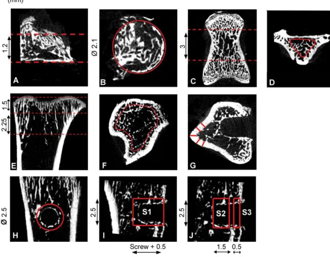
Figure 1: Regions of interest (ROI) for µCT analyses. The contents of the bone chamber , L5
vertebrae and the untraumatized proximal tibia were analyzed (A-F). In the bone chamber, a cylindrical region of interest with a diameter of 2.1 mm was defined with a 1.2 mm depth
(A-B). In the vertebra, the central cross-section was located, and the trabecular structural
parameters were analyzed for 3 mm (C-D). For the proximal tibia, a 2.25 mm section (starting 1.5 mm from the proximal end) was analyzed (E). Only trabecular bone within the region was analyzed (F). In traumatized tibia trabecular bone, not in the vicinity of the screw, was
analyzed similarly as described above but for1.5 mm instead of 2.25 mm (E). Cortical
thickness was measured in the traumatized tibia at five different locations (G) at the centre of the cylindrical ROI defined in (H). In the mechanical unloading experiment, the regions surrounding the screw were analyzed (H-J). A 2.5 mm cylindrical ROI coaxially with the screw was used for analysis (H). The largest ROI comprised both the entire intraosseous and cortical portions extending from the most superficial circular section at the periosteal surface down to 0.5 mm beyond the deep end of the screw (I, S1). The marrow portion of the cylinder was defined as the part of the cylinder extending from the deep end of the screw 1.5 mm toward the periosteum (J, S2).The cortical portion was defined as extending from the most superficial circular section 0.5 mm toward the marrow (J, S3). The portion of the cylinder made up by the screw was subtracted from all measurements.
Statistical analysis
Results are presented as mean and standard deviation (SD). Most results were analyzed using Student’s t-test, after checking that data appeared normally distributed and variances were similar. In order to describe the size of the treatment effect, we calculated the confidence intervals for differences between group means. The confidence limits were then expressed as a percentage of the mean of the corresponding control group. Pull-out forces in rats without Botox were tested using 2-way Anova, with time and treatment as fixed factors. When comparing loaded and unloaded sides in the same animal, paired t-test was used. Because each experiment has a pre-determined primary out-come variable, p-values for other, secondary variables were not corrected for multiple testing. A result was considered statistically significant if p< 0.05, and all tests were conducted in SPSS statistical software (SPSS v 18.0, SPSS Inc. Chicago Il).
Results
Screw fixation
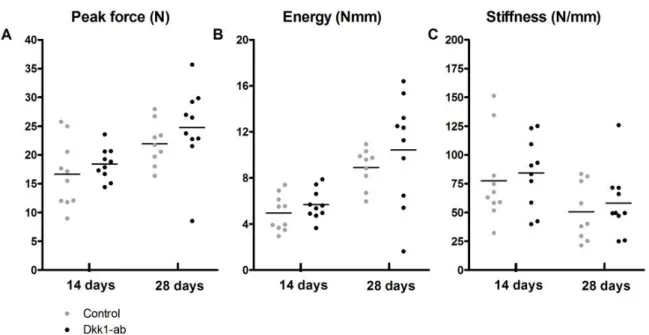
Screws were inserted to study how Dkk1-ab improved their fixation, as a measure of an improved metaphyseal healing response. Compared to controls, Dkk1-ab treatment increased the pull-out force by almost half after 14 days, and doubled it after 28 days (Figure 2A). Energy and stiffness were increased to a similar extent (Figure 2B, C). However, by two-way Anova, there was no significant increase in pull-out force over time. Screws inserted into the proximal tibia post-mortem showed no increase in pull-out force due to antibody treatment (Figure 3). One measurement was excluded because the tibia fractured during screw insertion.
Figure 2: Effect of Dkk1-ab treatment on screw fixation in traumatized bone. Pull-out testing
14 and 28 days after screw insertion. (A) Peak force was increased by 40% with the antibody compared to controls at 14 days (a, p= 0.043) and 100% at 28 days (b, p < 0.001). (B) Pull-out energy was increased by 80% (c, p = 0.005) and 160% (d, p < 0.001). (C) Stiffness was increased by 120% at 28 days (e, p=0.005). Control samples had a significantly lower peak force after 28 days compared to 14 day controls (p= 0.023). Student’s t-test without
Figure 3: Effect of Dkk1-ab treatment on screws inserted into the untraumatized
contra-lateral tibia. These screws had less than half the pull-out force of screws that had been
allowed to osseointegrate for 14 or 28 days. Compared to controls there no significant effects on peak force (A), pull-out energy (B) or stiffness (C
Student’s t-test without multiplicity correction.
Neo-formation of cancellous bone
Bone chambers were used to measure the regenerative capacity. In the experiment with histological evaluation at 28 days, Dkk1-ab treatment increased the bone volume fraction in the chamber by almost 70% (Figure 4A). By histology, ingrowth distance could not be shown to be significantly increased (Figure 4B). In both controls and Dkk1-ab treated animals, the chambers contained an undifferentiated callus-like tissue at the farthest distance from the ingrowth openings, followed by a layer of newly formed bone surrounding a marrow cavity with bone trabeculae, which made up most of the volume. The appearance was similar to controls in several previous studies [22, 24] and indicated that a frontier of membranous ossification followed behind the ingrowing fibrous callus. Behind the ossification frontier, bone had been resorbed to make space for bone marrow. Cartilage was never observed, although it was not specifically stained for. Five samples were excluded: three because the
content of the chamber was too small to be properly evaluated, and two due to technical errors during histological preparation.
Figure 4: Effect of Dkk1-ab treatment on intramembranous bone formation. Bone volume
fraction and bone tissue ingrowth distance inside the bone conduction chamber, as determined by histomorphometry after 28 days. (A) Dkk1-ab treatment caused a 70% increase in bone volume (a, p= 0.02) while not significantly affecting ingrowth distance (B).
Student’s t-test without multiplicity correction.
Based on µCT evaluation of the bone chamber experiment after 42 days, Dkk1-ab treatment increased the BV/TV in the chamber and the untraumatized proximal tibia to the same degree, whereas there was little effect on the vertebrae (Figure 5A). In the tibia, antibody treatment induced a small but statistically significant increase in trabecular thickness and a larger increase in trabecular number (Figure 5B-D). There was also a significant increase in bone ingrowth distance. (Figure 5E).
Figure 5: Effect of Dkk1-ab treatment on cancellous bone. Microstructural information from
bone chamber contents, tibiae and vertebrae after 42 days of treatment as determined by µCT. (A) Dkk1-ab increased bone volume fraction (BV/TV) by 40% in both in the untraumatized proximal tibia (a, t-test, p< 0.001) and inside the chamber (b, log-transform t-test, p = 0.03), but had no significant effect on cancellous bone in the vertebrae. In the trabecular bone in the tibia, Dkk1-ab treatment caused an 8 % increase in trabecular thickness (Tb.Th) (B; c, t-test, p = 0.004), a 25% decrease in trabecular separation (Tb.Sp) (C; d, log-transform t-test, p = 0.04), and a 35% increase in trabecular number (Tb.N) (D; e, log-transform t-test, p = 0.003). There were no significant changes in the vertebrae. In the bone chamber, the antibody
increased the bone ingrowth distance, as measured using µCT (E, f, log-transform t-test, p = 0.03).
Response to mechanical unloading
Screw region
Botox injections were used to measure the effects of load protection on the response to screw insertion (screw region), and on untraumatized bone (cancellous bone). Botox caused a clearly visible muscle atrophy, and the weight of the soleus and quadriceps muscles in the affected limb was reduced by half (data not shown). Dkk1-ab treatment had no effect on muscle loss. In mechanical tests, Botox treatment cause the peak force to drop by half in controls (Paired t-test, p = 0.002, Figure 6A). Similarly to the first experiment, Dkk1-ab increased the pull-out force in animals with normal loading, but there was no significant response to the Dkk1-ab in the Botox treated animals (Figure 6). The log-transformed ratio between the pull-out forces of the unloaded and loaded sides in the same animal was similar in Dkk1-ab and control groups. Two screws were excluded, one due to a technical error during mechanical testing and one because a tibia fractured during dissection.
Figure 6: Effect of Dkk1-ab treatment on screw fixation in loaded or unloaded traumatized
bone. Pull-out testing 28 days after screw insertion. Botox treatment clearly decreased the fixation of inserted screws. In the unloaded bone, Dkk1-ab treatment did not significantly increase either peak force (A) pull-out energy (B) or stiffness. In the loaded bone a 70% increase in peak force (a, p = 0.0013) was detected, as well as a 90% increase in pull-out energy (b, p < 0.001). Student’s t-test after log-transformation without multiplicity correction.
Based on µCT measurements (Figure 7), Dkk1-ab treatment increased the BV/TV around the screws (Figure 8A), without a clear difference between the cortical or cancellous region of interest. Statistically significant effects of the Dkk1-ab were found on BV/TV in the cortical region in both loaded and unloaded limbs and for the entire screw region in unloaded limbs (Figure 8B-C). However, the effect of the antibodies was not influenced by unloading. One screw was excluded since it had lost fixation and fallen out.
Figure 7: µCT results for mechanical unloading experiment. The median sample for each of
the four groups was chosen to visualize the findings. Scale bar 1 mm. The right two columns show the samples that were injected with Botox, and the first and the third column shows the samples that were treated with Dkk1-ab. A cross-section through the screw is visualized (a), and a 3D rendering (b) of the ROI surrounding the screw (Figure 1 H). The Dkk1-ab treated groups showed thicker cortex and higher BV/TV compared to controls, whereas the Botox treated groups showed thinner cortex, and lower BV/TV in the deep parts of the screws. (c) A cross-section through the analyzed trabecular bone is visualized, and a 3D rendering of the full trabecular ROI (d).
Figure 8:Effect of Dkk1-ab treatment on bone formation around implanted screws in either loaded or unloaded (Botox treated) bone. Bone volume fraction (BV/TV) after 28 days of treatment, assessed by µCT in the three regions of interest described in figure 1 (H-J). (A) Compared to controls, Dkk1-ab increased BV/TV immediately adjacent to the screw by 30% (a, p = 0.04) in the unloaded bone. No significant effects were detected in the loaded bone. In the cortical (B) and marrow (C) regions, the largest increase in BV/TV occurred in the cortical region, in which Dkk1-ab treatment increased BV/TV in both unloaded (b, p = 0.01) and loaded (c, p = 0.04) bone. The effect in the marrow region was not significant. Student’s t-test without multiplicity correction.
Cancellous bone
In the proximal tibia, Botox caused an obvious decrease in BV/TV (Figure 9A), trabecular thickness and trabecular number, and an increase in trabecular separation (Figure 9B-D). These effects were partly countered by Dkk1-ab treatment. Moreover, the ratio between unloaded and loaded sides regarding cancellous bone volume was increased by Dkk1-ab treatment (Table 1). In unloaded bone the BV/TV was increased by 233 % with Dkk1-ab treatment, mainly due to a large increase in trabecular number, whereas in loaded bone the increase was only 22 % (Figure 9A). BMD, however, was slightly decreased in both loaded and unloaded cancellous bone with Dkk1-ab treatment, probably because the newly formed bone was less mineralized (Figure 9E). For BV/TV, the Botox-treated and loaded sides in the same animal differed significantly between Dkk1-ab and control groups. This effect of
Figure 9: Effects of Dkk1-ab treatment on untraumatized cancellous bone. Microstructural information of tibia in loaded and unloaded (Botox treated) bone after 28 days of treatment as determined by µCT. (A) Botox lowered the bone volume fraction (BV/TV) to a third of the control value (paired t-test p < 0.001). Dkk1-ab treatment increased BV/TV in the unloaded bone by 230% (a, log-transformed t-test, p = 0.003) compared to controls, a much larger increase than the 40% (b, t-test, p = 0.01) detected in loaded bone. (B) Dkk1-ab treatment had no significant effect on trabecular thickness (Tb.Th), but Botox caused a decrease in thickness (paired test p < 0.001). (C) Dkk1-ab treatment decreased trabecular separation (Tb.Sp) in unloaded bone (c, t-test, p = 0.002), with no significant decrease in loaded bone. (D) Dkk1-ab caused a dramatic increase (230%) increase in trabecular number (Tb.N) unloaded bone (d, log-transformed t-test, p = 0.001). The increase in loaded bone was smaller (50%, e, t-test, p = 0.008). Botox decreased Tb.N by almost 75% (paired t-test p < 0.001). (E) Dkk1-ab caused a small (5%) but significant decrease in bone mineral density (BMD) in both loaded (f, t-test, p = 0.002) and unloaded (g, t-test, p = 0.007) bone. (F) Dkk1-ab treatment increased the thickness of the cortex both in the unloaded (h, t-test, p = 0.035) and loaded (i, t-test, p = 0.012) bone. Botox reduced the cortical thickness (paired t-test p = 0.01).
loading was not pronounced around the screws, where the loaded/unloaded ratio for BV/TV was not changed by Dkk1-ab treatment. Dkk1-ab had an effect on bone formation in the cortical surrounding of the screws (Figure 8) and in the other parts of the tibia, treatment increased cortical thickness both in loaded and unloaded bone to the same degree (Figure 9F).
Table 1: Ratio between BV/TV from µCT measurements on the loaded and unloaded sides. S1 and S2 refers to regions of interest shown in figure 1. P-values are based on log values, to avoid problems with different variation.
Region of
interest BV/TV Ratio Control Dkk1-ab P-value Tibia Loaded/Unloaded 5.4 (1.7) 2.3 (0.73) < 0.001 Entire screw surrounding (S1) Loaded/Unloaded 1.4 (0.36) 1.4 (0.55) 0.9 Cortical screw surrounding (S2) Loaded/Unloaded 1.2 (0.31) 1.4 (0.49) 0.3
Discussion
Our first hypothesis was that Dkk1 inhibition increases the response to trauma and improves screw fixation. We found that one single systemic dose of Dkk1 antibody was sufficient to improve the fixation of steel screws in cancellous bone. After 28 days and two doses, the pull-out force was almost doubled compared to controls. We also found an increase in bone volume fraction in the vicinity of the screws. The increase in pull-out force was similar to previous observations in this model with bisphosphonates [25], parathyroid hormone (PTH) [26] and sclerostin antibodies [21].
The pull-out testing of the untraumatized bone (where the screws were inserted after
euthanasia) exclude that the antibodies had a dramatic effect on the untraumatized bone. Thus, it appears that we had a specific response in traumatized bone. This could bedue to either that Dkk1 protein is more expressed during trauma or that it plays a different role in traumatized bone, influencing the availability of suitable progenitors. This would reflect on the differences in biology between traumatized and untraumatized bone. However, this finding is contrary to our previous results with a sclerostin-inhibiting antibody in a similar model [21]
Our second hypothesis was that Dkk1-ab would increase bone formation in untraumatized bone, both in the chamber and in the contra-lateral leg. This hypothesis was confirmed by histological and µCT data, showing an increased bone content and bone volume fraction in the chamber and in the trabecular bone of the tibia. The increase in BV/TV appeared to be caused by an increased number of trabeculae, and there was no corresponding increase in trabecular thickness. This is strikingly different from our previous experiment with sclerostin antibodies, where there was a dramatic increase in trabecular thickness [21]. Moreover, with
the sclerostin antibody there was an increase in proximal tibial BMD, whereas there was a slight decrease in this study. This finding points at different roles for sclerostin and Dickkopf signalling in cancellous bone. An explanation for this may be that Dkk1 and Sclerostin have different binding sites on LRP5 and 6 [27-28].
Dkk1 inhibition increased both bone volume fraction and bone ingrowth distance into the bone chamber by µCT data. With histology, we saw an increase in ingrowth distance but this was not statistically significant. This may be due to lack of power, or the difference in follow-up time. In previous studies using the chamber, PTH increased bone density but not ingrowth distance [24], whereas BMPs increased the ingrowth distance but not density [29]. This might reflect that PTH stimulates relatively differentiated cells, whereas BMPs stimulate stem cells and progenitors. The increased ingrowth into the chamber in the present experiment suggests that Dkk1 inhibition could have a positive effect on fracture callus growth.
Our third hypothesis was that the response to Dkk1 inhibition would depend on mechanical loading. This was apparently the case. For BV/TV in the cancellous tibial bone far away from the screw, there was a significant effect of the Dkk1 antibody on the ratio between the loaded and unloaded sides in the same animal. Without antibody, BV/TV was lowered by Botox treatment to about a fifth of the loaded bone. With antibody treatment, BV/TV was only lowered by half of the loaded bone. This suggests that antibody treatment can reduce bone loss due to unloading in untraumatized bone. The relatively larger effect of the antibody on unloaded bone implies that Dkk1 production is either up-regulated or plays a different role during unloading and that this change plays an important role. In contrast, loading had less influence on the effects of the antibody for screw fixation. These results suggest that trauma-induced Dkk1 signalling prevails over loading-dependent Dkk1 signalling.
The administration of Wnt ligand [30] or an antibody which blocks sclerostin leads to an increase in bone mass and strength in rodents [21, 31-33] and monkeys [33-34]. Both bone formation in response to trauma and in animals with induced osteoporosis was increased. Likewise, the administration of lithium chloride, a GSK3β inhibitor, leads to improved bone healing [12]. Similar results as those reported for sclerostin antibodies have also been reported with blocking or down regulation of Dkk1 expression. Dkk1 antibodies enhance endochondral bone healing in LRP5 deficient mice [16]. Inhibition of Dkk1 can counter some of the effects of oestrogen deficiency in rats, restoring bone mineral content and density [35]. Blocking of other Wnt-inhibitors could also influence bone formation: mice deficient in secreted frizzled related protein 1 (sFRP1) have higher bone mass and faster healing of diaphyseal fractures [36].
Several limitations of this study need to be addressed. We did not look at long term effects of antibody treatment. We studied effects of unloading, rather than effects of increased loading. Furthermore, we did not do any measurements to shed light on how Dkk1 is involved in the bone response to shifting mechanical load. Thus, we cannot offer any mechanistic explanation to the findings, although it is likely that the observed effects were due to changes in Wnt-signalling [19]. Further studies using histology and gene expression may contribute more data towards an understanding of the mechanisms involved.
There are several ways to unload bone in rodents, such as tail suspension, casting, and nerve severing. We chose to use Botox to paralyze the hind limb muscles. Being minimally
invasive, this method leads to significant bone loss [37-38]. Primarily, we were interested in the direct bone formation following trauma, since this is the primary healing response in
cancellous bone. Bone formation in the screw and chamber models occur under different premises. In the screw model, a trauma in cancellous bone leads to direct bone formation around the screw, influencing the degree of fixation. In the chamber model, the ingrowth distance can serve as a measurement of the regenerative potential of the bone. The bone volume fraction in the chamber is also influenced by resorption and the tendency to form a marrow cavity.
In previous experiments, we have repeatedly seen an increase in screw pull-out force over time in control animals [21, 39], whereas this time there was a slight decrease in spite of the fact the procedures were identical, and the surgeon the same (FA). This hampers comparison with previous experiments with the pull-out screws. The decrease in pull-out force could be due to the fact that we were forced to change rat breeder. For the same reason, the seemingly different effects of sclerostin and Dkk1 on trabecular thickness and number might possibly be due to differences between Sprague-Dawley rat strains from different breeders (Taconic Lille Skensved, Denmark and BK-universal, Stockholm, Sweden).
We have previously used PTH and bisphosphonates to increase bone formation and improve implant fixation in rodents [20, 25-26]. Improved implant fixation by bisphosphonates has also been shown in patients [40], and PTH appears to accelerate human fracture healing [41-42]. However, none of these drugs appears ideal. There are concerns regarding the long term effect of bisphosphonates on bone quality, and the effects of PTH on human fracture healing appear weak. Bone morphogenetic proteins (BMPs) are used to improve bone healing, but local side effects and the need for treatment to be given locally impairs their use. Modulation of Wnt signaling appears especially beneficial for intramembranous bone formation [30]. If
side effects can be avoided – which might be quite possible – Dkk1 antibodies could be used for orthopaedic applications to modulate the healing process.
In conclusion, antibodies inhibiting Dkk1 could improve the fixation of implants in rat cancellous bone and partly counter the effects of unloading in untraumatized bone. Apart from that this makes inhibition of Dkk1 signalling interesting for orthopaedic surgery, this information might also pertain to bone physiology. The results suggest that Dkk1 signalling in an important part of cancellous bone healing after trauma, and that Dkk1 is involved in
mechano-transduction.
Acknowledgement
Research was funded by Lilly and the Swedish research council (VR). Dkk1-ab was provided by Lilly Research Laboratories (Indianapolis, IN, USA). We thank Therese Andersson for assistance during animal surgery. We also thank Mats Christensson for the manufacturing of steel and PMMA screws used in this paper.
References
[1] Ryd L, Albrektsson BE, Carlsson L, Dansgard F, Herberts P, Lindstrand A, Regner L, Toksvig-Larsen S, Roentgen stereophotogrammetric analysis as a predictor of mechanical loosening of knee prostheses, J Bone Joint Surg Br. 77 (1995) 377-83.
[2] Glinka A, Wu W, Delius H, Monaghan AP, Blumenstock C, Niehrs C, Dickkopf-1 is a member of a new family of secreted proteins and functions in head induction, Nature. 391 (1998) 357-62.
[3] Mukhopadhyay M, Shtrom S, Rodriguez-Esteban C, Chen L, Tsukui T, Gomer L,
Dorward DW, Glinka A, Grinberg A, Huang SP, Niehrs C, Izpisua Belmonte JC, Westphal H, Dickkopf1 is required for embryonic head induction and limb morphogenesis in the mouse, Dev Cell. 1 (2001) 423-34.
[4] Bafico A, Liu G, Yaniv A, Gazit A, Aaronson SA, Novel mechanism of Wnt signalling inhibition mediated by Dickkopf-1 interaction with LRP6/Arrow, Nat Cell Biol. 3 (2001) 683-6.
[5] Balemans W, Devogelaer JP, Cleiren E, Piters E, Caussin E, Van Hul W, Novel LRP5 missense mutation in a patient with a high bone mass phenotype results in decreased DKK1-mediated inhibition of Wnt signaling, J Bone Miner Res. 22 (2007) 708-16.
[6] Choi HY, Dieckmann M, Herz J, Niemeier A, Lrp4, a novel receptor for Dickkopf 1 and sclerostin, is expressed by osteoblasts and regulates bone growth and turnover in vivo, PLoS One. 4 (2009) e7930.
[7] Mao B, Wu W, Li Y, Hoppe D, Stannek P, Glinka A, Niehrs C, LDL-receptor-related protein 6 is a receptor for Dickkopf proteins, Nature. 411 (2001) 321-5.
[8] Semenov MV, Zhang X, He X, DKK1 antagonizes Wnt signaling without promotion of LRP6 internalization and degradation, J Biol Chem. 283 (2008) 21427-32.
[9] Gong Y, Slee RB, Fukai N, Rawadi G, Roman-Roman S, Reginato AM, Wang H, Cundy T, Glorieux FH, Lev D, Zacharin M, Oexle K, Marcelino J, Suwairi W, Heeger S, Sabatakos G, Apte S, Adkins WN, Allgrove J, Arslan-Kirchner M, Batch JA, Beighton P, Black GC, Boles RG, Boon LM, Borrone C, Brunner HG, Carle GF, Dallapiccola B, De Paepe A, Floege B, Halfhide ML, Hall B, Hennekam RC, Hirose T, Jans A, Juppner H, Kim CA, Keppler-Noreuil K, Kohlschuetter A, LaCombe D, Lambert M, Lemyre E, Letteboer T, Peltonen L, Ramesar RS, Romanengo M, Somer H, Steichen-Gersdorf E, Steinmann B, Sullivan B, Superti-Furga A, Swoboda W, van den Boogaard MJ, Van Hul W, Vikkula M, Votruba M, Zabel B, Garcia T, Baron R, Olsen BR, Warman ML, LDL receptor-related protein 5 (LRP5) affects bone accrual and eye development, Cell. 107 (2001) 513-23.
[10] Case N, Rubin J, Beta-catenin--a supporting role in the skeleton, J Cell Biochem. 110 (2010) 545-53.
[11] Kim JB, Leucht P, Lam K, Luppen C, Ten Berge D, Nusse R, Helms JA, Bone regeneration is regulated by wnt signaling, J Bone Miner Res. 22 (2007) 1913-23.
[12] Chen Y, Whetstone HC, Lin AC, Nadesan P, Wei Q, Poon R, Alman BA, Beta-catenin signaling plays a disparate role in different phases of fracture repair: implications for therapy to improve bone healing, PLoS Med. 4 (2007) e249.
[13] Krishnan V, Bryant HU, Macdougald OA, Regulation of bone mass by Wnt signaling, J Clin Invest. 116 (2006) 1202-9.
[14] Li J, Sarosi I, Cattley RC, Pretorius J, Asuncion F, Grisanti M, Morony S, Adamu S, Geng Z, Qiu W, Kostenuik P, Lacey DL, Simonet WS, Bolon B, Qian X, Shalhoub V, Ominsky MS, Zhu Ke H, Li X, Richards WG, Dkk1-mediated inhibition of Wnt signaling in bone results in osteopenia, Bone. 39 (2006) 754-66.
[15] Morvan F, Boulukos K, Clement-Lacroix P, Roman Roman S, Suc-Royer I, Vayssiere B, Ammann P, Martin P, Pinho S, Pognonec P, Mollat P, Niehrs C, Baron R, Rawadi G,
Deletion of a single allele of the Dkk1 gene leads to an increase in bone formation and bone mass, J Bone Miner Res. 21 (2006) 934-45.
[16] Komatsu DE, Mary MN, Schroeder RJ, Robling AG, Turner CH, Warden SJ, Modulation of Wnt signaling influences fracture repair, J Orthop Res. 28 (2010) 928-36. [17] MacDonald BT, Joiner DM, Oyserman SM, Sharma P, Goldstein SA, He X, Hauschka PV, Bone mass is inversely proportional to Dkk1 levels in mice, Bone. 41 (2007) 331-9. [18] Lin C, Jiang X, Dai Z, Guo X, Weng T, Wang J, Li Y, Feng G, Gao X, He L, Sclerostin mediates bone response to mechanical unloading through antagonizing Wnt/beta-catenin signaling, J Bone Miner Res. 24 (2009) 1651-61.
[19] Robling AG, Niziolek PJ, Baldridge LA, Condon KW, Allen MR, Alam I, Mantila SM, Gluhak-Heinrich J, Bellido TM, Harris SE, Turner CH, Mechanical stimulation of bone in vivo reduces osteocyte expression of Sost/sclerostin, J Biol Chem. 283 (2008) 5866-75. [20] Skripitz R, Bohling S, Ruther W, Aspenberg P, Stimulation of implant fixation by parathyroid hormone (1-34)-A histomorphometric comparison of PMMA cement and stainless steel, J Orthop Res. 23 (2005) 1266-70.
[21] Agholme F, Li X, Isaksson H, Ke HZ, Aspenberg P, Sclerostin antibody treatment enhances metaphyseal bone healing in rats, J Bone Miner Res. 25 (2010) 2412-8.
[22] Skripitz R, Andreassen TT, Aspenberg P, Parathyroid hormone (1-34) increases the density of rat cancellous bone in a bone chamber. A dose-response study, J Bone Joint Surg Br. 82 (2000) 138-41.
[23] Wermelin K, Aspenberg P, Linderback P, Tengvall P, Bisphosphonate coating on titanium screws increases mechanical fixation in rat tibia after two weeks, J Biomed Mater Res A. 86 (2008) 220-7.
[24] Skripitz R, Andreassen TT, Aspenberg P, Strong effect of PTH (1-34) on regenerating bone: a time sequence study in rats, Acta Orthop Scand. 71 (2000) 619-24.
[25] Wermelin K, Suska F, Tengvall P, Thomsen P, Aspenberg P, Stainless steel screws coated with bisphosphonates gave stronger fixation and more surrounding bone.
Histomorphometry in rats, Bone. 42 (2008) 365-71.
[26] Skripitz R, Aspenberg P, Implant fixation enhanced by intermittent treatment with parathyroid hormone, J Bone Joint Surg Br. 83 (2001) 437-40.
[27] Bourhis E, Tam C, Franke Y, Bazan JF, Ernst J, Hwang J, Costa M, Cochran AG, Hannoush RN, Reconstitution of a frizzled8.Wnt3a.LRP6 signaling complex reveals multiple Wnt and Dkk1 binding sites on LRP6, J Biol Chem. 285 (2010) 9172-9.
[28] Ettenberg SA, Charlat O, Daley MP, Liu S, Vincent KJ, Stuart DD, Schuller AG, Yuan J, Ospina B, Green J, Yu Q, Walsh R, Li S, Schmitz R, Heine H, Bilic S, Ostrom L, Mosher R, Hartlepp KF, Zhu Z, Fawell S, Yao YM, Stover D, Finan PM, Porter JA, Sellers WR, Klagge IM, Cong F, Inhibition of tumorigenesis driven by different Wnt proteins requires blockade of distinct ligand-binding regions by LRP6 antibodies, Proc Natl Acad Sci U S A. 107 (2010) 15473-8.
[29] Jeppsson C, Astrand J, Tagil M, Aspenberg P, A combination of bisphosphonate and BMP additives in impacted bone allografts, Acta Orthop Scand. 74 (2003) 483-9.
[30] Minear S, Leucht P, Jiang J, Liu B, Zeng A, Fuerer C, Nusse R, Helms JA, Wnt proteins promote bone regeneration, Sci Transl Med. 2 (2010) 29ra30.
[31] Li X, Ominsky MS, Warmington KS, Morony S, Gong J, Cao J, Gao Y, Shalhoub V, Tipton B, Haldankar R, Chen Q, Winters A, Boone T, Geng Z, Niu QT, Ke HZ, Kostenuik PJ, Simonet WS, Lacey DL, Paszty C, Sclerostin antibody treatment increases bone formation, bone mass, and bone strength in a rat model of postmenopausal osteoporosis, J Bone Miner Res. 24 (2009) 578-88.
[32] Li X, Warmington KS, Niu QT, Asuncion FJ, Barrero M, Grisanti M, Dwyer D, Stouch B, Thway TM, Stolina M, Ominsky MS, Kostenuik PJ, Simonet WS, Paszty C, Ke HZ,
Inhibition of sclerostin by monoclonal antibody increases bone formation, bone mass, and bone strength in aged male rats, J Bone Miner Res. 25 (2010) 2371-80.
[33] Ominsky MS, Li C, Li X, Tan HL, Lee E, Barrero M, Asuncion FJ, Dwyer D, Han CY, Vlasseros F, Samadfam R, Jolette J, Smith SY, Stolina M, Lacey DL, Simonet WS, Paszty C, Li G, Ke HZ, Inhibition of sclerostin by monoclonal antibody enhances bone healing and improves bone density and strength of non-fractured bones, J Bone Miner Res. (2010). [34] Ominsky MS, Vlasseros F, Jolette J, Smith SY, Stouch B, Doellgast G, Gong J, Gao Y, Cao J, Graham K, Tipton B, Cai J, Deshpande R, Zhou L, Hale MD, Lightwood DJ, Henry AJ, Popplewell AG, Moore AR, Robinson MK, Lacey DL, Simonet WS, Paszty C, Two doses of sclerostin antibody in cynomolgus monkeys increases bone formation, bone mineral
density, and bone strength, J Bone Miner Res. 25 (2010) 948-59.
[35] Wang FS, Ko JY, Lin CL, Wu HL, Ke HJ, Tai PJ, Knocking down dickkopf-1 alleviates estrogen deficiency induction of bone loss. A histomorphological study in ovariectomized rats, Bone. 40 (2007) 485-92.
[36] Gaur T, Wixted JJ, Hussain S, O'Connell SL, Morgan EF, Ayers DC, Komm BS, Bodine PV, Stein GS, Lian JB, Secreted frizzled related protein 1 is a target to improve fracture healing, J Cell Physiol. 220 (2009) 174-81.
[37] Chappard D, Chennebault A, Moreau M, Legrand E, Audran M, Basle MF, Texture analysis of X-ray radiographs is a more reliable descriptor of bone loss than mineral content in a rat model of localized disuse induced by the Clostridium botulinum toxin, Bone. 28 (2001) 72-9.
[38] Warner SE, Sanford DA, Becker BA, Bain SD, Srinivasan S, Gross TS, Botox induced muscle paralysis rapidly degrades bone, Bone. 38 (2006) 257-64.
[39] Wermelin K, Tengvall P, Aspenberg P, Surface-bound bisphosphonates enhance screw fixation in rats--increasing effect up to 8 weeks after insertion, Acta Orthop. 78 (2007) 385-92.
[40] Hilding M, Aspenberg P, Local peroperative treatment with a bisphosphonate improves the fixation of total knee prostheses: a randomized, double-blind radiostereometric study of 50 patients, Acta Orthop. 78 (2007) 795-9.
[41] Aspenberg P, Genant HK, Johansson T, Nino AJ, See K, Krohn K, Garcia-Hernandez PA, Recknor CP, Einhorn TA, Dalsky GP, Mitlak BH, Fierlinger A, Lakshmanan MC, Teriparatide for acceleration of fracture repair in humans: a prospective, randomized, double-blind study of 102 postmenopausal women with distal radial fractures, J Bone Miner Res. 25 (2010) 404-14.
[42] Aspenberg P, Johansson T, Teriparatide improves early callus formation in distal radial fractures, Acta Orthop. 81 (2010) 236-8.