Fakulteten för veterinärmedicin och husdjursvetenskap
Pathogenesis and risk factors of
feline
infectious peritonitis
Kristian Arkela
Uppsala 2019
Pathogenesis and risk factors of feline
infectious peritonitis
Kristian Arkela
Handledare: Mikael Berg, Sveriges lantbruksuniversitet, institutionen för biomedicin och veterinär folkhälsovetenskap
Examinator: Maria Löfgren, Sveriges lantbruksuniversitet, institutionen för biomedicin och veterinär folkhälsovetenskap
Omfattning: 15 hp
Nivå och fördjupning: Grundnivå, G2E
Kurstitel: Självständigt arbete i veterinärmedicin
Kursansvarig institution: Institutionen för biomedicin och veterinär folkhälsovetenskap Kurskod: EX0862
Program/utbildning: Veterinärprogrammet
Kursansvarig institution: Institutionen för biomedicin och veterinär folkhälsovetenskap Utgivningsort: Uppsala
Utgivningsår: 2019
Elektronisk publicering: http://stud.epsilon.slu.se
Nyckelord: infektiös, peritonit, katt, patogenes, FIP, feline, coronavirus, risk Key words: FIP, feline, infectious, peritonitis, coronavirus, risk, pathogenesis
Sveriges lantbruksuniversitet
Swedish University of Agricultural Sciences Fakulteten för veterinärmedicin och husdjursvetenskap
Table of contents
SAMMANFATTNING ... 1
SUMMARY ... 2
INTRODUCTION ... 3
MATERIALS AND METHODS ... 3
LITERATURE REVIEW... 3 The virus ... 3 Spreading... 5 Pathogenesis ... 5 Diagnostics ... 6 Risk factors... 7 Genetics ... 7 Age ... 7 Stress ... 8 Infectious pressure ... 8 DISCUSSION ... 8 Risk factors ... 8 Different breeds ... 8
The male sex ... 9
Age ... 9
Neutered cats ... 9
Stress ... 9
Prevalence and infectious pressure ... 10
Diagnostics ... 10
1 SAMMANFATTNING
Feline infektiös peritonit (FIP) är en dödlig virussjukdom hos katter som utvecklas från Feline coronavirus (FCoV). Viruset muterar i den icke-strukturella 3c-genen och den strukturella Spike-genen (S) vilket leder till virulens och en oftast dödlig sjukdom, i kontrast till den milda enteriten som FCoV orsakar. FCoV sprids oftast fecal-oralt men kan även spridas via kattens saliv medan FIP inte verkar kunna spridas efter att mutationerna har skett. FCoV kan infektera värddjuret under hela livstiden och kan spridas hela tiden från de så kallade tysta bärarna, vilka kan vara helt symtomfria. FIPV kan påverka och infektera praktiskt taget vilket organ som helst, vilket ger en komplex sjukdomsbild och därför kan FIP blandas ihop med andra sjukdomar såsom rabies, vilket kan göra en diagnostisering extremt svår. FIPs patogenes är fortfarande oklar, vilket gör att de flesta riskfaktorer är mestadels baserat på statistiska sannolikheter och många studier motsäger varandra i avseende på ålder, kön, ras och populationstäthet. Svårigheten i diagnostisering kan förvränga statistiken, något som även påverkar studiernas precision, vilket gör att många aspekter på FIP är oklara.
2 SUMMARY
Feline infectious peritonitis (FIP) is a fatal disease amongst both domesticated and wild felines. Pathogenesis behind FIP is not completely clear, but the consensus states that Feline coronavirus (FCoV) acquires mutations in genes that code for example the Spike (S) protein and 3c (Bank-Wolf et al., 2014; Chang et al., 2012; Vennema, 1999). As there still are no treatment options or vaccines that would effectively protect felines from this disease, the pathogenesis and possible risk factors are the only ways to indirectly protect from the disease in domestic cat populations. If we can understand the risk factors to develop the disease the number of affected cats dying could be decreased, thus saving multiple pets’ lives. As we know quite much of how FCoV develops into FIPV there still is limited knowledge on why these mutations take place and that is why even research of pure statistical nature is of importance. Through statistics can individual elements, such as connections between prevalences of the viruses and individuals’ profiles, be observed from the FIP viewpoint and thus determine if they are significant in the diseases development or just anomalies which cannot be applied to a larger scale. The aim of this work is to identify possible risk factors for the development of FIP.
3
INTRODUCTION
Feline infectious peritonitis (FIP) is a fatal disease amongst both domesticated and wild felines. Pathogenesis behind FIP is not completely clear, but the consensus states that Feline coronavirus (FCoV) acquires mutations in genes that code for example the Spike (S) protein and 3c (Bank-Wolf et al., 2014; Chang et al., 2012; Vennema, 1999). As there still are no treatment options or vaccines that would effectively protect felines from this disease, the pathogenesis and possible risk factors are the only ways to indirectly protect from the disease in domestic cat populations. If we can understand the risk factors to develop the disease the number of affected cats dying could be decreased, thus saving multiple pets’ lives. As we know quite much of how FCoV develops into FIPV there still is limited knowledge on why these mutations take place and that is why even research of pure statistical nature is of importance. Through statistics can individual elements, such as connections between prevalences of the viruses and individuals’ profiles, be observed from the FIP viewpoint and thus determine if they are significant in the diseases development or just anomalies which cannot be applied to a larger scale. The aim of this work is to identify possible risk factors and the development processes of FIP.
MATERIALS AND METHODS
For this literature overlook the databases of Web of Science, Google Scholar, Pathos and Scopus were used. The keywords used were “feline coronavirus” or “FCoV” and “feline infectious peritonitis” or “FIP”, “feline infectious peritonitis” or “FIP” and “mutation”, “feline infectious peritonitis” or “FIP” and “stress”, “feline infectious peritonitis” or “FIP” and “risk”, “feline infectious peritonitis” or “FIP” and “risk factor”, “feline infectious peritonitis” or “FIP” and “immun*”, “feline coronavirus” or “FCoV” and “spreading”, “feline coronavirus” or “FCoV” and “prevalence”, “feline coronavirus” or “FCoV” or “feline enteric coronavirus” or “FECV” and “prevalence”, “feline coronavirus” or “FCoV” and “symptom*”, “feline coronavirus” or “FCoV” and “pathogenesis”, “feline coronavirus” or “FCoV” and “genome”, “feline infectious peritonitis” and “diagno*” and “clinic*”, “feline infectious peritonitis” and “diagno*”. SVAs website was also used.
LITERATURE REVIEW The virus
Feline infectious peritonitis (FIP) is one of the most common viral causes of death in domestic cats. It was first documented in 1963 in “Some important disorders of cats” by Holzworth. That the origin of the disease was a virus was confirmed in 1968. Recently Feline coronavirus FCoV has been separated in different types, such as serotypes FCoV I and FCoV II, where FCoV I is the original serotype which has been proven difficult to grow in vitro and FCoV II seems to be a recombinant between FCoV and Canine corona virus (CCV). A categorization to two different biotypes, feline enteric coronavirus FECV and feline infectious peritonitis virus (FIPV), to distinguish the enteric virus from the pathogenic virus, is also quite common (Jaimes and Whittaker, 2018).
4
The two serotypes of FCoV are quite different genomewise, as FCoV II seems to be a recombinant between the Canine coronavirus (CCV) and the type I FCoV with many genetic similarities with the CCV which the FCoV I does not possess. The recombination has happened in multiple loci, the most important of which is the S-gene as FCoV II has the spike-gene of CCV which tends to be the target for antibodies (Herrewegh et al., 1998; Vennema, 1999). S-protein is not however the only structural S-protein to activate an immune response; it is shown that even the nucleotide-binding N-protein caused a hypersensitivity-reaction in cats vaccinated with a N-recombinant (Hohdatsu et al., 2003).
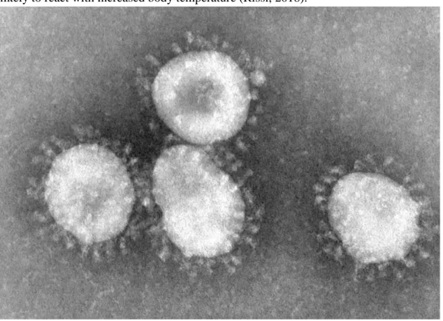
FCoV is an enveloped positive-stranded RNA-virus from the order Nidovirales and family Coronaviridae (Addie et al., 2003) with the largest known RNA genome of 29 200 nucleotides (Lin et al., 2013). The virus is on average 100 nm in diameter and has an appearance similar to a crown due to the Spike-proteins (Figure 1). This is the reason for the name coronavirus (Hartmann, 2005).
FIP can be manifested in different forms: an effusive form also called the “wet form”, a granulomatous non-effusive form also called “dry form” or a mixture of the two. The disease can affect almost any organ system, including central nervous system, thus creating a variable clinical diagnosis-profile. For example, fever is a common symptom but not present in all cases. Especially cats that developed symptoms associated with central nervous system where less likely to react with increased body temperature (Rissi, 2018).
Figure 1.Coronaviruses by CDC/Dr.Fred Murphy
https://commons.wikimedia.org/wiki/File:Coronaviruses_004_lores.jpg#/media/File:Coronavi
5
It is typical for coronaviruses to adapt their target-tissues’ tropism which leads to new and virulent diseases such as FIP that can affect any tissue type and cause massive systemic infections (Chang et al., 2012). As FCoV is prevalent in a significant amount of cats (Holst et
al., 2006) the risk of developing FIP becomes notable.
Spreading
The enteric form of the feline coronavirus (FECV) is highly infectious, extremely common and spreads mostly via feces of the infected animal but shedding via saliva is also a possibility (Addie and Jarrett, 2001). Usually the younger cats are affected but the elder individuals can spread the virus without showing any symptoms. FCoV is rather delicate as it can survive outside its host up to 48 hours and in dry conditions up to 7 weeks (Hartmann, 2005). As the virus acquires mutations to transform to FIPV it seems to lose the ability to infect other individuals, or rather, is not secreted in feces as the FECV anymore. As the animal possibly still carries the FECV can the latter still infect other felines around the carrier (SVA 2018; Hartmann, 2005). These carriers can spread the virus their entire lifetime and can suffer from a chronic diarrhea (Addie and Jarrett, 2001). In households with multiple cats can the animals be re-infected by the other household cats with the same or a different strain of the virus although this kind of superinfection is not common (Addie et al., 2003). Because of these silent carriers an infection in a closed breeding-facility leads often to a epidemic of FCoV as the cats can spread the disease long after they have stopped showing symptoms, even when kept in isolation from other cats (Herrewegh et al., 1997).
Pathogenesis
FECV most commonly infects and spreads via feces of both sick animals and silent carriers (SVA 2018) and is more prevalent in purebred cats than in house cats (Holst et al., 2006) Statistically up to 12% of the FCoV-infections develop into FIP and almost unexceptionally causing a lethal disease (Lloret, 2009). Even cats with a severe viremia of the FCoV do not necessarily develop FIP-symptoms (Fish et al., 2018).
FCoV does not cause serious symptoms by itself. The symptoms vary from mild enteritis to subclinical, but as the virus transforms into FIPV the symptoms become more severe and possibilities of survival decline. The form of the disease seems to depend on the host's immune-response as the “dry form” is characterized by a cell-mediated immunity while the “wet form” is a result of a vasculitis due to a type III immune reaction (Pesteanu-Somogyi et al., 2006). In the ”dry form” variation of FIP symptoms depend on which organ(s) are affected but weight loss, decreased energy levels and spiking fever are common in many of the cats. The variation of the disease is characterized by cell-mediated pyogranulomatous inflammatory reactions. The ”wet form” of the disease is more acute than the “dry form” and is characterized by the leaking of bodily fluids in to the breast- and abdominal cavities making it difficult for the animal to breathe. The leakage is caused by acute vasculitis in the abdominal veins and arteries. This is thought to be due to an immunological type III over reaction, also called Arthus reaction. The
6
actual disease tends to be a mixture of these two forms as even the dry form tends to develop into the wet form (SVA 2018, Hartmann, 2005).
The major biological difference between the two biotypes is that FECV replicates in the intestinal epithelial cells while FIPV can replicate in macrophages. The truncation of ORF3abc-genegroup has been proven to increase the replication of the FIPV in peripheral blood cells (Bálint et al., 2012; Chang et al., 2012). Specifically the truncation of the gene 3c as it is similar to the structure of the virulent 3a gene of SARS coronavirus, has been implicated in the development of the virulent virus. The full function of the 3c gene is still uncertain but evidence suggests its importance to viral replication as viruses with intact 3c-gene replicated more poorly or not at all in the gut (Chang et al., 2010). Also the Spike (S) gene has been implicated as the source of the ability to infect macrophages which is not present in the FECV (Bank-Wolf et al., 2014, Chang et al., 2012).The S protein is expressed as a precursor that needs to be cleaved by cellular proteases. Some data pointed out that the cleavage site of the S evolved from FECV to FIPV and became more easily cleavable and possibly changed its cell tropism. The S protein is responsible for the initial fusion of the virion and the host cell via binding to a receptor that in the case of FCoV II usually is aminopeptidase N (APN) and cleaving of the S precursor is a prerequisite to allow the fusion to take place. Different coronaviruses have different membrane-bound receptors they utilize. As serotype I is difficult to grow in vitro there is not data regarding the receptor affinity of the FCoV I and hence FCoV II has been used as a model for FCoV in
vitro (Jaimes and Whittaker, 2018). Mutated spike-protein does not however indicate a full
transformation to FIP, as there is a case reported with a cutaneous infection by FCoV carrying the S-protein mutation associated with FIP-infections with no typical symptoms of the fatal disease (Osumi et al., 2018).
The invasive nature of FIPV allows it to replicate itself in blood monocytes. The infected monocytes increase vasopermeability and dilate the blood vessels to attract more monocytes to the location which leads to a chain reaction that causes protein-rich fluids to leak into the body cavities. Infection of the blood cell enables a systemic infection whereas FECV causes more local effects (Dewerchin et al., 2005).
Diagnostics
FIP is a difficult disease to diagnose, partly because of the two morphologically very different forms of the disease partly due to the nonspecific symptoms the affected cats show. Any organ-specific reactions due to FIP are not pathognomonic either as the virus can affect organs at random due to the hematogenic spreading and invasive nature. A positive immunofluorescence test, RT-PCR from the effusions or FIP-typical lesions found in a post-mortem investigation can be regarded as definitive but require a specific suspicion for FIP or are done post-mortem. Antigene staining proved to be 100% specific and thus effective to verify diagnosis (Giori et
al., 2011; Hartmann et al., 2003). Immunohistochemistry (IHC) proved to be more accurate
7
FIP is significantly more common in cats under the age of 2 but cannot be excluded when diagnosing older cats as individuals as old as 19 have been documented to develop the disease. FIP can cause a variation of neurological symptoms and can be easily evoke a rabies suspicion due to these pathological changes that include neurophagia, hydrocephalus and cerebral swelling causing depression, ataxia, head tilt and other nonspecific clinical signs (Rissi, 2018). FIP can even cause elevated protein levels in the cerebrospinal fluid (Lloret, 2009) Also, different hematological anomalies were found in the majority of the cats suffering from FIP, for example anemia, lymphopenia and microcytosis. The effusions in the wet form are still dominant as they manifest in majority of the patients (Hartmann et al., 2003; Riemer et al., 2016).
It seems that FIP has a very specific acute phase reaction as well as it tends to have higher total globulin amounts and globulin fractions even compared to FCoV-infected cats (Giordano et al., 2004). In both FIP- and FCoV-cats were γ-globulins higher than in healthy cats (Giordano et
al., 2004; Hartmann et al., 2003). A heightened albumin to globulin fraction was four times
more likely to occur in FIP-cats than in healthy ones (Hartmann et al., 2003).
Risk factors Genetics
FIP has been documented to be slightly more prevalent in purebred cats than in mixed breed cats in some studies like Pesteanu-Somogyi et al. (2006) as they found that the prevalence of FIP in 11 535 cats was 0,52%, where mixed breeds had 0,35% and purebreds 1,3%. In the study all the breeds with a prevalence over 3% had a population under 30 cats partaking the study, which leaves those breeds’ prevalences lacking in statistical strength. However, Riemer et al. (2016) did not find any predisposition in purebred cats when the study population was compared with the clinic’s normal population. The prevalence of FCoV does not seem to be breed-bound, as Li et al. (2018) calculated a p-value of 0,994 for the significance of breed in FCoV prevalence, which shows practically no statistical significance at all. A study pointed out that male sex may be a predisposing factor. Riemer et al. (2016) stated that there is a statistically significantly bigger population of male cats being infected by FIPV, based on the p-value that was under 0,001. Non-neutered or non-sterilized cats had a higher risk to develop the disease in one study (Pesteanu-Somogyi et al., 2006) and in another no correlation with the sexual intactness was found (Riemer et al., 2016).
Age
Age was found to be a important factor in all the published studies regarding FIPVs prevalence. Cats younger than 2 years tended to be significantly more represented than the average, with a calculated p-value under 0,001 and cats older than 7 years tended to be significantly underrepresented than the average, with calculated p-value under 0,001 (Riemer et al., 2016; Pesteanu-Somogyi et al., 2006). Especially cats under 1 year in the study by Riemer et al. (2016) were overrepresented. Still by testing for FCoV antibodies cats under 1 year have a
8
lower prevalence of the virus, possibly due to maternal antibodies and their protection (Bell et
al., 2006).
Stress
Even stress in cats seropositive for FCoV has been implicated as an increased risk-factor as Riemer et al. (2016) documented that 131 of the 231 cats with FIP had a stressful event prior to the illness, such as operations, vaccinations or moving (Lloret, 2009; Riemer et al., 2016). As the study by Riemer et al. (2016) was a case study of cats with confirmed FIP, no healthy cats were included as a control group.
Infectious pressure
As the FCoV infects fecal-orally are cat groups housed compactly at bigger risk regarding FECV, while cats in one-cat households and outdoor cats had a lower risk (Hartmann, 2005). However, there are studies that did not find any statistical significance in housing density regarding FIPVs prevalence (Riemer et al., 2016). An infection of the Feline immunodeficiency virus (FIV) increased both the shedding of the virus and the risk to develop FIP (Vennema, 1999).
DISCUSSION
There is a lot of conflicting data relating to FIPV’s risk factors which include the prevalence of the virus behind the disease, FCoV, and the development of FIP from FECV.
Risk factors
Different breeds
Many studies found a correlation between increased risk and purebred cats and took up some particular breeds with a higher risk (Bell et al., 2006; Pesteanu-Somogyi et al., 2006) while some claimed that there is no significant difference (Riemer et al., 2016). The cause for these abnormalities may be popularity of some races, as in the study by Bell et al. 2006, the breeds with the lowest prevalence had the highest participation numbers. Especially extreme races that have been the target of breeding the longest, persians and siamese, were in the lowest prevalence numbers. This is in contrast to what one would expect if there was a genetic predisposition for the disease in purebred cats in comparison to mixed breeds. The genetic variation that is the strength in mixed breed population makes it also a difficult group to study. With a diversity like that the group cannot possibly be summarized as homogenous which makes it difficult to get statistical significance by studying the genetic risks, if not a relevant control group is included. Another problem when comparing mixed breeds and purebreds besides the difference in genetic variation is the different behavior of the owners. As purebred cats tend to cost multiple times as much as mixed breed cats, the owners may tend to take their purebred cats to the veterinarian more easily and even take part in studies more often than they would a mixed breed cat. There tends to be much more information given to the owners in case of adopting a purebred cat which may contribute to increasing the frequency of visits to the
9
veterinarian as the owners could have more information and knowledge how to spot and differentiate between everyday fluctuations in cats’ behavior and pathological signs.
The male sex
The sex of the cat is another possible risk factor. Pestenau-Somogyi et al. 2006 found a significant implication that male sex would be a predisposing element and Riemer et al. 2016 agree with their statistical analysis of the matter. There is no known aspect in the pathogenesis that could implicate the possible difference in sexes, but much is yet to be discovered. As no study implied that the female gender could be predisposed it is difficult to state that the studies that claim the significance of male sex as an increased risk to develop FIP is just a statistical anomaly. A large-scale statistical study of the disease is required to reveal the answer.
Age
A significantly increased risk was found in cats under the age of 2 or even 1 in some studies (Hartmann, 2005; Riemer et al., 2016), while there are studies that do not see any correlation regarding cats’ age and the antibody titers for FCoV (Bell et al., 2006; Fish et al., 2018). This would imply that younger individuals are not predisposed to FCoV but are to FIP which could mean that the mutations that FIP acquires may very well be host-derived and not completely due to virus’ virulence factors. This could even imply that the cats’ not yet developed immune system could be partly responsible for the mutation, but whether the involvement is of active nature or just lack of defending, is impossible to say.
Neutered cats
Sexual intactness can be seen as a combination of age and sex. Neutered cats have a higher age in average than non-neutered cats and the production of sex-specific hormones decreases strongly after the operation also changing the cats’ metabolism. Again, no unison could be found in the issue. Some studies found indications that neutered cats had a lower risk for FIP and some that there is no statistical significance (Pesteanu-Somogyi et al., 2006; Riemer et al., 2016). If there really is a statistical basis for increased risk in non-neutered cats there is a chance that it mirrors the age of the cats and not necessarily the intactness, as these cats tend to be younger.
Stress
Stress was implicated as a increased risk factor, but as the Riemer et al. 2016 study documented only the cats with FIP and no healthy cats were included. A control group of FCoV positive cats with no FIP symptoms would be optimal to assess if stress really increases the risk of developing FIP or if it just exposes cats to FCoV due to weakened immune response leading to increased prevalence of FCoV and ultimately FIP. There is also the possibility that stressful events in cats were just a coincidence or just indication for owners who tend to take their cats to the veterinarian more often.
10 Prevalence and infectious pressure
Another problematic issue when discussing the prevalence of FIP in different populations is the connection to FCoV. There have been no studies that have directly looked into the connection and differences between prevalences of FCoV and FIP in different cat breeds. This would not only give answers to the questions of the breed-based dispositions of the complex but even shed light to the background of the mutation process itself. If a significant difference in prevalences between FCoV and FIP should exist in a specific breed it would implicate that the mutation could be host-derived. Should the difference not exist a complete variable could be eliminated from the study of FIP’s pathogenesis as it would be just a question of exposure to FCoV. A combined infection of FIV increased the risk of developing FIP and shedding of FCoV (Vennema, 1999), which leaves open the question if FIV infection increases the risk just for developing FIP or does it happen indirectly via increased exposure to FCoV. Naturally the increased shedding would increase spreading of the viruses but if the actual risk for the virus to mutate increases too is still unclear. It could be possible that FIV would facilitate FIPVs pathogenesis as FIV strikes the immune system which could make the development easier for FIP.
Diagnostics
FIPs only difficulties do not lie in the complex risk-profiles and in the lack of treatment options; it is also a difficult illness to diagnose especially the non-effusive form. As the typical symptoms are associated with the exudative form can many cases of the dry FIP be missed due to the nonspecific disease-profile and ante-mortem diagnosis becomes problematic while post-mortem rarely provides solace to the owners. Even though the majority of the FIP-cases are documented to be effusive the statistics may be biased as the dry form is hard to diagnose and the cases are most probably missed when left without a post-mortem investigation. As FIP can cause a systemic infection and affect almost any organ system or systems it can be falsely taken for rabies or other neurological diseases (Rissi, 2018) which would lead to quite extreme measures. Measures that could in the worst case cause unnecessary suffering to the animals in the same household as well as to the owners, such as isolation that is not necessary in the case of FIP but vital epidemiologically when it comes to rabies.
In summary FIP is still a mystery for the most part as conflicting findings clarify little of the virus’ nature but progress is made towards a complete understanding of the fatal disease and possibly vaccines and treatments against it will be present in the near future. The most important thing to do as a cat owner, especially in multiple cat households, is to clean cats’ sandboxes as often as possible to minimize the contact with fecal matter. Unnecessary stress should also be avoided, particularly regarding individuals with FCoV.
11
REFERENCES
SVA (2018), Kattens coronavirus och FIP (felin infektiös peritonit) [online], SVA [Viewed 16. 2. 2019]. Available from: https://www.sva.se/djurhalsa/katt/infektionssjukdomar-katt/coronavirus-och-fip-katt
Addie, D. D., and O. Jarrett. ”Use of a Reverse-Transcriptase Polymerase Chain Reaction for
Monitoring the Shedding of Feline Coronavirus by Healthy Cats”. Veterinary Record 148, no. 21 (26. May 2001): 649–53. https://doi.org/10.1136/vr.148.21.649.
Addie, D. D., I. A. T. Schaap, L. Nicolson, and O. Jarrett. ”Persistence and transmission of natural type I feline coronavirus infection”. Journal of General Virology 84, no. 10 (2003): 2735–44.
https://doi.org/10.1099/vir.0.19129-0.
Amer, Al., A. S. Suri, O. A. Rahman, H. B. Mohd, B. Faruku, S. Saeed, and T. I. T. Azmi. ”Isolation and molecular characterization of type I and type II feline coronavirus in Malaysia”. Virology Journal 9, no. 1 (21. November 2012): 278. https://doi.org/10.1186/1743-422X-9-278. Bálint, Á., A. Farsang, Z. Zádori, Á. Hornyák, L. Dencső, F. Almazán, L. Enjuanes, and S. Belák.
”Molecular Characterization of Feline Infectious Peritonitis Virus Strain DF-2 and Studies of the Role of ORF3abc in Viral Cell Tropism”. Journal of Virology 86, no. 11 (1. June 2012): 6258– 67. https://doi.org/10.1128/JVI.00189-12.
Bank-Wolf, B. R., I. Stallkamp, S. Wiese, A. Moritz, G. Tekes, and H.-J.Thiel. ”Mutations of 3c and spike protein genes correlate with the occurrence of feline infectious peritonitis”. Veterinary Microbiology 173, no. 3 (10. October 2014): 177–88.
https://doi.org/10.1016/j.vetmic.2014.07.020.
Bell, E., R. Malik, and Jm Norris. ”The Relationship between the Feline Coronavirus Antibody Titre and the Age, Breed, Gender and Health Status of Australian Cats”. Australian Veterinary Journal 84, no. 1–2 (2006): 2–7. https://doi.org/10.1111/j.1751-0813.2006.tb13114.x.
Chang, H.-W., H. F. Egberink, R. Halpin, D. J. Spiro, and P. J.M. Rottier. ”Spike Protein Fusion Peptide and Feline Coronavirus Virulence”. Emerging Infectious Diseases 18, no. 7 (July 2012): 1089–95. https://doi.org/10.3201/eid1807.120143.
Chang, H.-W., Raoul J. de Groot, Herman F. Egberink, and Peter J. M. Rottier. ”Feline infectious peritonitis: insights into feline coronavirus pathobiogenesis and epidemiology based on genetic analysis of the viral 3c gene”. Journal of General Virology 91, no. 2 (2010): 415–20.
https://doi.org/10.1099/vir.0.016485-0.
Dewerchin, H. L., E. Cornelissen, and H. J. Nauwynck. ”Replication of Feline Coronaviruses in Peripheral Blood Monocytes”. Archives of Virology 150, no. 12 (1. December 2005): 2483–2500.
https://doi.org/10.1007/s00705-005-0598-6.
Fish, E. J, P. P. VP Diniz, Y.-C. Juan, F. Bossong, E. W. Collisson, Y. Drechsler, and B. Kaltenboeck. ”Cross-Sectional Quantitative RT-PCR Study of Feline Coronavirus Viremia and Replication in
12
Peripheral Blood of Healthy Shelter Cats in Southern California”. Journal of Feline Medicine and Surgery 20, no. 4 (1. April 2018): 295–301. https://doi.org/10.1177/1098612X17705227.
Giordano, A., V. Spagnolo, A. Colombo, and S. Paltrinieri. ”Changes in some acute phase protein and immunoglobulin concentrations in cats affected by feline infectious peritonitis or exposed to feline coronavirus infection”. The Veterinary Journal 167, no. 1 (1. January 2004): 38–44.
https://doi.org/10.1016/S1090-0233(03)00055-8.
Giori, L., A. Giordano, C. Giudice, V. Grieco, and S. Paltrinieri. ”Performances of Different Diagnostic Tests for Feline Infectious Peritonitis in Challenging Clinical Cases”. Journal of Small Animal Practice 52, no. 3 (2011): 152–57.
https://doi.org/10.1111/j.1748-5827.2011.01042.x.
Hartmann, K. ”Feline infectious peritonitis”. Veterinary Clinics of North America: Small Animal Practice, Topics in Feline Medicine, 35, no. 1 (1. January 2005): 39–79.
https://doi.org/10.1016/j.cvsm.2004.10.011.
Hartmann, K., C. Binder, J. Hirschberger, D. Cole, M. Reinacher, S. Schroo, J. Frost, H. Egberink, H. Lutz, and W. Hermanns. ”Comparison of Different Tests to Diagnose Feline Infectious
Peritonitis”. Journal of Veterinary Internal Medicine 17, no. 6 (2003): 781–90.
https://doi.org/10.1111/j.1939-1676.2003.tb02515.x.
Herrewegh, A.A.P.M., M. Mähler, H.J. Hedrich, B.L. Haagmans, H.F. Egberink, M.C. Horzinek, P.J.M. Rottier, and R.J. de Groot. ”Persistence and Evolution of Feline Coronavirus in a Closed Cat-Breeding Colony”. Virology 234, no. 2 (August 1997): 349–63.
https://doi.org/10.1006/viro.1997.8663.
Herrewegh, A. A. P. M., I. Smeenk, M. C. Horzinek, P. J. M. Rottier, and R. J. de Groot. ”Feline Coronavirus Type II Strains 79-1683 and 79-1146 Originate from a Double Recombination between Feline Coronavirus Type I and Canine Coronavirus”. Journal of Virology 72, no. 5 (1. May 1998): 4508–14.
Hohdatsu, T., H. Yamato, T. Ohkawa, M. Kaneko, K. Motokawa, H. Kusuhara, T. Kaneshima, S. Arai, and H. Koyama. ”Vaccine Efficacy of a Cell Lysate with Recombinant Baculovirus-Expressed Feline Infectious Peritonitis (FIP) Virus Nucleocapsid Protein against Progression of FIP”. Veterinary Microbiology 97, no. 1–2 (December 2003): 31–44.
https://doi.org/10.1016/j.vetmic.2003.09.016.
Holst, B. S., L. Englund, S. Palacios, L. Renström, and L. T. Berndtsson. ”Prevalence of Antibodies against Feline Coronavirus and Chlamydophila Felis in Swedish Cats”. Journal of Feline Medicine and Surgery 8, no. 3 (1. June 2006): 207–11.
https://doi.org/10.1016/j.jfms.2005.12.004.
Jaimes, J. A., and G. R. Whittaker. ”Feline coronavirus: Insights into viral pathogenesis based on the spike protein structure and function”. Virology, Nidovirus Research, 517 (1. April 2018): 108–21.
https://doi.org/10.1016/j.virol.2017.12.027.
Li, C., Q. Liu, F. Kong, D. Guo, J. Zhai, M. Su, and D. Sun. ”Circulation and Genetic Diversity of Feline Coronavirus Type I and II from Clinically Healthy and FIP-Suspected Cats in China”.
13
Transboundary and Emerging Diseases 0, no. 0 (13. July 2018).
https://doi.org/10.1111/tbed.13081.
Lin, C.-N., R.-Y. Chang, B.-L. Su, and L.-L. Chueh. ”Full Genome Analysis of a Novel Type II Feline Coronavirus NTU156”. Virus Genes 46, no. 2 (1. April 2013): 316–22.
https://doi.org/10.1007/s11262-012-0864-0.
Lloret, A. ”The Process of Evidence-Based Medicine”. Journal of Feline Medicine and Surgery 11, no. 7 (July 2009): 529–529. https://doi.org/10.1016/j.jfms.2009.05.001.
Oguma, K., M. Ohno, M. Yoshida, and H. Sentsui. ”Mutation of the S and 3c genes in genomes of feline coronaviruses”. The Journal of Veterinary Medical Science 80, no. 7 (July 2018): 1094– 1100. https://doi.org/10.1292/jvms.17-0704.
Osumi, T., I. Mitsui, C. M. Leutenegger, R. Okabe, K. Ide, and K. Nishifuji. ”First identification of a single amino acid change in the spike protein region of feline coronavirus detected from a coronavirus-associated cutaneous nodule in a cat”. JFMS Open Reports 4, no. 2 (20. September 2018). https://doi.org/10.1177/2055116918801385.
Pesteanu-Somogyi, L. D., C. Radzai, and B. M. Pressler. ”Prevalence of Feline Infectious Peritonitis in Specific Cat Breeds”. Journal of Feline Medicine and Surgery 8, no. 1 (1. February 2006): 1– 5. https://doi.org/10.1016/j.jfms.2005.04.003.
Riemer, F., K. A. Kuehner, S. Ritz, C. Sauter-Louis, and K. Hartmann. ”Clinical and Laboratory Features of Cats with Feline Infectious Peritonitis – a Retrospective Study of 231 Confirmed Cases (2000–2010)”. Journal of Feline Medicine and Surgery 18, no. 4 (April 2016): 348–56.
https://doi.org/10.1177/1098612X15586209.
Rissi, D. R. ”A Retrospective Study of the Neuropathology and Diagnosis of Naturally Occurring Feline Infectious Peritonitis”. Journal of Veterinary Diagnostic Investigation 30, no. 3 (May 2018): 392–99. https://doi.org/10.1177/1040638718755833.
Vennema, H. ”Genetic drift and genetic shift during feline coronavirus evolution”. Veterinary Microbiology 69, no. 1 (1. September 1999): 139–41. https://doi.org/10.1016/S0378-1135(99)00102-9.