Mälardalen University Press Licentiate Theses No. 131
NON-INVASIVE TECHNIQUES FOR ASSESSMENT OF
PERIPHERAL BLOOD FLOW AT DIFFERENT VASCULAR DEPTHS
Jimmie Hagblad 2011
Copyright © Jimmie Hagblad, 2011 ISBN 978-91-7485-014-7
ISSN 1651-9256
Printed by Mälardalen University, Västerås, Sweden
Non-invasive techniques for assessment of
peripheral blood flow at different vascular
depths
Copyright © Jimmie Hagblad, 2011
Non-invasive techniques for assessment of
peripheral blood flow at different vascular
depths
Abstract
Missing or reduced blood flow can lead to pressure ulcers. Monitoring blood flow in areas prone to pressure ulcer development would be a valuable tool for prevention of pressure ulcers.
PPG and LDF are both established non invasive optical techniques that can be used to estimate the changes in blood flow in different tissue vo-lumes. The aim of this thesis was to combine the established techniques, LDF and PPG, into one flexible silicon probe intended for evaluation of peripheral blood flow in tissue volumes prone to pressure ulcer development. Further, a temperature sensor was integrated.
Two probe configurations combining LDF and PPG has been evaluated regarding the ability to separate between shallow, medium and deep blood flow variations, and skin temperature changes. Further, techniques to reduce or totally avoid interference between LDF and PPG have been investigated.
The probes can be used to discriminate between blood flows at different vascular depths. The vascular depths reached by the different channels cor-respond to the depths of interest when investigating pressure ulcer aetiology. The probe itself has shown not to affect the skin surface temperature, neither due to the silicon sheet or the light. The skin temperature can be expected to rise and approach the body core temperature by lying in supine position alone, which has to be taken into consideration when designing studies.
By switching between the different light sources, interference can be to-tally avoided. When rapid blood flow variations at several vascular depths are of interest to evaluate, a peripheral placement of the LDF-fibre and an increased illumination power of the PPG-LEDs might be used to minimize the interference between LDF and PPG.
Both probes have shown potential to be used for investigation of the processes in pressure ulcer development.
List of Papers
This thesis is based on the following papers, which are referred to in the text by their Roman numerals.
I Hagblad J., Lindberg L-G., Kaisdotter Andersson A.,
Bergstrand S., Lindgren M., Ek A-C., Folke M. and Lindén M. (2010) A technique based on laser Doppler flowmetry and photoplethysmography for simultaneously monitoring blood flow at different tissue depths. Medical
and Biological Engineering and Computing, 48(5):
415-422
II Hagblad J., Folke M., Lindberg L.-G. and Lindén M.
(2011) Long-term monitoring of blood flow at different depths using LDF and PPG – issues to consider,
Submit-ted to journal
III Hagblad J., Folke M., Lindberg L.-G. and Lindén M.
(2011) Assessment of the optical interference in a PPG-LDF system used for estimation of tissue blood flow
Accepted for presentation at Nordic – Baltic Conference on Biomedical Engineering and Medical Physics – NBC15 June 14-17 2011, Aalborg, Denmark
Author’s contribution
Paper I:
Part of designing the study and performing the measurements. Analyzed the result and part of writing.
Paper II:
Major part of design and performing the study. Analyzed the result and major part of writing.
Paper III:
Major part of design and performing the study. Analyzed the result and major part of writing.
Contents
Introduction ... 11
Background ... 13
Photoplethysmography ... 15
Laser Doppler flowmetry ... 17
Aims ... 19
Method ... 21
Assessing blood flow variation at several tissue depths ... 21
Methods to avoid interference between the two techniques LDF and PPG ... 22
Designing a probe for placement between the person and the support surface with minor influence on the physiological environment ... 23
Results ... 25
Assessing blood flow variation at several tissue depths ... 25
Methods to avoid interference between the two techniques LDF and PPG ... 26
Designing a probe for placement between the person and the support surface with minor influence on the physiological environment ... 26
Discussion ... 27
Conclusions ... 31
References ... 33
Introduction
The main function of the blood is to transport nutrient and oxygen to the tissue and to remove waste products. Missing or reduced blood flow can lead to tissue damage.
Pressure ulcers are examples of damages occurring due to lack of blood flow after prolonged time under pressure or by mechanical destruction of the tissue. The aetiology of pressure ulcer development is however not fully understood. By monitoring the blood flow in areas prone to pressure ulcers these processes can be investigated. Further, methods and equipment de-signed for prevention of pressure ulcers (such as regular repositioning of the patients and use of different support surfaces) are possible to evaluate by blood flow monitoring.
The aim of this thesis was to combine the established techniques, laser Doppler flowmetry (LDF) and photoplethysmography (PPG), into one probe intended for evaluation of peripheral blood in tissue volumes prone to pres-sure ulcer development.
My contribution is the development and evaluation of optical probes de-signed for placement between the person and the support surface with minor influence on the physiological environment and with the ability to assess blood flow variations at different vascular depths in the tissue. The contribu-tion is also to evaluate the design of the probes and to investigate methods to avoid interference between the two techniques LDF and PPG in one of these probes.
Background
In order to sustain a normal function of the cells, a continuously supply of oxygen and nutrient, and removal of carbon dioxide and other waste prod-ucts is necessary. The transport system for this is the circulatory system with the blood as the carrier. The circulation time of the blood is approximately 1 minute. The exchange between the blood and cells occurs in the capillary network, the smallest type of blood vessel (fig 1), in which the blood flows slowly, at less than 1 mm per second. [12]
When a temporary regional occlusion of tissue is released, an increase in blood flow, called reactive hypermia, will occur in healthy subjects.
Temperature regulation compensates for variations in the surrounding en-vironment by changes in the skin blood flow. The differences in blood flow due to temperature can be considerable. In extremely cold condition the blood flow is strongly reduced and the subcutaneous fat layer will act as an isolating layer. In warm condition, the flow can be considerably higher, both to transport heat away and to supply raw material for the sweat production. Further, the metabolism of the tissue will increase with the temperature, thus requiring an increase in blood flow to persist the demand of nutrient and oxygen supply and the removal of waste products.
The regulatory functions are active in healthy tissue. In elderly or fragile persons, these functions might be reduced or acting differently.
All of these functions are relying on well functioning regulation processes, especially of the blood flow regulation. Due to external factors, such as pressure, the blood flow can be impeded, which can lead to tissue damage.
To investigate these factors with respect to their part in the development of pressure ulcers several techniques are available.
Skin temperature measurements with thermistors or similar techniques can be used to detect deviations of temperature in the superficial tissue.
Thermography has been used for classification of wound sites to estimate the risk of delayed healing of pressure ulcers [13].
Near Infrared Spectroscopy (NIRS) can be used to evaluate changes in blood flow and has been used to differentiate between healthy and spinal cord injured subjects [8]. Using NIRS, differences in specific frequency bands of the flow motion signal can be found depending on the status of the tissue, which could be used to detect pressure ulcers.
Pressure at sites prone for the development of pressure ulcers can be eva-luated using pressure sensors, such as interface pressure mapping systems. These systems are capable of producing a view of the variations in pressure using a matrix of sensors integrated in a flexible sheet (fig 2). This is the current common method for evaluation and comparison of mattresses.
Figure 2. Example of pressure mapping (XSENSOR Technology Corp Calgary, Canada).
PPG and LDF are two non invasive optical techniques
estimate the changes in blood flow in tissue volumes. These techniques are both established techniques. In order to achieve a system capable to invest gate and reduce the risk of pressure ulcers, development, a combination of these two techniques has been chosen, with a temperature sensor integrated in the probe. Both these techniques are described more in detail below.
Photoplethysmography
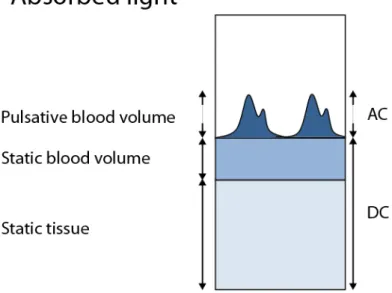
Photoplethysmography (PPG) is based on emitting monochromatic light into tissue and collecting the light reflected back, or through, and not absorbed by tissue and blood. Variation in the signal correlates to several parameters of which pulsative changes in blood flow and blood volume are regarded as most important.
The PPG signal consists of a dominat
(fig 3). The AC-part is usually filtered out and amplified.
By using different wavelengths and distance from light source to detector, reflectance PPG can be designed to estimate the changes in blood flow in different vascular beds [10]. Non invasive monitoring of skin blood flow has
Example of pressure mapping (XSENSOR Technology Corporation,
PPG and LDF are two non invasive optical techniques that can be used to estimate the changes in blood flow in tissue volumes. These techniques are both established techniques. In order to achieve a system capable to investi-gate and reduce the risk of pressure ulcers, development, a combination of
techniques has been chosen, with a temperature sensor integrated in the probe. Both these techniques are described more in detail below.
Photoplethysmography (PPG) is based on emitting monochromatic light into light reflected back, or through, and not absorbed by tissue and blood. Variation in the signal correlates to several parameters of which pulsative changes in blood flow and blood volume are regarded as The PPG signal consists of a dominating DC-part and a pulsative AC-part
part is usually filtered out and amplified.
By using different wavelengths and distance from light source to detector, reflectance PPG can be designed to estimate the changes in blood flow in Non invasive monitoring of skin blood flow has
When a temporary regional occlusion of tissue is released, an increase in blood flow, called reactive hypermia, will occur in healthy subjects.
Temperature regulation compensates for variations in the surrounding en-vironment by changes in the skin blood flow. The differences in blood flow due to temperature can be considerable. In extremely cold condition the blood flow is strongly reduced and the subcutaneous fat layer will act as an isolating layer. In warm condition, the flow can be considerably higher, both to transport heat away and to supply raw material for the sweat production. Further, the metabolism of the tissue will increase with the temperature, thus requiring an increase in blood flow to persist the demand of nutrient and oxygen supply and the removal of waste products.
The regulatory functions are active in healthy tissue. In elderly or fragile persons, these functions might be reduced or acting differently.
All of these functions are relying on well functioning regulation processes, especially of the blood flow regulation. Due to external factors, such as pressure, the blood flow can be impeded, which can lead to tissue damage.
To investigate these factors with respect to their part in the development of pressure ulcers several techniques are available.
Skin temperature measurements with thermistors or similar techniques can be used to detect deviations of temperature in the superficial tissue.
Thermography has been used for classification of wound sites to estimate the risk of delayed healing of pressure ulcers [13].
Near Infrared Spectroscopy (NIRS) can be used to evaluate changes in blood flow and has been used to differentiate between healthy and spinal cord injured subjects [8]. Using NIRS, differences in specific frequency bands of the flow motion signal can be found depending on the status of the tissue, which could be used to detect pressure ulcers.
Pressure at sites prone for the development of pressure ulcers can be eva-luated using pressure sensors, such as interface pressure mapping systems. These systems are capable of producing a view of the variations in pressure using a matrix of sensors integrated in a flexible sheet (fig 2). This is the current common method for evaluation and comparison of mattresses.
Figure 2. Example of pressure mapping (XSENSOR Technology Corp Calgary, Canada).
PPG and LDF are two non invasive optical techniques
estimate the changes in blood flow in tissue volumes. These techniques are both established techniques. In order to achieve a system capable to invest gate and reduce the risk of pressure ulcers, development, a combination of these two techniques has been chosen, with a temperature sensor integrated in the probe. Both these techniques are described more in detail below.
Photoplethysmography
Photoplethysmography (PPG) is based on emitting monochromatic light into tissue and collecting the light reflected back, or through, and not absorbed by tissue and blood. Variation in the signal correlates to several parameters of which pulsative changes in blood flow and blood volume are regarded as most important.
The PPG signal consists of a dominat
(fig 3). The AC-part is usually filtered out and amplified.
Example of pressure mapping (XSENSOR Technology Corporation,
PPG and LDF are two non invasive optical techniques that can be used to estimate the changes in blood flow in tissue volumes. These techniques are both established techniques. In order to achieve a system capable to investi-gate and reduce the risk of pressure ulcers, development, a combination of
techniques has been chosen, with a temperature sensor integrated in the probe. Both these techniques are described more in detail below.
Photoplethysmography (PPG) is based on emitting monochromatic light into light reflected back, or through, and not absorbed by tissue and blood. Variation in the signal correlates to several parameters of which pulsative changes in blood flow and blood volume are regarded as The PPG signal consists of a dominating DC-part and a pulsative AC-part
Figure 3. PPG: The origin of the signal (AC, DC) and absorbed light.
By using suitable combinations of distances between the light source and the photo detector and suitable choice of wavelength, various ranges of vascular depths can be investigated. By using a short distance from a green LED (560 nm) to a photo detector, a shallow penetration depth is achieved. By using near-infrared light (810 nm) placed at a further distance from the sen-sor, 25 mm, a deeper penetration depth can be reached [6, 18]. The absorp-tion of light at different wavelengths is presented in fig 4.
1 10 100 1000 10000 Hb02 Hb
Laser Doppler flowmetry
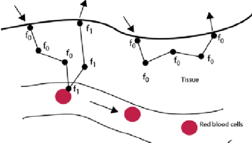
LDF uses monochromatic laser light for assessing the microcirculation of a small volume, less than 1 mm3 according to Monte Carlo simulations [7].
In LDF, the light is emitted through an optical fibre and scattered. Some of the light is reflected by moving red blood cells and the frequency is shifted. Light scattered by static tissue is not shifted in frequency. This fre-quency shift is detected in the reflected light and presented in arbitrary units as the perfusion (fig 5). The scattering will involve interaction with tissue multiple times making the frequency shift more complex to interpret.
Figure 5, Principles of LDF. Emitted light changing frequency due to reflection by moving blood cells but not by scattering in static tissue.
The perfusion scales linearly with the average velocity (<vRBC>) and the
concentration of moving red blood cells (cRBC), providing a low blood cell
concentration to avoid multiple scattering [14-16]:
The perfusion is calculated as the -weighted power spectra of the photocur-rent:
Figure 3. PPG: The origin of the signal (AC, DC) and absorbed light.
By using suitable combinations of distances between the light source and the photo detector and suitable choice of wavelength, various ranges of vascular depths can be investigated. By using a short distance from a green LED (560 nm) to a photo detector, a shallow penetration depth is achieved. By using near-infrared light (810 nm) placed at a further distance from the sen-sor, 25 mm, a deeper penetration depth can be reached [6, 18]. The absorp-tion of light at different wavelengths is presented in fig 4.
100 1000 10000
Hb02 Hb
Laser Doppler flowmetry
LDF uses monochromatic laser light for assessing the microcirculation of a small volume, less than 1 mm3 according to Monte Carlo simulations [7].
In LDF, the light is emitted through an optical fibre and scattered. Some of the light is reflected by moving red blood cells and the frequency is shifted. Light scattered by static tissue is not shifted in frequency. This fre-quency shift is detected in the reflected light and presented in arbitrary units as the perfusion (fig 5). The scattering will involve interaction with tissue multiple times making the frequency shift more complex to interpret.
Figure 5, Principles of LDF. Emitted light changing frequency due to reflection by moving blood cells but not by scattering in static tissue.
The perfusion scales linearly with the average velocity (<vRBC>) and the
concentration of moving red blood cells (cRBC), providing a low blood cell
concentration to avoid multiple scattering [14-16]:
The perfusion is calculated as the -weighted power spectra of the photocur-rent:
The possibility to quantify LDF-signals to give a physiologically relevant unit has been investigated [7]. Initial evaluations in clinical studies have been done, but still further investigation remains.
Aims
The aim of this thesis was to combine the established techniques, LDF and PPG, into one probe intended for evaluation of peripheral blood flow in tissue volumes prone to pressure ulcer development.
Special important aspects were:
- the possibility to assess blood flow variations at several tissue depths - to design the probe for placement between the person and the support
surface with minor influence on the physiological environment
- methods to avoid interference between the two techniques LDF and PPG.
Method
In this theses, two established techniques for evaluation of peripherial blood flow have been combined into two various probe configurations. These probes have both been evaluated.
Assessing blood flow variation at several tissue depths
To investigate the possibility to discriminate between blood flow variations at different tissue depths, a study was conducted using a provocation proce-dure that increased the blood flow at a known depth [Paper I]. These flow variations were evaluated by the first probe configuration, comprising three PPG-channels and one LDF-channel.
The provocation consisted of dynamic muscle contractions performed by the subject by raising the right shoulder upward while grasping a dumbbell. The aim of this was to increase the blood flow in the muscle but not in the skin. The purpose was to investigate if the system could be used to separate between shallow, medium and deep blood flow variations.
The probe used in this study consisted of a central photo detector sur-rounded by different light sources for PPG and a LDF fibre tip. The optical components were embedded in silicon fixated in a wooded frame, left part of Fig 5.
Three combinations of wavelength of the light and distance from light source to detector were used: 560 nm at a distance of 4 mm, 810 nm at a distance of 10 mm and 810 nm at 25 mm distance.
The distance from the skin to the trapezius muscle was measured using ultrasound. The measured distances ranged from 3.8 to 23 mm in the 11 persons that was involved in the study.
Using the optical probe, blood flow was evaluated before and after the provocation procedure.
The ability of each of the PPG-channels and the LDF-channel to detect this induced change in blood flow at a known depth was evaluated. By
com-Method
In this theses, two established techniques for evaluation of peripherial blood flow have been combined into two various probe configurations. These probes have both been evaluated.
Assessing blood flow variation at several tissue depths
To investigate the possibility to discriminate between blood flow variations at different tissue depths, a study was conducted using a provocation proce-dure that increased the blood flow at a known depth [Paper I]. These flow variations were evaluated by the first probe configuration, comprising three PPG-channels and one LDF-channel.
The provocation consisted of dynamic muscle contractions performed by the subject by raising the right shoulder upward while grasping a dumbbell. The aim of this was to increase the blood flow in the muscle but not in the skin. The purpose was to investigate if the system could be used to separate between shallow, medium and deep blood flow variations.
The probe used in this study consisted of a central photo detector sur-rounded by different light sources for PPG and a LDF fibre tip. The optical components were embedded in silicon fixated in a wooded frame, left part of Fig 5.
Three combinations of wavelength of the light and distance from light source to detector were used: 560 nm at a distance of 4 mm, 810 nm at a distance of 10 mm and 810 nm at 25 mm distance.
The distance from the skin to the trapezius muscle was measured using ultrasound. The measured distances ranged from 3.8 to 23 mm in the 11 persons that was involved in the study.
The results of this study were then used to improve the probe (fig 6, right side). The new probe was thereafter used for later studies [Paper 2 and Pa-per 3].
The changes in skin temperature and blood flow variations at the multiple depths reached by the new probe was investigated with subjects lying in a supine position for 60 min [Paper 3].
Figure 6, Sketch of the probes and the tissue volumes reached. The first probe on the left, and the new probe on the right.
Methods to avoid interference between the two
techniques LDF and PPG
To address the interference between LDF and PPG, the system presented in Paper I was equipped with optical filters to minimize the cross technique interference.
In the first probe, the separation in wavelength between the light sources of the LDF- and PPG-techniques was considerable. The PPG-channels were designed using 560 nm and 810 nm light, whereas the LDF-channel used
LDF-system. The PPG-system switches between the two wavelengths, which eliminates any risk of interference between the two channels.
In the design of the second probe, the wavelengths used for the PPG-channels were kept but the LDF-system was replaced by a LDF-system us-ing a solid state laser with a wavelength at 780 nm, which is closer to one of the wavelengths used by the PPG-system. The new probe was design with-out any optical filters. Instead, a switching algorithm was used, activating only one of the techniques at a time, thus totally removing the risk of interfe-rence between the light sources.
This new approach was evaluated and compared to using both LDF and PPG simultaneously (Paper III). To assess the influence from the PPG-LEDs on the LDF-system, LDF first was recorded with only the laser light on and then with the laser and PPG-LEDs active. The PPG-channels were recorded in three ways; using only laser from the LDF as light source, only the PPG-LEDs active and with both systems active. This was repeated using two dif-ferent placements of the LDF- fibre in relation to the area covered by the PPG-light. The recordings were also repeated using three different illumina-tion power of the PPG-LEDs.
The partial influence of the LDF-light source to the PPG-channels and vice versa was then investigated.
Designing a probe for placement between the person
and the support surface with minor influence on the
physiological environment
The first probe was stiff and had to be mounted into a wooden frame. In order to be able to place it under a subject in lying position, a slot was made in the support surface, enabling measurements under the subject. Further, the laser Doppler fibre had to be inserted through the probe. Due to that limita-tion it was not possible to use it for evalualimita-tion of different mattresses.
To minimize the influence of the probe to the subject and the surface, a new flexible probe was developed. This new probe was made using a flexi-ble circuitry board embedded in flexiflexi-ble silicon. The probe includes a matrix of photo detectors and light sources of two different wavelengths to produce two PPG-channels and one LDF-channel. A temperature probe could be inserted into the probe. To minimize the amount of electronic components and thereby increasing the flexibility of the probe, the number of
PPG-The results of this study were then used to improve the probe (fig 6, right side). The new probe was thereafter used for later studies [Paper 2 and Pa-per 3].
The changes in skin temperature and blood flow variations at the multiple depths reached by the new probe was investigated with subjects lying in a supine position for 60 min [Paper 3].
Figure 6, Sketch of the probes and the tissue volumes reached. The first probe on the left, and the new probe on the right.
Methods to avoid interference between the two
techniques LDF and PPG
To address the interference between LDF and PPG, the system presented in Paper I was equipped with optical filters to minimize the cross technique
LDF-system. The PPG-system switches between the two wavelengths, which eliminates any risk of interference between the two channels.
In the design of the second probe, the wavelengths used for the PPG-channels were kept but the LDF-system was replaced by a LDF-system us-ing a solid state laser with a wavelength at 780 nm, which is closer to one of the wavelengths used by the PPG-system. The new probe was design with-out any optical filters. Instead, a switching algorithm was used, activating only one of the techniques at a time, thus totally removing the risk of interfe-rence between the light sources.
This new approach was evaluated and compared to using both LDF and PPG simultaneously (Paper III). To assess the influence from the PPG-LEDs on the LDF-system, LDF first was recorded with only the laser light on and then with the laser and PPG-LEDs active. The PPG-channels were recorded in three ways; using only laser from the LDF as light source, only the PPG-LEDs active and with both systems active. This was repeated using two dif-ferent placements of the LDF- fibre in relation to the area covered by the PPG-light. The recordings were also repeated using three different illumina-tion power of the PPG-LEDs.
The partial influence of the LDF-light source to the PPG-channels and vice versa was then investigated.
Designing a probe for placement between the person
and the support surface with minor influence on the
physiological environment
The first probe was stiff and had to be mounted into a wooden frame. In order to be able to place it under a subject in lying position, a slot was made in the support surface, enabling measurements under the subject. Further, the laser Doppler fibre had to be inserted through the probe. Due to that limita-tion it was not possible to use it for evalualimita-tion of different mattresses.
To minimize the influence of the probe to the subject and the surface, a new flexible probe was developed. This new probe was made using a flexi-ble circuitry board embedded in flexiflexi-ble silicon. The probe includes a matrix
time. The study also involved the differences in temperature at the placement of the probe (Paper II).
The probe was used to collect blood flow and temperature data during 60 minutes. Skin temperatures were also measured by a contactless thermo-meter before and after the measurements procedures.
Temperature measurements were conducted using both active and passive probes and without probe.
Results from the different probe configurations were then compared to de-termine if the probe itself or the light from the PPG-LEDs increased the temperature.
T-Tests on paired data were used to investigate the differences in temper-ature and blood flow.
Results
Two probes combining LDF and PPG has been evaluated (Paper I and Pa-per II). Both probes have shown potential to be used as a tool in the investi-gation of the processes in pressure ulcer development.
Assessing blood flow variation at several tissue depths
The result from Paper I show that it is possible to discriminate between blood flow at different tissue depths. The PPG-channel using the wavelength of 810 nm placed 25 mm from the sensor registers an increase of the signal in all cases, thus reaching depths of more than 22 mm. The other PPG-channel using the wavelength of 810 nm, but with a shorter distance between the light source and detector (10 mm), only registered the change on depths to the muscle less than 10-15 mm. In no case did the LDF or the PPG-channel (wavelength of 560 nm, 4 mm distance between LED and photode-tector), show any increase due to the provocation. This indicated a measure-ment depth of less than 4 mm.
When the flexible probe was used to monitor blood flow and temperature variation during a period of 60 min, it was found that the variations over time differed between the channels, representing different measurement depths, Paper II.
The result shows an increase in the tissue volume reach by the LDF-channel from the beginning and throughout the measurement time in the subjects used for this study. In the beginning of the measurement, the PPG-channels, 810 nm placed 25 mm and 560 nm placed 4 mm from the detector, both showed an increase in blood flow of the reached tissue volumes. The flow measured by the PPG-channels levelled out and continued to increase in the end of the measurement.
time. The study also involved the differences in temperature at the placement of the probe (Paper II).
The probe was used to collect blood flow and temperature data during 60 minutes. Skin temperatures were also measured by a contactless thermo-meter before and after the measurements procedures.
Temperature measurements were conducted using both active and passive probes and without probe.
Results from the different probe configurations were then compared to de-termine if the probe itself or the light from the PPG-LEDs increased the temperature.
T-Tests on paired data were used to investigate the differences in temper-ature and blood flow.
Results
Two probes combining LDF and PPG has been evaluated (Paper I and Pa-per II). Both probes have shown potential to be used as a tool in the investi-gation of the processes in pressure ulcer development.
Assessing blood flow variation at several tissue depths
The result from Paper I show that it is possible to discriminate between blood flow at different tissue depths. The PPG-channel using the wavelength of 810 nm placed 25 mm from the sensor registers an increase of the signal in all cases, thus reaching depths of more than 22 mm. The other PPG-channel using the wavelength of 810 nm, but with a shorter distance between the light source and detector (10 mm), only registered the change on depths to the muscle less than 10-15 mm. In no case did the LDF or the PPG-channel (wavelength of 560 nm, 4 mm distance between LED and photode-tector), show any increase due to the provocation. This indicated a measure-ment depth of less than 4 mm.
When the flexible probe was used to monitor blood flow and temperature variation during a period of 60 min, it was found that the variations over time differed between the channels, representing different measurement depths, Paper II.
The result shows an increase in the tissue volume reach by the LDF-channel from the beginning and throughout the measurement time in the subjects used for this study. In the beginning of the measurement, the PPG-channels, 810 nm placed 25 mm and 560 nm placed 4 mm from the detector, both showed an increase in blood flow of the reached tissue volumes. The flow measured by the PPG-channels levelled out and continued to increase
Methods to avoid interference between the two
techniques LDF and PPG
The LDF-channel did not show any changes depending on the presence of light from the PPG-LEDs or the placement of the LDF-fibre used in the study, paper III.
The sensitivity of the PPG-channels to be influenced by the laser light was dependent of the illumination power used for the PPG-LEDs. Using a higher illumination power decreased the relative influence on the PPG-channel, but did not fully remove it. The influence from the laser light to the PPG-channels was less when using the LDF-fibre in a peripheral position.
Using the switching between the systems removed the risk of inference since only the designated light source is active during measurement.
Designing a probe for placement between the person
and the support surface with minor influence on the
physiological environment
The flexible probe itself has shown not to have any effect on the skin surface temperature, neither due to the light used by the LDF and PPG techniques or by the silicon sheet. The results in paper II, where changes in temperature and blood flow was investigated showed a significant increase in skin perature regardless of the presence of a probe, active or not. The skin tem-perature rise was however less than 0.1 °C/min after 30 min.
A significant increase in blood flow is also found by the LDF- and PPG-channels after 60 min with the subject in a static position.
The blood flow measured by the LDF-channel has a significant increase from the start which continues throughout the whole hour of measurement time. The PPG-channels show a significant increase at the beginning and later parts of the measurement time but levelling out in the time between.
Discussion
In this thesis, a probe combining the techniques LDF and PPG, capable of assessing blood flow variations at multiple vascular depths without interfe-rence between the techniques, has been presented.
The aetiology of pressure ulcer development is not fully understood, and therefore technologies allowing monitoring of blood flow variations at vari-ous vascular depths would be an important help to understand the process. LDF has been used in several studies since a long time to investigate pres-sure ulcer development [9-11], but does only allow assessment of the more superficial vascular structures.
Pressure ulcers often occur in the sacral area. This tissue has shown to have a thickness between 4 mm and 22 mm measured by ultrasound when loaded by the body [2]. Similar depths are discussed also in other studies regarding pressure ulcer aetiology [3, 11].
Based on results from other studies using PPG with similar configurations [17, 19] it can be assumed that pulsatile blood flow variations may be moni-tored from approximate depths of 2 mm using 560 nm, 8–10 mm using 810 nm (source-to-detector separation 10 mm) and a depth of 20 mm using 806 or 880 nm (source-to-detector separation 20–25 mm).
Based on these studies, the first version of the probe was designed with PPG using 560 nm detector separation 4 mm), 810 nm (source-to-detector separation 10 mm) and 810 nm (source-to-(source-to-detector separation 25 mm) in combination with a LDF-fibre. The resulting system thus was expected to be able to monitor and discriminate between blood flows from the most shallow blood flow down to flows at tissue depths of at least 20 mm. Results from Paper I indicated that the deepest reaching PPG confi-guration (810 nm, separation 25 mm) reached at least 23 mm. The PPG-channel using a wavelength of 810 nm and source-to detector separation 10 mm was found to reach a vascular depth of approximately 10–15 mm. The vascular depths reached by the PPG-channel with 560 nm at 4 mm separa-tion and the LDF-channel were found to be more shallow than 4 mm. The expected measuring depth of the LDF is however less than 1 mm.
Methods to avoid interference between the two
techniques LDF and PPG
The LDF-channel did not show any changes depending on the presence of light from the PPG-LEDs or the placement of the LDF-fibre used in the study, paper III.
The sensitivity of the PPG-channels to be influenced by the laser light was dependent of the illumination power used for the PPG-LEDs. Using a higher illumination power decreased the relative influence on the PPG-channel, but did not fully remove it. The influence from the laser light to the PPG-channels was less when using the LDF-fibre in a peripheral position.
Using the switching between the systems removed the risk of inference since only the designated light source is active during measurement.
Designing a probe for placement between the person
and the support surface with minor influence on the
physiological environment
The flexible probe itself has shown not to have any effect on the skin surface temperature, neither due to the light used by the LDF and PPG techniques or by the silicon sheet. The results in paper II, where changes in temperature and blood flow was investigated showed a significant increase in skin perature regardless of the presence of a probe, active or not. The skin tem-perature rise was however less than 0.1 °C/min after 30 min.
A significant increase in blood flow is also found by the LDF- and PPG-channels after 60 min with the subject in a static position.
The blood flow measured by the LDF-channel has a significant increase from the start which continues throughout the whole hour of measurement time. The PPG-channels show a significant increase at the beginning and later parts of the measurement time but levelling out in the time between.
Discussion
In this thesis, a probe combining the techniques LDF and PPG, capable of assessing blood flow variations at multiple vascular depths without interfe-rence between the techniques, has been presented.
The aetiology of pressure ulcer development is not fully understood, and therefore technologies allowing monitoring of blood flow variations at vari-ous vascular depths would be an important help to understand the process. LDF has been used in several studies since a long time to investigate pres-sure ulcer development [9-11], but does only allow assessment of the more superficial vascular structures.
Pressure ulcers often occur in the sacral area. This tissue has shown to have a thickness between 4 mm and 22 mm measured by ultrasound when loaded by the body [2]. Similar depths are discussed also in other studies regarding pressure ulcer aetiology [3, 11].
Based on results from other studies using PPG with similar configurations [17, 19] it can be assumed that pulsatile blood flow variations may be moni-tored from approximate depths of 2 mm using 560 nm, 8–10 mm using 810 nm (source-to-detector separation 10 mm) and a depth of 20 mm using 806 or 880 nm (source-to-detector separation 20–25 mm).
Based on these studies, the first version of the probe was designed with PPG using 560 nm detector separation 4 mm), 810 nm (source-to-detector separation 10 mm) and 810 nm (source-to-(source-to-detector separation 25 mm) in combination with a LDF-fibre. The resulting system thus was expected to be able to monitor and discriminate between blood flows from the most shallow blood flow down to flows at tissue depths of at least 20 mm. Results from Paper I indicated that the deepest reaching PPG confi-guration (810 nm, separation 25 mm) reached at least 23 mm. The PPG-channel using a wavelength of 810 nm and source-to detector separation 10
When designing the second probe, the limitations of the first probe were considered. First, a flexible circuit board was used, and the components were embedded in a silicon sheet with a thickness of 5 mm, thus making the probe flexible. Further, the measurement area was increased to cover 10 cm x 10 cm. Even though the circuitry board of the new probe was flexi-ble, the amount of components had to be minimised to archive maximum compliance. Thus, one of the changes made when designing the new probe was to reduce the number of different PPG-channels to a total of two. When selecting wavelengths and distances for the PPG-channels, one aspect was to certify that the deepest vascular structure of interest was reached. Thus the configurations 810 nm with source-to detector separation 25 mm and 560 nm and source-to detector separation 4 mm were included in the probe. Further, the LDF was changed to a solid state laser of wavelength 780 nm, which allowed a new flat probe to be inserted into the silicon sheet.
The larger area of the new probe means that it is possible to capture flow changes integrated over a larger area, which might have been missed using the smaller first probe. The drawback is, however, that it is no longer possi-ble to monitor local blood flow variations in a specific point. Then measur-ing with the new larger probe, it is important that there is a good contact between the person and the whole probe, which might be difficult on certain persons and on certain measurement sites, as the heels. Sometimes the flex-ibility of the probe is not enough to provide good contact even though the probe should adapt to the shape of the body, and sometimes the body part does not cover the size of the probe.
The new probe was evaluated and could monitor flow variations at vari-ous depths.
The result of the blood flows measured and presented in paper II include several different types of variations during the 60 min. Some subjects showed synchronized changes in some channels.
The presence of these differences and similarities between different chan-nels show that the probe can be used to discriminate between blood flows at different vascular depths and be used to separate them.
When placing something, in this case a probe, between the person and the support surface, the presence of the probe might affect the tissue. To investi-gate these possible effects, the probe was used for 60 min to record the changes in the signals of the PPG and LDF without further provocation than lying down with the probe between the person and the mattress and the skin temperature was measured.
According to the result of Paper II the light sources did not produce any
temperature was, the temperature rise will be reduced to less than 0.1 °C/min within 30 min.
Results of the blood flow measurements showed generally that the total change after 60 min of measurement compared to the starting level was a relative increase of the blood flow.
The fact that a rise in both temperature and blood flow was found at the interface area of the subjects in this study is something that has to be taken into consideration not only when designing studies using this particular probe placement but on all long time measurements including, or dependent of, blood flow or temperature variations using this, or similar, probes on subject in this position.
When combining multiple optical measurement systems in one probe aiming to using them simultaneously one factor to consider is the risk of interference between the techniques.
One solution is the use of optical filters, which was the approach used for the first probe. This approach is not optimal since the filters will to some extent reduce the light intensities also for the wavelengths used for the mea-surements.
The new probe was designed to allow for two different placements of the LDF in relation to the area covered by the PPG, one inner and one peripher-al. Further, a switching algorithm was implemented to the system, totally removing the risk of interference.
The interference was compared using different illumination power of the PPG and the two placements of the LDF-fibre. The interference was found to be minimised by using high illumination power and by using a peripheral placement of the LDF-fibre.
To monitor reasonably slow variations the switching mode is favourable. Switching also allows for the LDF-fibre to be kept in the inner position eva-luating the blood flow variations in the same tissue volume as the PPG but at various depths. Rapid flow changes however cannot be fully monitored in the whole tissue volume, since only one of the techniques is active at a time.
To further investigate the influence of the probe on the physiological en-vironment, the measurement procedure could be repeated using a probe acti-vated intermittent, thus minimizing possible influences of the light, and al-lowing for measurement of blood flow and temperature over time.
Incorporating additional parameters, such as pressure, moisture and shear forces in the measurement system (not necessary into the probe), would also provide valuable information in the context of pressure ulcer prevention.
When designing the second probe, the limitations of the first probe were considered. First, a flexible circuit board was used, and the components were embedded in a silicon sheet with a thickness of 5 mm, thus making the probe flexible. Further, the measurement area was increased to cover 10 cm x 10 cm. Even though the circuitry board of the new probe was flexi-ble, the amount of components had to be minimised to archive maximum compliance. Thus, one of the changes made when designing the new probe was to reduce the number of different PPG-channels to a total of two. When selecting wavelengths and distances for the PPG-channels, one aspect was to certify that the deepest vascular structure of interest was reached. Thus the configurations 810 nm with source-to detector separation 25 mm and 560 nm and source-to detector separation 4 mm were included in the probe. Further, the LDF was changed to a solid state laser of wavelength 780 nm, which allowed a new flat probe to be inserted into the silicon sheet.
The larger area of the new probe means that it is possible to capture flow changes integrated over a larger area, which might have been missed using the smaller first probe. The drawback is, however, that it is no longer possi-ble to monitor local blood flow variations in a specific point. Then measur-ing with the new larger probe, it is important that there is a good contact between the person and the whole probe, which might be difficult on certain persons and on certain measurement sites, as the heels. Sometimes the flex-ibility of the probe is not enough to provide good contact even though the probe should adapt to the shape of the body, and sometimes the body part does not cover the size of the probe.
The new probe was evaluated and could monitor flow variations at vari-ous depths.
The result of the blood flows measured and presented in paper II include several different types of variations during the 60 min. Some subjects showed synchronized changes in some channels.
The presence of these differences and similarities between different chan-nels show that the probe can be used to discriminate between blood flows at different vascular depths and be used to separate them.
When placing something, in this case a probe, between the person and the support surface, the presence of the probe might affect the tissue. To investi-gate these possible effects, the probe was used for 60 min to record the
temperature was, the temperature rise will be reduced to less than 0.1 °C/min within 30 min.
Results of the blood flow measurements showed generally that the total change after 60 min of measurement compared to the starting level was a relative increase of the blood flow.
The fact that a rise in both temperature and blood flow was found at the interface area of the subjects in this study is something that has to be taken into consideration not only when designing studies using this particular probe placement but on all long time measurements including, or dependent of, blood flow or temperature variations using this, or similar, probes on subject in this position.
When combining multiple optical measurement systems in one probe aiming to using them simultaneously one factor to consider is the risk of interference between the techniques.
One solution is the use of optical filters, which was the approach used for the first probe. This approach is not optimal since the filters will to some extent reduce the light intensities also for the wavelengths used for the mea-surements.
The new probe was designed to allow for two different placements of the LDF in relation to the area covered by the PPG, one inner and one peripher-al. Further, a switching algorithm was implemented to the system, totally removing the risk of interference.
The interference was compared using different illumination power of the PPG and the two placements of the LDF-fibre. The interference was found to be minimised by using high illumination power and by using a peripheral placement of the LDF-fibre.
To monitor reasonably slow variations the switching mode is favourable. Switching also allows for the LDF-fibre to be kept in the inner position eva-luating the blood flow variations in the same tissue volume as the PPG but at various depths. Rapid flow changes however cannot be fully monitored in the whole tissue volume, since only one of the techniques is active at a time.
To further investigate the influence of the probe on the physiological en-vironment, the measurement procedure could be repeated using a probe acti-vated intermittent, thus minimizing possible influences of the light, and al-lowing for measurement of blood flow and temperature over time.
face, but the increase in temperature and blood flow must be taken into
con-sideration.
Conclusions
In this licentiate theses, two probes combining the two techniques PPG and LDF, has been presented. Both probes have shown potential to be used as a tool in the investigation of the processes in pressure ulcer development. The probes can be used to discriminate between blood flows at different vascular depths. The vascular depths reached by the different channels correspond to the depths of interest when investigating pressure ulcer aetiology.
The probe itself has shown not to affect the skin surface temperature, nei-ther due to the silicon sheet or the light. The skin temperature can be ex-pected to rise and approach the body core temperature by lying in supine position alone.
A rise in both temperature and blood flow has to be expected and taken into consideration, not only when designing studies using this particular probe placement but on all long time measurements on subject in this posi-tion.
By switching between the different light sources, interference can be to-tally avoided. When rapid blood flow variations at several vascular depths are of interest to evaluate, a peripheral placement of the LDF-fibre and an increased illumination power of the PPG-LEDs might be used to minimize the interference between LDF and PPG.
Acknowledgements
I would like to express my gratitude towards everyone who has helped
me on the way to this thesis.
Especially I would like to thank:
My supervisors Maria Lindén and Mia Folke for all the time, support and encouragement around the clock.
Lars-Göran Lindberg at Linköping University for your invaluable help in understanding and using both the equipment and results.
The rest of the research group in Linköping for your input on the clinical aspects on my work, Anna-Christina, Margareta, Sara and Ulrika.
All who has participated as test subjects in the studies, nothing could have been done without you.
Martin for the almost nine years we have spent together at MDH, first work-ing together as students and now as colleagues sharwork-ing office.
And all other colleagues at IDT for making the time spent at work so plea-sant, mostly Marcus, Nikola and Micke.
Jesper with family, for inviting me in to your life, allowing me to take a break from work and experience some reality once in a while.
This work was supported by grants from Sparbanksstiftelsen Nya and the EU Objective-2 project NovaMedTech.
References
1. Allen J (2007) Photoplethysmography and its application in clinical physiologi-cal measurement. Phys meas 28:R1-R39
2. Bergstrand S (2010) Tissue blood flow responses to external pressure using LDF and PPG. Testing a system developed for pressure ulcer research. Licen-tiate thesis, Linköping University, Linköping, Sweden
3. Clark M, Rowland LB, Wood HA, Crow RA (1989) Measurement of soft tissue thickness over the sacrum of elderly hospital patients using B-mode ultrasound. J Biomed Eng 11(3):200–202
4. Ek AC, Gustavsson G, Lewis DH (1987) Skin blood flow in relation to external pressure and temperature in the supine position on a standard hospital mattress. Scand J Rehab Med 19:121–126
5. Ek AC, Lewis DH, Zetterqvist H, Svensson PG (1984) Skin blood flow in an area at risk for pressure sore. Scand J Rehabil Med. 16(2):85-9.
6. Hagblad J, Lindberg L-G, Kaisdotter Andersson A, Bergstrand S, Lindgren M, Ek A-C, Folke M, Lindén M (2010) A technique based on laser Doppler flow-metry and photoplethysmography for simultaneously monitoring blood flow at different tissue depths. Med Biol Eng Comput 48:5:415-422
7. Fredriksson I (2009) Quantitative Laser Doppler Flowmetry. PhD thesis, Linköping University, Linköping, Sweden
8. Li Z, Zhang M, Wang Y, Wang Y, Xin Q, Li J, Lu C (2011) Wavelet analysis of sacral tissue oxygenation oscillations by near-infrared spectroscopy in per-sons with spinal cord injury. Microvasc Res 81:81-87
9. Liaoa F, Garrisona DW, Jan YK (2010) Relationship between nonlinear proper-ties of sacral skin blood flow oscillations and vasodilatory function in people at risk for pressure ulcers. Microvasc Res 80(1):44-53
10. Lindberg LG, Öberg PA° (1991) Photoplethysmography: Part 2. Influence of light source wavelength. Med Biol Eng Comput 29:48–54
11. Linder-Ganz E, Shabshin N, Itzchak Y, Yizhar Z, Siev-New I, Gefen A (2007) Strains and stresses in sub-dermal tissues of the buttocks are greater in parapleg-ics than in healthy during sitting. J Biomech 41:267–580
12. Mellander S. (1976) Perifer cirkulation, Blodkärlens fysiologi. Sandoz
13. Nakagami G, Sanada H, Lizaka s, Kadono T, Higasino T, Koyanagi H, Haga N (2010) Predicting delayed pressure ulcer healing using thermography: a pros-pective cohort study. Journal of Wound Care, 19(11):465-472
14. Nilsson GE, Salerud EG, Strömberg T, Wårdell K (2003) Laser Doppler perfu-sion monitoring and imaging. In: Tuan V-D (ed) Biomedical photonics
hand-Acknowledgements
I would like to express my gratitude towards everyone who has helped
me on the way to this thesis.
Especially I would like to thank:
My supervisors Maria Lindén and Mia Folke for all the time, support and encouragement around the clock.
Lars-Göran Lindberg at Linköping University for your invaluable help in understanding and using both the equipment and results.
The rest of the research group in Linköping for your input on the clinical aspects on my work, Anna-Christina, Margareta, Sara and Ulrika.
All who has participated as test subjects in the studies, nothing could have been done without you.
Martin for the almost nine years we have spent together at MDH, first work-ing together as students and now as colleagues sharwork-ing office.
And all other colleagues at IDT for making the time spent at work so plea-sant, mostly Marcus, Nikola and Micke.
Jesper with family, for inviting me in to your life, allowing me to take a break from work and experience some reality once in a while.
This work was supported by grants from Sparbanksstiftelsen Nya and the EU Objective-2 project NovaMedTech.
References
1. Allen J (2007) Photoplethysmography and its application in clinical physiologi-cal measurement. Phys meas 28:R1-R39
2. Bergstrand S (2010) Tissue blood flow responses to external pressure using LDF and PPG. Testing a system developed for pressure ulcer research. Licen-tiate thesis, Linköping University, Linköping, Sweden
3. Clark M, Rowland LB, Wood HA, Crow RA (1989) Measurement of soft tissue thickness over the sacrum of elderly hospital patients using B-mode ultrasound. J Biomed Eng 11(3):200–202
4. Ek AC, Gustavsson G, Lewis DH (1987) Skin blood flow in relation to external pressure and temperature in the supine position on a standard hospital mattress. Scand J Rehab Med 19:121–126
5. Ek AC, Lewis DH, Zetterqvist H, Svensson PG (1984) Skin blood flow in an area at risk for pressure sore. Scand J Rehabil Med. 16(2):85-9.
6. Hagblad J, Lindberg L-G, Kaisdotter Andersson A, Bergstrand S, Lindgren M, Ek A-C, Folke M, Lindén M (2010) A technique based on laser Doppler flow-metry and photoplethysmography for simultaneously monitoring blood flow at different tissue depths. Med Biol Eng Comput 48:5:415-422
7. Fredriksson I (2009) Quantitative Laser Doppler Flowmetry. PhD thesis, Linköping University, Linköping, Sweden
8. Li Z, Zhang M, Wang Y, Wang Y, Xin Q, Li J, Lu C (2011) Wavelet analysis of sacral tissue oxygenation oscillations by near-infrared spectroscopy in per-sons with spinal cord injury. Microvasc Res 81:81-87
9. Liaoa F, Garrisona DW, Jan YK (2010) Relationship between nonlinear proper-ties of sacral skin blood flow oscillations and vasodilatory function in people at risk for pressure ulcers. Microvasc Res 80(1):44-53
10. Lindberg LG, Öberg PA° (1991) Photoplethysmography: Part 2. Influence of light source wavelength. Med Biol Eng Comput 29:48–54
11. Linder-Ganz E, Shabshin N, Itzchak Y, Yizhar Z, Siev-New I, Gefen A (2007) Strains and stresses in sub-dermal tissues of the buttocks are greater in parapleg-ics than in healthy during sitting. J Biomech 41:267–580
12. Mellander S. (1976) Perifer cirkulation, Blodkärlens fysiologi. Sandoz
16. Nilsson GE, Tenland T, Öberg PÅ (1980) Evaluation of a laser Doppler flow-meter for measurement of tissue blood flow. IEEE Trans Biomed Eng 27(10):597–604b
17. Sandberg ML, Sandberg MK, Dahl J (2007) Blood flow changes in the trape-zius muscle and overlying skin following transcutaneous electrical nerve stimu-lation. Phys Ther 87(8):1047–1055
18. Sandberg M, Zhang Q, Styf J, Gerdle B, Lindberg L G (2005) Non-invasive monitoring of muscle blood perfusion by photoplethysmography: evaluation of a new application. Acta Physiol Scand 183 335–343
19. Zhang Q, Lindberg LG, Kadefors R, Styf J (2001) A non-invasive measure of changes in blood flow in the human anterior tibial muscle. Eur J Appl Physiol 84(5):448–452