http://www.diva-portal.org
Postprint
This is the accepted version of a paper published in Clinical Implant Dentistry and Related Research. This paper has been peer-reviewed but does not include the final publisher proof-corrections or journal pagination.
Citation for the original published paper (version of record):
Toia, M., Stocchero, M., Becktor, J P., Chrcanovic, B., Wennerberg, A. (2019) Implant vs abutment level connection in implant supported screw#retained fixed partial dentures with cobalt#chrome framework: 1#year interim results of a randomized clinical study
Clinical Implant Dentistry and Related Research, 21(2): 238-246 https://doi.org/10.1111/cid.12717
Access to the published version may require subscription. N.B. When citing this work, cite the original published paper.
Permanent link to this version:
Title:
Implant vs Abutment level connection in implant supported screw-retained fixed partial dentures with Cobalt-Chrome framework: 1-year interim results of a randomized clinical study.
Running Head:
Implant level in fixed partial dentures.
Authors:
Marco Toia, DDS, PhD Student, Department of Oral and Maxillofacial Surgery and Oral Medicine, Malmö University, Sweden.
Michele Stocchero, DDS, PhD, Department of Oral and Maxillofacial Surgery and Oral Medicine, Malmö University, Sweden.
Jonas P. Becktor, Docent, DDS, PhD, Head, Department of Oral and Maxillofacial Surgery and Oral Medicine, Malmö University, Sweden.
Bruno Chrcanovic, DDS, PhD, Department Prosthodontics, Malmö University, Sweden.
Ann Wennerberg, Professor, DDS, PhD, Department of Prosthodontics, Sahlgrenska Academy, University of Gothenburg, Sweden.
Corresponding author:
Marco Toia Dep. of Oral and Maxillofacial Surgery and Oral Medicine. Faculty of Odontology, Malmö University, Carl Gustavs väg 34, SE 205 06, Malmö, Sweden Email: marco.toia@mau.se
ABSTRACT
Background: Screw-retained fixed partial dentures (FPD) have shown a lower incidence of bio- logic complications and an easier retrievability compared with cemented FPD.
Purpose: To compare the marginal bone loss at conical connection implant restored with a screw retained cobalt-chrome FPD in an implant-level (IL) or an abutment-level (AL) setup. Materials and Methods: Patients with at least two adjacent missing teeth were randomly allo- cated to be restored with IL or AL FPD. Periapical radiographs and clinical examination were taken at implant placement, prosthetic connection, 6 and 12 months to evaluate marginal bone loss (MBL), and soft tissue conditions. Complications were used to calculate prognostic indexes.
Results: Fifty patients were treated with 50 FPD supported by 119 implants. The difference of MBL between the IL and AL groups was statistically significant (P = 0.003). At 1 year, MBL was 0.086 ± 0.313 mm and 0.005 ± 0.222 mm in the IL and AL groups, respectively. The presence of BoP increased with time in IL, whereas it decreased in AL group (P < 0.001). A minor compli- cation was encounted in one FPD.
Conclusions: A low grade of MBL was present after 1 year. IL showed greater amount of MBL and soft tissue inflammation indexes than AL. In FPD, AL may be a safer procedure than IL setup in order to preserve a healthy periimplant tissue.
KEYWORDS
INTRODUCTION
Long-term survival rates and high patient satisfaction have been observed with implant-supported fixed partial dentures (FPD).1,2 Compared with cemented solutions, screw-retained FPD have shown a lower incidence of biologic complications and an easier retrievability.3 According to the original protocol,4 the interposition of an abutment between the implants and the framework was recommended. This abutment-level (AL) setup would protect the implant from overload and counterbalance potential misfit between framework and the implants.5 Nevertheless, in the clinical reality, a framework is often screwed directly to the implants, avoiding the use of a screw-retained abutment.6 This implant-level (IL) setup is often used in cases due to limited vertical spaces and to reduce costs.7 Very few mechanical complications were reported in a 5-year prospective multicentre study in which total and partial fixed dentures were directly screwed to the implants.8 However, IL restoration may be more susceptible to inaccuracy between the components. The presence of misfit, although inevi- table during the prosthetic procedures,9 could generate uneven stresses and strains between the framework and implant, which may cause relevant complications, such as screw fracture, framework frac- ture, implant fracture, marginal bone loss (MBL), and implant loss.10 In contrast to AL setup, the accuracy of IL framework seems to be nega- tively affected by implant disparallelism, when using systems with internal connection.11 Thus, it was advocated not to have excessive disparallelism between the implants in this setup.12 Moreover, to allow the insertion and a passive engagement of an IL FPD, the framework is often manufactured without antirotational interlocking indexes. As a consequence, the length of implant- framework interface is reduced both in conical as well as in internal hexagon connection designs. This modified configuration may prevent the benefits of the original internal implant design. It was previously reported that shortened implant-framework interface may introduce higher risk of microgaps between implant and FPD, which may influence both the mechanical engagement and the biological stability of the soft and hard periimplant tissues.13
Based on the current literature, the IL setup has an unclear clinical recommendation,14 and the choice between AL and IL setups is debatable.6 A 1-year prospective clinical study by Gothberg et al.,15 used implants with external hexagonal connection. They observed greater marginal bone resorption on IL restorations compared with an AL setup. Compared to external or internal butt joints, conical implant- abutment connections exhibit better continuity in yield forces16,17 and possess high rigidity with low risk for leakage.18,19 Nevertheless, internal conical connection may present mechanical disadvantages because of the reduced coronal wall thickness of the implant and may therefore have a decreased bearing capacity.20,21 However, there is no clinical evidence comparing IL and AL setups for screw-retained restoration in implants with conical connection.
The primary objective of this preliminary report of a 5-year study was to present results of MBL at implants with conical connection restored with a screw-retained cobalt-chrome (Co-Cr) FPD in an IL or AL setup after 1 year of follow-up. Clinical parameters, such as soft tissue status, mechanical, technical, and biological com- plications, were evaluated as secondary objectives. The null hypoth- esis of the study was that there is no difference in soft and hard tissue changes as well as no difference in prosthetic complications between the two groups.
MATERIALS AND METHODS
Study Design
This prospective investigation was performed in one clinical center affiliated to the Department of Oral and Maxillofacial Surgery and Oral Medicine, Malmö University, Sweden. One (MT) clinician per- formed all the surgical as well as the prosthetic steps of the treatment, whereas an external examiner, not involved in the study, collected data on the clinical parameters.
The study protocol was approved by the Medical Ethics Committee “Azienda Ospedaliera di circolo”, Italy ((VA) number 0001832). The clinical trial was registered at the U.S. National Institutes of Health (Clinicaltrials.gov):NCT02789956. The CONSORT 2010 check- list was used as a guideline
to report on the outcomes. The study flowchart is presented in Figure 1.22
Patient selection
Patients were selected to be included in the study based on the fol- lowing criteria:
1. Age ≥18 years.
2. Willingness to comply with all study requirements and to sign an informed consent. 3. Absence of systemic medical condition: patients had to be ASA-1 or ASA-2.23 4. Good oral hygiene defined as full mouth plaque score ≤25%.24
5. Patients with at least two adjacent teeth to replace in any quadrant of the jaws. Patients were excluded based on the following criteria:
1. Implant-treated patients with bone defects in need of major bone augmentation; 2. Patients with bone defects resulting from tumor resection;
3. Smoking more than 10 cigarettes per day; 4. Severe renal and liver disease;
5. History of radiotherapy in the head and neck region; 6. Chemotherapy at the time of the surgical procedure; 7. Uncontrolled diabetes;
8. Mucosal disease (such as oral lichen planus and epidermolysis bullosa), in the areas to be treated;
9. Noncompliant patients;
10. Situations that the main investigator considered unsuitable for the surgical treatment.
Patients were thoroughly informed about the treatment. The study was conducted in accordance with the Helsinki declaration.
Patient data, medical history, clinical and radiographic examination were recorded. Periodontal, endodontic, and open caries lesions were treated prior to implant installation. All patients received careful oral hygiene instructions and training in self-performed plaque control measures before the surgery. Orthopantomogram at constant magnification and CT scan exams were carried out.
Surgical procedures
Each patient received antibiotic prophylaxis (Amoxicillin 2 g) 1 hour prior to implant placement (IP). A different antibiotic was prescribed for the penicillin-allergic patients. Patients rinsed chlorexidine 0.2% for 1 minute prior to surgery and local anesthesia was induced. After a crestal incision, a full thickness flap was elevated and 2, 3, or 4 implants (OsseoSpeed EV Astra Tech Implant System, Dentsply Sir- ona Implants, Mölndal, Sweden) were placed in each patient according to the drilling protocol described by Toia et al.,25 using a two-stage procedure. The implant bone site was classified according to Lekholm and Zarb classification (L&Z)26 and insertion torque value (ITV) was registered using SA-310 W&H Elcomed implant unit (W&H, Burmoos, Austria). Implant spinning during the positioning of the cover screw was registered. The operation was finalized by a primary adaption of the flaps by means of an accurate suture using absorbable 5-0 suture (Vicryl, Ethicon J&J International, St-Stevens-Woluwe, Belgium) to obtain full periosteal coverage. The patients were instructed to rinse twice daily with chlorexidine and to have a soft diet for 14 days. Sutures were removed after 2 weeks. The patients were advised to take nonsteroidal antiinflammatory drugs for pain relief at their own discretion.
Second Stage Surgery and Randomization
At the second-stage surgery, 6 weeks after IP, the randomization was performed. Computer randomization block software was used to assign the patients in two groups: one group was prosthetically restored at the AL and the other group at the IL. In the AL group, the cover screws were removed and a screw-retained index-free abut- ment (Uni Abutment 33° EV Dentsply Sirona
Implants, Mölndal, Swe- den) was secured using a torque wrench at 25 Ncm. A healing cap (Uni Abutment EV Heal Cap Dentsply Sirona Implants, Mölndal, Swe- den) was positioned onto the abutment, and a suture was applied if necessary. In the IL group, the cover screws were replaced with heal- ing abutments (Healing abutments, Heal Design (TM) EV, Dentsply Sirona Implants, Mölndal, Sweden).
Prosthetic and technical procedures
Impression was taken, using a customized impression tray and silicone elastomeric dental impression material (Aquasil Ultra, Dentsply-Sirona, Milford, Connecticut), 1 week after the second-stage surgery.
After the master cast was poured, the technician produced both a gypsum index, connecting temporary index-free abutments to validate the model as well as an anatomical wax-up. The index was fixed and its final position checked with a periapical radiograph whereas the wax-up was regulated according to the occlusion. In case the index would break, a new impression was taken, and a new index was produced and checked in the patient's mouth. The master model and the anatomical wax-up were then sent to a milling center (Atlantis suprastructures, Dentsply Sirona Implants, Hasselt, Bel- gium). The model and the wax-up were scanned, and a cut-back digi- tal superstructure design was produced and uploaded in the software (Atlantis suprastructures viewer, Dentsply Sirona Implants, Hasselt, Belgium) for final review and approval prior to fabrication.27 Framework was produced in Co-Cr with an additive manufacturing process on the milled implant connection interface.28
Four (±2) weeks after the impression, the definitive screw-retained FPD was installed: baseline timepoint (BL). In the AL group, the screw-retained index-free abutment was secured using a torque wrench at 25 Ncm before seating the framework. The screw hole was closed with polytetrafluoroethylene (PTFE) and composite.29 Each patient received a careful hygiene and motivational instruction after the delivery and was recalled for examination and professional cleaning
at 6 months (6M) and 1 year (1Y) follow-up visits.
Radiographic Parameters
Standardized periapical radiographs were taken after the surgery at IP, BL, 6M, and 1Y with an X-ray apparatus supplied with a long cone and a Rinn Universal Collimator (Dentsply RINN, York, Pennsylvania) (Figure 2). The digital radiographs were analyzed using a software (IllustratorCS, Adobe Systems, Inc. San Jose, California) on a 24-inch high-resolution screen (iMacApple Inc, Cupertino, California) to measure periimplant MBL. The distance from the mesial and distal interproximal bone to the implant-abutment connection (bevel) was measured to the nearest 0.1 mm, and a mean of these two measurements was calculated for each implant. If the bevel was at or below the margin of the crestal bone, that is, subcrestal, the value was considered zero. The known implant diameter and length were used to calibrate the measurements for any distortions (screen resolution was 1920 × 1200 pixels). The radiographs were analyzed at the Department of Radiology, Sahlgrenska Academy, Gothenburg University, Sweden, by one experi- enced radiologist who did not take part in any of the clinical procedures.
Clinical examination
Clinical measurements were performed at BL, 6M, and 1Y. The same examinations performed at BL were conducted at each visit.
Plaque index (PI), probing pocket depth (PPD), bleeding on prob- ing (BoP), and presence of keratinized mucosa (KM) was examined and registered.
PI was scored as 0 (no detection of plaque), 1 (plaque only recog- nized by running around with a probe the abutment/framework), 2 (visible plaque), 3 (abundance of plaque).
PPD was calculated by measuring the distance in millimeter (approximated to the nearest 0.5 mm) from the periimplant mucosal margin to the bottom of the mucosal pocket, using a periodontal cali- brated probe (PCP 15; Hu-Friedy Manufacturing Co, Chicago, Illinois).
The BoP was assessed as “0” (no bleeding), “1” (minute bleeding), and “2” (abundant bleeding).30 Keratinized mucosa (KM) was measured on four implant sites (mesial, buccal, distal, and lingual/palatal) from the gingival margin till the muco-gingival junction. It was assed as “0” (no KM meaning that the gingival margin was lining mucosa), “1” (partial presence of KM <2 mm) and “2” (complete presence KM >2 mm).
Additionally, the papilla fill was registered mesial and distal at each implant site according to Jemt's classification.31
Complications
Mechanical, technical, and biological complications were investigated. For mechanical complications following events were registered: implant failure or fracture; abutment and bridge screw mobile mobility or fracture. Technical complications were considered: framework frac- ture; ceramic layer chipping or fracture. Biological complications were considered: mucositis, fistula, hyperplasia, and perimplantis. The com- plications were reported and used to calculate the prognostic indexes.32
Statistical Analysis
MBL was set as the primary outcome, and prosthetic and clinical com- plications (implant failure, fracture of prosthetic components, bacterial plaque accumulation, BoP, PPD) were set as the secondary outcomes. The mean, SD, and percentage were calculated for all variables. The Kolmogorov-Smirnov test was performed to evaluate the normal distribution of the variables, and Levene's test evaluated homoscedasticity. Differences between control and test groups were compared with the student's t-test or Mann-Whitney test for continuous variables, depending on the normality. Pearson's chi-squared or Fisher's exact test was used in the analysis of contingency tables of categorical data. Survival analyses were performed. A life table was presented with cumulative survival rate (CSR) of both implants and prostheses, besides Kaplan-Meier analysis. The software used was the Statistical Package for the Social Sciences (SPSS) version 25 (SPSS Inc., Chicago,
Illinois). The degree of statistical significance was considered P < 0.05
RESULTS
Patient Demographics
Fifty patients (21 males, 4 smokers), with a mean age of 58.9 years (SD 12.1, range 35.6-93.5) were treated with 50 screw-retained FPD supported by 119 implants between January 2015 and October 2016. The IL group included 25 patients (8 males, 3 smokers), with a mean age of 57.3 years (SD 9.9, range 41.4-73.2) and were treated with 58 implants. The AL group consisted of 25 patients (13 males, 1 smoker), mean age 65 years (SD 13.9, range 35.6-93.5) were treated with 61 implants. The summary of the number of implants in each patient and number of FPD units is reported in Table 1. Sixty-six implants were placed in the mandible and 53 in the maxilla. In the AL group, the abutments were divided in to three lengths resulting in: 21 abutments of 1 mm, 33 of 2 mm and 7 of 3 mm.
Surgery Specifications
ITV resulted in a mean of 26.5 Ncm (SD 10.2; 9-58) and its specifications according to jaws, bone quality and drilling protocol are reported in Table 2.
There was a moderate correlation between the last drill used at the implant surgery installation and the bone quality of the implant site (R = −0.638, R2 = 0.407, P < 0.001; Spearman correlation). Suture were removed after 2 weeks, No implants presented signs of inflammation.
Marginal Bone Loss (MBL)
The difference of MBL between the IL and AL groups was statistically
significant at 6M and 1Y in relation to baseline, as reported in Table 3. There was no difference in MBL, between smokers and non-smokers or between implants of different diameters.
Plaque Index (PI)
PI scored 1 in 0.4% of 476 of sites at BL (2 sites); 9.9% at 6M (47 sites); 13.9% at 1Y (66 sites). No PI scored 2 or 3 were registered.
Bleeding on Probing (BoP)
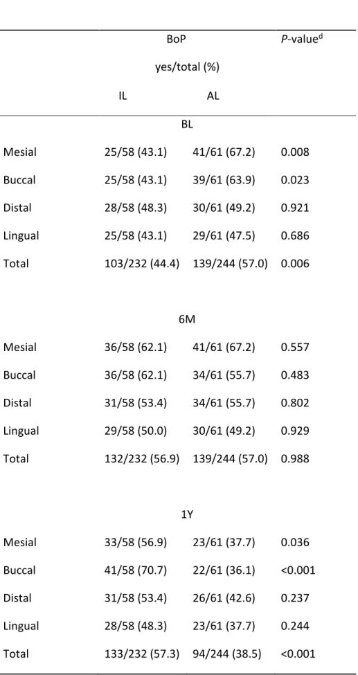
BoP was present at BL in 242(50.8%) out of 476 sites. At 6M and 1Y BoP was registered in 271(56.9%) and in 227(47.7%) out of 476 sites respectively. The difference between the two groups was statistically significant (Table 4). The presence of BoP increased with time in IL group while it decreased in AL group. The main changes happened between BL and 6M in the IL sites, and between 6M and 1Y in the AL sites.
Of the 476 sites, 66 presented plaque and 227 presented BoP, and 54 presented at the same time plaque and BoP at 1Y. This means that 81.8% (54/66) of the sites with plaque had BoP, but only 23.8% of the sites with BoP presented plaque. The correlation between BoP and plaque was weak (R = 0.274, R2 = 0.075, P < 0.001; Spearman correlation).
Probing pocket depth (PPD)
The mean PPD at BL, 6M and 1Y and the difference between the two groups are reported in Table 5. There was a statistically significant difference of PPD between IL and AL group for all the sites at all follow- up time points. Moreover, the PPD significantly decreased with time in the AL group but not in the IL group.
Keratinized mucosa (KM)
In general, there was a tendency towards decrease of buccal keratinized mucosa with time. Only the molar sites presented a statistically significant difference in width of the buccal keratinized mucosa between the two groups. The correlation between the width of keratinized mucosa and MBL at 1Y
was very weak (R = −0.038, R2 = 0.001, P = 0.700; Pearson correlation). Data are presented in Table 6.
Complications
One abutment loosening occurred at BL. The abutment was secured using a torque wrench, before the final FPD delivery. One chip-off fracture was reported in the IL group (position 23) and it was resolved by polishing. Thus, the overall failure rate of the screw retained FPD at 1Y was 2%.
DISCUSSION
In this preliminary 1-year report of a randomized clinical trial, the authors investigated possible differences in hard and soft tissue response between IL and AL screw-retained FPD supported by implants with conical connection.
A greater amount of MBL is reported to occur in the early stage of healing. During the first year, periimplant bone remodeling could be influenced by several factors such as surgical procedures, numbers of surgical stages, repetitive disconnections during prosthetic procedures, type of implant connection and the establishment of the supra- crestal tissue.33–35
According to present findings, it was observed better results in marginal bone maintenance and soft-tissue parameters around AL implants both at 6 and 12 months after loading, thus null hypothesis was rejected. These results highlight how periimplant tissue response could be influenced by the FPD design after 1 year of function.
Even though the magnitude of MBL between the groups may be of limited clinical relevance, a statistically significant difference was observed between the implants in the IL group in comparison to the AL group. This trend is corroborated by a 5-year prospective study which investigated the role of the interposition of the abutment in the fixation implant-supported FPD,36 in which statistically higher bone resorption during the first year of follow-up was reported around abutment-free implants. This initial alteration of the marginal bone level, though, was not followed by further modification
after 3 and 5 years, regardless of the fixation mode.
Concerning the soft tissue, the IL group presented a significant worsening in BoP and PPD with time, while there was a significant improvement in the AL group. After 6 months and 1 year, the difference between the groups resulted to be statistically significant.
This finding is in line with a 5-years clinical trial investigating the use of an intermediary abutment for a full-arch rehabilitation. After 5 years of loading BoP was statistically higher in the abutment free group.37
In clinical terms, these results suggest that periimplant soft tissue shows less inflammation and higher stability in AL group in the early follow-up phase. Interestingly, it was found that a greater number of sites were positive to BoP in AL compared to IL at BL. However, the design of the present study could not explain such finding.
The difference in hard and soft tissue between the groups can be justified by a number of possible explanations.
Firstly, the IL group was subjected to a prosthetic procedure that included the repeated unscrewing and screwing of the components. In an in vivo study, Abrahamsson38 reported that recurrent abutment change interferes with the biological width, damaging the peri-implant connective tissue. It was hypothesized that this repetitive soft tissue disruption would expose the peri-implant compartment to bacterial contamination, resulting in a worsening of the inflammatory status and eventually in a more pronounced marginal bone resorption,39 according to the host response.40 Differently, in the AL group, the definitive abutment was placed at the second surgery and then never removed. This procedure may allow the soft tissue to create a mucosal barrier and protect the osseointegration. The maturation of the soft tissue is a gradual process. Histological studies in vivo41 and on human tissue42
reported that the connective fibers became organized in 4-6 weeks, while the epithelium layer is completed in 6-8 weeks. This means that the prosthetic procedures in the IL group might have interfered with the maturation process.
the material of the components. Peri-implant tissues in the AL group were in direct contact to the titanium abutment, while in the IL group, tissues faced the Co/Cr frame- work structure. Even if the Co/Cr is considered biocompatible, an in vitro study reported that the viability of the epithelium cells as well as fibroblast was superior when in contact to titanium than to Co/Cr.43 However, there is still no strong evidence on the clinical response of hard and soft tissues to Co/Cr frameworks.44
A third explanation of the results could be related to the implant- abutment connection. IL screw retained restorations were produced with a modified implant-abutment interface design. To allow a frictionless seating of the framework into its final contact with the implant, index-free interfaces were manufactured. Moreover, according to the existing inter implant inclination, an additional reduction of the height interface contact was requested. Thus, a shortened contact between the framework and implant walls can prevent the benefits provided by a conical connection since, this modification is often arbitrary and it may not be validated from the implant manufacturer.13 Potential mechanical
impairments and/or leakage phenomena may have caused the peri-implant soft tissue inflammation in the IL group.16 On the other hand, the AL group can take the full advantage of the complete implant-abutment engagement.45 The scarce correlation between the PI and BoP would confirm that the peri-implant inflammation was associated to the type of prosthetic connection. However, it must be said that according to previous reports, soft tissue parameters, such as BoP and PPD, may not reflect the actual perimplant health condition.46
A limitation of the present study was that the inter-implant angulation was not considered. It is acknowledged that IL screw-retained reconstruction requires a great effort in order to have a passive fit, especially when implants are not placed parallel.5,47 As reported previously,48 misfit generates areas of high peak stresses in the implant walls. This condition could cause mechanical complications, such as screw loosening or implant fracture.
A long-term follow-up period is needed to investigate the potential soft and hard tissue reaction at the two set-ups tested in this clinical trial.
CONCLUSION
Within the limitations of this study we can conclude that a low grade of MBL was present in implant supported FPD after 1 year. Implant level connection showed significantly greater amount of MBL and soft tissue inflammation indexes compared with abutment level connected FDP. The results of this 1-year RCT suggest that, FPD at abutment level may be a safer procedure than at implant level setup in order to preserve a peri-implant tissue health. Long-term results are needed to confirm the present results.
ACKNOWLEDGEMENTS
This study was supported in part Dentsply Sirona Implants (Mölndal Sweden) (IIS-D-2013-021).
CONFLICT OF INTEREST The authors declare that they have no conflict of interests. This publication’s content is solely the responsibility of the authors.
References
1. Jemt T, Lekholm U. Oral implant treatment in posterior partially edentulous jaws: a 5-year follow-up report. Int J Oral Maxillofac Implants. 1993;8(6):635-640.
2. Donati M, Ekestubbe A, Lindhe J, Wennstrom JL. Marginal bone loss at implants with different surface characteristics - A 20-year follow-up of a randomized controlled clinical trial. Clin Oral Implants Res. 2018;29(5):480-487.
3. Sailer I, Muhlemann S, Zwahlen M, Hammerle CH, Schneider D. Cemented and screw-retained implant reconstructions: a systematic review of the survival and complication rates. Clin Oral Implants Res. 2012;23 Suppl 6:163-201.
4. Adell R, Lekholm U, Branemark PI, Lindhe J, Rockler B, Eriksson B, Lindvall AM, Yoneyama T, Sbordone L. Marginal tissue reactions at osseointegrated titanium fixtures. Swed Dent J Suppl. 1985;28:175-181.
5. Hellden LB, Derand T. Description and evaluation of a simplified method to achieve passive fit between cast titanium frameworks and implants. Int J Oral Maxillofac Implants. 1998;13(2):190-196.
6. Worni A, Kolgeci L, Rentsch-Kollar A, Katsoulis J, Mericske-Stern R. Zirconia-Based Screw-Retained Prostheses Supported by Implants: A Retrospective Study on Technical Complications and Failures. Clin Implant Dent Relat Res. 2015;17(6):1073-1081.
7. Lewis SG, Llamas D, Avera S. The UCLA abutment: a four-year review. J Prosthet Dent. 1992;67(4):509-515.
8. Hellden L, Ericson G, Elliot A, Fornell J, Holmgren K, Nilner K, Olsson CO. A prospective 5-year multicenter study of the Cresco implantology concept. Int J Prosthodont. 2003;16(5):554-562. 9. Wee AG, Aquilino SA, Schneider RL. Strategies to achieve fit in implant prosthodontics: a review of the literature. Int J Prosthodont. 1999;12(2):167-178.
10. Eckert SE, Meraw SJ, Cal E, Ow RK. Analysis of incidence and associated factors with fractured implants: a retrospective study. Int J Oral Maxillofac Implants. 2000;15(5):662-667.
11. Gracis S, Michalakis K, Vigolo P, Vult von Steyern P, Zwahlen M, Sailer I. Internal vs. external connections for abutments/reconstructions: a systematic review. Clin Oral Implants Res. 2012;23 Suppl 6:202-216.
12. Barbier L, Abeloos J, De Clercq C, Jacobs R. Peri-implant bone changes following tooth extraction, immediate placement and loading of implants in the edentulous maxilla. Clin Oral Investig. 2012;16(4):1061-1070.
13. Sasada Y, Cochran DL. Implant-Abutment Connections: A Review of Biologic Consequences and Peri-implantitis Implications. Int J Oral Maxillofac Implants. 2017;32(6):1296-1307.
14. Wismeijer D, Bragger U, Evans C, Kapos T, Kelly JR, Millen C, Wittneben JG, Zembic A, Taylor TD. Consensus statements and recommended clinical procedures regarding restorative materials and techniques for implant dentistry. Int J Oral Maxillofac Implants. 2014;29 Suppl:137-140.
15. Gothberg C, Andre U, Grondahl K, Ljungquist B, Thomsen P, Slotte C. Immediately loaded implants with or without abutments supporting fixed partial dentures: 1-year results from a prospective, randomized, clinical trial. Clin Implant Dent Relat Res. 2014;16(4):487-500.
16. Berberi A, Tehini G, Rifai K, Bou Nasser Eddine F, Badran B, Akl H. Leakage evaluation of original and compatible implant-abutment connections: In vitro study using Rhodamine B. J Dent Biomech. 2014;5:1758736014547143.
17. Schmitt CM, Nogueira-Filho G, Tenenbaum HC, Lai JY, Brito C, Doring H, Nonhoff J. Performance of conical abutment (Morse Taper) connection implants: a systematic review. J Biomed Mater Res A. 2014;102(2):552-574.
18. Suzuki H, Hata Y, Watanabe F. Implant fracture under dynamic fatigue loading: influence of embedded angle and depth of implant. Odontology. 2016;104(3):357-362.
19. Sumi T, Braian M, Shimada A, Shibata N, Takeshita K, Vandeweghe S, Coelho PG, Wennerberg A, Jimbo R. Characteristics of implant-CAD/CAM abutment connections of two different internal connection systems. J Oral Rehabil. 2012;39(5):391-398.
20. Chrcanovic BR, Kisch J, Albrektsson T, Wennerberg A. Factors influencing the fracture of dental implants. Clin Implant Dent Relat Res. 2018;20(1):58-67.
21. Lee JH, Huh YH, Park CJ, Cho LR. Effect of the Coronal Wall Thickness of Dental Implants on the Screw Joint Stability in the Internal Implant-Abutment Connection. Int J Oral Maxillofac Implants. 2016;31(5):1058-1065.
22. Schulz KF, Altman DG, Moher D, Group C. CONSORT 2010 Statement: updated guidelines for reporting parallel group randomised trials. BMC Med. 2010;8:18.
23. Wolters U, Wolf T, Stutzer H, Schroder T. ASA classification and perioperative variables as predictors of postoperative outcome. Br J Anaesth. 1996;77(2):217-222.
24. O'Leary TJ, Drake RB, Naylor JE. The plaque control record. J Periodontol. 1972;43(1):38. 25. Toia M, Stocchero M, Cecchinato F, Corra E, Jimbo R, Cecchinato D. Clinical Considerations of Adapted Drilling Protocol by Bone Quality Perception. Int J Oral Maxillofac Implants. 2017;32(6):1288-1295.
26. Lekholm U, Zarb G. Patient selection and preparation In: Proceedings of the Branemark,
P.-I., Zarb, G.A., Albrektsson, T.: Tissue Integrated Prostheses: Osseointegration in Clinical Dentistry.
Quintessence Publishing Company, 1985: 199-209.
27. Kapos T, Evans C. CAD/CAM technology for implant abutments, crowns, and superstructures. Int J Oral Maxillofac Implants. 2014;29 Suppl:117-136.
28. Bremen S, Meiners W, Diatlov A. Selective laser melting: a manufacturing technology for the future? Laser Technik Journal. 2012;9(2):33-38.
29. Moráguez OD, Belser UC. The use of polytetrafluoroethylene tape for the management of screw access channels in implant-supported prostheses. The Journal of prosthetic dentistry. 2010;103(3):189-191.
30. Mombelli A, Muhle T, Bragger U, Lang NP, Burgin WB. Comparison of periodontal and peri-implant probing by depth-force pattern analysis. Clin Oral Implants Res. 1997;8(6):448-454.
31. Jemt T. Regeneration of gingival papillae after single-implant treatment. Int J Periodontics Restorative Dent. 1997;17(4):326-333.
32. Salvi GE, Bragger U. Mechanical and technical risks in implant therapy. Int J Oral Maxillofac Implants. 2009;24 Suppl:69-85.
33. Qian J, Wennerberg A, Albrektsson T. Reasons for marginal bone loss around oral implants. Clin Implant Dent Relat Res. 2012;14(6):792-807.
34. Stocchero M, Toia M, Jinno Y, Cecchinato F, Becktor JP, Naito Y, Halldin A, Jimbo R. Influence of different drilling preparation on cortical bone: A biomechanical, histological, and micro‐ CT study on sheep. Clinical oral implants research. 2018.
35. Borges T, Leitão B, Pereira M, Carvalho Á, Galindo‐Moreno P. Influence of the abutment height and connection timing in early peri‐implant marginal bone changes: A prospective randomized clinical trial. Clinical oral implants research. 2018;29(9):907-914.
36. Gothberg C, Grondahl K, Omar O, Thomsen P, Slotte C. Bone and soft tissue outcomes, risk factors, and complications of implant-supported prostheses: 5-Years RCT with different abutment types and loading protocols. Clin Implant Dent Relat Res. 2018.
37. Abrahamsson I, Berglundh T, Lindhe J. The mucosal barrier following abutment dis/reconnection. An experimental study in dogs. J Clin Periodontol. 1997;24(8):568-572.
38. Lazzara RJ, Porter SS. Platform switching: a new concept in implant dentistry for controlling postrestorative crestal bone levels. Int J Periodontics Restorative Dent. 2006;26(1):9-17.
39. Trindade R, Albrektsson T, Galli S, Prgomet Z, Tengvall P, Wennerberg A. Osseointegration and foreign body reaction: Titanium implants activate the immune system and suppress bone resorption during the first 4 weeks after implantation. Clin Implant Dent Relat Res. 2018;20(1):82-91.
40. Berglundh T, Abrahamsson I, Welander M, Lang NP, Lindhe J. Morphogenesis of the peri-implant mucosa: an experimental study in dogs. Clin Oral Implants Res. 2007;18(1):1-8.
41. Tomasi C, Tessarolo F, Caola I, Wennstrom J, Nollo G, Berglundh T. Morphogenesis of peri-implant mucosa revisited: an experimental study in humans. Clin Oral Implants Res. 2014;25(9):997-1003.
42. Hjalmarsson L, Smedberg JI, Aronsson G, Wennerberg A. Cellular responses to cobalt-chrome and CP titanium--an in vitro comparison of frameworks for implant-retained oral prostheses. Swed Dent J. 2011;35(4):177-186.
43. Hjalmarsson L, Smedberg JI, Wennerberg A. Material degradation in implant-retained cobalt-chrome and titanium frameworks. J Oral Rehabil. 2011;38(1):61-71.
44. Binon PP. Implants and components: entering the new millennium. Int J Oral Maxillofac Implants. 2000;15(1):76-94.
45. Coli P, Christiaens V, Sennerby L, Bruyn HD. Reliability of periodontal diagnostic tools for monitoring peri‐implant health and disease. Periodontology 2000. 2017;73(1):203-217.
46. Hedkvist L, Mattsson T, Hellden LB. Clinical performance of a method for the fabrication of implant-supported precisely fitting titanium frameworks: a retrospective 5- to 8-year clinical follow-up study. Clin Implant Dent Relat Res. 2004;6(3):174-180.
47. Jimbo R, Halldin A, Janda M, Wennerberg A, Vandeweghe S. Vertical fracture and marginal bone loss of internal-connection implants: a finite element analysis. Int J Oral Maxillofac Implants. 2013;28(4):e171-176.
TABLES
Table 1. Summary of the number of implants in each patient and number of FPD units.
Patients FPD Units 2 3 4 5 6 Number of implants 2 35 IL 20 14 6 AL 15 11 4 3 11 IL 2 1 1 AL 9 3 4 1 1 4 4 IL 3 1 2 AL 1 1 Total 50 50 25 14 6 4 1
IL: implant level AL: abutment level
Table 2. Results for ITV(Ncm) in jaws, bone quality and drilling protocol.
ITV
N Mean (SD;Min-Max) P-value
Jaw <.001
Mandible 66 31.7 (9.8;12-58)
Maxilla 53 20.1 (6.6; 9-33)
Bone Quality (L&Z) <.001
1 6 50.7 (4.2;47-58) 2 49 32.0 (6.4;21-48) 3 47 22.3 (5.9;11-33) 4 17 13.9 (3.8;9-25) Drilling protocol <.001 S 31 21.2 (7.9;10-48) S-A 17 20.3 (6.0;10-32) S-B 18 29.3 (9.5;9-45) S-X-B 53 30.7 (10.6;11-58) Total 119 26.5 (10.2;9-58) N (Frequency). SD (Standard deviation). Min (Minimum). Max (Maximum).
L&Z (Lekholm & Zarb classification)26
Table 3. Comparison of marginal bone loss (MBL) between the groups implant level and abutment level at
different time.
Group MBL (mm)*
mean±SD (min, max)
IP – BL BL – 6M BL – 1Y IL -0.148±0.312 (-1.66, 0.00) (n=57) -0.106±0.334 (-0.78, 1.42) (n=51) -0.086±0.313 (-0.81, 1.17) (n=58) AL -0.103±0.234 (-0.90, 0.01), (n=60) -0.018±0.214 (-0.95, 0.58), (n=59) -0.005±0.222 (-0.99, 0.58), (n=61) P-value ** 0.178 0.002 0.003
* Negative values indicate vertical bone loss and positive values indicate vertical bone gain. ** Mann-Whitney test.
MBL (marginal bone loss) IL (Implant level). AL (abutment level). IP (Implant placement). BL (Final prostheses delivery). 6M (Six months).
Table 4. Comparison of bleed on probing of different implant faces between the implant level and abutment
level groups at different time points.
BoP yes/total (%) P-valued IL AL BL Mesial 25/58 (43.1) 41/61 (67.2) 0.008 Buccal 25/58 (43.1) 39/61 (63.9) 0.023 Distal 28/58 (48.3) 30/61 (49.2) 0.921 Lingual 25/58 (43.1) 29/61 (47.5) 0.686 Total 103/232 (44.4) 139/244 (57.0) 0.006 6M Mesial 36/58 (62.1) 41/61 (67.2) 0.557 Buccal 36/58 (62.1) 34/61 (55.7) 0.483 Distal 31/58 (53.4) 34/61 (55.7) 0.802 Lingual 29/58 (50.0) 30/61 (49.2) 0.929 Total 132/232 (56.9) 139/244 (57.0) 0.988 1Y Mesial 33/58 (56.9) 23/61 (37.7) 0.036 Buccal 41/58 (70.7) 22/61 (36.1) <0.001 Distal 31/58 (53.4) 26/61 (42.6) 0.237 Lingual 28/58 (48.3) 23/61 (37.7) 0.244 Total 133/232 (57.3) 94/244 (38.5) <0.001
P-valued P-valued
BL – 6M a <0.007 1.000
6M – 1Y b 0.925 <0.001
BL – 1Y c <0.005 <0.001
a Comparison of the incidence of BoP sites of between BL and 6M for the respective groups b Comparison of the incidence of BoP sites of between 6M and 1Y, for the respective groups c Comparison of the incidence of BoP sites of between BL and 1Y, for the respective groups d Pearson’s chi-squared test
BoP (bleed on probing) IL (Implant level). AL (abutment level). IP (Implant placement). BL (Final prostheses delivery). 6M (Six months).
Table 5. Comparison of probing pocket depth of different implant faces between implant level and abutment
level groups at different time points.
PPD mean±SD (min-max) P-valued
IL AL BL Mesial 3.76±0.885 (2-6) 3.41±1.230 (1-6) 0.043 Buccal 3.31±0.568 (2-5) 2.87±1.072 (1-6) 0.001 Distal 3.48±0.903 (2-6) 2.89±0.950 (1-6) <0.001 Lingual 3.40±0.857 (2-6) 2.72±0.819 (2-6) <0.001 Total 3.49±0.827 (2-6) (n=232) 2.97±1.056 (1-6) (n=244) <0.001 6M Mesial 3.67±0.747 (2-5) 3.27±1.362 (1-8) 0.007 Buccal 3.40±0.894 (2-5) 2.73±1.080 (1-7) <0.001 Distal 3.51±0.879 (2-6) 2.98±1.106 (1-6) 0.001 Lingual 2.91±0.674 (2-4) 2.53±1.023 (0-6) 0.001 Total 3.37±0.848 (2-6) (n=220) 2.88±1.177 (0-8) (n=236) <0.001 1Y Mesial 3.81±1.011 (2-6) 2.64±1.119 (1-6) <0.001 Buccal 3.37±1.186 (1-7) 2.45±0.829 (1-6) <0.001 Distal 3.50±1.005 (2-6) 2.77±1.191 (1-6) <0.001 Lingual 3.15±0.979 (1-6) 2.52±1.191 (1-7) <0.001 Total 3.46±1.069 (1-7) (n=216) 2.59±1.092 (1-7) (n=224) <0.001 P-value d P-value d
BL – 6M a 0.230 0.217 6M – 1Y b 0.571 <0.001 BL – 1Y c 0.594 <0.001 SD – standard deviation Min (Minimum). Max (Maximum).
a Comparison of PPD between BL and 6M, for the respective groups b Comparison of PPD between 6M and 1Y, for the respective groups c Comparison of PPD between BL and 1Y, for the respective groups d Mann-Whitney test
PPD (probing pocket depth) IL (Implant level).
AL (abutment level). IP (Implant placement). BL (Final prostheses delivery). 6M (Six months).
Table 6. Comparison of the width of the buccal keratinized mucosa (mm) the groups implant level and
abutment level at different time points. KM mean±SD (min-max) P-value a IL AL BL Incisive - 2.00±0.00 (2-2) (n=5) - Canine 2.00±0.00 (2-2) (n=3) 2.00±0.00 (2-2) (n=2) 1.000 Premolar 1.96±0.209 (1-2) (n=23) 1.87±0.344 (1-2) (n=23) 0.301 Molar 1.81±0.397 (1-2) (n=32) 1.71±0.461 (1-2) (n=31) 0.342 6M Incisive - 2.00±0.00 (2-2) (n=5) - Canine 2.00±0.00 (2-2) (n=2) 2.00±0.00 (2-2) (n=2) 1.000 Premolar 1.95±0.213 (1-2) (n=22) 1.62±0.740 (0-2) (n=21) 0.063 Molar 1.94±0.250 (1-2) (n=31) 1.48±0.677 (0-2) (n=31) 0.001 1Y Incisive - 2.00±0.00 (2-2) (n=4) - Canine 2.00±0.00 (2-2) (n=2) 2.00±0.00 (2-2) (n=2) 1.000 Premolar 1.85±0.366 (1-2) (n=20) 1.45±0.759 (0-2) (n=20) 0.059 Molar 1.86±0.351 (1-2) (n=29) 1.43±0.626 (0-2) (n=30) 0.003 SD – standard deviation Min (Minimum). Max (Maximum).
KM (keratinized mucosa buccal site) IL (Implant level).
AL (abutment level). IP (Implant placement). BL (Final prostheses delivery). 6M (Six months).
FIGURES
Figure 1
Figure 2
X-rays at each time point in abutment level patient (AL) and in implant level patient (IL). Implant placement (IP), prostheses delivery baseline (BL), six months follow-up (6M), one year follow-up (1Y)