Depicting the role of CD2AP during
Old World alphavirus infection
Ainhoa Moliner Morro
__________________________________________
Master Degree Project in Infection Biology, 45 credits. Spring
Semester 2017
Department of Microbiology, Tumor and Cell Biology, Karolinska
Institutet
Supervisor: Gerald McInerney
Co-supervisor: Cecilia Smedberg
ACKNOWLEDGEMENTS
I would first like to thank my supervisors Gerald McInerney and Cecilia Smedberg for everything they have taught me over the past months. They have helped me with every question and problem I have had and have made me a better scientist.
I would also like to thank the rest of the alphavirus group for their help, but more importantly for the nice atmosphere they have created, making these months an amazing and fulfilling time. Last but not least, I would like to express my gratitude to my friends and family for believing in me and their endless encouragement. I could not have made it without them. Thank you.
Table of contents 1. Abstract ... 4 2. Popular science ... 4 3. Abbreviations ... 5 4. Introduction ... 7 5. Aim ... 10
6. Materials and methods ... 11
6.1. Cell culture ... 11
6.2. Viruses and plasmids. ... 11
6.3. Virus growth curve experiments... 11
6.4. Cell lysis, SDS-PAGE, Western Blotting and immunoprecipitation ... 12
6.5. Immunofluorescence and confocal microscopy... 12
7. Results ... 14
7.1. CHIKV and SFV nsP3 protein binds the cellular CD2AP ... 14
7.2. Growth curves. ... 14
7.3. CD2AP and nsP3 co-localization ... 15
7.4. Characterization of CD2AP-nsP3 foci ... 17
7.5. Actin rearrangement upon infection ... 20
7.6. Internalization of replication complexes ... 21
8. Discussion ... 23
1. Abstract
Alphaviruses are group of positive sense single-stranded RNA viruses including the human pathogen chikungunya virus and the model virus Semliki Forest virus. Viruses are pathogens that interact with different cellular proteins to ensure efficient viral replication. Recently, CD2-associated protein (CD2AP), a scaffolding protein of 80-kDa, has been observed to bind the non-structural protein 3 (nsP3) of chikungunya virus. Here we report that viral mutants lacking the ability to bind to CD2AP are attenuated, which indicates that the CD2AP interaction is important for normal viral replication. We show that replication complexes of SFV unable to bind CD2AP, are delayed in internalization to the perinuclear area. After treatment with Wortmannin, a drug inhibiting internalization of replication complexes, the replication complexes were found aligning with actin fibers, suggesting that replication complexes of SFV mutant unable to bind CD2AP either bypass the effect of Wortmannin or are formed at other membranous structures in the cell.
Key words: CD2AP, nsP3, chikungunya virus, Semliki Forest virus
2. Popular science
Have you ever heard of chikungunya fever? It’s a viral disease transmitted, just as Zika or Dengue, by mosquitoes. It causes arthritic disease and its name, chikungunya, comes from a Makonde word that describes the hunched posture of people suffering from the disease. Does it not intrigue you how these tiny identities can make us so sick? Viruses use the cellular machinery to their own advantage, and that can lead to some of the symptoms. The ultimate goal of viral research is to prevent (vaccination) or treat (antivirals) the infection. To be able to do this, it is important to understand better the interaction between these small pathogens and their host cells.
Chikungunya virus has for a total of 9 proteins, four of them are so called non-structural proteins, which are involved in viral multiplication, and the other five are structural proteins that will make the new viruses. The non-structural proteins have different functions; however, the function of non-structural protein 3 (nsP3) is not completely understood. In this project, we assessed its interaction with a cellular protein called CD2AP, which has been studied in relation to kidney failure and Alzheimer disease.
We used wild type virus and mutants lacking the ability to bind CD2AP. First, we checked viral growth for which we infected cells with either wild type or mutant virus, harvested samples every 2 hours and determined viral production rate. We observed that mutant viruses were highly impaired in growth. Following, we investigated co-localization of both proteins during infection by immunofluorescence, a microscopy technique that allows specific visualization of different molecules. CD2AP and nsP3 were seen in punctate structures during infection with the wild type virus, but CD2AP was diffused in the inside the cell during infection with the mutant virus. Next, we checked if the foci were part of the replication complexes (membranous structures created by the virus where the replication takes place). Part of the foci were found in these structures, but some were independent of replication complexes.
What makes the interaction between nsP3 (virus) and CD2AP (cell) important? CD2AP is known to reorganize the actin cytoskeleton, a cellular dynamic structure that, among other functions, is involved in intracellular transport or formation of specific structures on the cell membrane. We addressed this question by immunofluorescence, visualizing actin (a protein
that helps to keep the shape of the cell and transport proteins), nsP3 and CD2AP during infection. However, we did not observe acting rearrangement, suggesting that CD2AP is not important for this feature during viral replication. Aside from actin reorganization, CD2AP is involved in other cellular mechanisms: internalization of cellular receptor and intracellular transport. These possibilities will be assessed in the future to find out why chikungunya targets the cellular CD2AP.
3. Abbreviations
AUD Alphavirus unique domain BHK Baby hamster kidney cells CD2AP CD2 associated protein CHIKV Chikungunya virus
CHIKV-P Chikungunya virus carrying P423A substitution
CHIKV-PR Chikungunya virus carrying P423A and R428A substitution CHIKV-R Chikungunya virus carrying R428A substitution
CHIKV-WT Chikungunya virus wild type CIN85 Cbl-interacting protein of 85kDa CMS Cas ligand with multiple SH3 domains CPV-I Intracellular cytopathic vacuoles DMEM Dulbecco's modified Eagle's medium dsRNA Double-stranded RNA
FBS Fetal bovine serum
G3BP Ras GTPase-activating protein-binding protein 1 GMEM Glasgow's modified Eagle's medium
h p.i. Hours post infection HOS Human osteosarcoma HVD Hypervariable domain
IF Immunofluorescence
IP Immunoprecipitation MOI Multiplicity of infection
NC Nucleocapsid nsP1 Non-structural protein 1 nsP2 Non-structural protein 2 nsP3 Non-structural protein 3 nsP4 Non-structural protein 4 nsPs Non-structural proteins NTPase Nucleoside triphosphatase ORFs Open Reading Frame PFU Plaque forming units
PI3K Phosphatidylinositol-3-kinase RC Replication complex
RTK Receptor tyrosine kinases SFV Semliki Forest virus
SFV-noCD2AP
Semliki Forest virus carrying P428A, R433A, P435A, P439A, R440A and R444A substitutions
SFV-WT Semliki Forest virus wild type
SH3 Src Homology 3
SILAC Stable Isotope Labelling by Amino acids in Cell culture U2OS Human bone osteosarcoma epithelial cells
4. Introduction
Alphaviruses are single-stranded positive sense RNA viruses belonging to the Togaviridae family1,2. Depending on their geographical distribution alphaviruses are divided into Old World and New World (Fig. 1)1,2. Old World alphaviruses were initially found in Africa and East Asia and have spread in the recent decades to Australia and Europe. Among them, several human pathogens such as Ross river virus, O’nyong’nyong virus and chikungunya virus (CHIKV) can be found3. Semliki Forest virus (SFV), which affects rodents, is an Old World alphavirus used as a model tool for these human pathogens1. Meanwhile, New-World alphaviruses such as Venezuelan equine encephalitis virus or
East equine encephalitis virus are found in the Americas and Pacific Islands and cause life-threatening encephalitis in animals and humans4.
Alphaviruses are mainly transmitted by Aedes mosquitoes and, as other arboviruses, are usually maintained in a sylvatic cycle between mosquitoes and primates5,6. However, outbreaks
in humans or livestock can occur, which in turn may lead to economic losses and public health concerns2. Arboviruses in general have re-emerged in recent years due to changes in their vectors’ distribution due to several factors such as international transport of goods or the mosquito ability to adapt to new environments7, which has led to an increase of exposed population and at risk of disease.
Although these viruses are known to cause arthritic disease in humans, CHIKV infection in particular, leads to a debilitating disease named after the Makonde word “chikungunya”, which describes the hunched posture due to the joint pain of patients suffering from the disease6. Common symptoms of chikungunya disease are fever, rash, nausea and the distinctive polyarthritis, which appear between 3 to 12 days after infection5,6,8. Although chikungunya
fever has a low mortality rate, the disease and sequelae can be severely debilitating5. The lack of vaccines and antiviral treatment against this virus together with its debilitating characteristics urge scientists to study and understand the underlying mechanisms of viral infection.
4.1. Viral structure and genome
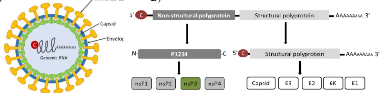
Alphaviruses are enveloped viruses, which have an icosahedral capsid of about 70 nm with a symmetry of T = 41. In the envelope, 6K proteins are embedded and also the glycoproteins E1, E2 and E3 that form 80 spikes, also arranged in a T = 4 symmetry1. Underneath the envelope
the nucleocapsid core (NC) is found, which is formed by the capsid protein and the genomic RNA (Fig. 2A).
The viral genome is, as mentioned previously, a positive sense single-stranded RNA (+ssRNA) molecule of 11.5 kb, which contains a 5’ 7-methyl-GpppA cap and a 3’ poly(A) tail structures (Fig. 2B)2,4. The alphavirus genome has two open reading frames (ORFs) and codes for a total of 9 proteins (Fig. 2A)2. The non-structural proteins (nsPs 1 to 4), which are involved
Figure 1. Origin of Old World and New World alphaviruses. Old World alphaviruses (red) were first
found in Africa, Southeast Asia and the Pacific. New World alphaviruses (blue) were firstly found in the Americas and some pacific islands.
in viral replication, are encoded in the first ORF2. The second ORF is expressed through production of a sub-genomic mRNA and encodes the structural proteins: glycoproteins E1, pE2, E3, 6K and capsid (C) that will form new viral particles2.
4.2. Alphavirus life cycle
The ability of alphaviruses to infect diverse hosts, vertebrate and invertebrate, is most likely facilitated by either the use of a receptor which is conserved among different cells, or the ability to interact with different cellular receptors9. The glycoprotein E2 is responsible for this virus:host interaction, after which virions are internalized by clathrin-dependent endocytosis1,9. A drop in endosomal pH triggers conformational change of the glycoprotein E1 which is found on the viral envelope, displaying the hidden fusion loop, therefore inducing the fusion of the viral and endosomal membranes, allowing the release of the NC into the cytoplasm1.
Once in the cytoplasm, the alphavirus genome is used directly as an mRNA to translate the nsPs into a polyprotein. However, translation of the alphavirus genome usually yields two different polyproteins, P123 or P1234, the later one containing all the non-structural proteins1. The polyprotein is processed by the non-structural protein 2 (nsP2), found within the polyprotein, which contains a papain-like cysteine protease that is active both, as an individual protein and when resident within the polyprotein; nevertheless, when nsP2 is found in the polyprotein, only cleavage at the nsP3/nsP4 site occurs in cis, while cleavage at the other cleavage sites occurs only in trans1. In this way, production of the different nsPs is regulated temporally during the infection. The different non-structural proteins have diverse functions which will be discussed later.
The nsPs stay connected by protein-protein interactions throughout the infection and form, together with the genome, the so-called replication complexes (RCs) or spherules1,2,10,11. RCs are cytoplasmic vacuoles formed at the plasma membrane by an unknown mechanism, and are bound to the plasma membrane by nsP14,10,11. Genome replication takes place in these
structures, producing complementary negative strands and full-length positive strands; thereby creating double-stranded RNA (dsRNA) replication intermediates11. RCs stay at the plasma membrane during CHIKV infection; but, in SFV infection these complexes are internalized forming larger structures called intracellular cytopathic vacuoles (CPV-I); however, the reason for this difference is not completely understood10,11. Internalization of the spherules is a multistep mechanism dependent on the phosphatidylinositol-3-kinase (PI3K)-Akt signaling
A) B)
Figure 2. Virion and genome structure. A) Alphavirus virion is enveloped and has a T=4 symmetry,
trimers E1-E2 are embedded into the envelope and are usually known as spikes. The nucleocapsid (NC) core is formed by the capsid protein and the genomic RNA. B) Alphavirus genome is a positive sense single-stranded RNA molecule containing two open reading frames (ORFs). ORF1 encodes the non-structural proteins, which are translated into a polyprotein that is cleaved by nsP2 into the different nsPs. ORF2 encodes for the structural proteins that will give rise to new virus particles.
pathway (which is strongly activated by SFV but not by CHIKV), actin cytoskeleton and microtubules10,11.
After genome replication, sub-genomic mRNA that encodes for an additional polyprotein containing the structural proteins (capsid, E1, E2, E3 and 6K proteins) is translated on rough endoplasmic reticulum1. The capsid protein is the first one to be synthesized; then it is released from the structural-polyprotein by autoproteolysis and transported to the plasma membrane. The new N-terminus of the polyprotein contains a signal sequence for translocation into the endoplasmic reticulum; thereby, the polyprotein containing E1, E2, E3 and 6K proteins is directly transported to the endoplasmic reticulum where the proteins will be cleaved, processed and, finally, send to the plasma membrane1. Full-length positive strand together with the
structural proteins will give rise to new virus particles, which will bud from the plasma membrane in a reaction that requires both the nucleocapsid and the spike (E1, E2 and E3 glycoproteins)1.
4.3. Non-structural proteins
The non-structural proteins of alphaviruses have functions that are essential for replication of the viral genome. As described previously, the non-structural proteins are synthesized as a polyprotein, which is cleaved by the nsP2, a protein of about 90 kDa that functions as a protease, helicase and nucleoside triphosphatase (NTPase)2. The nsP4 protein, of 70 kDa, is an RNA dependent RNA polymerase (RdRP), which is responsible for the synthesis of viral RNA2. Meanwhile, nsP1 is involved in viral capping since it contains a methyltransferase in its N-terminal domain2. Moreover, nsP1 is known to anchor the polyprotein and replication complexes to the host cellular membrane through an amphipathic helix and a palmitoylation on its C-terminal domain2,12.
Even though the non-structural proteins have been largely studied, little is known about the function of nsP3 during viral replication. This protein can be divided into three distinct domains: the macrodomain, the alphavirus unique domain (AUD) and the hypervariable domain (HVD) (Fig. 3A)2. The macrodomain, conserved amongst alphaviruses, contains
nucleic acid binding and phosphatase abilities2. On the other hand, the HVD presents poor conservation, although certain elements are conserved between different alphaviruses, indicating that those regions have an important function for viral replication11,13. Binding partners for the conserved regions on the HVD have been detected, and their functional relationships during viral infection addressed4,14–16. Recently, a novel binding partner, the CD2-associated protein (CD2AP), has been found by Stable Isotope Labelling by Amino acids in Cell culture (SILAC) (Merits research group, unpublished).
4.4. The CD2-Associated Protein
The CD2-associated protein (CD2AP) or Cas-ligand with multiple SH3 domains (CMS, human orthologous) is a scaffolding protein of 80 kDa17. It is part of the CIN85/CMS family of
adaptor proteins. CD2AP was first discovered in T-cells, where, by binding to the receptor CD2, it induces formation of the so called immunological synapse17. It has been largely studied in relation to glomeruli dysfunction, where CD2AP has a critical role in formation and maintenance of kidney structure and function, anchoring nephrin to the cytoskeleton, thereby creating the slit diaphragm (a cell adhesive structure)17–20. Furthermore, CD2AP is thought to be involved in Alzheimer’s disease21,22. It has also been linked to induction of apoptosis in glial
and neuronal cells and maintenance of blood-brain barrier integrity17,22.
Molecularly, CD2AP is implicated in several cellular mechanisms. One of the most important and highly studied mechanisms of CD2AP is its role in actin cytoskeleton
rearrangement23. It interacts with proteins such as dendrin, synaptopodin and dynamin to form stress fibers4. It is also involved in actin capping by its interaction with CAPZ proteins and the capping protein24 or formation of lamellipodia in the cell periphery25. Formation of structures such as the immune synapse or the slit diaphragm mentioned earlier are also linked to its role in actin rearrangement. Moreover, this protein is involved in clathrin-dependent endocytosis17. Aside from this, CD2AP is involved in the regulation of receptor tyrosine-kinase (RTK) internalization, by interaction with E3 ubiquitin-protein ligases (c-Cbl and Cbl3) as well as GTPases18,26. Regulation of RTKs internalization leads to balancing RTKs signaling, therefore regulating different pathways such as cell survival.
CD2AP contains three SH3 domains, a proline-rich region, four actin binding sites and a C-terminal coiled-coil domain17. In the same family of adaptor proteins the closely related CIN85 is found, which differs structurally from CD2AP in the four actin binding sites17,27. CD2AP SH3 domains have PX(P/A)XPR-motif as a preferred binding site, which is found in both CHIKV and SFV nsP3’s amino acid sequence (Fig. 3 and 4)28,29. Aside from the similarity in
structure, CIN85 and CD2AP have similar cellular functions. They are tightly regulated in the cell and changes in the balance of CIN85 and CD2AP can lead to, i.e., apoptosis26. Moreover, CIN85 has been found to interact with CHIKV nsP3 (Merits, unpublished). Nevertheless, during this project we focus on the interaction of CD2AP and nsP3. In order to investigate the importance of the recently found interaction between CD2AP and the viral nsP3, several mutants lacking the ability to bind CD2AP were developed and kindly supplied by a collaborator (material and methods).
A)
B)
Figure 3. nsP3 and CD2AP structure. A) nsP3 has three domains: the macrodomain, conserved
amongst alphaviruses; the alphavirus unique domain (AUD) and the hypervariable domain (HVD). Some regions of the HVD are conserved between alphaviruses. The preferred binding site of CD2AP [PX(P/A)XPR] is found in the proline-rich region. B) CD2AP is a scaffolding protein of 80 kDa, which contains three SH3 domains, a proline-rich region, four actin-binding sites and a coiled-coil domain in the C-terminal.
5. Aim
The aim of this study was to:
- Expand our knowledge of the hypervariable domain in the alphavirus genome in order to further our understanding of the pathogenesis of Semliki Forest virus, chikungunya virus and other pathogenic alphaviruses.
- In particular, to study the interaction between a short motif in the hypervariable domain of the non-structural protein 3 and the cellular protein CD2AP.
6. Materials and methods
6.1. Cell culture
Baby hamster kidney-21 (BHK) cells were grown at 37ºC in 5% CO2 at 95% humidity in
Glasgow’s modified Eagle’s medium (GMEM) supplemented with 10% fetal bovine serum (FBS), 10% tryptose phosphate broth (TPB), 20 mM HEPES, 1 mM L-glutamine, and penicillin-streptomycin. Human osteosarcoma (HOS) cells and human bone osteosarcoma epithelial cells (U2OS) were kept in Dulbecco’s modified Eagle’s medium (DMEM) supplemented with 10% FBS, 2 mM L-glutamine, and penicillin-streptomycin at 37°C in 5% CO2 at 95% humidity.
6.2. Viruses and plasmids.
Wild type CHIKV and SFV6 (CHIKV-WT and SFV-WT) stocks were maintained in-house.
Infectious clones pCMV-CHIKV-P423A, pCMV-CHIKV-R428A, pCMV-CHIKV-PR carrying the mutation Pro to Ala, Arg to Ala or both, respectively; and pCMV-SFVnoCD2AP, which carries six substitutions to Ala in the following amino acids, P428, R433, P435, P439, R440, R444. All these plasmids were kindly provided by Anders Merits lab at the Univeristy of Tartu. For virus production;
BHK cells were transfected with either of the plasmids described above using an in-house CaPO4 transfection kit. Solution B (HEPES 50mM, 10mM KCl, 12mM dextrose, 280mM NaCl,
1,5mM Na2PO4 in H2O [HBS 2X]) was added into solution A (36 µL of CaPO4, 20 µg of DNA
in H2O), the mixture was added drop-wise onto the cells immediately after mixing. Cells were
then incubated at 37ºC in 5% CO2 at 95% humidity for 24 hours. The medium was harvested
24hour post transfection, centrifuged at 14000 g for 10 minutes at 4ºC to remove cell debris, aliquoted and kept at -80ºC until use. Virus titers were determined by plaque assay in BHK cells as described previously15. Plaque assay is a robust method commonly used in quantification of infectious particles.
6.3. Virus growth curve experiments
For virus growth curve experiments 0.4x10⁶ BHK cells were seeded per well in 12-well plates. Virus inoculum was diluted in infection media (DMEM with 0.2% bovine serum albumin [BSA] and 20 mM HEPES). Growth media was removed and virus inoculum added to the cells for 1 hour at MOI 1 and 0.1 for single- and multi-step growth curve. Cells were washed in PBS and growth media was added. Supernatants were collected at 4, 8, 10, 12, 24 and 32-hour post infection, and stored at -80ºC until further processing. Virus growth rates were determined by plaque assay in BHK cells.
Figure 4. Mutant viruses. Three mutants lacking the ability to bind to CD2AP were developed for CHIKV.
N- PMASVR -C N- AMASVR -C N- PMASVA -C N- AMASVA -C CHIKV-WT CHIKV-P CHIKV-R CHIKV-PR CD2AP binding site CD2AP binding
2017/05/31 Ainhoa Moliner Morro 4
N- -C
N- -C
PVPAPRKPTAAPRTAFR AVPAPAKPTAAAATAFA SFV-WT
6.4. Cell lysis, SDS-PAGE, Western Blotting and immunoprecipitation
For Western Blotting and immunoprecipitation (IPs) experiments, cells were infected at MOI (multiplicity of infection) 1. Cells were washed in ice-cold PBS and lysed with 300 µL of Cristea lysis buffer (20 mM HEPES, pH 7.4, 110 mM potassium acetate, 2 mM MgCl2, 0.1% Tween 20, 1% Triton X-100, 0.5% sodium deoxycholate, 0.5 M NaCl) supplemented with protease inhibitor and PhosphoSTOP® on ice. Cell debris was removed from the lysates by centrifugation at 14.000 x g for 10 min at 4°C. Supernatant was divided into two fractions, 30 µL were used for assessment of whole-cell lysates and 250 µL were used for immunoprecipitation experiments. For whole-cell lysates, 30 µL were mixed with 4X reducing NuPAGE lithium dodecyl sulfate (LDS) sample buffer (Life Technologies), and heated (80°C) for 10 min. Simultaneously, 250 µL of the supernatants were incubated with 500 ng of rabbit anti-CHIKV-nsP3 or rabbit anti-SFV-nsP3 antibodies for 15 min at room temperature (RT) in rotation, followed by incubation with Protein G magnetic beads (previously washed in Cristea lysis buffer) overnight at 4°C in constant rotation. Immunoprecipitation samples were washed with Cristea lysis buffer and proteins were eluted with 2X reducing NuPAGE LDS sample buffer, and heated for 10 min at 80ºC. Whole-cell lysates and/or IPs were electrophoresed in NuPAGE Bis-Tris polyacrylamide gel (Invitrogen) and transferred onto Hybond P polyvinylidene difluoride (PVDF) membranes (GE Healthcare). After transfer, membranes were blocked in 3% BSA in PBS with 0.1% Tween 20 (PBST) or 5% skim milk powder in Tris-buffered saline with 0.1% Tween20 (TBST). Membranes were then incubated with primary antibodies: rabbit CHIKV-nsP3, rabbit SFV-nsP3, rabbit CD2AP, mouse anti-CIN85 or goat anti-actin overnight at 4°C, followed by washing in PBST or TBST and incubation with horseradish peroxidase-coupled secondary antibody (Sigma) 1 hour at RT (antibodies dilutions described in Table 1). Detection was performed by enhanced chemiluminescence using Hyperfilm chemiluminescence films (GE Healthcare) and a Curix 60 film developer (AGFA).
6.5. Immunofluorescence and confocal microscopy
To study possible co-localization of CD2AP with different viral proteins and visualization of actin rearrangement upon infection, 0.1x10⁶ HOS or BHK cells per well were seeded on coverslips on 12-well plates, respectively; and infected at MOI 1 with either CHIKV-WT, CHIKV-PR, SFV-WT or SFV-noCD2aP. Cells were fixed at 2, 4, 6, 8, 10 or 16 h p.i. in 4% formaldehyde in PBS for 10 min at RT, washed three times in PBS, treated with 0,5% Triton X-100 for 3 to 5 minutes at RT, washed three times in PBS and treated with 5% horse serum (Sigma) in PBS overnight. Cells were stained with different combinations of primary antibodies for 1 hour at 4ºC, washed in PBS and incubated with secondary antibodies for 30 minutes at RT (primary and secondary antibodies were diluted in 5% horse serum as described in Table 1). Then, the coverslips were washed in PBS and mounted on microscope slides. Cells were visualized by confocal laser scanning microscopy using a Leica TCS SP5 X microscope. Images were processed in Adobe Photoshop®.
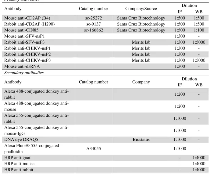
Table 1. Antibodies and dyes
Primary antibodies
Antibody Catalog number Company/Source Dilution
IF WB
Mouse anti-CD2AP (B4) sc-25272 Santa Cruz Biotechnology 1:500 1:500 Rabbit anti-CD2AP (H290) sc-9137 Santa Cruz Biotechnology 1:500 1:500 Mouse anti-CIN85 sc-166862 Santa Cruz Biotechnology 1:500 1:100
Mouse anti-SFV-nsP1 1:300 -
Rabbit anti-SFV-nsP3 Merits lab 1:300 1:5000
Rabbit anti-CHIKV-nsP1 Merits lab 1:300 -
Rabbit anti-CHIKV-nsP2 Merits lab 1:300 -
Rabbit anti-CHIKV-nsP3 Merits lab 1:300 1:5000
Mouse anti-dsRNA 1:300 -
Secondary antibodies
Antibody Catalog number Company Dilution
IF WB
Alexa 488-conjugated donkey
anti-rabbit 1:200 -
Alexa 488-conjugated donkey
anti-mouse 1:200 -
Alexa 555-conjugated donkey
anti-rabbit 1:1000 -
Alexa 555-conjugated donkey
anti-mouse-IgG 1:1000 -
DNA dye DRAQ5 Biostatus 1:1000 -
Alexa Fluor® 555-conjugated
phalloidin A34055 1:1000 -
HRP anti-goat - 1:4000
HRP anti-mouse - 1:4000
7. Results
7.1. CHIKV and SFV nsP3 protein binds the cellular CD2AP
To determine whether the interaction of CD2AP and nsP3 of SFV was also present during viral infection, HOS cells were infected at MOI 1 with either SFV-WT or SFV-noCD2AP. Cells were lysed at 4 and 8 h p.i., and lysates were immunoprecipitated with anti-SFV-nsP3 antibody. Cell lysates were subjected to SDS/PAGE and precipitates were probed with CD2AP, anti-SFV-nsP3 and anti-actin antibodies. As shown in Figure 5, nsP3 and CD2AP interacted in cells infected with WT; the interaction was, however, lost in cells infected with noCD2AP. In a parallel experiment cells were infected at MOI 1 with either WT or SFV-noCD2AP and lysed at 6 h p.i. Whole-cell lysates were subjected to SDS/PAGE and probed with anti-CD2AP, anti-SFV-nsP3 and anti-actin antibodies. All proteins, CD2AP, nsP3 and actin were found in whole-cell lysate (Fig. 5).
Figure 5. nsP3 and CD2AP binding. Binding of CD2AP and nsP3 was assessed by immunoprecipitation
experiments. Cells were infected at MOI 1 with either SFV-WT or SFV-noCD2AP, lysed at 4 or 8 h p.i. IPs showed that nsP3 bound CD2AP only in cells infected with wild-type virus. Whole-cell lysates were analyzed in a parallel experiment. Cells were infected at MOI 1 with either SFV-WT or SFV-noCD2AP and lysed at 6 h p.i. Lysates showed that CD2AP was present for all conditions and nsP3 only in cells infected with either virus. Similarly, nsP3 could bind CD2AP only during infection with SFV-WT.
7.2. Growth curves.
Once the interaction between nsP3 and CD2AP during viral infection was confirmed by immunoprecipitation assays and SILAC (un-published), the growth of the different mutant viruses was assessed to determine whether the inability to interact with CD2AP caused an attenuated phenotype. Cells were infected with either WT, P423A, CHIKV-R428A, CHIKV-PR, SFV-WT or SFV-noCD2AP at MOI 1 or MOI 0.1 for single-step and
multi-step growth curve experiments, respectively (for differences between the viruses refer to fig. 4). Viral infection rates were, then, determined by plaque assay in BHK cells. Results showed that all mutants were attenuated when comparing with corresponding wild type virus. CHIKV-PR, CHIKV-P423A and CHIKV-R428A were highly impaired with 2-log, 1-log or 0.5-log reduction, respectively. On the other hand, SFV-noCD2AP was not severely attenuated when comparing with SFV-WT (Fig. 6).
7.3. CD2AP and nsP3 co-localization
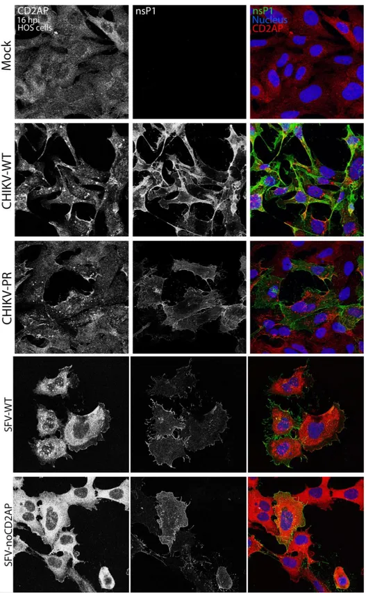
Following assessment of nsP3 and CD2AP binding, we wanted to check the cellular localization of both proteins during viral infection. For this purpose, cells were infected at MOI 1 with either of the following viruses: CHIKV-WT, CHIKV-PR, SFV-WT or SFV-noCD2AP (for differences between the viruses see fig. 4). Cells were fixed at 16 or 8 h p.i. for CHIKV and SFV, respectively; and CD2AP and nsP3 were visualized by confocal microscopy. As observed for mock-infected cells, CD2AP is normally diffused in the cytoplasm. However, when cells were infected with either CHIKV-WT or SFV-WT, CD2AP was shown to aggregate in foci together with nsP3 (Fig. 7). On the other hand, it remained diffused in cytoplasm upon infection with either of the mutants (Fig. 7). These results, together with results from IPs, confirmed interaction of CD2AP and nsP3 during viral infection for the wild type viruses (CHIKV-WT and WT) and the lack of binding for the mutants CHIKV-PR and SFV-noCD2AP.
Figure 6. Viral growth. BHK cells were infected with either CHIKV-WT, CHIKV-P423A, CHIKV-R428A,
CHIKV-PR, SFV-WT or SFV-noCD2AP at MOI 0.1 or 1 for multi-step or single-step growth curve experiments, respectively. Mutant viruses were attenuated compared with either CHIKV-WT or SFV-WT. MOI (multiplicity of infection), PFU (plaque forming units).
Figure 7. CD2AP and nsP3 co-localization during infection. HOS cells were infected at MOI 1 with one
of the following viruses: CHIKV-WT, CHIKV-PR, SFV-WT and SFV-noCD2AP. Cells were fixed and permeabilized at 16 or 8 h p.i., for CHIKV- and SFV-infected cells, respectively. CD2AP (red) and nsP3 (green) were visualized by confocal microscopy*. CD2AP was found in foci co-localizing with nsP3 in wild-type infected cells, while CD2AP was diffused in cells infected with either mutant virus or mock-infected. * Cells were stained with a mix of primary antibodies including: mouse anti-CD2AP antibody (1:500) and rabbit anti-CHIKV-nsP3 or rabbit anti-SFV-nsP3 (1:300); and secondary antibodies: Alexa 488-conjugated donkey anti-rabbit antibody (1:200) and Alexa 555-conjugated donkey anti-mouse antibody (1:1000).
For analysis of foci production, HOS cells were infected at MOI 1 with CHIKV-WT or SFV-WT. Cells were fixed at 8, 10 or 16 h p.i. for CHIKV, and at 4, 6 and 8 h p.i. for SFV. Both, nsP3 and CD2AP were visualized by confocal microscopy. Foci were already present at the earliest time point tested and were shown to not dissolve over time in infected cells (Fig. 8). Interestingly, the foci increased in size over time when cells were infected with CHIKV-WT compared with infection with SFV-WT where they did not grow (Fig. 8).
Figure 8. CD2AP and nsP3 foci over time. HOS cells were infected at MOI 1 with either CHIKV-WT
or SFV-WT. CD2AP and nsP3 were visualized by confocal microscopy*. Cells infected with CHIKV-WT were fixed at 8, 10 or 16 h p.i. CD2AP foci were already present at 6 h p.i and grew over time. Cells infected with SFV-WT were fixed at 4, 6 or 8 h p.i. CD2AP foci were present at 4 h p.i; however, foci did not grow over time in SFV-infected cells. * Cells were stained with a mix of primary antibodies including: mouse anti-CD2AP antibody (1:500) and rabbit anti-CHIKV-nsP3 or rabbit anti-SFV-nsP3 (1:300); and secondary antibodies: Alexa 488-conjugated donkey anti-rabbit antibody (1:200) and Alexa 555-conjugated donkey anti-mouse antibody (1:1000).
7.4. Characterization of CD2AP-nsP3 foci
To determine whether CD2AP-nsP3 foci observed were replication complexes (RCs), HOS cells were infected at MOI 1 with either CHIKV-WT, CHIKV-PR, WT or SFV-noCD2AP, and were stained to visualize CD2AP and dsRNA or nsP1, the last two considered markers for RCs. As expected, CD2AP did not co-localize with dsRNA or nsP1 in cells infected with either mutant virus (Fig. 9 and 10). Interestingly, CD2AP foci were shown to co-localize with dsRNA for both, CHIKV and SFV wild type viruses (Fig. 10). Nonetheless, some CD2AP foci did not co-localize with dsRNA in CHIKV infection. Analysis of the nsP1 and CD2AP staining showed no co-localization for any of the infections (Fig. 9).
Figure 9. CD2AP and nsP1 co-localization. HOS cells were infected at MOI 1 with either CHIKV-WT,
CHIKV-PR, SFV-WT or SFV-noCD2AP. Cells were fixed at 16 or 8 h p.i for CHIKV and SFV, respectively. CD2AP and nsP1 were visualized by confocal microscopy*. No co-localization of CD2AP and nsP1 was observed for any virus. *Cells were stained with a mix of primary antibodies including: mouse anti-CD2AP antibody (1:500) and rabbit anti-CHIKV-nsP1 or rabbit anti-SFV-nsP1 (1:300); and secondary antibodies: Alexa 488-conjugated donkey anti-rabbit antibody (1:200), Alexa 555-conjugated donkey anti-mouse antibody (1:1000) and DNA dye DRAQ5 (1:1000).
Figure 10. CD2AP and dsRNA co-localization. HOS cells were infected at MOI 1 with either CHIKV-WT,
CHIKV-PR, SFV-WT or SFV-noCD2AP. Cells were fixed at 16 or 8 h p.i for CHIKV and SFV, respectively and CD2AP and dsRNA visualized by confocal microscopy*. Co-localization of CD2AP and dsRNA was observed for both CHIKV-WT and SFV-WT while no co-localization was shown in cells infected with either mutant virus. * Cells were stained with a mix of primary antibodies including: mouse anti-CD2AP antibody (1:500) and rabbit anti-dsRNA or rabbit anti-CIN85 (1:300); and secondary antibodies: Alexa 488-conjugated donkey anti-rabbit antibody (1:200) and Alexa 555-conjugated donkey anti-mouse antibody (1:1000).
Next, we investigated, co-localization of CD2AP foci with CIN85 in cells infected with CHIKV. CIN85 is an adaptor protein of 85 kDa which shares 54% similarity to CD2AP in the amino acid sequence and has a similar binding site. After visualization by confocal microscopy, foci positive for both CD2AP and CIN85 were found in cells infected with CHIVK-WT (Fig. 11). As expected no foci were observed in cells infected with CHIKV-PR, which supports the hypothesis that nsP3 can interact with both CD2AP and CIN85 and that mutation in CD2AP binding site abrogates interaction, not only with CD2AP but also with CIN85.
Figure 11. CD2AP and CIN85 co-localization. HOS cells were infected at MOI 1 with either CHIKV-WT,
CHIKV-PR, SFV-WT or SFV-noCD2AP. Cells were fixed at 16 or 8 h p.i for CHIKV and SFV, respectively; CD2AP and CIN85 were visualized by confocal microscopy*. Co-localization of CD2AP and CIN85 was observed for both CHIKV-WT while no co-localization was shown in cells infected with CHIKV-PR virus. * Cells were stained with a mix of primary antibodies including: mouse anti-CD2AP antibody (1:500) and rabbit anti-dsRNA or rabbit anti-CIN85 (1:300); and secondary antibodies: Alexa 488-conjugated donkey anti-rabbit antibody (1:200), Alexa 555-conjugated donkey anti-mouse antibody (1:1000) and DNA dye DRAQ5 (1:1000).
7.5. Actin rearrangement upon infection
CD2AP is known to regulate the actin machinery in cells and has been shown to be targeted during different viral infections. To understand whether Old World alphaviruses interact with CD2AP to modify the actin organization, BHK cells were infected at MOI 1 with either
CHIKV-WT, CHIKV-PR, SFV-WT or SFV-noCD2AP and fixed at 2, 4, 6, 8, 10 and 16 h p.i. Actin, nsP3 and dsRNA were visualized by confocal microscopy. At late time points the actin cytoskeleton was altered, with loss of well-defined stress fibers and increase of transverse arcs for SFV infection (Fig. 12). However, the alteration of the actin cytoskeleton was only shown in 10% of wild-type infected cells for SFV and was absent in cell infected with CHIKV.
Virus SFV-WT SFV-noCD2AP Normal stress fibres 156 120 Loss of stress fibres 20 - Total infected cells 176 120 % cells with loss of stress fibres 11% 0%
Figure 12. Actin rearrangement upon viral infection. BHK cells were infected at MOI 1 with either
SFV-WT or SFV-noCD2AP. Cells were fixed at 8 h p.i. Actin and nsP3 or dsRNA were visualized by confocal microscopy*. Actin rearrangement phenotype (lower panel): loss of stress fibers and increased in arcs (cortical actin) was observed in 11% of cells infected with SFV-WT. The table shows the number of infected cells which presented normal actin stress fibers, number of infected cells which presented loss on stress fibers and increase cortical actin staining, total number of infected cells and percentage of cells with loss of stress fibers. Quantification was determined in stained cells and blinded when possible. * Cells were stained with a mix of primary antibodies including: mouse anti-dsRNA and rabbit anti-SFV-nsP3 (1:300); and secondary antibodies: Alexa 488-conjugated donkey anti-rabbit antibody (1:200) and Alexa 555-conjugated donkey anti-mouse antibody (1:1000) and Alexa Fluor® 555-conjugated phalloidin
7.6. Internalization of replication complexes
Internalization of replication complexes varies among different alphaviruses. SFV RCs are formed at the plasma membrane and internalized in a multi-step mechanism, whereas CHIKV RCs are not internalized. To assess whether CD2AP binding is important for the replication complexes internalization, cells were infected at MOI 1 with either SFV-WT or SFV-noCD2AP and cells were fixed at 2, 4 and 6 h p.i and nsP3, dsRNA and actin were visualized by confocal microscopy. A parallel experiment using Wortmannin, a drug that blocks internalization of spherules by inhibiting phosphatidylinositol 3-kinase (PI3K), an enzyme known to be involved in internalization of replication complexes, was performed.Spherules were found in plasma membrane and scattered around the cytoplasm at early times after infection. As the infection went on, the replication complexes were internalized to be found in the perinuclear area at late times (Fig. 13A). However, internalization of spherules in cells infected with SFV-noCD2AP seemed to be delayed (Fig. 13A). Upon Wortmannin treatment spherules are expected to be found on the plasma membrane, since the internalization is abrogated by this drug. During SFV-WT infection replication complexes were mostly found at the plasma membrane even at late times after infection (only 2 h p.i is shown, Fig. 13B). Surprisingly, replication complexes of SFV-noCD2AP were found aligning with actin fibers throughout the infection (Fig. 13B and 14).
A)
B)
Figure 13. Replication complexes internalization. Cells were infected with SFV-noCD2AP at MOI 1. A)
Cells were fixed at 2, 4 and 6 h p.i., and actin, nsP3 and dsRNA were visualized by confocal microscopy*. RCs were found scattered at the cytoplasm at 2 h p.i., were found in the perinuclear area from 4 h p.i., in SFV-WT infection. However, RCs were found at the plasma membrane at 2 h p.i., scattered throught the cytoplasma at 4 h p.i, and in the perinuclear area at 6 h p.i., for SFV-noCD2AP infection. B) Cells were treated with 400 nMs of Wortmannin (low row) or not treated (upper row), fixed at 2 h p.i. and actin, nsP3 and dsRNA were visualized*. Without treatment RCs were found scattered in the cytoplasm for SFV-WT infection and at the plasma membrane for SFV-noCD2AP infection. After treatment with Wortmannin, RCs were found at the plasma membrane in SFV-WT infection while they were aligning with actin fibers in SFV-noCD2AP infection. * Cells were stained with a mix of primary antibodies including: mouse anti-dsRNA and rabbit anti-SFV-nsP3 (1:300); and secondary antibodies: Alexa 488-conjugated donkey anti-rabbit antibody (1:200) and Alexa 555-conjugated donkey anti-mouse antibody (1:1000) and Alexa Fluor® 555-555-conjugated phalloidin
Figure 14. Replication complexes internalization. Cells were infected with SFV-noCD2AP at MOI 1 and
treated with 400 nM of wortmannin during the length of the experiment. Cells were fixed at 2, 4, 6 or 8 hpi and actin, nsP3 and dsRNA were visualized by confocal microscopy*. Replication complexes were found aligning with actin fibers from the 2 h p.i. Zoom images of the selected areas is shown. * Cells were stained with a mix of primary antibodies including: mouse anti-dsRNA and rabbit anti-SFV-nsP3 (1:300); and secondary antibodies: Alexa 488-conjugated donkey rabbit antibody (1:200) and Alexa 555-conjugated donkey anti-mouse antibody (1:1000) and Alexa Fluor® 555-conjugated phalloidin
8. Discussion
The interaction between the scaffolding protein CD2AP and nsP3 of CHIKV has recently been observed (Merits lab, unpublished data). Interaction of CD2AP with other viruses has also been shown, in particular it has been reported to interact with Venezuelan equine encephalitis virus (VEEV) a New World alphavirus, which is thought to be important due to CD2AP’s role in maintenance of blood-brain barrier4,22. In this work, we have demonstrated the interaction between nsP3 and CD2AP during infection of SFV by co-immunoprecipitation experiments. However, we were not able to demonstrate interaction of CD2AP and nsP3 by immunoprecipitation during CHIKV infection. We encountered difficulties in obtaining signals
of nsP3 and CD2AP proteins during and Western Blot experiments investigating both immunoprecipitation and whole-cell lysate samples. We believe that the primary anti-CD2AP antibody could have suffered degradation because the signal has been decreasing gradually and the brand is known to be not reliable. An alternative explanation for the absence of signal for both CD2AP and nsP3 could be due to slight differences in blocking of membranes after protein transfer, which could have been too short, and washing. Both could have affected the outcome of the experiment.
Despite the inability to detect CD2AP:nsP3 interaction by immunoprecipitation experiments, the decrease of 5 to 100-fold in viral production of mutant viruses lacking the ability to bind CD2AP indicates that the interaction between CD2AP and nsP3 during CHIKV and SFV infection is important for efficient viral replication. Here, we showed that CD2AP and nsP3 co-localize during CHIKV and SFV infection (Fig. 7). CD2AP containing foci grew over time in CHIKV infection while their size remained the same during SFV infection (Fig. 8). During CHIKV infection, nsP3 is found both in replication complexes (vesicles including dsRNA and non-structural proteins) and in other punctate structures in the cytoplasm, while most nsP3 is found in replication complexes during SFV infection. This could be the reason for the difference observed regarding the size of CD2AP foci during infection. Moreover, CD2AP co-localized with dsRNA during infection for both viruses (Fig. 10). Nevertheless, during infection with CHIKV independent CD2AP foci, which did not co-localize with dsRNA, were observed. We believe that these CD2AP foci co-localize with nsP3 in CHIKV infection; however, this could not be tested due to species restrictions (only mouse and rabbit antibodies were available for CD2AP, nsP3 and dsRNA) of the available primary antibodies. Nonetheless, no co-localization of CD2AP and nsP1 proteins was observed in CHIKV or SFV infections. The nsP1 protein is known to be found at the plasma membrane and in filopodia formed upon viral infection3. On the other hand, the nsP3 found in the RCs is thought to be further away
from these structures. The lack of co-localization between nsP1 and CD2AP upon infection with wild type viruses, even though CD2AP co-localized with RCs, could be explained by the organization of the proteins within the RCs.
Moreover, we observed co-localization of CD2AP and CIN85 in all mock-, CHIKV-WT and CHIKV-PR-infected cells (Fig. 11). These two proteins are highly related and are part of the same family of adaptor proteins; both have three SH3 domains17, whose preferred binding
site is PX(P/A)XPR. This binding motif is found in both CHIKV and SFV nsP3 (Fig. 3). Furthermore, CD2AP and CIN85 have been observed to form homo- and heterodimers through interaction between their SH3 domains17. Interestingly, both proteins co-localized in punctate
structures during CHIKV-WT infection (Fig. 11). Our results suggest that both CD2AP and CIN85 interact with nsP3 during CHIKV infection; however, studies to verify if CIN85 co-localizes with nsP3 during viral infection as well as immunoprecipitation experiments to assess binding should be carried out. At the same time, the lack of foci during CHIKV-PR infection suggests that the mutation in CD2AP binding site, prevents not only interaction of nsP3 with CD2AP but also with CIN85. In addition to the experiments in CHIKV, we plan to study this interaction in SFV.
Similar foci to those observed during this study have been observed previously during alphavirus infection 14. These foci contain nsP3 and also G3BP (Ras GTPase-activating
by nsP3 abrogates formation of stress granules upon infection 15. We want to determine whether the foci observed during this project also include G3BP and if their formation is driven by G3BP presence or if CD2AP and nsP3 interaction is enough to create them. Visualization of CD2AP and G3BP by immunofluorescence after infection with CHIKV and SFV, as well as studies of CD2AP foci formation upon infection in knock-out cells for G3BP could help determine whether CD2AP and nsP3 interaction is enough to produce foci.
Actin rearrangement is important for many cellular processes such as internalization, transport and formation of cell-to-cell contact structures31. Rearrangement of the actin cytoskeleton is used by different viruses to ensure a high efficiency during viral replication or budding; i.e., vaccinia virus and simian virus facilitate their intracellular movements and budding by promoting actin polymerization32,33. CD2AP is known to be involved in actin rearrangement, interacting with proteins such as dendrin, synaptopodin, dynamin to form stress fibres4. CD2AP is also involved in actin capping by its interaction with acting capping proteins
such as CAPZ24, or formation of lamellipodia by linking capping proteins to cortactin in the cell periphery25. Therefore, we studied whether nsP3 interaction with CD2AP caused rearrangement of the actin cytoskeleton upon CHIKV and SFV infection. Loss of stress fibers and an increase of arcs (cortical actin) was observed in 10% of SFV-infected cells (Fig. 12); however, no actin rearrangement was observed in CHIKV-infected cells. The low percentage of cells presenting this phenotype and the absence of it during CHIKV infection could indicate that it is a response to viral infection rather than an alphavirus-induced phenotype; on the other hand, the low multiplicity of infection used could also explain the low percentage seen. Nevertheless, this phenotype has recently been observed in CD2AP knock-out cells with high levels of TFG-β120, which could suggest that Semliki Forest virus is capable of inducing actin rearrangement by sequestration of CD2AP.
Intracellular transport is needed for efficient viral replication; i.e., replication complexes of Semliki Forest virus are formed at the plasma membrane early in infection and are internalized in a multistep mechanism to the perinuclear area10,11. Since CD2AP is known to be involved in cellular trafficking, we studied whether nsP3 and CD2AP interaction during SFV infection is important for transport of RCs. Our results suggest that internalization of the spherules is delayed in infection with SFV-noCD2AP; however viral replication is still efficient with a decrease of less than 5-fold (Fig. 6). The first step of the internalization process is mediated by activation of the phosphatidylinositol 3-kinase (PI3K), an enzyme that can be blocked by Wortmannin. To understand how RC internalization was delayed in cells infected with SFV-noCD2AP, we decided to perform experiments using Wortmannin. Replication complexes were found at the plasma membrane in cells infected with SFV-WT after treatment with Wortmannin (Fig 13B), as it has been reported previously10. Meanwhile, RCs were found aligning with actin fibres throughout the experiment for SFV-noCD2AP. The alignment of replication complexes with actin fibres for SFV infection has been observed previously10. However, we report here a more prominent pattern for SFV-noCD2AP, which is even clearer after treatment with Wortmannin (Figs. 13B and 14). Replication complexes of SFV-noCD2AP seem to escape the effect of Wortmannin, which could suggest that internalization of these complexes is independent of PI3K. Could, otherwise, be explained if the replication complexes were not formed at the plasma membrane, but at other membranous structures in the cell, and after treatment with Wortmannin, these replication complexes formed at other membranes would be
motionless at their site of formation. Therefore, we propose that CD2AP could be involved in the transport of the non-structural protein and mRNA to the plasma membrane.
Besides actin rearrangement, CD2AP is involved in many other cellular mechanisms. For example, CD2AP is known to mediate signaling, internalization and degradation of receptor tyrosine kinases (RTKs), which are involved i.e., in activation of survival pathways; thus, RTKs are tightly regulated in the cell18,26,34. Once RTKs bind their ligand, they become activated and the signaling pathway that will lead to a certain response will start. In order to be deactivated, RTKs are ubiquitinated and then internalized and targeted either for recycling to the cell surface or for degradation18. CD2AP has been linked to endosomal trafficking, and thereby to the degradative pathway35. We hypothesized that nsP3 could bind CD2AP in order to prevent internalization of RTKs, therefore prolonging their effect. Studying internalization of RTKs in infected- and non-infected cells with wild type and mutant viruses could help determine whether sequestration of CD2AP prevents internalization of RTKs. Moreover, activation of PI3K, which leads to an increase of phosphorylated Akt (p-Akt), could be a marker for this phenomenon.
Summarizing, we report that mutations in CD2AP binding site give an attenuated growth phenotype in both CHIKV and SFV. Moreover, CD2AP was observed to co-localize with nsP3, dsRNA and CIN85 during viral infection (Figs. 8, 10 and 11). Interestingly, internalization of Semliki Forest virus replication complexes was delayed upon mutation of CD2AP binding site (Fig 13A). In addition, after treatment with Wortmannin, replication complexes of SFV-noCD2AP were not found at the plasma membrane as expected, but aligning with actin fibers inside the cell (Fig. 13B and 14). Hence, we propose that CD2AP could be involved in the transport of non-structural proteins and the viral genome to the plasma membrane before formation of the replication complexes. This finding could contribute to better understanding of the viral infection, which in turn could facilitate development of antiviral treatments and/or vaccines. Chikungunya fever symptoms, as mentioned previously, are debilitating and long-lasting which leads to a decrease in quality of life for patients suffering from the disease. Furthermore, there are no vaccines or antiviral treatments available, which has a great impact in low-income countries, preventing populations from further economic progression. Altogether, there is a need to study further the biology of this virus.
9. References
1. Jose, J., Snyder, J. E. & Kuhn, R. J. Replication and Assembly. Rev. Lit. Arts Am. 837– 856 (2009). doi:10.2217/fmb.09.59
2. Rupp, J. C., Sokoloski, K. J., Gebhart, N. N. & Hardy, R. W. Alphavirus RNA synthesis and non-structural protein functions. J. Gen. Virol. 96, 2483–2500 (2015).
3. Suhrbier, A., Jaffar-Bandjee, M.-C. & Gasque, P. Arthritogenic alphaviruses—an overview. Nat. Rev. Rheumatol. 8, 420–429 (2012).
4. Kim, D. Y. et al. New World and Old World Alphaviruses Have Evolved to Exploit Different Components of Stress Granules, FXR and G3BP Proteins, for Assembly of Viral Replication Complexes. PLoS Pathog. 12, 1–31 (2016).
5. Bala Murugan, S. & Sathishkumar, R. Chikungunya infection: A potential re-emerging global threat. Asian Pac. J. Trop. Med. 9, 933–937 (2016).
6. Schwartz, O. & Albert, M. L. Biology and pathogenesis of chikungunya virus. Nat. Rev.
Microbiol. 8, 491–500 (2010).
7. Kraemer, M. U. G. et al. The global distribution of the arbovirus vectors Aedes aegypti and Ae. Albopictus. Elife 4, 1–18 (2015).
8. Lo Presti, A., Cella, E., Angeletti, S. & Ciccozzi, M. Molecular epidemiology, evolution and phylogeny of Chikungunya virus: An updating review. Infect. Genet. Evol. 41, 270– 278 (2016).
9. Fros, J. J. & Pijlman, G. P. Alphavirus infection: Host cell shut-off and inhibition of antiviral responses. Viruses 8, (2016).
10. Spuul, P. et al. Phosphatidylinositol 3-Kinase-, Actin-, and Microtubule-Dependent Transport of Semliki Forest Virus Replication Complexes from the Plasma Membrane to Modified Lysosomes. J. Virol. 84, 7543–7557 (2010).
11. Thaa, B. et al. Differential Phosphatidylinositol-3-Kinase-Akt-mTOR Activation by Semliki Forest and Chikungunya Viruses Is Dependent on nsP3 and Connected to Replication Complex Internalization. J. Virol. 89, 11420–11437 (2015).
12. Salonen, A. et al. Properly folded nonstructural polyprotein directs the semliki forest virus replication complex to the endosomal compartment. J. Virol. 77, 1691–702 (2003). 13. Panas, M. D., Ahola, T. & McInerney, G. M. The C-terminal repeat domains of nsP3 from the Old World alphaviruses bind directly to G3BP. J. Virol. 88, 5888–93 (2014). 14. Panas, M. D. et al. Viral and Cellular Proteins Containing FGDF Motifs Bind G3BP to
Block Stress Granule Formation. PLoS Pathog. 11, 1–22 (2015).
15. Panas, M. D. et al. Sequestration of G3BP coupled with efficient translation inhibits stress granules in Semliki Forest virus infection. Mol. Biol. Cell 23, 4701–4712 (2012).
16. Schulte, T. et al. Combined structural, biochemical and cellular evidence demonstrates that both FGDF motifs in alphavirus nsP3 are required for efficient replication. Open
Biol. 6, 160078 (2016).
17. Dikic, I. CIN85/CMS family of adaptor molecules. FEBS Lett. 529, 110–115 (2002). 18. Tsui, C. C. & Pierchala, B. a. CD2AP and Cbl-3/Cbl-c constitute a critical checkpoint in
the regulation of ret signal transduction. J. Neurosci. 28, 8789–800 (2008).
19. Lu, C. et al. EGF-recruited JunD/c-fos complexes activate CD2AP gene promoter and suppress apoptosis in renal tubular epithelial cells. Gene 433, 56–64 (2009).
20. Yaddanapudi, S. et al. CD2AP in mouse and human podocytes controls a proteolytic program that regulates cytoskeletal structure and cellular survival. J. Clin. Invest. 121, 3965–3980 (2011).
21. Harrison, B. J. et al. The Adaptor Protein CD2AP Is a Coordinator of Neurotrophin Signaling-Mediated Axon Arbor Plasticity. J. Neurosci. 36, 4259–4275 (2016).
22. Cochran, J. N., Rush, T., Buckingham, S. C. & Roberson, E. D. The Alzheimer’s disease risk factor CD2AP maintains blood-brain barrier integrity. Hum. Mol. Genet. 24, 6667– 74 (2015).
23. Kirsch, K. H., Georgescu, M. M., Ishimaru, S. & Hanafusa, H. CMS: an adapter molecule involved in cytoskeletal rearrangements. Proc. Natl. Acad. Sci. USA 96, 6211–6 (1999). 24. Hutchingst, N. J., Clarkson, N., Chalkley, R., Barclay, A. N. & Brown, M. H. Linking the T cell surface protein CD2 to the actin-capping protein CAPZ via CMS and CIN85.
J. Biol. Chem. 278, 22396–22403 (2003).
25. Zhao, J. et al. CD2AP links cortactin and capping protein at the cell periphery to facilitate formation of lamellipodia. Mol. Cell. Biol. 33, 38–47 (2013).
26. Tossidou, I. et al. CD2AP/CIN85 balance determines receptor tyrosine kinase signaling response in podocytes. J. Biol. Chem. 282, 7457–7464 (2007).
27. Ceregido, M. A. et al. Multimeric and differential binding of CIN85/CD2AP with two atypical proline-rich sequences from CD2 and Cbl-b. FEBS J. 280, 3399–3415 (2013). 28. Ortega Roldan, J. L. et al. Distinct ubiquitin binding modes exhibited by SH3 domains:
Molecular determinants and functional implications. PLoS One 8, 1–14 (2013).
29. Rouka, E. et al. Differential recognition preferences of the three Src Homology 3 (SH3) domains from the adaptor CD2-associated Protein (CD2AP) and Direct Association with Ras and Rab Interactor 3 (RIN3). J. Biol. Chem. 290, 25275–25292 (2015).
30. Laakkonen, P., Auvinen, P., Kujala, P. & Kääriäinen, L. Alphavirus replicase protein NSP1 induces filopodia and rearrangement of actin filaments. J. Virol. 72, 10265–10269 (1998).
31. Disanza, A. et al. Actin polymerization machinery: The finish line of signaling networks, the starting point of cellular movement. Cell. Mol. Life Sci. 62, 955–970 (2005).
32. Taylor, A. et al. Effects of an In-Frame Deletion of the 6k Gene Locus from the Genome of Ross River Virus. J. Virol. 90, 4150–4159 (2016).
33. Frischknecht, F. & Way, M. Surfing pathogens and the lessons learned for actin polymerization. Trends Cell Biol. 11, 30–38 (2001).
34. Li, N., Hill, K. S. & Elferink, L. A. Analysis of receptor tyrosine kinase internalization using flow cytometry. Methods Mol. Biol. 457, 305–17 (2008).
35. Cormont, M. et al. CD2AP/CMS Regulates Endosome Morphology and Traffic to the Degradative Pathway Through its Interaction with Rab4 and c-Cbl. Traffic 4, 97–112 (2003).