Design and synthesis of inhibitors
of the ADP ribosylating toxin
ExoS: Targeting the Type III
Secretion
System
(T3SS)
of
Pseudomonas aeruginosa.
Öznur Aglar
Öznur Aglar
Master Thesis 30 ECTS
Report passed: 21 June, 2016 Supervisor: Prof. Mikael Elofsson Examiner: Bertil Eliasson
Abstract
Pseudomonas aeruginosa, a gram-negative bacterium, is one of the most challenging
pathogens due to intrinsic resistance to antibiotics. It has the ability to rapidly create new resistance mechanisms. Therefore, the lack of useful anti-pseudomonal agents forces us to develop new appropriate therapeutic agents via alternative strategies such as targeting the major virulence factors of the bacteria. These bacteria have a variety of virulence factors and one of them is type III secretion system (T3SS) which is the most attractive target for the anti-pseudomonal drug discovery. T3SS in P.
aeruginosa is essential virulence factor that is responsible for the secretion of four
effector proteins into the host cell which are ExotoxinS (ExoS), ExoT, ExoU, and ExoY. ExoS is a bifunctional enzyme and has a GTPase activating (GAP) domain and an ADP-ribosyl transferase (ADPRT) domain that are important for the pathogenicity of P. aeruginosa according to several studies. Herein, we target to inhibit ExoS ADPRT activity via small organic compounds as a new therapeutic strategy. For this reason, we designed and synthesized a set of compounds to investigate the role of the different side chains and scaffold to the activity against ADPRT activity of ExoS and also to improve activity. The side chain and main scaffold were modified with various groups that have different length and rigidity in order to establish structure-activity relationships (SAR) of compounds. Besides, all designed and synthesized compounds were tested against ExoS-ADPRT activity in an enzymatic assay which was developed by our collaborators in Karolinska Institute.
Keywords
ADPRT, antibiotic, antibiotic resistance, anti-pseudomonal, bacterial virulence, enzymatic assay, ExotoxinS (ExoS), Pseudomonas aeruginosa, type III secretion system (T3SS).
III
List of Abbreviations
ADP Adenosine diphosphate
ADPRT ADP-ribosyltransferase
Cdc42 Cell division control protein 42 homolog
CF Cystic Fibrosis
DMF Dimethylformamide
DMSO Dimethyl sulfoxide
eq Equivalent
εNAD+ 1,N6 -etheno NAD
EtOAC Ethyl acetate
EtOH Ethanol
ExoS P. aeruginosa exotoxin S
ExoT P. aeruginosa exotoxin T
ExoU P. aeruginosa exotoxin U
ExoY P. aeruginosa exotoxin Y
GAP GTPase activating protein
IC50 Half maximal inhibitory concentration
HPLC High-performance liquid chromatography
M Molar concentration
MLD Membrane localization domain
MS Mass Spectrometry
NAD Nicotinamide adenine dinucleotide
P. aeruginosa Pseudomonas aeruginosa
QS Quorum Sensing
Rf Retention factor
TBAI 1-(p-Toluenesulfonyl)imidazole
TEA Triethylamine
TLC Thin layer chromatography
TsIm Tetrabutylammonium iodide
T3SS Type III Secretion System
V
Table of Contents
Abstract ... I
List of Abbreviations ...III
1. Introduction ... 1
1.1. Pseudomonas aeruginosa ... 1
1.2 Drug-Resistance of Pseudomonas aeruginosa... 1
1.3 The Type III Secretion System and Exotoxin S ... 2
1.4 STO1101 and ME0569: Inhibitors of ExoS-ADPRT ... 2
1.5 Aim of the diploma work ... 3
2. Popular scientific summary including social and ethical aspects ... 3
2.1 Popular scientific summary ... 3
2.2 Social and ethical aspects... 3
3. Experimental ... 4
3.1 Chemistry Section ... 4
3.2 Biology Section ... 8
3.2.1 Dose-response experiment: ... 8
4. Results and Discussion ...10
5. Conclusions ...15
6. Future Plan ...16
7. Appendix I ...17
8. Appendix II ...19
9. Appendix III ...21
10. Acknowledgement ...23
11. References ...24
1
1. Introduction
1.1. Pseudomonas aeruginosa
Pseudomonas aeruginosa is a gram-negative bacterium and an opportunistic human pathogen that causes serious infections and can be fatal among immunocompromised individuals such as HIV, cancer and organ transplant patients and infants who have an undeveloped immune system.1 It is species of Pseudomonas and grows on a wide
range of environments.2 The first genome sequence of the wild type of these bacteria
(PA01) was completed in 2000 and provided useful information which showed the bacteria contains various paralogous gene families encoding significant adaptive functions.3 Genetic capacity of P. aeruginosa makes the bacteria capable of adapting
to extreme environmental conditions.3 Generally, it exists in water, air, soil, skins of
animals, plants and humans.4 Mostly, it lives in moist habitat. These bacteria are
motile via polar flagella. P. aeruginosa strains play an significant role in nature during the decomposition and biodegradation of the organic materials and also carbon and nitrogen cycle. They are known as an aerobe but also can survive under anaerobic condition. It has been shown that they are highly adaptive within various enviromental alterations.5
P. aeruginosa has natural ability to develop resistance against all marked antibiotic classes.6 For that treatment of P. aeruginosa infection remains
challenging.5 Clinically, infections of P. aeruginosa is very important due to high rates
of drug resistance and mortality among patients with burn wounds, cystic fibrosis (CF) and compromised immunity.2 It infects a numerous animal, plant and person
with immunodeficiency.4 In addition, P. aeruginosa infection may occur within
healthy individuals, as well.7 It is a healthcare-associated infection which can be
acquired from contaminated sources and transmission of person to person.1
This pathogen has numerous virulence factors that are responsible for its pathogenesis.8 Pili and flagella, quorum sensing (QS), Exotoxin A, the type III
secretion system, phospholipase, and pyoverdin are the most important virulence factors of this pathogen. Except pili and flagella other virulence factors that mentioned above, contribute to cell and tissue destruction of host and also responsible for impaired host defences. Pili and flagella allow host cell adherence, motility, and biofilm formation.9
1.2 Drug-Resistance of Pseudomonas aeruginosa
The treatment of P. aeruginosa infections is extremely problematic because of the intrinsic resistant to a broad range of antibiotics.6 It can easily develop resistance by
means of several mechanisms which are associated with low membrane permeability, efflux pump and genetic mutations.10 Multi-factorial mechanism of antibiotic
resistance in P. aeruginosa is based on low outer membrane permeability for the antibiotics, multiple antibiotic modifying enzymes like ß-lactamases, metallo-ß-lactamases and aminoglycoside modifying enzyme, efflux pumps. Such as, MexAB-OprM, MexEF-OprN, MexCD-OprJ and MexXY-OprM and acquisition of encoded antibiotic resistance genes via plasmid and chromosomal mutations.6 The first
resistance mechanism is low outer membrane permeability, which serves to decrease the rate of uptake of most antibiotics and the second mechanisms is energy dependent multidrug efflux and ß-lactamase.11
The resistance rates of P. aeruginosa strains are significantly higher than the other gram-negative pathogens because it has large and versatility genome which are able to develop new resistance mechanisms to antibiotics and also contributes its pathogenicity. They also cause higher rate of mortality compare to other gram negative bacteria.7
The therapy for P. aeruginosa infections is more challenging since it has also multi-drug resistance which means the bacteria has resistance more than two of the antibiotics that were once effective for the combating of infectious. Multi-drug
2 resistance is making combination therapy of P. aeruginosa infections useless, difficult and unsuccessful.12 Unfortunately, extensive use of antibiotics are leading to increase
the amount of drug-resistant strains of P. aeruginosa.11
Hopefully, there are several alternative approaches in order to overcome difficulties in treating P. aeruginosa infections.
1.3 The Type III Secretion System and Exotoxin S
There are numerous virulence factors that contribute to the pathogenicity of P. aeruginosa. A major virulence factor of P. aeruginosa is the type III secretion system (T3SS) that is responsible of secretion effector toxins.13 These toxins are
ExotoxinS (ExoS), ExoT, ExoU and ExoY which play an important roles in the pathogenesis of the bacteria.13b T3SS is a needle-like complex structure and has five
groups of proteins, which are the needle complex, the translocation apparatus, regulator proteins, chaperons and effector toxins.13b, 14
Only four effector proteins have been identified in T3SS of P. aeruginosa. Most strains of P. aeruginosa do not have the four effector encoding genes.14 For this reason,
strains of P. aeruginosa have either ExoS or the ExoU gene but not both of them. This characteristic feature can help to define phenotype of strains during the infection.14
ExoS is a bifunctional toxin and has GTPase activating protein (GAP) activity and adenosine diphosphate ribosyl transferase (ADPRT) activity.15 The amino-terminus
ExoS possess membrane localization domain (MLD) that is responsible for the temporary localization of ExoS to the plasma membrane of the host cell.14 This
localization of ExoS is very important for the useful modification of its substrates. Targets of GTP domain of ExoS are Rho, Rac and Cdc42.14 GTP domain of ExoS
causes disruption of the actin cytoskeleton of the host cell. ADPRT domain of ExoS has a wide number of negative effects on the host cell such as cell death, disruption of the actin cytoskeleton via cell rounding, inhibition of DNA synthesis and endocytosis. Both domain of ExoS lead to irreversible damage to the host cell by disruption of the cytoskeleton.13b, 14-16
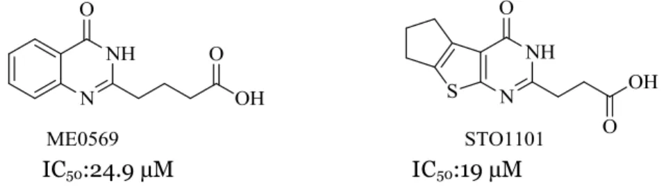
1.4 STO1101 and ME0569: Inhibitors of ExoS-ADPRT
A recent study published by our group in collaboration with researchers at Karolinska Institute has identified STO1101 and ME0569 (Figure 1) as potent inhibitors of ExoS-ADPRT.17
STO1101 is a competitive inhibitor of ExoS with an IC50 value of 19 µM. In the
structure of this molecule, there are two adjacent rings which are pyrimidone and cyclopente[b]thiophene and this ring system was substituted with propionic acid. The IC50 value of ME0569 is 25 µM and it has quinazolinone ring which substituted
with butyric acid.17
IC
50= 19 µM IC
50= 25 µM
3
1.5 Aim of the diploma work
The main aim of the study was to establish structure-activity relationship (SAR) of the identified inhibitors of ExoS ADPRT (STO1101, ME0569).17
We also aimed to improve the potency of the identified inhibitors of ADPRT activity of ExoS as well as the pharmacokinetics and pharmacodynamics properties.
2. Popular scientific summary including social and
ethical aspects
2.1 Popular scientific summary
Pseudomonas aeruginosa is one of the most common pathogen that causes of serious infections such as pneumonia, meningitis, soft tissue infections, chronic lung infections and corneal infections. P. aeruginosa infections associated with high rate of morbidity and mortality especially among immunocompromised individuals such as cystic fibrosis (CF), burn wound, or cancer.11 Due to intrinsic resistance to a broad
range of antibiotics, P. aeruginosa infections are very difficult to eradicate compared with other gram-negative pathogens infections.6 Currently, P. aeruginosa infections
may be treated by a combination of anti-pseudomonal agents in order to tackle resistance issue. However, this standard combination therapy remains problematic and not effective, as it leads to the increase of the drug-resistant strains.18 Therefore,
there are not many useful anti-pseudomonal drugs, P. aeruginosa infections are still one of the most dangerous infection disease in clinic. To combat the difficulties in treating P. aeruginosa infections, there are several approaches to develop new anti-pseudomona drugs. Targeting virulence factors, the ability of the bacteria to causes disease, is one of those novel strategies to fight against antibiotic resistance by disarming the bacteria.11 Virulence factors of P. aeruginosa are responsible for the
severity of infections because they cause irreversible host cell damage.11 Especially
exotoxins and proteases are associated with cell and tissue damage by disrupting the cytoskeletal structure and to develop of chronic infections.11 P. aeruginosa has five
protein secretion systems and among them, the type III secretion system transfers toxins (ExoS-T-U-Y) into the host cell.15 ExoS is a bifunctional enzyme and possesses a
GTPase-activating protein (GAP) activity and a ADP-ribosyl transferase (ADPRT) activity.15 These activities work to disrupt the actin cytoskeleton and cause to cell
death. The ADPRT domain of ExoS is responsible for the irreversible cell damage and it is highly toxic to cultured cells.15
2.2 Social and ethical aspects
Infection diseases are still major problem of clinic and P. aeruginosa causes severe and life-threating infections. Infections of P. aeruginosa are clinically challenged since it can readily develop antibiotic resistance during the therapy.18 If the resistance
occurs during the therapy, it will cause the length of hospital stay, additional medical procedures, surgery, chronic care and overall cost of antibiotics and even it will lead to the death of patients.18 The high rate of drug-resistant strains of P. aeruginosa is
increasing health threats facing the nation.
For this reason, there is an urgent need to develop new therapeutic agents against this pathogen for the combating P. aeruginosa infections. This project will help us to develop a new effective chemical probe which will be used as a chemical tool for the discovery of anti-pseudomonal drugs. Because of that this project is very important for the public health.
4
3. Experimental
3.1 Chemistry Section
General. All reactions were carried out under nitrogen atmosphere. Chemicals and reagents were purchased from Aldrich, Alfa Aesar, AK Scientific, Matrix Scientific, or Apollo Scientific. Organic solvents were dried using the dry solvent system (Glass Contour Solvent Systems, SG Water USA) except CH3CN, EtOH and PhCH3, which
were dried over activated molecular sieves 3 Å or 4 Å. Flash chromatography was performed on Biotage Isolera One using appropriate SNAP Cartridge KP-Sil or SNAP Ultra HP-Sphere 25µm Cartridge and UV absorbance at 254 nm. TLC was performed on Silica gel 60 F254 (Merck) with detection by UV light unless staining solution is
mentioned. Preparative HPLC separation were performed on Gilson System HPLC, using a VP 250/21 NUCLEODUR C18 column HTEC 5 μm with a flow rate 18 mL/min,
detection at 214 nm and eluent system: A. aq. 0.075% HCOOH, and B. 0.075% HCOOH in CH3CN. The NMR spectra were recorded at 298 K on Bruker-DRX 400
MHz and 600 MHz using the residual peak of the solvent DMSO-d6 (δH 2.50 ppm) or
CDCl3 (δH 7.26 ppm) as internal standard for 1H, and DMSO-d6 (δc 39.50 ppm) and
CDCl3 (δc 77.16 ppm) as internal standard for 13C. LCMS were recorded by detecting
positive/negative ion (EC+/EC-) with an electrospray Water Micromass ZG 2000
instrument using XTerra MS C18 (5 μm 19x50 mm column) and H2O/CH3CN (0.2%
HCOOH) as the eluent system, or with Agilent 1290 infinity – 6150 Quadrupole using YMC Triart C18 (1.9 μm 20x50 mm column) and H2O/CH3CN (0.1% HCOOH) as the
eluent system.
General Synthetic procedure19
Compounds ME0820 to ME0828 were synthesized according to the above general procedure reported earlier by Öznur Aglar’s degree report 2015 (Appendix II Method B)19
3-(4-oxo-4,6,7,8-tetrahydro-3H-cyclopenta[g]quinazolin-2-yl)propanoic acid- Compound 1 - ME0820 (30oa_36)
Synthesis: Methods B (3 % yield) Chromatography: A: aq. 0.75% HCOOH in H2O,
B: aq. 0.75% HCOOH in CH3CN, organic phase gradient 25→100% over 25 min, white
foam. 1H NMR (600MHz, DMSO-d6): δH 12.67 (s, 1H), 12.14 (s, 1H), 7.88 (s, 1H), 7.40
(s, 1H), 2.96 (p, J= 7.8Hz, 4H), 2.82 (t, J= 6.6Hz, 2H), 2.71 (t, J= 6.6Hz, 2H), 2.06(p, 7.4, 2H) ppm. 13C NMR (150MHz, DMSO-d6): δC 174.08, 162.26, 152.04, 148.26,
142.94, 127.29, 122.36, 120.86, 119.72, 33.02, 32.22, 30.50, 29.51, 25.79 ppm. LC-MS m/z calcd for C14H14N2O3 258.27 [M+H+]; 259.3 observed.
5
2-(4-oxo-3,4-dihydroquinazolin-2-yl)benzoic acid- Compound 2 - ME0822
(30oa_38)
Synthesis: Methods B (4 % yield) Chromatography: A: aq. 0.75% HCOOH in H2O,
B: aq. 0.75% HCOOH in CH3CN, organic phase gradient 25→100% over 25 min white
foam. 1H NMR (600MHz, DMSO-d6): δH 12.10 (s, 2H), 7.88 (s, 1H), 7.43 (s, 1H), 2.96
(p, J= 7.1Hz, 4H), 2.61 (t, J= 7.4Hz, 2H), 2.28 (t, J= 7.3Hz, 2H ), 2.06 (p, J= 7.3Hz, 2H), 1.94 (p, J= 7.4Hz, 2H) ppm. 13C NMR (150MHz, DMSO-d6): δC 174.65, 162.39,
156.20, 152.01, 142.93, 122.33, 120.78, 119.66, 33.919, 33.52, 33.03, 32.23, 25.81, 22.43 ppm. LC-MS m/z calcd for C15H16N2O3 272.30 [M+H+]; 273.3 observed.
3-(4-oxo-3,4,6,7,8,9-hexahydrobenzo[g]quinazolin-2-yl)propanoic acid- Compound 3- ME0823 (30oa_39)
Synthesis: Methods B (2 % yield) Chromatography: A: aq. 0.75% HCOOH in H2O,
B: aq. 0.75% HCOOH in CH3CN, organic phase gradient 25→100% over 25 min, white
foam. 1H NMR (600MHz, DMSO-d6): δH 12.20 (s, 2H), 7.75 (s, 1H), 7.27 (s, 1H),
2.87-2.83 (m, 4H), 2.80 (t, J= 6.9Hz, 2H), 2.69 (t, J= 6.9Hz, 2H), 1.76 (appear as p, J= 3.1Hz, 4H) ppm. 13C NMR (150MHz, DMSO-d6): δC 174.15, 162.01, 155.92, 146.93,
144.68, 135.83, 126.61, 125.68, 119.106, 30.65, 29.65, 29.61, 29.00, 22.98, 22.72 ppm.
LC-MS m/z calcd for C15H16N2O3 272.30 [M+H+]; 273.3 observed.
4-(4-oxo-3,4,6,7,8,9-hexahydrobenzo[g]quinazolin-2-yl)butanoic acid- Compound 4 - ME0824 (30oa_40)
Synthesis: Methods B (2 % yield) Chromatography: A: aq. 0.75% HCOOH in H2O,
B: aq. 0.75% HCOOH in CH3CN, organic phase gradient 25→100% over 25 min.,
white foam. 1H NMR (600MHz, DMSO-d6): δH 12.07(s, 2H), 7.76 (s, 1H), 7.30 (s, 1H),
2.87-2.84 (m, 4H), 2.60 (t, J= 7.4Hz, 2H), 2.28 (t, J= 7.3Hz, 2H), 1.93 (p, J= 7.3Hz, 2H), 1.76 (appear as p, J= 3.1Hz, 4H) ppm. 13C NMR (150MHz, DMSO-d6): δC 174.64, 162.18, 156.34, 147.12, 144.67, 135.84, 126.64, 125.65, 119.08, 33.93, 33.55, 29.64, 29.01, 22.98, 22.73, 22.37 ppm. LC-MS m/z calcd for C16H18N2O3 286.33 [M+H+]; 287.3 observed. 3-(1-oxo-1,2,7,8,9,10-hexahydrobenzo[f]quinazolin-3-yl)propanoic acid - Compound 5 - ME0825 (30oa_41)
Synthesis: Methods B (5 % yield) Chromatography: A: aq. 0.75% HCOOH in H2O,
B: aq. 0.75% HCOOH in CH3CN, organic phase gradient 25→100% over 25 min, white
foam. 1H NMR (600MHz, DMSO-d6): δH 12.46 (s, H), 11.95 (s, 2H), 7.39 (d, J=
8.3Hz, 1H), 7.27 (d, J= 8.2Hz, 1H), 3.35- 3.33 (m, 2H), 2.79- 2.76 (m, 4H), 2.69 (t, J= 7.0Hz, 2H), 1.75- 1.68 (m, 4H) ppm. 13C NMR (150MHz, DMSO-d6): δC 174.06,
6 163.08, 155.61, 149.20, 138.74, 135.63, 135.008, 124.68, 119.16, 30.39, 30.17, 29.09, 28.29, 23.18, 22.15 ppm. LC-MS m/z calcd for C15H16N2O3 272.30 [M+H+]; 273.30
observed.
4-(1-oxo-1,2,7,8,9,10-hexahydrobenzo[f]quinazolin-3-yl)butanoic acid-
Compound 6 - ME0826 (30oa_42)
Synthesis: Methods B (14 % yield) Chromatography: A: aq. 0.75% HCOOH in
H2O, B: aq. 0.75% HCOOH in CH3CN, organic phase gradient 25→100% over 25 min,
white foam. 1H NMR (600MHz, DMSO-d6): δH 11.91 (s, 1H), 7.39 (d, J= 8.3Hz, 1H),
7.29 (d, J= 8.2Hz, 1H), 3.35-3.32 (m, 2H), 2.78 (t, J= 5.8Hz, 2H), 2.55 (t, J= 7.4Hz, 2H), 2.26 (t, J= 7.3Hz, 2H), 1.91 (p, J= 7.4Hz, 2H), 1.76- 1.66 (m, 4H) ppm. 13C NMR (150MHz, DMSO-d6): δC 174.66, 163.22, 156.23, 149.41, 138.71, 135.62, 134.98, 124.71, 119.15, 33.53, 33.50, 30.18, 28.94, 23.19, 22.37, 22.16 ppm. LC-MS m/z calcd for C16H18N2O3 286.33 [M+H+]; 287.3 observed. 3-(1-oxo-2,7,8,9-tetrahydro-1H-cyclopenta[f]quinazolin-3-yl)propanoic acid- Compound 7 - ME0827 (30oa_43)
Synthesis: Methods B (14 % yield) Chromatography: A: aq. 0.75% HCOOH in
H2O, B: aq. 0.75% HCOOH in CH3CN, organic phase gradient 25→100% over 25 min,
white foam. 1H NMR (600MHz, DMSO-d
6): δH 12.41 (s, 1H), 7.55 (d, J= 8.1Hz, 1H), 7.30 (d, J= 8.1Hz, 1H), 3.35- 3.33 (m, 2H), 2.87 (t, J= 7.5Hz, 2H), 2.75 (t, J= 6.8Hz, 2H), 2.61 (t, J= 6.9Hz, 2H), 2.05 (p, J= 7.5, 2H) ppm. 13C NMR (150MHz, DMSO-d6): δC 174.55, 162.58, 156.28, 148.90, 144.26, 142.32, 130.37, 125.46, 118.04, 34.05, 32.17, 31.51, 30.00, 25.40 ppm. LC-MS m/z calcd for C14H14N2O3 258.27 [M+H+]; 259.3 observed. 4-(1-oxo-2,7,8,9-tetrahydro-1H-cyclopenta[f]quinazolin-3-yl)butanoic acid- Compound 8 - ME0828 (30oa_44)
Synthesis: Methods B (11 % yield) Chromatography A: aq. 0.75% HCOOH in H2O,
B: aq. 0.75% HCOOH in CH3CN, organic phase gradient 25→100% over 25 min, white
foam. 1H NMR (600MHz, DMSO-d 6): δH 12.10 (s, 1H), 7.58 (d, J = 8.1Hz, 1H), 7.35 (appeared as d, J = 8.1Hz, 1H), 3.37- 3.35 (m, 2H), 2.89 (t, J= 7.5Hz, 2H), 2.57 (t, J = 7.4Hz, 2H), 2.25 (appear as t, J = 7.3Hz, 2H), 2.07 (p, J = 7.5Hz, 2H), 1.92 (p, J = 7.4Hz, 2H) ppm. 13C NMR (150MHz, DMSO-d6): δC 174.80, 162.70, 156.39, 148.98, 144.25, 142.39, 130.42, 125.55, 117.99, 34.04, 33.88, 33.82, 32.18, 25.42, 22.59 ppm.
LC-MS m/z calcd for C15H16N2O3 272.30 [M+H+]; 273.3 observed.
4-(5-fluoro-4-oxo-3,4-dihydroquinazolin-2-yl)butanoic acid - 9 - ME0850
7
Synthesis: 2-amino-6-fluorobenzamide (150 mg, 0.97 mmol) and glutaric anhydride
(5 equiv.) were suspended in toluene (0.25M, 3.88 ml) and irradiated to microwave at 150 degree for 30 minutes. After the end of the first step of reaction, NaOH (2M, 20 equiv.) was added to the reaction mixture and irradiated to microwave at 100 ⁰C for 30 minutes. The each reaction’s steps were monitored by TLC and LCMS. The reaction mixture was washed with NaOH (1M). The aqua phase was collected and acidified with HCl (6M) until the formation of precipitates. The precipitates were collected by filtration and washed with water and evaporated. The product was dried under vacuo as a pure white powder (69 % yield). 1H NMR (600MHz, DMSO-d
6): δH 12.19- 12.10 (m, 2H), 7.75- 7.70 (m, 1H), 7.40 (appeared as d, J = 7.4Hz, 1H), 7.18 (t, J= 8.9Hz, 1H), 2.60 (t, J= 7.2Hz, 2H), 2.30 (t, J = 6.9Hz, 2H), 1.93 (appear as t, J = 7.1Hz, 2H) ppm. 13C NMR (150MHz, DMSO-d6): δC 174.51, 160.97 (d, JCF=262Hz), 159.54, 158.48, 151.52, 135.26 (d, JCF=10Hz), 123.36, 112.76 (d, JCF=20Hz), 110.84 (d, JCF=6Hz), 33.78, 33.24, 22.12 ppm. LC-MS m/z calcd for C12H11FN2O3 250.23
[M+H+]; 251.2 observed. NMR data of the compound has been shown in Appendix
III.
2-(4-oxo-4,5,6,7-tetrahydro-3H-cyclopenta[4,5]thieno[2,3-d]pyrimidin-2 yl) cyclopropanecarboxylic acid - Compound 10 - ME0876 (30oa_63)
Synthesis: 2-Aminothiophene-3-carboxamide (100 mg, 0.54mmol) and 3-
oxabicyclo[3.1.0]hexane-2,4-dione (307.55 mg, 2.74 mmol) were suspended in toluene (0.25M) and irradiated to microwave at 150 degree for 30 minutes. After the end of the first step of reaction, NaOH (2M, 20 equiv.) was added to the reaction mixture and irradiated to microwave at 100 ⁰C for 30 minutes. The each reaction’s steps were monitored by TLC and LCMS. The reaction mixture was washed with NaOH (1M). The aqua phase was collected and acidified with HCl (6M) until the formation of precipitates. The precipitates were collected by filtration and washed with water and evaporated. The product was dried under vacuo and the final product was purified by HPLC as a pure white powder. 1H NMR (600MHz, DMSO-d6): δH
12.52 (s, 1H), 12.17 (s, 1H), 2.91- 2.87 (t, J=7.2 Hz, 4H), 2.41- 2.34 (m, 3H), 2.06- 2.00 (m, 1H), 1.69- 1.65 (appeared as dd, J = 11.2Hz, 1H), 1.36 – 1.31 (m, 1H) ppm. 13C NMR (150MHz, DMSO-d6): 13C NMR 172.36, 168.57, 158.66, 139.80, 136.96, 118.79, 60.99, 30.78, 29.40, 29.12, 27.90, 23.22, 22.14 ppm. LC-MS m/z calcd for C13H12N2O3S 226.31 [M+H+]; 227.3 observed. 2-(4-oxo-4,5,6,7-tetrahydro-3H-cyclopenta[4,5]thieno[2,3-d]pyrimidin-2-yl)cyclopent-1-enecarboxylic acid - Compound 11 - ME0877 (30oa_64)
Synthesis: 2-Aminothiophene-3-carboxamide (100 mg, 0.54mmol) and 1-
cyclopentene-1,2-dicarboxylic anhydride (378.88 mg, 2.74 mmol) were suspended in toluene (0.25M) and irradiated to microwave at 150 degree for 30 minutes. After the end of the first step of reaction, NaOH (2M, 20 equiv.) was added to the reaction mixture and irradiated to microwave at 100 ⁰C for 30 minutes. The each reaction’s steps were monitored by TLC and LCMS. The reaction mixture was washed with NaOH (1M). The aqua phase was collected and acidified with HCl (6M) until the
8 formation of precipitates. The precipitates were collected by filtration and washed with water and evaporated. The product was dried under vacuum and the final product was purified by HPLC as a pure white powder. 1H NMR (600MHz,
DMSO-d6): δH 13.29 (s, 1H), 2.91- 2.88 (t, J = 7.2Hz, 6H), 2.78- 2.74 (m, 2H), 2.40- 2.33 (appeared as p, J = 7.3Hz, 2H), 1.89 – 1.82 (p, J= 7.6Hz, 2H) ppm. 13C NMR (150MHz, DMSO-d6): δC 168.17, 167.63, 157.94, 150.02, 144.48, 140.28, 139.10, 119.63, 37.54, 36.31, 29.56, 29.07, 29.90, 21.27 ppm. LC-MS m/z calcd for C15H14N2O3S 302.35 [M+H+]; 303.3 observed. 2,3,7,8-tetrahydro-1H-cyclopenta[4,5]thieno[2,3-d]pyrrolo[1,2-a]pyrimidin-10(6H)-one – Compound 12- (30oa_65)
Synthesis :A mixture,
(3-hydroxypropyl)-6,7-dihydro-3H-cyclopenta[4,5]thieno[2,3-d]pyrimidin-4(5H)-one ME07872 (0.01 mmol), TsIm (1.5 eq.),TEA (2 eq.), NaN3 (3
eq.) and a catalytic amount of TBAI (1.1 mg) was refluxed in DMF (30 mL). Reflux was continued until TLC monitoring indicated no further improvement in the conversion. The solvent was evaporated under vacuum and the remaining foam was dissolved in DMSO (2 mL) and the crude product was purified by HPLC.
1H NMR (600MHz, DMSO-d6): δH 4.02 (t, J= 7.3Hz, 2H), 3.08 (t, J = 8.0Hz, 2H), 2.91 (t, J= 7.3Hz, 4H), 2.38 (p, J = 7.3Hz, 2H), 2.18 (p, J= 7.6Hz, 2H) ppm. 13C NMR (150MHz, DMSO-d6): δC 168.80, 160.65, 157.01, 150.02, 139.42, 136.80, 117.75, 46.68, 32.04, 29.47, 29.16, 27.78, 19.63 ppm. LC-MS m/z calcd for C12H12N2OS 232.30 [M+H+]; 233.1 observed.
3.2 Biology Section
3.2.1 Dose-response experiment:
All compounds were dissolved in DMSO to make a stock solution of 40mM, which was used to prepare a dilution series of 8 concentration points. The highest concentration was 200 µM and the lowest concentration was 1.562 µM. The dilution series was prepared in 96-well polypropylene compound plate, before it was transferred into the assay plate (Figure 2).
Protein master mix (ExoS + Ras + 14.3.3) which were obtained from research group of Herwig Schüler in Karolinska Institute was prepared including controls as following:
Master mix without 14.3.317 : 0 % active enzyme
DMSO: 100 % inactive enzyme
STO1101: 30 µM concentration of inhibitor of ExoS ADPRT activity was used as a control.
Enzymatic assay:
Ɛ-NAD+ is a fluorescent analog of NAD and used as a substrate for ExoS.
Ras is a small GTPase protein subfamily and used as a co-substrate.
14.3.3 is a cofactor of ExoS and is required for activity of enzyme (ExoS). The enzymatic assay is described in the discussion part.
9
Figure 2. General dose-response assay format, in which compounds are
screened against ExoS ADPRT activity in 384-well plates.
Table 1. Dose–response analysis of ExoS ADPRT activity inhibition. Dose–response
curves of the compounds have been shown (Appendix I).
Compound No IC50 (µM) ME0815 27 ME0816 585 ME0817 ND ME0818 106 ME0819 32 ME0820 98 ME0821 141 ME0822 86 ME0823 ND ME0824 ND ME0825 ND ME0826 ND ME0827 54 ME0828 275 ME0850 25 ND: Not determined
10
4. Results and Discussion
Due to antibiotic resistance problem, there is an urgent need for useful alternative therapeutics methods. There are several attractive approaches based on targeting bacterial virulence systems or disrupting interaction between the host cell and the bacteria.20 T3SS which is responsible for the pathogenesis of the bacteria is one of the
major virulence factors of P. aeruginosa. Our strategy is inhibiting ADPRT activity of ExoS of T3SS in P. aeruginosa via small organic compounds.
Recently, two potent inhibitors of ExoS-ADPRT have been described17, which are
STO1101 and ME0569 as shown in figure 1. Therefore, we designed a small library
of analogues of both compounds. In this study, our attention was to synthesize analogues of ME0569.
IC50:24.9 µM IC50:19 µM
Figure 1. Structure of the ME0569 and STO1101.
Primary structure-activity relationship (SAR) of the hit compounds (STO1101 and
ME0569) has been showed that carboxylic acid, amide proton, and pyrimidine ring
are needed for the activity.17 On this basis, we focused our efforts to investigate the
side chain and the main scaffold of ME0569. For that, we designed 15 analogues in which the flexibility and the length of the side chain were investigated by modifying the length and the rigidity of the linker (Table 1). Different building blocks were selected including ether linker, hexyl and pentyl linkers, phenyl ring and furan ring. In comparing with STO1101 we also decided to introduce different fused alkyl ring to the main scaffold of ME0569. For that the building blocks 1e, 2e, 1f and 2f were synthesized in 3 steps starting from iodination as shown in scheme 1 and the compounds 1a-f and 2a-f have been described in previous report.19
Scheme 1. The synthetic route to intermediates.19
Two reaction methods were performed to synthesize analogues (method A and B)19
(Appendix II). Method A is an efficient method in which corresponding aldehydes
used one pot reaction in order to get the final product. Method B is two steps one pot microwave reaction in the presence of corresponding amides and anhydrides. Due to the solubility of the product and polarity of carboxylic acid, it was not possible to
11 purify the crude product by silica gel column chromatography. All compounds were purified by HPLC which resulted in low yield of the pure products.
For the synthesis of building blocks, the compounds 1a-f, and 2a-f, need to be synthesized because they were not commercially available. It was started from iodination and then nucleophilic substitution on the aromatic ring with nitrile and after that, hydrolysis of 1c-d and 2c-d for the synthesis of corresponding amides. In order to prepare the corresponding amides (1e, 1f, 2e and 2f) several hydrolysis reactions were attempted with different reactions including Cs2CO3 (cesium
carbonate), K2CO3 (potassium carbonate), K2CO3-peroxide and urea peroxide-K2CO3
and KOH (potassium hydroxide) and concentrated H2SO4. It turns out that, H2SO4
hydrolysis is an efficient method in very good yield.
Overall, a total of 15 analogues of ME0569 were synthesized and tested in the enzymatic assay for their inhibition of ExoS-ADPRT activity. Dose-response experiments were performed to determine IC50 values of the compounds as described
earlier in section 3.2.1 (Appendix I).
The assay is based on measurement the increase in fluorescence intensity when the ADP-ribosyl moiety is transferred from εNAD+ (a fluorescent analog of NAD) to the
target protein Ras. For the enzyme activity a co-factor, 14.3.3 is required. εNAD+ is a
co-substrate, while, Ras is substrate. In the presence of 14.3.3, ExoS is an active enzyme and it catalyzes the cleavage of the glycoside bond of 1,N6 -etheno NAD which
results in a 10-fold increase in fluorescence intensity (Figure 3).
Figure 3. The scheme is illustrating the reaction in the enzymatic assay.
As a result, we earlier identified two compounds ME0815 and ME0850 that inhibited ExoS activity with IC50 values of 27 µM and 25 µM. However, the majority of
the synthesized analogues did not allow us to improve the inhibition activity of this compound class. One clear issue with this class of compounds is their autofluorescence activity, which interferes with the assay measurement and a background correction had to be considered. Compound, ME0850 a very close analogue to the lead compound ME0569 has been proven to be as the lead compound. This result may imply that fluorine atom did not add any additional interaction with the target enzyme.
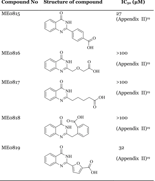
Nevertheless, structure-activity relationship (SAR) analysis (Table 2) allowed us to study the importance of the main features of the lead compound ME0569. We proved that longer chain (ME0817) was not favored as it reduces the inhibitory activity significantly (>100 µM). Also introducing a linker with hetero atom such as etheric oxygen (ME0816) did not contribute to any gained interaction with the target
12 enzyme or any favored conformation change. A flexible linker was shown to be more favored. That might be due to conformational change and preferably intermolecular hydrogen bonding between the terminal carboxylic acid and the amide bond in the core scaffold. This result concludes that the need for a flexible linker with three carbon atoms between the carboxylic acid and the core scaffold. Quinazolinone ring, on the other hand, was substituted on different positions with fused alkyl rings including six- and five-membered rings. Our results have shown that a fused alkyl ring larger than 5-membered ring (i.e six-5-membered ring) was not tolerated, which confirm earlier results on the analogue STO1101.17
However, fused 5-membered ring compounds ME0820, ME0822, were tolerated, they did not show any improved activity. These results indicate a limited modification or substitutions pattern on the quinazolinone ring.
As parallel study on STO1101 SAR has led to the identification of two more potent inhibitors ME0800 (IC50= 14.5 µM) and ME0805 (IC50= 10.6 µM) (data not shown).
Therefore, our group decided to focus the effort trying to improve the activity of those two analogues of STO1101. Thereby, we designed and synthesized a small set of analogues in order to finalize this SAR with ExoS inhibitors (data are not shown). All promising inhibitors of ExoS-ADPRT will eventually be tested the cell-based assay.
Table 2. Structure-activity relationship analysis (SAR) of analogues.
Compound No Structure of compound IC50 (µM)
ME0815 27 (Appendix II)19 ME0816 >100 (Appendix II)19 ME0817 >100 (Appendix II)19 ME0818 >100 (Appendix II)19 ME0819 32 (Appendix II)19
13 ME0821 >100 (Appendix II)19 ME0850 25 ME0820 98 ME0822 86 ME0823 >100 ME0824 >100 ME0827 54 ME0828 >100
14
ME0825 >100
ME0826 >100
ME0876 Not tested
15
5. Conclusions
In the present study, we successfully designed and synthesized a total of 17 analogues of STO1101 and ME0569 for further development of inhibitors. All designed compounds were tested in the enzymatic assay and their IC50 values were determined
via concentrated-response experiments. We identified two potent analogues (ME0815 & ME0850) of ME0569 as inhibitors of ExoS ADPRT activity with IC50
value of 27µM and 25µM respectively. Furthermore, all promising analogues will be tested in a cell-based assay for their activity.
It was found that some analogues possess solubility problems in DMSO during the dose-response experiments and also most of the analogues fluoresce naturally. Dose-response experiments indicate that the solubility and theirs fluorescence properties in a fluorescence-based assay for ExoS activity are a significant challenge for such compounds.
In conclusion, this study allowed us to better understand the structural activity of this compound class, and to determine the important structural features such as the length of the linker and the substitution of the main scaffold.
Accordingly, we believe that these results make possible to develop a chemical probes, which can be used as a research tool to discover novel antibacterial agent against the P. aeruginosa infections.
16
6. Future Plan
A small set of potent hits (ME0800 and ME0805) has been designed in order to improve potency of inhibitors and finalize SAR analyze of STO1101.17 In this library,
two compounds (ME0876 and ME0877) were synthesized. In addition, two more compounds will be synthesized (Figure 4) and all synthesized compounds to be tested in enzymatic assay.
(a) (b)
17
7. Appendix I
Concentration–response analysis of ExoS ADPRT activity inhibition. Compound
numbers shown with corresponding dose–response curves. Data shown for ExoS
ADPRT activity for promising compounds between the analogues: ME0815 and
ME0850.
18
19
8. Appendix II
Method A:21
2-Aminobenzamide (0.50 mmol), corresponding aldehydes (0.525 mmol) and CuCl2.2H2O (1 mmol) in dry ethanol (4ml) were refluxed for 16 hours. The reaction
was monitored with TLC and LCMS. After completion of reaction, the reaction mixture was cooled to room temperature and distilled water was added until the formation of precipitates. The precipitates were filtered and washed with distilled water. Product was isolated as a white solid and dried under vacuo. Final product was purified by HPLC (A: aq. 0.75% HCOOH in H2O, B: aq. 0.75% HCOOH in CH3CN,
organic phase gradient 25→100% over 25 min).
Method B
:
Scheme 2 Synthesis of derivatives of quinazolinones via two steps one-pot reaction.
Derivatives of 2-aminobenzamide (0.9 mmol, 1 equiv.) was suspended in toluene (0.25M, 3.6 ml) in the presence of corresponding anhydride (5 equiv.) and irradiated to microwave at 150 °C for 30 minutes. The reaction mixture, was monitored by TLC and LCMS. Then it was concentrated and NaOH (2M, 20 equiv.) was added to the mixture and irradiated to microwave at 100 ⁰C for 30 minutes. The reaction was monitored by TLC (Heptane/ EtOAc, 1:2) and LCMS till completion. After completion, the mixture was washed with NaOH (1M). The aqueous phase was collected and acidified with HCl (6M) till pH<7 and extracted with EtOAc, dried on anydrous Na2SO4 and evaporated. The product was dried under vacuo. HPLC was
performed on the final products (A: aq. 0.75% HCOOH in H2O, B: aq. 0.75% HCOOH
in CH3CN, organic phase gradient 25→100% over 25 min).
4-(4-oxo-3,4-dihydroquinazolin-2-yl)benzoic acid- ME0815 (30oa_03) Synthesis: Methods A (4 % yield) Chromatography: A: aq. 0.75% HCOOH in H2O,
B: aq. 0.75% HCOOH in CH3CN, organic phase gradient 25→100% over 25 min.,
white foam. 1H NMR (600MHz, DMSO-d6): δH 13.28 (s, 1H), 12.68 (s, 1H), 8.28
(appeared as d, J = 8.4Hz, 2H), 8.18 (appear as dd, J = 7.8Hz, 1.12Hz, 1H), 8.08 (d, J = 8.4Hz, 2H), 7.87 (ddd, J = 8.1Hz, 7.0Hz, 1.3Hz, 1H), 7.78 (appeared as d, J = 7.9Hz, 1H), 7.56 (ddd, J = 7.9Hz, 6.9Hz, 0.9Hz 1H) ppm. 13C NMR (150MHz, DMSO-d6): δC
167.27, 162.59, 152.12, 149.03, 136.89, 135.19, 129.87, 128.50, 128.13, 127.45, 126.37, 121.60 ppm. LC-MS m/z calcd for C15H10N2O3 266.25 [M+H+]; 267.3 observed.
2-((4-oxo-3,4-dihydroquinazolin-2-yl)methoxy)acetic acid - ME0816
20
Synthesis: Methods B Chromatography: A: aq. 0.75% HCOOH in H2O, B: aq.
0.75% HCOOH in CH3CN, organic phase gradient 25→100% over 25 min., white
foam. 1H NMR (600MHz, DMSO-d6): δH 12.226 (s, 1H), 12.91 (s, 1H), 8.119 (dd J=7.9Hz, 1.2Hz, 1H), 7.82 (ddd J=6.8Hz, 6.9Hz, 1.5Hz, 1H), 7.65 (d, J= 8.0Hz, 1H), 7.52 (ddd J=7.0Hz, 7.0Hz, 0.8Hz, 1H), 4.52 (s, 2H), 4. 22 (s, 2H) ppm. 13C NMR (150MHz, DMSO-d6): δC172.14, 161.89, 154.11, 148.71, 134.93, 127.49, 127.12, 126.32, 121.90, 70.77, 68.43 ppm. LC-MS m/z calcd for C11H10N2O4 234.21 [M+H+]; 235.2 observed.
5-(4-oxo-3,4-dihydroquinazolin-2-yl)pentanoic acid- ME0817 (30oa_09) Synthesis: 2-Aminobenzamide (50.43 mg, 0.37mmol) and methyl adipoyl chloride
(82.67mg, 0.4628mmol) were suspended in toluene (0.25M) and irradiated to microwave at 150 degree for 30 minutes. After completion of first step, NaOH (2M, 10eq) was added to the reaction mixture and irradiated to microwave at 100 °C for 30 minutes. The reaction mixture was monitored by TLC and LCMS. Then it was washed with NaOH (1M) and aqua layer was collected, acidified with HCl (6M) and extracted with EtOAc, dried over Na2SO4 and evaporated. The product was dried under vacuo as
a yellow solid. The final product was purified using HPLC (A: aq. 0.75% HCOOH in H2O, B: aq. 0.75% HCOOH in CH3CN, organic phase gradient 25→100% over 25 min)
to yield pure. Chromatography: A: aq. 0.75% HCOOH in H2O, B: aq. 0.75%
HCOOH in CH3CN, organic phase gradient 25→100% over 25 min, white foam. 1H
NMR (600MHz, DMSO-d6): δH 12.18 (s, 1H), 8.08 (dd, J= 7.9Hz, 1.3Hz, 1H), 7.77 (ddd,J= 6.9Hz, 6.8Hz, 1.4Hz, 1H), 7.59 (d, J= 8.0Hz, 1H), 7.46 (ddd J= 7.1Hz, 7.1Hz, 0.7Hz, 1H), 2.60 (t, J= 7.5Hz, 2H), 2.25 (t, J= 7.3Hz, 2H), 1.74 (p, J= 7.6Hz, 2H), 1.56 (p, J= 7.5Hz, 2H) ppm. 13C NMR (150MHz, DMSO-d 6): δC 174.84, 162.32, 157.79, 149.36, 134.80, 127.27, 126.42, 126.17, 121.26, 116.56, 114.87, 34.64, 33.86, 26.77, 24.46 ppm. LC-MS m/z calcd for C13H14N2O3 246.26 [M+H+]; 247.3 observed.
2-((4-oxo-3,4-dihydroquinazolin-2-yl)methyl)benzoic acid- ME0818
(30oa_12)
Synthesis: Methods B Chromatography: A: aq. 0.75% HCOOH in H2O, B: aq.
0.75% HCOOH in CH3CN, organic phase gradient 25→100% over 25 min, white foam.
1H NMR (600MHz, DMSO-d6): δH 12.87 (s, 1H), 12.39 (s, 1H), 8.07 (dd, J= 7.9Hz, 0.8Hz, 1H), 7.89 (d, J= 7.6Hz, 1H), 7.70 (ddd, J= 6.9Hz, 7.0Hz, 1.3Hz 1H), 7.52 (ddd, J= 6.8Hz,6.5Hz, 0.8Hz, 1H), 7.443-7.368 (m, 4H), 4.33 (s, 2H) ppm. 13C NMR (150MHz, DMSO-d6): δC 169.07, 162.19, 157.04, 149.27, 137.47, 134.68, 132.35, 131.58, 130.90, 129.89, 128.57, 127.53, 127.28, 126.45, 126.14, 121.29 ppm. LC-MS m/z calcd for C16H12N2O3 280.28 [M+H+]; 281.3 observed.
5-(4-oxo-3,4-dihydroquinazolin-2-yl)furan-2-carboxylic acid- ME0819
(30oa_15)
Synthesis: Methods A Chromatography A: aq. 0.75% HCOOH in H2O, B: aq.
0.75% HCOOH in CH3CN, organic phase gradient 25→100% over 25 min. white foam.
1H NMR (600MHz, DMSO-d6): δH 13.60 (s, 1H), 12.66 (s, 1H), 8.15-7.40 (m, 6H)
ppm. 13C NMR (150MHz, DMSO-d6): δC 161.97, 159.50, 148.84, 147.52, 143.90,
135.34, 128.09, 127.64, 126.45, 122.00, 119.37, 115.95 ppm. LC-MS m/z calcd for C13H8N2O4 256.21 [M+H+]; 257.2 observed.
2-(4-oxo-3,4-dihydroquinazolin-2-yl)benzoic acid- ME0821 (30oa_37) Synthesis: Methods B Chromatography: A: aq. 0.75% HCOOH in H2O, B: aq.
0.75% HCOOH in CH3CN, organic phase gradient 25→100% over 25 min, white foam.
1H NMR (600MHz, DMSO-d 6): δH 13.01 (s, 1H), 12.74 (s, 1H), 8.16 (dd, J= 7.9Hz, 1.278Hz, 1H), 7.95 (d, J= 7.5Hz, 1H), 7.82 (ddd, J= 8.3Hz, 1.5Hz, 1H), 7.70-7.63 (m, 4H), 7.53 (ddd, J= 7.9Hz, 0.9Hz, 1H) ppm. 13C NMR (150MHz, DMSO-d6): δC 167.87, 162.12, 155.45, 149.37, 135.64, 134.77, 131.96, 131.61, 130.41, 130.28, 127.66, 126.87, 126.24, 121.57 ppm. LC-MS m/z calcd for C15H10N2O3 266.25 [M+H+]; 267.3 observed.
21
9. Appendix III
NMR data of ME0815 and ME0850.
1HNMR spectrum of ME0815
13CNMR spectrum of ME0815
22
23
10. Acknowledgement
With great pleasure, I take this opportunity to present my special gratitude to the following people who directly or indirectly contributed to this work and helped me to finish my master degree thesis project.
I would like to thank to especially to my supervisor, Prof Mikael Elofsson for the opportunity to complete my master thesis with you and for providing an excellent work environment.
A special thanks to Michael Saleeb, who is a great co-supervisor and supportive person. Thank you for your kindness, your guidance, suggestions and support during my master degree thesis project.
Many thanks to my lab mates especially to Remi Caraballo, Naresh Sunduru and Duc Duy Vo who helped and encouraged me a lot during my lab work.
I would like to thank my officemates, Liena Qin and Charlotta Sundin. Thank you for your kindness, encouragement, and your great friendship.
Also, I would like to thank Caroline Zetterstrom, Sara Spjut, Emil Johansson, Anders Lindgren, Shannon Hinch, Tommy Orre and the other members of the Elofsson Research Group. Thank you for everything.
24
11. References
1. Jefferies, J.; Cooper, T.; Yam, T.; Clarke, S., Pseudomonas aeruginosa outbreaks in the neonatal intensive care unit–a systematic review of risk factors and environmental sources. Journal of medical microbiology 2012, 61 (8), 1052-1061.
2. Gellatly, S. L.; Hancock, R. E., Pseudomonas aeruginosa: new insights into pathogenesis and host defenses. Pathogens and disease 2013, 67 (3), 159-173. 3. El Solh, A. A.; Alhajhusain, A., Update on the treatment of Pseudomonas aeruginosa
pneumonia. Journal of Antimicrobial Chemotherapy 2009, dkp201.
4. Wiehlmann, L.; Wagner, G.; Cramer, N.; Siebert, B.; Gudowius, P.; Morales, G.; Köhler, T.; van Delden, C.; Weinel, C.; Slickers, P., Population structure of Pseudomonas aeruginosa. Proceedings of the National Academy of Sciences 2007, 104 (19), 8101-8106.
5. Liu, D., Molecular detection of human bacterial pathogens. CRC press: 2011.
6. Breidenstein, E. B.; de la Fuente-Núñez, C.; Hancock, R. E., Pseudomonas aeruginosa: all roads lead to resistance. Trends in microbiology 2011, 19 (8), 419-426.
7. Rossolini, G.; Mantengoli, E., Treatment and control of severe infections caused by multiresistant Pseudomonas aeruginosa. Clinical Microbiology and Infection 2005, 11 (s4), 17-32.
8. Japoni, A.; Farshad, S.; Alborzi, A., Pseudomonas aeruginosa: Burn infection, treatment and antibacterial resistance. Iranian Red Crescent Medical Journal 2009, 2009 (3), 244-253.
9. Vasil, M. L., Pseudomonas aeruginosa: biology, mechanisms of virulence, epidemiology. The Journal of pediatrics 1986, 108 (5), 800-805.
10. Mesaros, N.; Nordmann, P.; Plésiat, P.; Roussel‐Delvallez, M.; Van Eldere, J.; Glupczynski, Y.; Van Laethem, Y.; Jacobs, F.; Lebecque, P.; Malfroot, A., Pseudomonas aeruginosa: resistance and therapeutic options at the turn of the new millennium. Clinical Microbiology and Infection 2007, 13 (6), 560-578.
11. Chatterjee, M.; Anju, C.; Biswas, L.; Kumar, V. A.; Mohan, C. G.; Biswas, R., Antibiotic resistance in Pseudomonas aeruginosa and alternative therapeutic options. International Journal of Medical Microbiology 2016, 306 (1), 48-58.
12. Hancock, R. E.; Speert, D. P., Antibiotic resistance in Pseudomonas aeruginosa: mechanisms and impact on treatment. Drug resistance updates 2000, 3 (4), 247-255. 13. (a) Galle, M.; Carpentier, I.; Beyaert, R., Structure and function of the Type III
secretion system of Pseudomonas aeruginosa. Current Protein and Peptide Science
2012, 13 (8), 831-842; (b) Galle, M.; Jin, S.; Bogaert, P.; Haegman, M.; Vandenabeele,
P.; Beyaert, R., The Pseudomonas aeruginosa type III secretion system has an exotoxin S/T/Y independent pathogenic role during acute lung infection. PLoS One
2012, 7 (7), e41547.
14. Engel, J.; Balachandran, P., Role of Pseudomonas aeruginosa type III effectors in disease. Current opinion in microbiology 2009, 12 (1), 61-66.
15. Hauser, A. R., The type III secretion system of Pseudomonas aeruginosa: infection by injection. Nature Reviews Microbiology 2009, 7 (9), 654-665.
16. Coburn, B.; Sekirov, I.; Finlay, B. B., Type III secretion systems and disease. Clinical microbiology reviews 2007, 20 (4), 535-549.
17. Pinto, A. F. E., M. ; Saleeb, M. ; Forsberg, A. ; Elofsson, M. ; Schu ler, H., Identification of Inhibitors of Pseudomonas aeruginosa Exotoxin-S ADP-Ribosyltransferase Activity. Journal of Biomolecular Screening 2016, 1-6.
18. Lister, P. D.; Wolter, D. J., Resistance Challenges Threatening the Treatment of Pseudomonas aeruginosa Infections with Levofloxacin: The Role of a Levofloxacin-Imipenem Combination for Prevention of Resistance.
25 20. Lee, J.-H.; Kim, Y.-G.; Cho, M. H.; Kim, J.-A.; Lee, J., 7-fluoroindole as an antivirulence compound against Pseudomonas aeruginosa. FEMS microbiology letters 2012, 329 (1), 36-44.
21. Khan, K. M.; Saad, S. M.; Shaikh, N. N.; Hussain, S.; Fakhri, M. I.; Perveen, S.; Taha, M.; Choudhary, M. I., Synthesis and β-glucuronidase inhibitory activity of 2-arylquinazolin-4 (3H)-ones. Bioorganic & medicinal chemistry 2014, 22 (13), 3449-3454.