Theoretical Studies on Photophysics and
Photochemistry of DNA
Yuejie Ai
Doctoral Thesis in Theoretical Chemistry and Biology
School of Biotechnology
Royal Institute of Technology
Theoretical studies on photophysics and photochemistry of DNA Thesis for Philosophy Doctor degree
Department of Theoretical Chemistry and Biology School of Biotechnology
Royal Institute of Technology Stockholm, Sweden 2011
© Yuejie Ai, 2011
ISBN 978-91-7415-977-6 ISSN 1654-2312
TRITA-BIO Report 2011:15
Printed by Universitetsservice US-AB Stockholm, Sweden 2011
Theoretical studies on biological systems like nucleic acid and protein have been
widely developed in the past 50 years and will continue to be a topic of interest in
forefronts of natural science. In addition to experimental science, computational
modeling can give useful information and help us to understand biochemical issues at
molecular, atomic and even electronic levels.
Deoxyribonucleic acid (DNA), the hereditary basis of life’s genetic identity, has
always been major topic of discussions since its structure was built in 1953. However,
harmful UV radiation from sunlight can make damage to DNA molecules and
eventually give rise to DNA damaging biological consequences, like mutagenesis,
carcinogenesis, and cell death. Photostability, photodamage, and photorepair are of
vital importance in the photophysics and photochemistry of DNA. In this thesis, we
have applied high level computer-aided theoretical methods to explore the underlying
mechanisms for these three critical issues of DNA. Special attentions are paid to the
following aspects: the properties of the excited states, the design of relevant
computational models and the effects of biological environments.
We have systematically studied the excited state properties of DNA from single
base to base pair and oligonucleotides, where the concerted base pairing and base
stacking effects was found to play important roles in DNA photostability. The
UV-light induced isomerization mechanism between two photoproducts of DNA
photodamage has been revealed in different biological environments. In association
with DNA photodamage, the related photorepair processes have been proposed for
different lesions in photolyase which is a catalytic enzyme for DNA, and the
calculated results well explained the experimental observations. In particular, the
internal and external properties of flavin cofactors have been extensively studied by
combining the electronic structure and spectroscopic calculations. We have examined
the effects of the intramolecular hydrogen bond on spectroscopic properties of flavins.
The good agreements with the experimental spectra indicated that the biological
II
Preface
The work presented in this thesis has been carried out at the Department of Theoretical Chemistry and Biology, School of Biotechnology, Royal Institute of Technology, Stockholm, Sweden.
Papers included in the thesis
Paper I. Yuejie Ai, Ganglong Cui, Weihai Fang and Yi Luo. Exploring concerted effects of base pairing and stacking on the excited-state nature of DNA oligonucleotides by DFT and TD-DFT studies. Int. J. Quantum. Chem. 2010, DOI 10.1002/qua.22524
Paper II. Feng Zhang, Yuejie Ai , Yi Luo, and Weihai Fang. Nonradiative decay of the lowest excited singlet state of 2-aminopyridine is considerably faster than the radiative decay. J. Chem. Phys. 2009, 130, 144315.
Paper III. Yuejie Ai, Feng Zhang, Ganglong Cui,Yi Luo and Weihai Fang. Ultrafast deactivation processes in 2-aminopyridine dimer and the A-T base pair: similarities and differences. J. Chem. Phys. 2010, 133, 064302.
Paper IV. Yuejie Ai, Rongzhen Liao, ShuFeng Chen, Yi Luo and Weihai Fang. Theoretical studies on photoisomerizations of (6-4) and Dewar photolesions in DNA. J. Phys. Chem. B 2010, 114, 14096–14102.
Paper V. Yuejie Ai, Feng Zhang, Shufeng Chen, Yi Luo, and Weihai Fang. Importance of the intra-hydrogen bonding on the photochemistry of anionic hydroquinone (FADH-) in DNA photolyase. J. Phys.Chem. Lett. 2010, 1, 743–747. Paper VI. Yuejie Ai, Rongzhen Liao, Shilv Chen, Yi Luo, and Weihai Fang. Repair of DNA dewar photoproduct to (6-4) photoproduct in (6-4) photolyase. Manuscript.
Paper VII. Yuejie Ai, Guangjun Tian, Rongzhen liao, Qiong Zhang, Weihai Fang, and Yi Luo. Intrinsic natural property of flavin mononucleotide controls its optical spectra in three redox states. Manuscript.
Paper VIII. Ganglong Cui, Yuejie Ai and Weihai Fang. Conical intersection is responsible for the fluorescence disappearance below 365 nm in cyclopropanone.
J. Phys. Chem. A. 2010, 114, 730–734.
Paper IX. Feng Zhang, Yuejie Ai ,Yi Luo, Weihai Fang. Nonadiabatic histidine dissociation of hexacoordinate heme in neuroglobin protein. J. Phys. Chem. A. 2010, 114, 1980–1984.
Paper X. Juan Li, Yuejie Ai, Zhizhong Xie, Weihai Fang. How CO binds to hexacoordinated heme in neuroglobin protein. J. Phys. Chem. B. 2008, 112, 8715–8723.
Comments on My Contribution to the Papers Included
·
I have taken the major responsibility for the calculations and writing of paper I, III, IV, V, VI,VII.IV
It is an honor for me to give my acknowledgements here to all the people who
provided their unselfish assistance.
First and foremost, I owe my deepest gratitude to my supervisor Prof. Yi Luo,
whose guidance and kind encouragements enabled me to complete this thesis. I have
benefited a great deal from not only his valuable scientific insights but also his
positive views on life. With his support, I am much more confident of my academic
research and also my future life. Also lots of thanks to his family: Dr. Kezhao Xing,
dear Linda and Oscar. I cherish all the happy time we have spent together.
My special thanks go to Prof. Wei-Hai Fang, my former supervisor in Beijing
Normal University. Thank you for giving me the opportunity to study in KTH. He is
always my respectful teacher who firstly takes me to the fantastic theoretical world.
I would like to give my heartily thanks to Prof. Hans Ågren, the head of the
department of theoretical chemistry and biology, for the kind consideration of our
academic environment and requirements. Thanks for the lessons and help from Dr.
Ying Fu, Prof. Fahmi Himo, Prof. Kersti Hermansson, Prof. Faris Gel'mukhanov, Dr.
Yaoquan Tu, Dr. Olav Vahtras, Dr. Zilvinas Rinkevicius.
This thesis would not have been possible without the shared discussion and
collaboration with my dear classmates. They are Feng Zhang, Rongzhen Liao, Shilv
Chen, Guangjun Tian. I am grateful for their unselfish work and shared assistance.
I would like to show my great thanks to my colleagues and friends: Qiong Zhang,
Feng Zhang and Xing Chen, Igor Ying Zhang, Sai Duan, Lili Lin, Yuping Sun, Xiao
Chen, Kai Fu, Guangjun Tian, Weijie Hua, Xin Li(both two), Zhijun Ning, Qiang Fu,
Xiuneng Song, Yongfei Ji, Qiu Fang, …. The friendship between us is my most
precious experience in Sweden.
Lastly, I offer my thanks and blessings to beloved parents and little brother, for
their endless love and support over these years, and also my dear Kerry, for his
Abstract………...I
Chapter 1 Introduction………...1
Chapter 2 Photochemistry 2.1 Photophysical and Photochemical Processes………...4
2.2 Popular Computational Methods for Excited states……….8
Chapter 3 Deoxyribonucleic acid (DNA) and its photochemistry 3.1 The Building Blocks of DNA………..………16
3.2 DNA Double Helix………...………..………...17
3.3 DNA Photostability………...19
3.4 DNA Photodamage………...25
3.5 DNA Photorepair………...28
Chapter 4 Summary of Papers 4.1 Design of Theoretical Models for Biological Systems………40
4.2 Computational Photophysical and Photochemical Studies on UV-induced DNA issues………...51
Chapter 1
Introduction
Light is one of the most important elements to life. The exploring of light has
promoted the source of life perception since ancient times.1 For instance, the searching for the age-old question why plants can grow in the light finally leads to the
discovery of the famous photosynthesis effect of plant, which is the most important
photo-induced chemical reaction that involves the energy conversion between
sunlight and human being.1 The photosynthesis effect is the early exploration for the photochemistry. Since then, scientists began to study extensive photochemical and
photophysical changes when different substances interact with light and finally
enriched the field of photochemistry. The development of photochemistry is closely
related to two aspects: spectroscopic technology and related photochemical theories.
From the flash photolysis technique2-4 to crossed molecular beam technique5-6 and femtosecond laser pulse technology7-8, the advanced experimental techniques provide a basis for the development of the photochemical experimental studies. At the same
time, the theorists established first and second laws of photochemistry9, Lamber-Beer’s law9, Frank-Condon principle10, Jablonski diagram11, Fermi’s Golden rule9 that laid a firm foundation for understanding the complicated photophysical and photochemical processes. However, photochemical studies had been largely limited
for small organic molecules for many years. The emergence of novel ultrafast
femtosecond spectroscopy and supercomputer marks beginning of new era of the
photochemistry, which has extended into many new areas, including artificial
photosynthesis, visualization, light cycle (clock), solar energy, biosensor, and
bio-imaging, just to name a few. The successful application of photochemistry to
biology was highlighted by the Nobel Prize in chemistry 2008, which was awarded
- 2 -
their outstanding contributions in Green Fluorescent Protein (GFP). An important
branch of biological photochemistry, the nucleic acid photochemistry has always
drawn much attention; Actually, the interaction between light and DNA is a thematic
issue as old as time. Nevertheless, the photo science of DNA is inactive until the
double-helical structure of DNA was discovered in 1953.12 Especially in recent years, the development of spectroscopic and computational techniques have brought
immense interests into this field.13-15
When exposed to ultraviolet (UV)-light, the nucleic acids are promoted to the
excited electronic states. Fortunately, DNA is intrinsically photo-stable and it can
dissipate the excess electronic energy before photo-reaction happens.15 It is one kind of self-protection that keeps the maintenance of life. This photostability arises from
the intrinsic geometric and electronic structures, which are the result of evolution.
DNA is a double helix composed of base, pentose and phorsphoricacid. Both the
structural units and its specific space structure may be responsible for the
photostability of DNA. There are great efforts by different groups in understanding
the relaxation pathway of excited electronic states in DNA, and lots of proposed
factors, such as base pairing, base stacking, exciton and excimers, or charge and
proton transfer, are still under debate.14-15 A much clearer picture of the underlying mechanism for DNA photostability is needed. On the other hand, although we are
lucky most of the time, external hazard like UV radiation from sunlight or chemicals can sometimes make damage to DNA’s integrity and eventually give rise to DNA mutations and cancer risk.16 For instance, the high risk for skin cancer which is caused by the photodamage of skin cells’ DNA from UV radiation has attracted much more
public attention nowadays.17 As estimated by American Cancer Society, skin cancer accounts for nearly half of all cancers in the United States. Photodamage of DNA is
another practical topic in DNA photochemistry. Photochemical reactions in the
excited states can produce harmful photoproducts. The absorption of the UV light by
DNA results in mainly three photochemical lesions: cyclobutane pyrimidine dimers
(CPDs), pyrimidine (6-4) pyrimidone [(6-4)PP] and its valence photoisomer, Dewar
been less studied. In response to UV photo-damage, cells can repair the photo-lesions
via the photorepair process by photolyase.13 As the repair process is assisted by specific photolyase and the catalytic cofactors (flavins), things become much more
complicated since both internal and external environmental factors are involved. It is
a challenging task to explore the related static and dynamic features in those
processes.
To identify new therapeutic strategies for the treatment of cancer arising from DNA
photodamage and for the anti-cancer drug design, we need to know clearly the
structures and properties of these harmful photolesions. More importantly, intensive
studies on the formation mechanism of the DNA photolesions and DNA repair can
prevent cancers and enhance the quality of our life. However, the underlying
mechanisms of these behaviors are not always well understood. As a matter of
common practice and the complexity of DNA systems, the current available
experimental technology on DNA photochemistry has practical difficulties. Therefore,
computer-aided theoretical studies can come in to play an important role in the
practical application. In conjunction with experiments, we have employed high level
computational methods such as the complete active-space self-consistent field
(CASSCF)25-26, complete active space with second-order perturbation theory
(CASPT2)27, CASPT2//CASSCF/Amber (QM/MM), time-dependent density
functional theory (TDDFT),28-30 to investigate the structural properties and the possible mechanisms of the DNA photochemistry. For several systems, the actual
profiles of optical spectra have been simulated with the inclusion of the vibronic
coupling by using the linear coupling model (LCM). 31
The thesis is organized in the following way: In chapter 2, we will give a brief
introduction to the basic photochemical concepts and the widely used computational
methods for the excited states of photo-induced molecules. Research background and
contents for the UV-induced DNA photochemistry are presented in chapter 3. Finally,
- 4 -
Chapter 2
Photochemistry
2.1 Photophysical and Photochemical Processes
As an important branch of chemistry, photochemistry mainly studies the
photo-induced chemical phenomenon in molecules, atoms, and atomic ions, and so
forth when they absorb and emit the energy of photons.1,9,32-36 Usually, photochemical processes are accompanied by photophysical processes. We have summarized those
processes in the Jablonski diagram, see Figure 2.1.A Jablonski diagram is usually
used to illustrate the varying molecular electronic states, energy levels, possible
photophysical and photochemical processes. 11,33-36
Figure 2.1 Jablonski Diagram
ET, ELT, CR ET, ELT, CR ISC IC F A A IC ISCP S0 T1 S2 S1 CR
In Jablonski diagram, the thick horizontal lines represent the electronic energy
levels, while the thin and short lines are vibrational energy levels. In most cases,
according to Pauli exclusion principle, electrons in the electronic ground state are
paired (+1/2,-1/2). That is to say, S=0, M=2S+1=1. Most of the electronic ground
states of molecules are singlet states, represented as S0. With the absorption of
photons, one of the paired electrons in the outmost layer is excited. According to the
different spin directions of electrons, we classify them into different electronic excited
states. If the electron keep the spin orientation along the excitation, M=2S+1=1, the
excited state is still singlet state—―S‖ state. We use symbols S1, S2 to represent the
first excited singlet state and the second excited singlet state, which are ordered by the
magnitude of energies. On the other hand, the spin multiplicity M equals to 3 when
they are parallel spins. In this case, the excited states are triplet states (T), represented
as T1, T2….and so on. They are grouped according to multiplicity into horizontally
displaced columns.
Usually, molecules can be excited to their electronic excited states by absorbing
energy from ultraviolet light or visible light. In the electronic excited states, molecules
are very active. Chemical reactions (CR) may occur if the excess energy can’t be
released through radiative transitions, nonradiative transitions, and intermolecular
energy transfer (ET).36
Radiative transitions are transitions involve the absorption (A) and emission (F and
P). They are displayed by straight solid arrows. The photons carry the different energy
between the involved energy levels. Transition from the lowest excited electronic state
is associated with emission of photons (light). If the transition occurs between states
in the same spin, like from lowest singlet excited state (S1) to the ground state (S0),
fluorescence may occur:
) (
0
1 S h fluorescence
S
The fluorescence is short-lived with the lifetime of 10-9-10-6s. The radiative transition between different spin states, for example, transition from triplet T1 state to
- 6 - ) ( 0 1 S h phosphorescence T
The lifetime of phosphorescence is 10-4-10-2s, which is much longer than that of the fluorescence.
Nonradiative transitions are transitions not involved light emission. The excess
energy is then dissipated in three main processes: vibrational relaxation, internal
conversion (IC) in same spin states, and intersystem crossing (ISC) with different spin
multiplicities. In vibrational relaxation, the molecule in a higher excited vibrational
states returns to a lower vibrational state through energy transfer to the surroundings
caused by collisions. IC and ISC are all radiationless transitions. It is worth to
mention that the nonradiative transitions are essential for the deactivation process of
the excited states, unless their rates are negligible compared with the radiative ones.
These are effective photophysical channels by which the excited molecule can
return to the ground state. In competition with those processes, the photochemical
reactions in terms of cleavage and formation of chemical bonds can occur in the
excited state directly or in the ground state via nonradiative transitions.
To describe the photophysical and photochemical processes qualitatively, we need
to determine the equilibrium structures and energies of the electronic excited states.
The information of excited state is important to understand the properties of
spectroscopy and other photochemical phenomena. The geometric relaxation and
changes upon electronic excitation may reflect on the shape of spectra, fine structure,
etc.. Energies and other molecular properties of excited states can be calculated for the
analysis of spectroscopy and reaction mechanisms.
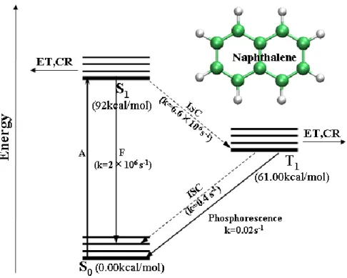
We take naphthalene for example in Figure 2.2.37 The lowest singlet and triplet excited states of naphthalene are S1 (1ππ*) and T1 (1ππ*) shown in Figure 2.2. After
singlet-singlet absorption (A) from ground state S0 to the S1 state, the first excited
singlet state (S1) of naphthalene may decay to the T1 state through the intersystem
crossing (ISC) or undergo the fluorescence (F) to the ground state. The rate constant
for the radiationless ISC process is 6.6×10⁻⁶ s-1 and it is comparable with the rate constant of fluorescence (2×10⁻⁶ s-1).
Once the molecule is in the T1 state, there are two ways for it to release the excess
energy and return to the S0 state. By spontaneously emitting a photon, the molecule
Figure 2.2 Jablonski diagram for naphthalene, data from Ref.33
can decay to the ground state by phosphorescence. The rate constant for this process
is 0.02s-1. Another intersystem crossing from T1 to S0 is much faster (k=0.4 s-1) than
the phosphorescence. The fluorescence and the green phosphorescence can be readily
observed by experimental technology. There is energy transfer process in the T1 state
involves a collision and electron exchange. In the excited states (S1 and T1 states), a
series of photoreduction, photoaddition, and other photochemical reactions (CR) may
take place.38 For example, the photochemical displacement of hydride ion by cyanide can take place after irradiation of naphthalene.
- 8 -
2.2 Popular Computational Methods for Excited States
Most of the photo-induced chemical reactions involve the electronic excited states of
the molecules. Generally, there are two categories of quantum chemical approaches
for calculation of excited states. 39-42 First kind is the wave-function-based method, such as Configuration Interaction (CI) methods (Configuration Interaction with Single
excitation (CIS), with Single and Double excitations (CISD), with Single, Double and
Triplet excitations (CISDT)), Multi-Configurational Self-Consistent Field (MCSCF)
methods (Complete Active Space Self-Consistent Field (CASSCF), Complete Active
Space Perturbation Theory of second order (CASPT2)). The other kind is
electron-density-based methods, such as time-dependent density functional theory
(TDDFT) method. In recent years, CASSCF and TDDFT methods are widely used to
deal with the photochemical properties from small to medium molecules and even
large biological systems. In the next sections, we will focus on these two popular
computational methods for excited states.
2.2.1 Time-Dependent Functional Theory (TDDFT)
To solve the Schrödinger’s equation, traditional quantum chemical approaches such
as ab initio methods, semi-empirical methods depend on the determination of the
wavefunction.42 When the number of electrons increases, the wavefunctions become much more complicated and it will cost more computing time. However, in density
functional theory, the electron density of a molecule is used to determine the energy
and derivative properties of molecules.43-45 The electron density only depends on three spatial coordinates. It is a function with three variables: x-position, y-position, and
z-position of the electrons (( , , )x y z ). The energy of the molecule is then a functional of the electron density.
[ ( , , )]
EF x y z (2.1)
So there is a one-to-one mapping between the electron density of a system and the
energy. We can get numerous information of a molecule if we can determine its
initio methods, the electron density function is only dependent on three coordinates,
independently of the system size. This approach is much faster than ab initio methods.
To look back at density functional theory’s history, from the Thomas–Fermi
model46 (developed by Thomas and Fermi in 1927) to Hohenberg-Kohn theorem47 which is thought to be the real birth of density functional theory, DFT has come a
long way.
In 1964, Hohenberg and Kohn set up the famous Hohenberg-Kohn theorem.47 The first Hohenberg-Kohn theorem ensures that the external potential is uniquely
determined by the electron density of the ground state. The second Hohenberg-Kohn
theorem guarantees the existence of a variational principle for electron densities.
Kohn and Sham introduced the Kohn-Sham orbitals and developed the Kohn-Sham
theory which brings the density functional theory into a more practical version.48 In Kohn-Sham theorems, the total energyE[ ] is shown as follows:
] [ ] [ ] [ ] [ ] [ TS J Ene EXC E (2.2) Where, TS[ ] is the kinetic energy of a non-interaction system, J[ ] is the classical electron-electron repulsive (Coulombic) energy, Ene[ ] is the nuclear-electron attraction energy. The exchange-correlation term EXC[ ] contains the exchange and correlation effects. In density functional theory, all the
approximations lie in the exchange-correlation term.
The Kohn-Sham equation:
HˆKSiKS i iKS (2.3) Where, the Halmiton HˆKS can be expressed as:
( ) 2 1 ) ( ˆ 2 r V r HKS KS (2.4) Where,
' ( ) ' ) ' ( ) ( ) ( dr V r r r r r V r VKS ne XC (2.5) [ ] XC- 10 -
exchange-correlation functionals can be divided into three types: local spin density
approximation (LSDA) functionals,49,50 generalized gradient approximation (GGA) functionals,51-53 and hybridized functionals.54,55
However, equation (2.3) is limited to time-independent systems. The Runge-Gross Theorem56 is an analogous time-dependent version of the first Hohenberg-Kohn theorem. It states that two external potentials v(r1,t)and v(r2,t) differed by more
than a time-dependent constant C t( )result in two different electron densities, that is:
1 2 1 2
( , ) ( , ) ( ) ( , ) ( , )
v r t v r t C t r t r t
So there still exists a unique relationship between time-dependent potentials
( , )
V r t and time-dependent densities ( , ) r t . Therefore the property of system can be written as a functional of the time-dependent density. The Runge-Gross Theorem is
rigorous basis of TDDFT. This is the first step for the extension to the time-dependent
domain. The next step is the existence of a time-dependent variational principle that is
analogous to the second Hohenberg-Kohn theorem.30, 57-58
If wave function ( , )r t is the solution of the time-dependent Schrödinger equation, (r,t) Hˆ(r,t) (r,t) t i (2.6)
It is the stationary point of the action integralA:
1 0 ˆ ( ) ( ) ( ) t t A dt t i H t t t
(2.7) According to the Runge-Gross theorem, the action integral can be written as afunctional of the time-dependent density:
1 0 ) , ]( [ ) , ( ˆ ) , ]( [ ] [ t t dt r t i t H r t r t A (2.8)To derive the time-dependent Kohn-Sham equation, a time-dependent
noninteracting reference system exists according to van Leeuwen. The noninteracting
density is equal to the exact density of the real interacting system. The time-dependent
, 1 2 ( , ) 3 ( , )
( , ) 2 ( , ) i XC i i r t r t A i v r t d r r t t r r r t
(2.9)In this case, the A part contains all the exchange and correlation effects. There XC
are different approximations for this functional. The widely-know one is the adiabatic
local density approximation (ALDA).
By now, the DFT theory is extended to the time-dependent domain and developed
to time-dependent density functional theory (TDDFT). The TDDFT in linear response
is thought to be another milestone of TDDFT theory. Using the linear response of the
time-dependent Kohn-Sham equation, we can obtain useful information, such as
excitation energies and oscillator strengths of excited states.
TDDFT has become one of the most popular quantum chemical tools from first
principles to calculate excited-state properties of medium-sized or even large
biological molecules. Since accurate high level calculations such as CASPT2 and
CASSCF employing large active spaces are tedious and time consuming; the TDDFT
calculation can reach the accuracy of sophisticated quantum chemical methods with
moderate computational cost. However, there are shortcomings in TDDFT. The
chosen of the right exchange-correlation functional for the given excited state
property is crucial.The hybrid functional B3LYP is unsuccessful in some applications.
For instance, the lack of the long range correlation causes the charge transfer (CT)
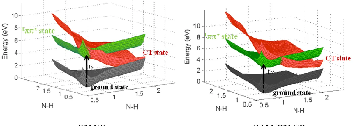
problem. We did TDDFT calculations for 2-aminopyridine dimer with different
functionals. The results are compared in Figure 2.3. Coulomb-attenuated hybrid
exchange correlation functional CAM-B3LYP59-60 is specially designed for treating the long-range CT transitions. Compared to the CAM-B3LYP functional, one can see
that the TDDFT/B3LYP underestimates the energies of CT states which are shown in
- 12 -
Figure 2.3 Potential energy surfaces of ground state, charge transfer state (CT) and localized excited states (1ππ*) for 2-AP dimer, calculated by TDDFT method with different functionals.
2.2.2 Complete Active Space Self-Consistent Field (CASSCF) method
First of all, we give a brief introduction of the Configuration Interaction (CI)
method and Multi Configurational Self-Consistent Field (MCSCF) method.40,42
Configuration Interaction method uses molecular orbitals based on a Hartree-Fock
calculation. The wave function is a linear combination of HF wave function and other
electronic configurations. The contributions of excited states are included in a linear
variation function for the ground state.
0 , , , ,
abc ijk ab ij a i CI (3.0) abc ijk ab ij a i , , are single(S), double(D) and triple (T) configurations respectively. The CI wavefunction can be represented as:
0 0 , ... a a ab ab abc abc CI i i ij ij ijk ijk i a i j i j k a b a b c C C C C
(3.1)Full configuration interaction (Full-CI) includes all the possible excitations from the
HF ground state to all the virtual orbitals. However, full CI is computationally
extremely expensive because of the large majority of the basis sets and configurations.
So it is only possible for small systems and small basis sets. To put that into a more
For instance, Configuration Interaction with Single Excitation (CIS) is one of the
easiest ways to get excited state energies. It starts from the Hartree-Fork wave
function and promotes one electron to one of the virtual orbitals. There are also other
methodologies considering the multi-configurations interactions, such as CISD
(Configuration Interaction with Single and Double excitations) and CISDT which
includes the triple excitations.
The CI calculations take the Hartree-Fock ground state wavefunctions as the
reference states of molecules. If it encounters the case that the Hartree-Fock
determinants are not adequate, e.g. when the coefficient C is very small, we need to 0
choose more electronic configurations for the reference states. Multi-configurational
self consistent field (MCSCF) calculations are used as an improvement of
configuration interaction method. 39 The wavefunction of MCSCF method MCSCF is a linear combination of different configuration state functions (CSFs) which is written
asKin equation 3.2.
MCSCF K K
K
A
(3.2) Where Kis again written as determinants of molecular orbital (i) which is linear combination of the atomic orbital () also.1 ! N K i i k Det N
(3.3) i Ci
(3.4) So in the MCSCF calculations, the coefficients of both the configuration statefunctions and the basis functions in the molecular orbitals are optimized so as to
minimize the energy. The MCSCF method has faster convergence than that of CI and
MCSCF wavefunctions are widely used as reference states in Multireference
configuration interaction (MRCI), complete active space SCF (CASSCF) method, or
complete active space with second-order perturbation theory (CASPT2) to calculate
- 14 -
The Complete Active Space Self-Consistent Field (CASSCF) method is a
particularly widely used approach of MCSCF and it was first propounded by
Ruedenberg and Roos. 61 It is one of the few popular methods that are suitable for the optimizations of both the ground states and excited states. Similar to other MCSCF
methods, the most important problem in CASSCF is how to choose the configurations.
The orbitals used in CASSCF situation are usually classified into inactive orbitals and
active orbitals. The inactive orbitals include the so-called core orbitals which are
doubly occupied and also the virtual orbitals. The active orbitals are the most
important orbitals involved in the reactions of the systems. Electrons that occupy
active orbitals called active electrons. Usually the number of the electron occupation
in active orbital is in the range from 0 to 2. Then the active electrons and active
orbitals constitute the active space. The CASSCF wavefunction consists of a linear
combination of all the configurations that arise from distribution of the active
electrons among active orbitals under given spatial and spin symmetry. For example,
the CASSCF wave function of active space CAS (10, 8) is built by distributing of 10
active electrons in 8 active orbitals. That is to say, 10 active electrons are distributed
between all configurations that can be constructed from 8 active orbitals. The active
space is the key concept of the CASSCF method. Different orbitals and electrons in
the active space may have significant impact on the relative and absolute energy of
the studied system. Generally, the active space should include all the valence orbitals
and valence electrons. However, along with the expansion of the active space, the
computational cost increases quickly. The larger active space is, the more
computational resource costs. So in practical application, we only select relevant
orbitals and electrons according to the chemical reaction process that we are interested.
In contrast to other MCSCF approaches, the CASSCF method avoids the randomicity
of the composition of the configuration functions and involves no selection of
individual configurations. It is reasonably accurate for the equilibrium structures and
properties of the ground states and electronic excited states under appropriate basis
sets. Meanwhile, the CASSCF method can also give good description for the
Therefore, it is frequently used to solve the complex photo-induced chemical
reactions that always involve the molecular excited states.
However, the CASSCF method also has some limitations. The CASSCF method
itself only includes a small fraction of the electronic correlation energy and does not
include electronic dynamic correlations outside the active space. So there is error in
the electronic energies. Therefore, if we want to get more accurate excitation energy,
we need to include correction to dynamic correlation. Starting from the CASSCF
wave functions, there are some ways to consider the dynamic correlations, such as
MR-CI, MRPT (MR-MP2 and CAS-PT2) and MRCC approaches. An overall protocol
named CASPT2//CASSCF is widely used. What’s more, the CASSCF method is
limited by the system’s size. Especially in biological systems, due to the large number
of atoms, the CASSCF method is always combined with classical molecular
- 16 -
Chapter 3
Deoxyribonucleic acid (DNA) and its
photochemistry
3.1 The Building Blocks of DNA
Figure 3.1 The basic components of a nucleic acid and a deoxynucleotide Nucleic acids are the basic biological macromolecules of heredity. Their structural
unit is nucleotide, consisting of phorsphoric acid, pentose sugar (ribose or ribodesose),
and base (purine or pyrimidine) as shown in Figure 3.1. According to the different
types of the pentose sugar, nucleic acids are classified into the ribonucleic acids
(RNAs) and deoxyribonucleic acids (DNAs). In RNA, the pentose sugar contains a
hydroxyl group at the 2’ carbon atom of the five membered-ring. In DNA, the 2’-OH
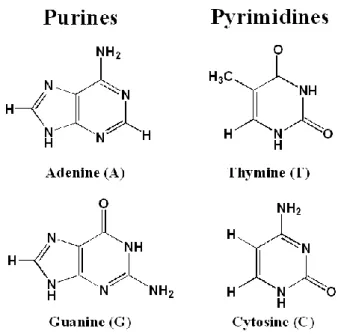
is replaced by hydrogen as shown in Figure 3.1. The genetic code in DNA is mainly
stored in four bases: adenine (A), thymine (T), cytosine (C), and guanine (G), see
Figure 3.2 Four main nucleobases in DNA
3.2 DNA double helix
Each base of DNA is linked to the backbone, composed of sugars (2-deoxyribose in
DNA) and phosphates. And repeated units of deoxynucleotide constitute the
polynucleotide, which is termed as one DNA strand. Because the sugar units is
bonded with phosphate groups by the formation of ester bonds between C3’ and C5’
of adjacent sugar rings, the terminals of DNA strand are often defined as 3’ and 5’
terminals.
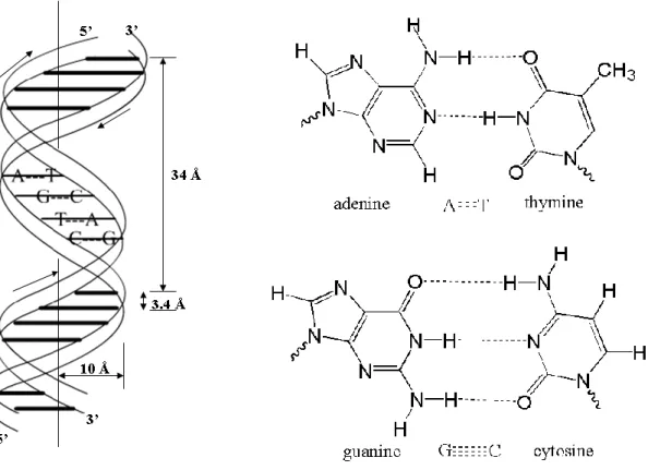
In 1953, Watson and Crick discovered the famous double helix structure of the
DNA (see Figure 3.3).12 In this structure, the double-strand DNA is built up by two anti-parallel, right handed strands running in opposite directions along the same axis.
The two adjacent base pairs are in average separated by 3.4 Å and a complete turn
covers 10 base pairs with ca. 34 Å, while the radius of the helix is 10 Å.
The four main DNA bases pair up with each other following the ―base-pair rule‖
which states that A only bonds with T and C only bonds with G. See the base pair A-T
- 18 -
bases (A-T: two hydrogen bonds, G-C: three hydrogen bonds) which connect the two
DNA single strands together.
Figure 3.3 The structures of DNA double helix and base pairs A-T and C-G.
The discovery of the double helix structure of DNA is thought to be one of the most
significant milestones in the human science history. After that, the DNA science ranks
as one of the most popular fields of the last few decades, and it will continue to be the
frontier science since it leads the way to a better understanding of the basic units of
3.3 DNA photostability
As the genetic material of living organisms, DNA is somehow vulnerable and can
easily be damaged by external perturbations such as UV radiation from the sun.13 The DNA bases are good chromophores that have intense absorptions at UV-light around
260 nm.62 The DNA nucleobases can be promoted to the excited states by absorbing radiation energy. The excess energy needs to dissipate via several pathways to the
ground states, or it will initiate photochemical reactions that finally results in the
formation of harmful photolesions and bring out mutations and carcinogenesis.
Fortunately, the quantum yield of photolesion formation is less than 1%.15 This indicates that most excited DNA molecules just relax back to their ground states,
avoiding harmful photochemical reactions which lead to photo-damaged lesions. In
this sense, DNA has its own resistance to UV-light induced photochemical damage.
This property is called photostability of DNA, which is the potential choice for
biological evolution and is major topic of debates for decades.15
As mentioned above, starting from the single bases which are the building blocks of
DNA, base pairing and base stacking and other interactions ultimately constitute the
double helix structure. There is therefore great interest in which effect leads to the
photostability of DNA.
- 20 -
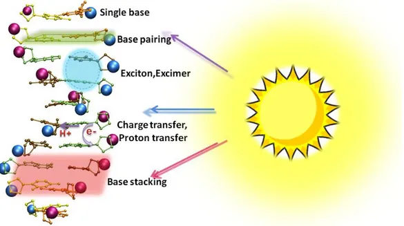
Figure 3.4 represents some suggested effects for the photostability, which will be
discussed in following paragraphs. For the DNA base monomers, relative low
fluorescence quantum yields (about 10-4) in aqueous solution demonstrate that there exists a highly efficient nonradiative decay pathway after UV-light absorption.63-64 Experimentally, in 2000, the first accurate ultrafast femtosecond transient absorption
measurements showed that the 1ππ* states of DNA and RNA base monomers decay to the ground state within a subpicosecond or even femtosecond timescale.65 At present, there is good consensus between experimentalists and theoreticians that the ultrafast
nonradiative decay of DNA single base is mostly caused by efficient internal
conversions through conical intersections (CIs).65-68 Theoretically, conical intersections have been found in almost all natural DNA nucleobases (A, T, C, G) and
many other of their derivatives.67 The conical intersections in different bases are of different characters, such as 1ππ*/S0, 1nπ*/S0, 1πσ*/S0, 1ππ*/1nπ*, 1ππ*/1πσ*, and so on.
These low-lying CIs or even high-lying CIs can be accessed from the Franck-Condon
region through nearly barrierless paths, and favorable internal conversion from the
excited singlet states to the ground state happens. The diverse CIs are usually
possessed of typical ring deformation structures that involve in out-of-plane
motions.67,69
(a) (b)
Figure 3.5 Structure of 2-aminopyridine (a) and the energy profile for the ring deformation reaction (b). Selected from Paper II, reprinted with permission from
We use ab initio methods and RRKM theory to study the radiative and nonradiative
decay of 2-aminopyridine which is a frequently used model system for nucleobase,
see Figure 3.5 (a).
Compared to other possible nonradiative photochemical pathways (like aromatic
ring opening, intramolecular hydrogen transfer, hydrogen detachment), our
calculations shown that an ultrashort nonradiative ring deformation pathway on S1
state is the most efficient nonradiative deactivation for 2-aminopyridine.
We have located the conical intersection between the S1 excited state and the
ground state, denoted as S1/S0 in Figure 3.5(b). One carbon atom in the aromatic ring
is out of plane, accompanied by twisting of the C-C and C-N bonds. From the energy
profile in Figure 3.5(b), we can see that when the molecule is excited upon to the first
excited state S1, it can access the conical intersection S1/S0 via a low energy barrier
along the ring deformation coordinate. At the same time, the S0 state rises sharply and
finally it meets the surface of S1 state at the point S1/S0. It is explained by Kohler et al.
that in the ring deformation structure, the aromaticity is destroyed.15 The ground state energy increases sharply due to the loss of π-bond stabilization; while the
excited-state energy is relatively insensitive to the ring deformation. 15
In summary, the conical intersection plays an important role in the nonradiative
decay pathways for the single base. By means of this efficient internal conversion, the
excited monomers can release their initial energy and relax to the ground state
avoiding injurious photodamage. Some excellent reviews emphasized again that the
CIs are responsible for the ultrafast internal conversion in different kinds of single
base monomers.14,67,70 Bern Kohler and co-workers stressed the nonradiative decay mechanisms in DNA from the experimental point of view.14,70 In the meantime, theoretical calculations on DNA natural bases and tautomers emphasized the
importance of the CIs by using accurate quantum chemical approaches.67 Those studies indicate that the special photochemical feature of DNA nucleobases may
attribute to the natural selection of the molecular evolution.
Different bases can assemble together horizontally (base pairing) or vertically (base
- 22 -
For instance, base pairing effect originates from the hydrogen bonds between single
bases. Domcke et al.71 used Femtosecond time-resolved mass spectroscopy and detected an excited state of 65±10 ps for the 2-aminopyridine dimer which is a
model system for the nucleic acid base pairs, as shown in Figure 3.6 (a).
(a) (b)
Figure 3.6 The electron-driven proton-transfer process in (a) 2-aminopyridine dimer and (b) Adenine-Thymine base pair. Selected from paper III, reprinted with
permission from American Institute of Physics.
Their later ab initio calculations proposed that the conical intersection between
low-lying 1ππ* local excited state (LE state in Figure 3.6(a)) and a charge transfer state (CT1 and CT2 states in Figure 3.6(a)) along the N-H proton transfer coordinate
was responsible for the observed short lifetime. This electron-driven proton-transfer
process was also revealed in the A-T and G-C base pairs.72-73 As seen in Figure 3.6 (b), after excitation to the LE state of the A-T base pair, the molecule can overcome small
barrier and reach the crossing seams between the LE state and CT states. There are
more favorable than that from T to A, denoted as CT (T→A) in Figure 3.6 (b). The
charge transfer from adenine to thymine leads to spontaneous proton transfer in the
same direction to neutralize the charge separation. Along the proton transfer
coordinate, the energy of ground state increases while the CT states are essentially
repulsive. At longer N-H distance, internal conversion will occur and finally the
excited molecule decays back to the ground state. As has been shown above, the
electron-driven proton charge-transfer states always play a decisive role in the
relaxation processes of the base pairing system. There are also experimental studies
which support this light-induced proton transfer mechanism.71,74,75 However,
questions are raised when the base stacking effect is included into the oligo- and
polynucleotides that makes the situation more complicated.
The base stacking effect is a significant interaction to the stability of DNA. It
includes overlapping of π orbitals, dispersion attraction and electrostatic interactions,
and so on. Since the nucleobases possess the purine or pyrimidine rings which are
suitable for stacking, the base stacking generally exists in the single- and
double-stranded DNA. For DNA oligo- and polynucleotides, in addition to the fast
nonradiative decay rate in those single bases, experimental evidence of one or two
orders of magnitude slower rate also exists.76 It suggests that there are long-lived
states which compete with the ultrafast monomer-like decay channels.
Crespo-Hernández and Kohler studied the single-stranded (dA)1876, and indicated that
there were both ultrafast (τ≈ 1 ps) and a slower (τ≈ 126 ps) components exist in the
single strand. It’s surprising that same kinetics has been observed in the duplex
(dA)18·(dT)18. So they concluded the characteristic long-lived signals in DNA oligo-
and polynucleotides were ascribed to the stacking effect instead of the base pairing.
This work thereby evokes a series of controversy about the primary and secondary
roles of the base pairing and base stacking effects. 14,15,70,77 Besides, there are other
possible explanations for the slow nonradiative decay in DNA oligo- and
polynucleotides.
Some researchers concluded that the excimer, exciplex or excitonic states formed
- 24 -
that it was because of the delocalized excited states, such as Frenkel excitons or
interbase charge-transfer states, exist in the DNA single and double strands.70, 78, 79 But the delocalization length is unknown. In Figure 3.7, we illustrate the detachment
(blue color) and attachment (red color) electron densities calculated at
PCM/TD-B3LYP for the excited singlet state S11 for the A3 and T3. It is obvious that
the S11 state of A3 displays much delocalized property while the S11 state of T3 is a
charge transfer state. What’s more, interstrand proton transfer and other channels via
dark states like 1nπ* states have also been suggested.70 However, there are lots of arguments and great uncertainty about the nonradiative decay in the above
mechanisms. This is not only because of the complexity of the DNA double helix
itself but also the intricate biological environment around the real DNA molecules.
Since the underlying nonradiative decay mechanisms of the DNA oligo- and
polynucleotides are poorly understood, more challenges and opportunities in this
active topic will continually make it a subject of great interest both experimentally
and theoretically.
A3 (S11) T3 (S11)
Figure 3.7 Detachment (blue) and attachment (red) electron densities for the selected S11 (π→π*) states for A3 and T3.
5' 3' 2A 3A 4A P 1A 5A P P P 5' 3' 2T 3T 4T P 1T 5T P P P
3.4 DNA photodamage
The photostability of DNA can prevent most of the photodamage formations in
DNA. However, once a chemical bond in one of three billion nucleotides of human
being is modified, the outcome will be catastrophic. From this perspective, studies on
DNA photodamage have great practical significance in the DNA photochemistry.
Since the skin cancer induced by UV radiation has drawn much attention these years
because of its high incidence rate, the photodamage appears to be crucial in
photocarcinogenesis. UV exposure results in a variety of potentially mutagenic
cellular lesions, referred as photoproducts. Exposed by carcinogenic far-UV
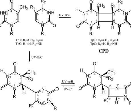
wavelengths brings in three characteristic lesions, resulting in cyclobutane pyrimidine
dimers (CPD), pyrimidine (6–4) pyrimidone photoproducts [(6–4)PP] and their Dewar
valence photoisomers (DewarPP).80-82
The CPDs come from the covalent linkage between two adjacent pyrimidines by
the formation of a four-membered ring structure, see Figure 3.8, the C5-C6 double
bonds of pyrimidines are linked and saturated in CPDs. CPDs are the most abundant
photoproducts of all three photoproducts. It is said that the CPDs and the (6-4)PPs
constitute around 75% and 25% of the DNA photodamage products, respectively.18 Since described as early as 1960, the reaction and repair mechanism of CPDs have
been extensively studied by means of both experimental techniques and theoretical
computations.23,83-85 Now, It is well recognized that the mutagenic and carcinogenic potential of UV-induced damage is expected to arise mainly from CPDs. Studies have
shown that CPDs are the main trigger for apoptosis in nucleotide excision repair
- 26 -
Figure 3.8 Structures of three DNA photoproducts. Selected from paper IV, reprinted with permission from American Chemical Society.
Different from the CPDs, as illustrated in Figure 3.8, an incorrect covalent linkage
between the C6 atom of the one pyrimidine and the C4’ atom of the adjacent
pyrimidine results in the (6-4)PPs. Although smaller proportion than CPDs, in some
cases, the (6-4)PPs are predicted to be more potentially lethal than CPDs. For instance,
in the NER-deficient cells, experimental data indicated that the (6-4)PP lesions were
more toxic and mutagenic compared with CPDs, 86 which made it currently a subject
of intensive studies.87
When exposed to UV-A/B light, the formation of the N3’-C6’ bond in the
pyrimidone ring leads to its valence isomer called DewarPP.24 The quantum yield of
DewarPP is quite low and this hampers the study of experiments and theoretical
calculations on this naturally formed photoproduct. So compared to the other two
photoproducts, the DewarPP has been less studied. The cytotoxic and mutagenic
properties of this photoproduct are not well established to date. In particular, the N N O O CH3 H R2 N N O R1 H R R N N O O CH3 H R2 H R N N O R1 H R UV-A/B 5 4 3 2 1 4' 3' 2' 1' 6' 5' 6 4 3 2 1 6 5 4' 3' 2' 6' 5' 1' HN N O O CH3 N N N N O O R2 O CH3 R1 H H R R H H CPD DewarPP (6-4)PP N NH R2 R1 O R R TpT: R1=CH3, R2=O TpC: R1=H, R2=NH TpT: R1=CH3, R2=O TpC: R1=H, R2=NH UV-B/C UV-B/C UV-C TpT: R1=CH3, R2=OH TpC: R1=H, R2=NH2 TpT: R1=CH3, R2=OH TpC: R1=H, R2=NH2 1 5 4 3 2 6 5 6 1 4 3 2 1 5 4 3 2 6 5 6 1 4 3 2
underlying mechanisms of the photoisomerization between the (6-4)PP and DewarPP
remain unclear at the molecular level.
To sum up, the above three photoproducts are considered to be responsible for
mutation induction in critical genes, tumorigenesis, cell death, and so on. These will
drastically impair the transcription and replication of DNA and finally lead to
carcinogenesis like skin cancer. Therefore, if the photo-damaged DNA lesions cannot
be repaired or removed, they will infect the normal cellular processing of DNA and
cause mutations or even death.
- 28 -
3.5 DNA photorepair
In order to maintain the regular operation for the biophysical activities, especially
for the genetic stability and continuance of life, the organisms have a set of restoration
system to repair specific damaged lesions. Especially for DNA photodamage, there
are several approaches for organisms to repair the UV-induced photo-lesions. First,
the excision repair, which includes the base excision repair (BER) and nucleotide
excision repair (NER), is one kind of common repair mechanisms.88 In this mechanism, the damaged DNA is replaced with new nucleotides by cutting off and
then interlinking nucleotides in the damaged part. Second, using the energy of light,
many organisms can undergo photoreactivation process which involves in particular
enzymes called CPD photolyase and (6-4)PP photolyase.13,88 The photoreactivation refers to the reversal process of the photodamage reactions by the enzyme upon
irradiation with blue or near-UV light (wavelength 300-450 nm).13 The DNA photolyases are monomeric enzymes found in a variety of organisms including higher
plants and bacteria. The photolyases have 450-550 amino acids in length, with the
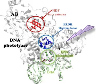
total molecular weight of 55-65 kDal.88 The X-ray structure of the CPD photolyase found in Escherichia coli (PDB entry:1TEZ) is shown in Figure 3.9. The photolyase
has two domains which are labeled as AB and A domains in the N-terminal and
C-terminal, respectively. In A domain, the enzyme folds up a spatial cavity which is
the binding position for the damaged DNA lesions and cofactor. In Figure 3.9, the
photolyase binds to the oligomer DNA (in green) duplex with a photodamaged CPD
flipped out and inserted into the active site.
There are two kinds of important chromophore cofactors in the photolyase. First
is so-called the light-harvesting cofactor, which can be either a
methenyltetrahydrofolate (MTHF) or an 8-hydroxy-5-deazariboflavin (HDF), as
colored red in Figure 3.9. People gives them a lively description — light antenna that
absorbs a photon and transfers the energy to the other redox active cofactor and the
repair rate will increase by ten to hundred-fold under light limitation by these
Figure 3.9Structure of DNA photolyase bound to DNA with a CPD. (PDB
code:1TEZ) The solvated water molecules are not shown for clarity.
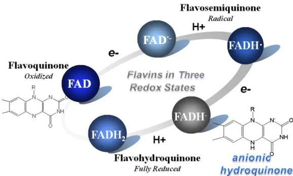
The second cofactor is the flavin adenine dinucleotide (FAD) which is shown in
blue in Figure 3.9. The DNA photolyases are thus also called flavoproteins. There are
three different oxidation states in the flavin molecules: the oxidized form
(flavoquinone, FAD), radicals (flavosemiquinone, FADH·and FAD·-), and the two-electron-reduced flavin-adenine dinucleotide (flavohydroquinone, FADH2 and
FADH-). We present the oxidation-reduction cycle in Figure 3.10. The redox reaction is also accompanied by the electron and proton transfer processes between different
redox states. The flavins thus have indispensable functions in the enzyme repair
- 30 -
Figure 3.10 The three redox states in the oxidation-reduction cycle of flavins.
It is now well recognized that in the photorepair process, the catalytic active
form in vivo is the anionic hydroquinone (FADH-), see Figure 3.11. We will discuss
its significant role in the photorepair mechanism of photolyase in the following
chapter.
Figure 3.11 Structure and labeling of the FADH- molecule. Selected from paper V,
reprinted with permission from American Chemical Society.
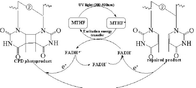
We summarize the photoreactivation process for CPD lesions in Figure 3.12. In
by twisting the CPD dimer out of the DNA normal oligomer into the active pocket.13 Then, the cofactor MTHF (or HDF) absorbs photon from the UV light and transfers
energy to excite the FADH- molecule (FADH*-) through a Förster resonance energy transfer (FRET) mechanism.89 The excited FADH*- has a sufficiently low redox potential to transfer an electron to the thymine dimer with the formation of a radical
anion that subsequently undergoes cycloreversion. This electron transfer process has
been observed by Kao et al. via femtosecond synchronization method and the electron
transfer rate was measured to be in the range of 5.5×10⁹ - 3×10¹⁰ s-1. 90 The photo-induced electron transfer from FADH*- to the thymine dimer triggers a [2+2] cycloreversion which leads to the monomerization of the thymine dimer. The splitting
of thymine dimer forms a thymine and a thymine radical anion. The latter one donates
an electron back to the semiquinone flavin radical (FADH·) to regenerate FADH- which is responsible for another catalytic cycle. The repair process was observed to be
completed in 560 and 589 ps for E.coli and A. nidulans photolyase, respectively.90,91 The repair process involves light and thus the DNA photolyase is also called the ―photo-driven‖ flavoprotein. The whole photoreactivation is a very complex process with both electron and energy transfer.
Figure 3.12 Proposed reaction mechanism for the repair of thymine dimer by DNA photolyase.
- 32 -
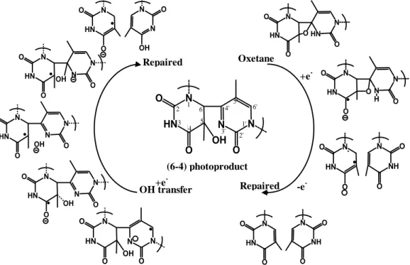
The photoreactivation process for the (6-4)PPs in (6-4) photolyase is quite
similar to the CPDs. However, after the photo-induced electron transfer from FADH-, the successive electron-induced repair step for (6-4)PPs goes in different direction.In
contrast to CPDs, numerous studies proposed that a thermal four-membered ring
oxetane intermediate is formed in the repair mechanism of (6-4) PP.92 The subsequent ring opening of this intermediate then splits the (6-4)PP into two base monomers, as
shown in the right column of Figure 3.13.
Figure 3.13 Oxetane-mediated and the non-oxetane repair mechanisms for the (6-4)PPs.
However, some experimental and theoretical evidences supported a non-oxetane
repair mechanism. Direct OH-transfer from 5’- to 3’-base avoiding the formation of
oxetane intermediate was proposed by Domratcheva et al. 93. It was suggested that electron transfer occurs directly from protein to lesion but not to a strained oxetane
intermediate as previously postulated.In this mechanism, the hydroxyl group of the
pyrimidone transfers directly as depicted in Figure 3.13. However, the transition state
N HN O O OH N N O N HN O O N N O OH N HN O O N N O OH N HN O O N N O OH N HN O O N N OH O Repaired OH transfer N HN O O O HN N O N HN O O O N N O H N HN O O N NH O O N HN O O N NH O O Oxetane +e --e -Repaired N HN O O OH N N O 3' 4' 5 6 5' 6' 1' 2' 1 2 3 4 (6-4) photoproduct +e
-has relative high energy. A barrierless non-oxetane pathway via the conical
intersection involved in the excited lesion radical anion was found theoretically by
Yamamoto and co-workers.94 However, it was then questioned by Philipp that the (6-4) lesion radical anion was in the ground state instead of the excited state due to the
insufficient absorbed photon energy.95 Besides, a transient-water-molecule-formation model was suggested by several groups and the two adjacent histidine residues were
suggested to play important roles in the proton transfer process.96-98
Figure 3.14 Model for the study of (6-4)PP repair mechanism constructed from crystal structure (PDB code: 3CVU).
In order to reveal the underlying repair mechanism, we check the above
suggested mechanisms based on the cluster model (Figure 3.14) by using the cluster
approach99-105 at the B3LYP/6-31G(d) level, where the photoproduct T(6-4)T is colored mauve in the active site. Several functional groups around the photo lesion are
included. Two histidines (green) and a water molecule WAT369 which is quite close
to the pyrimidine ring, are chosen in the cluster model. The flavin FADH- is simplified by the Adenine and 2’-OH. Other residues are collected to account for the
- 34 -
role of the residue His365 which is suggested to be the proton donor in the repair
mechanism, we treat His365 in both neutral (N) and protonated (P) forms, labeled as
HIE365 and HIP365, respectively. Larger basis set 6-311++G(2d,2p) is used in the
single-point calculations based on the optimized structures. The polarizable
continuum model (PCM) with the dielectric constant of 4 is used to consider the
solvent effect and the rest part of the enzyme. 100-105
We first discuss the model with HIE365 inside. The optimized structures and
energy profiles are illustrated in Figure 3.15, where the reactant with HIE365 is
represented as N-react. The WAT369 forms a strong hydrogen bond (1.83Å) to
5-hydroxyl group and it is further stabilized by the Y306 residue with a hydrogen
bond (distance of 1.80 Å). Meanwhile, there is also a hydrogen bond between HIE365
and pyrimidone. The HIE365 and H369 are stabilized by 2’-OH and Y234 by
hydrogen bonds, respectively. Starting from this reactant, we obtain the transition
state for the non-oxetane repair mechanism which involves the direct hydroxyl
transfer process. We investigate the stepwise hydroxyl transfer process (denoted as
path A). The C5-O bond is increased to 1.85 Å in the transition state N-A-TS1 where
the hydroxyl group is stabilized by HIE365 and WAT369. However, the barrier for
the C-O cleavage is 31.6 kcal/mol at the PCM//B3LYP/6-31G(d) level. The hydroxyl
group forms a hydrogen bond with His369 in following intermediate N-A-int which is
3.2 kcal/mol above the N-react. The next nucleophilic attack on the C4’ atom of
pyrimidone produces a transition state N-A-TS2, with a reaction barrier of 20.3
kcal/mol. The product of the hydroxyl group transfer is represented as int2 in Figure
3.15. The C4’-O distance is 1.49 Å and the C6-C4’ bond is weakened from 1.53 Å of
N-react to 1.67 Å of int2, which implies that the linked pyrimidines are gradually
N-react N-A-TS1
N-A-int1 N-A-TS2
int2 PES
Figure 3.15 Optimized geometries depicted by XYZViewer and relative energies for stationary points and transition states calculated at B3LYP/6-311++G(2d,2p) and