• No results found

Medication-related osteonecrosis of the jaw : occurence, risk factors, pathogenesis & treatment

N/A
N/A
Protected

Academic year: 2021

Share "Medication-related osteonecrosis of the jaw : occurence, risk factors, pathogenesis & treatment"

Copied!
136
0
0

Loading.... (view fulltext now)

Full text

(1)

DOCT OR AL DISSERT A TION IN ODONT OL OG Y FREDRIK HALLMER MALMÖ UNIVERSIT

MEDIC

A

TION-REL

A

TED

OS

TEONECR

OSIS

OF

THE

JA

W

FREDRIK HALLMER

MEDICATION-RELATED

OSTEONECROSIS OF THE JAW

(2)
(3)
(4)

ISBN 978-91-7104-983-4 (print) ISSN 978-91-7104-984-1 (pdf) Holmbergs, Malmö 2019
(5)

Department of Oral and Maxillofacial Surgery and

Department of Oral Diagnostics

Faculty of Odontology

Malmö University, 2019

FREDRIK HALLMER

MEDICATION-RELATED

OSTEONECROSIS OF THE JAW

Occurence, Risk Factors, Pathogenesis & Treatment

(6)

Publikationen finns även elektroniskt,

se muep.mau.se

(7)
(8)
(9)

TABLE OF CONTENTS

ABSTRACT ... 9

LIST OF PUBLICATIONS ... 12

INTRODUCTION ... 13

MRONJ ... 14

Pathogenesis ... 15

Risk factors ... 15

Treatment ... 16

Bone ... 16

Bone remodeling ... 17

Bisphosphonates ... 18

Denosumab ... 19

Osteoporosis ... 19

Glucocorticoid-induced osteoporosis ... 20

Skeletal-related events in metastatic disease ... 20

Multipel myeloma ... 21

Rationale and background for the thesis ... 22

AIMS ... 25

MATERIAL AND METHODS ... 26

Patients ... 26

Study I ... 26

Study II ... 28

Study III ... 29

Study IV ... 31

Ethical consideration ... 33

Statistical analyses ... 33

(10)

Study I and III ... 33

Study IV ... 33

RESULTS ... 34

Study I ... 34

Prevalence ... 34

Underlying disease and type of bisphosphonates ... 34

Initiating factor ... 34

Site ... 35

Stage ... 35

Treatment ... 35

Study II ... 37

Study III ... 38

Prevalence ... 40

Underlying disease ... 40

Site ... 40

Stage ... 42

Initiating factor ... 42

Pain intensity ... 45

Study IV ... 47

Incidence ... 47

Risk factors ... 48

DISCUSSION ... 51

CONCLUSIONS ... 58

FUTURE RESEARCH ... 59

POPULÄRVETENSKAPLIG SAMMANFATTNING ... 61

ACKNOWLEDGEMENTS ... 63

REFERENCES ... 65

PAPERS I-IV ... 79

(11)

ABSTRACT

The antiresorptive drugs bisphosphonates and denosumab are

widely used to preserve bone strength by inhibiting osteoclast

me-diated bone resorption. High dose intravenous bisphosphonates

and high dose denosumab are mainly used to treat skeletal related

events in patients with metastatic bone disease such as metastatic

breast cancer and metastatic prostate cancer or in patients with

multiple myeloma. In patients with osteoporosis, oral

bisphospho-nates are mostly used. Medication-related osteonecrosis of the jaw

(MRONJ) is a serious side effect associated both with

bisphospho-nates and denosumab treatment. The first report on MRONJ was

published in 2003. At first, the condition was believed to be a new

disease, but it was later proved to be the same disease, which

exist-ed more than 100 years ago in match factory workers, and

phos-phate miners, in whom the disease was recognized as phossy jaw.

The overall aim of this thesis was to study the occurrence, risk

fac-tors, pathogenesis and treatment of MRONJ.

Study I describes prevalence, trigger factors and treatment

out-comes of MRONJ retrospectively between the years 2003-2010 in

Region Skåne. During this time, 55 patients had been diagnosed

with MRONJ. Of these patients, 31 patients with a malignant

dis-ease were treated with high dose intravenous bisphosphonates and

24 patients with osteoporosis were treated with oral

bisphospho-nates. The prevalence of MRONJ was estimated to be 0.024% for

patient on oral bisphosphonates and 2.8% for patients on high

(12)

dose intravenous bisphosphonates. Tooth extraction was the most

common trigger factor. After treatment of MRONJ, healing

oc-curred more frequently in patients with osteoporosis treated with

oral bisphosphonates than in patients with a malignant disease

treated with high dose intravenous bisphosphonates.

Study II investigated the association between microflora and

MRONJ by using 16S rRNA pyrosequencing technique for the

de-tection of bacteria in necrotic bone lesions. Included were 18

con-secutive patients with MRONJ, ten with osteoporosis and eight

with metastatic disease. Bone biopsies were retrieved with two

sep-arate 3 mm sterile trepan burrs from the centre of the necrotic

bone and from visible healthy bone. The necrotic bone lesions

con-tained mainly anaerobic bacteria, representative for periodontal

microflora, suggesting that a periodontal infection in combination

with antiresorptive treatment could initiate the osteonecrosis.

Study III is a prospective cohort study where the prevalence and

initiating factors of MRONJ and the outcome of surgical therapy

were analysed. All new cases of MRONJ between 2012 and 2015

in Region Skåne were included. Fifty-five patients with MRONJ

were identified. The prevalence of MRONJ for patients on oral

bisphosphonates was 0.043%, on high dose intravenous

bisphos-phonates 1.03% and on high dose denosumab 3.64%. Periodontal

disease preceded development of MRONJ in 41 patients. Fifty

pa-tients were treated surgically and followed up for at least 2

months. Lesions progressed to remission or healing in 80.0% of

patients treated with sequestrectomy and in 92.5% of patients

treated with block resection.

In study IV, the aim was to prospectively determine the incidence

and define risk factors for MRONJ in patients with metastatic

breast cancer treated with zoledronic acid and/or denosumab.

Breast cancer patients diagnosed between 2012 and 2015 in the

(13)

re-therapy or corticosteroid use, diabetes and smoking habits) were

recorded. Sixteen patients of 242 (6.6%) developed MRONJ

dur-ing the 77 months study period (from 1st of January 2012 to 31st

of May 2018). The incidence of MRONJ in patients treated with

high dose zoledronic acid was 4.1%, and for patients with high

dose denosumab 13.6%. The risk of developing MRONJ in

pa-tients on denosumab was significantly higher compared to papa-tients

treated with zoledronic acid. Corticosteroid use was associated

with a significant decreased risk of MRONJ and diabetes was

as-sociated with a significantly increased risk of MRONJ.

Chemo-therapy or smoking was not associated with a significant increased

risk of MRONJ.

In conclusion, the incidence of MRONJ is more than three times

higher in breast cancer patients treated with denosumab compared

to breast cancer patients treated with zoledronic acid. The

preva-lence in patients with osteoporosis on oral bisphosphonates is low,

< 0.05%. Corticosteroid use decreases the risk of developing

MRONJ whilst diabetes increases the risk. The most common local

risk factor is a periodontal disease. Periodontal bacteria play a

cen-tral role in the pathogenesis and development of MRONJ. The

treatment outcome of MRONJ demonstrates healing in most

pa-tients treated surgically.

(14)

LIST OF PUBLICATIONS

This thesis is based on the following articles, referred to in the text

by their Roman numerals. The articles are appended at the end of

the thesis.

I.

Osteonecrosis of the jaw in patients treated with oral and

intravenous bisphosphonates: experience in Sweden.

Hallmer F, Bjørnland T, Nicklasson A, Becktor JP,

Anders-son G. Oral Surg Oral Med Oral Pathol Oral Radiol. 2014

Aug;118(2):202-8.

II.

Bacterial diversity in medication-related osteonecrosis of

the jaw. Hallmer F, Bjørnland T, Andersson G, Becktor JP,

Kristoffersen AK, Enersen M. Oral Surg Oral Med Oral

Pathol Oral Radiol. 2017 Apr;123(4):436-444.

III.

Prevalence, initiating factor, and treatment outcome of

medication-related osteonecrosis of the jaw-a 4-year

pro-spective study. Hallmer F, Andersson G, Götrick B,

Warfvinge G, Anderud J, Bjørnland T. Oral Surg Oral Med

Oral Pathol Oral Radiol. 2018 Dec;126(6):477-485.

IV.

Incidence of and risk factors for medication-related

(15)

metasta-INTRODUCTION

Medication-related osteonecrosis of the jaw (MRONJ) is a serious

side effect associated with both bisphosphonates and denosumab

treatment (Ruggiero et al., 2014) . In 2003 and 2004, oral and

maxillofacial surgeons began to report cases of non-healing of

ex-posed necrotic bone in the maxillofacial region in patients treated

with bisphosphonates (Marx, 2003; Ruggiero et al. 2004). At first,

the condition was believed to be a new disease, but it actually

proved to be a disease that had existed more than 100 years ago in

match factory workers and phosphate miners in whom the disease

was known as phossy jaw (Marx, 2008; Miles, 1972; Myers &

McGlothlin, 1996). The “strike anywhere” match was invented in

the 1860s, after white phosphorus was added to the sulfur-based

matchstick coating to trigger a more volatile flame. The

phospho-rus-based vats released toxic fumes into the factory air and

“dip-pers and handlers” were exposed to the fumes. Many match

facto-ry workers developed inflammation and sclerosis of the bones and

of the periosteum of the face, due to chronic phosphorus

poison-ing, later resulting in a suppurative process and osteonecrosis. The

osteonecrosis most often started in the alveolar process of the

jaw-bone and was most common in the mandible. Preventive methods

were addressed including excluding workers with dental caries

from the match factories, offering them tooth extraction and after

that at least two weeks’ exclusion from work to minimise the risk

of developing phossy jaw (Ward, 1928). It has been estimated that

phossy jaw developed in a significant percentage of workers,

(16)

United States, a law was passed 1914 prohibiting the importation

and exportation of white phosphorus matches. It seemed, then,

that phossy jaw was a thing of the past (Ward, 1928).

In the mid-1990s, the new potent phosphorous containing drugs

zoledronic acid and pamidronic acid was introduced to enhance

the quality of life by reducing the frequency of morbid

skeletal-related events caused by osteolytic metastases in the most common

cancer form in women, breast cancer, and the second most

com-mon in men, prostate cancer (Body, 1997; DeSantis et al., 2014;

Ferlay et al., 2015; Gilbert et al., 2003; Lundqvist et al., 2016).

Bone metastases can cause substantial morbidity and mortality in

metastatic cancer. The metastases can cause pain and might cause

fractures and spinal cord compression (Saylor & Smith, 2010).

Surprisingly, when introducing a new phosphorous drug, no one

took in an account the knowledge of phossy jaw and that this

could cause osteonecrosis of the jaw as seen in match factory

workers 100 years ago.

MRONJ

MRONJ is defined by the American Association of Oral and

Max-illofacial Surgeons (AAOMS) as “exposed necrotic bone or bone

that can be probed through an intraoral or extraoral fistula in the

maxillofacial region after current or previous treatment with

anti-resorptive agents. It must have persisted for longer than eight

weeks with no history of radiation therapy to the jaws or obvious

metastatic disease of the jaws” (Ruggiero et al., 2014).

MRONJ is staged from 0-3 according to the recommendations of

the AAOMS 2014 (Ruggiero et al., 2014). Stage 0 is defined as no

clinical evidence of necrotic bone but non-specific clinical findings,

radiographic changes and symptoms; Stage 1 as exposed necrotic

bone or fistulas that could be probed to bone without pain or signs

of infection (asymptomatic); Stage 2 as exposed necrotic bone or

(17)

more of the following: pathological fracture, oral-cutaneous fistula,

involvement of the maxillary sinus or necrosis extending to the

in-ferior border of the ramus of the mandible.

Pathogenesis

The pathogenesis of MRONJ has not yet been fully elucidated.

One hypothesis is that MRONJ is a result of

bisphosphonate-induced suppression of the remodeling of the bone in the jaws

(Al-len & Burr, 2009). The hypothesis is based on studies showing that

the jaw has a high remodeling rate, that bisphosphonates suppress

remodeling and that remodeling is considerably higher in the jaw

compared with other skeletal sites (Russell et al., 2008; Russell et

al., 2007). An animal study showed the mandible remodeling rates,

specifically within the alveolar region, to be more than ten times

higher than those within the long bones (Allen & Burr, 2008).

However, the question of how the suppression of remodeling

re-sults in necrotic bone is not answered by this hypothesis.

Another theory is that MRONJ is caused by a dental infection. In

an animal study, ligature-periodontal inflammation was induced in

rats treated with zoledronic acid. Osteonecrosis was observed

asso-ciated with periodontitis in the group of rats treated with

zoledron-ic acid, suggesting that periodontal disease and zoledronzoledron-ic acid

therapy are sufficient for MRONJ development (Aghaloo et al.,

2011).

Risk factors

Risk factors reported for the development of MRONJ include

gen-eral risk factors such as corticosteroid treatment, chemotherapy,

diabetes and smoking (Khan et al., 2016; Marx et al., 2005; Otto

et al., 2012) as well as local risk factors such as anatomic locations

predisposed to trauma (Rasmusson & Abtahi, 2014). The trigger

factor in a majority of cases is tooth extraction (Otto et al., 2012).

(18)

Treatment

Treatment choice for MRONJ is still controversial and

recommen-dations from AAOMS suggest antibacterial mouth rinses,

sympto-matic treatment with antibiotics in the early stages and, in more

severe cases, superficial debridement for long term palliation of

in-fection and pain (Ruggiero et al., 2014). Conservative treatment

include procedures such as minor local debridement with

elimina-tion of sharp bone edges, local hygiene of the area of exposed

bone, the use of topical antibacterial agents, and systemic

antibiot-ics for infection and pain control. In a study long-term antibiotantibiot-ics

resulted in complete or partial healing in 18% of the patients

(Lazarovici et al., 2009). Similar findings were obtained in studies

where conservative treatment showed poor treatment outcome

with a high recurrence rate of MRONJ (Magopoulos et al., 2007;

Zervas et al., 2006).

Surgical interventions have previously been reported to be capable

of exacerbating bone exposure and a conservative approach was

recommended, (Ruggiero et al., 2006; Van den Wyngaert et al.,

2009). More recent reports suggest a more radical treatment

strat-egy with surgical removal of the necrotic bone and primary closure

in combination with antibiotic treatment and in severe cases

seg-mental resection. (Bedogni et al., 2011; Pautke et al., 2011;

Pichardo & van Merkesteyn, 2016; Stockmann et al., 2014;

Wil-liamson, 2010).

Bone

Bone consists of extracellular matrix (crystallised mineral salts),

collagen fibers and bone cells. Bone tissue consists of four different

types of bone cells (osteoblasts, osteoclasts, osteocytes and

osteo-genic cells) (Tortora, 2011). Osteoosteo-genic cells are bone stem cells

from the mesenchyme turning into osteoblasts. The osteoblast is a

bone cell with mesenchymal origin from osteoprogenitor cells.

Os-teoblasts synthesise collagen and the proteins osteocalcin and

(19)

oste-are mature osteoblasts located in lacunae trapped inside the bone

tissue and responsible for maintaining the metabolism of bone

tis-sue. The osteoclast is a multinucleated cell with a hematopoietic

origin in precursor cells from self-fusion mononuclear

mono-cytes/macrophages. Osteoclasts are responsible for bone resorption

(Boyle et al., 2003).

Bone remodeling

Through remodeling of the bone matrix the bone is continuously

renewed. The remodeling cycle starts with resorption of collagen

and minerals by the osteoclasts at the surface of the bone and

oste-oblasts then deposits new mineral and collagen fibers to rebuild the

bone tissue (Boyle et al., 2003).

To start the remodeling cycle, osteoclasts must be recruited.

Oste-oblasts produce a transmembrane protein, RANKL (Receptor

Ac-tivator of Nuclear factor Kappa B-Ligand) that binds to a receptor

on the surface of the osteoclast precursor cells, RANK (Receptor

Activator of Nuclear factor Kappa B). The pre-osteoclasts fuse

to-gether to form large multinuclear cells and are thereby activated

and differentiated into mature osteoclasts (Boyle et al., 2003).

Re-sorption is initiated when the osteoclasts attaches to the bone

sur-face with developed ruffled border. The osteoclasts then release

protons (H+) to lower the pH on the bone surface and thereby

start a breakdown of minerals such as hydroxyapatite crystals.

En-zymes are also released that decompose the bone matrix such as

the protein collagen. During the resorption the cytokines

insulin-like growth factor 1 (IGF-1) and transforming growth factor

(TGF), embedded in the bone, are released and activate osteoblasts

to initiate new bone formation. The osteoblasts can also

down-regulate the osteoclast activation by secreting osteoprotegerin

(OPG) which binds to RANKL and thereby prevents the ligand

from binding to RANK. Thus, RANKL and OPG both affect the

degree of activated osteoclasts by up and down regulation and

thereby balance and control the amount of bone resorption (Boyle

et al., 2003).

(20)

Bisphosphonates

Bisphosphonates are drugs that inhibit bone resorption, bone

me-tabolism and bone remodeling through inhibition of osteoclasts

(Allen & Burr, 2009, 2011; Russell et al., 2008). Bisphosphonates

have high affinity for hydroxyapatite crystals of the bone (Drake et

al., 2008) and they bind preferentially to bones which have high

turnover rates. They are liberated again only when the bone in

which they are deposited is resorbed (Lin, 1996). Due to an

irre-versible binding to the hydroxyapatite crystals in bone mineral,

bisphosphonates have a half-life of approximately eleven years

(Lasseter et al., 2005). The true mechanism of action of

bisphos-phonates is still unknown. One theory is that bisphosbisphos-phonates are

absorbed by the osteoclasts during bone remodeling, causing

apop-tosis of the osteoclast and decreasing osteoclast progenitor

devel-opment and recruitment (Drake et al., 2008; Hughes et al., 1995).

Another theory is that bisphosphonates interfere with and inhibit

proteins on osteoclasts’ cell surface, necessary for the attachment

of the osteoclast to the bone surface (Drake et al., 2008).

Bisphosphonates can be divided into two groups,

non-nitrogen-containing and nitrogen-non-nitrogen-containing. The presence of a nitrogen

group increases the bisphosphonates antiresorptive potency by ten

times (Drake et al., 2008). It is mainly nitrogen-containing

bisphosphonates that have been associated with the development

of MRONJ. The explanation for this is probably that the

nitrogen-containing side chains increase the potency and perhaps toxicity

(Otto et al., 2010, 2012).

In general, when orally administered the bisphosphonates

alen-dronic acid and pamialen-dronic acid are poorly absorbed from the

gastrointestinal tract as a result of their poor lipophilicity which

prevents transcellular transport across epithelial barriers. This

means a low bioavailability of 0.3-0.8%. (Daley-Yates et al., 1991;

Gertz et al., 1995).

(21)

Intravenously administered bisphosphonates on the other hand

lead to a rapid uptake in bone tissue. To date, all bisphosphonates

studied show no evidence of metabolism. Renal excretion is the

only route of elimination (Lin, 1996).

Denosumab

Denosumab is an IgG2 monoclonal antibody with an affinity and

specificity for RANK ligand (RANKL) that inhibits RANKL from

activating its receptor, RANK, on the surface of osteoclasts and

their precursors. Prevention of RANKL-RANK interaction inhibits

osteoclast formation, function and survival, thereby decreasing

bone resorption (Cummings et al., 2009).

Denosumab is administered subcutaneously. The mean half-life of

denosumab is 26 days (Chen et al., 2018). After cessation of

deno-sumab treatment normal osteoclast function can be expected after

approximately 12 months (Iranikhah et al., 2018; McClung et al.,

2017).

Osteoporosis

Osteoporosis is a systemic skeletal disease characterised by low

bone mass leading to increased fracture risk and reduced bone

strength. Osteoporotic fractures predominantly occur at major

skeletal site such as vertebrae, proximal femur, distal forearm, or

scapulae (Awasthi et al., 2018).

The main treatment goal of osteoporosis is prevention of

osteopo-rotic fracture, by achieving maximal bone mineral density with

ad-equate calcium and vitamin D intake, a balanced diet, and weight

bearing exercise (Nguyen, 2017). Estrogen therapy can also be an

important therapy for osteoporosis (Awasthi et al., 2018). Both

bisphosphonates and denosumab are effective in reducing the

inci-dence of clinical vertebral fractures, as well as other clinical

frac-tures in patients suffering from osteoporosis (Black & Rosen,

2016). Alendronic acid has been shown to increase bone mineral

density and reduce clinical fractures with 14% in women with

os-teoporosis (Cummings et al., 1998).

(22)

In patients with osteoporosis, oral bisphosphonates (alendronic

ac-id, ibandronic acid or risedronic acid) are mostly used. In some

pa-tients that do not tolerate oral bisphosphonates, zoledronic acid (5

mg) can be used as an annual infusion or denosumab (60 mg) as

subcutanous injection twice a year (Cummings et al., 2009; Jacques

et al., 2012).

Glucocorticoid-induced osteoporosis

Glucocorticoids play an important role in the treatment of many

inflammatory conditions, for example rheumatoid arteritis

(Buck-ley et al., 2017). However, glucocorticoid use causes a decline in

bone mass during treatment with bone loss and/or fractures as a

result (Lane & Lukert, 1998; Saag et al., 1994).

Skeletal-related events in metastatic disease

The molecular mechanisms by which tumour cells metastasise to

bone are likely to involve invasion, cell adhesion to bone, and the

release of soluble mediators from tumor cells that stimulate

osteo-clast-mediated bone resorption. As both bisphosphonates and

denosumab are powerful inhibitors of osteoclast activity, they are

used in the treatment of patients with osteolytic metastases

(Boissi-er et al., 2000). Pain is the most frequent consequence of bone

me-tastases for patients with advanced cancer and can significantly

in-crease the emotional and physical burden of the metastatic disease

(Body et al., 1998; Janjan et al., 1998).

Intravenous bisphosphonates or denosumab are used to reduce

skeletal-related events such as painful pathologic fractures as well

as to treat hypercalcemia associated with bone metastases in

pa-tients with metastatic breast cancer or prostate cancer (Saylor &

Smith, 2010; Stopeck et al., 2010). In a study by Berenson et al,

zoledronic acid at doses of 2 and 4 mg and pamidronic acid at a

dose of 90 mg each significantly reduced the need for radiation

(23)

ly in patients treated with 2 or 4 mg zoledronic acid or pamidronic

acid than with 0.4 mg zoledronic acid (Berenson et al., 2001).

Denosumab have been shown to be superior to bisphosphonates

when it comes to preventing skeletal-related events among patients

with breast cancer and bone metastases (Stopeck et al., 2010). The

greater effect of denosumab over bisphosphonate in preventing the

worsening of pain may be explained by its greater ability to inhibit

osteoclast formation and function, thereby suppressing

cancer-induced bone destruction (Cleeland et al., 2013).

Multipel myeloma

Multiple myeloma is a haematological malignancy. The

pathogene-sis of multiple myeloma is characterised by the clonal proliferation

of plasma cells in the bone marrow. In more than 80% of patients

with multiple myeloma, a myeloma bone disease develops as a

re-sult of multiple myeloma plasma cells-­‐mediated activation of

oste-oclast activity and suppression of osteoblast activity. Multiple

mye-loma spread to the skeleton is characterised by the formation of

os-teolytic lesions, which increase the risk of skeletal-related events

such as pathologic fracture, spinal cord compression and

hypercal-caemia (Lee et al., 2017). Bisphosphonates prevent, reduce and

de-lay multiple myeloma-related skeletal-related events. Intravenous

pamidronic acid and zoledronic acic and oral clodronate are used

in the management of multiple myeloma (Engelhardt et al., 2014).

The European Myeloma Network recommends the use of

bisphos-phonates in multiple myeloma patients suffering from lytic bone

disease or severe osteoporosis since they are an essential

compo-nent for reducing skeletal morbidity (Engelhardt et al., 2014). In a

recent Cochrane review on bisphosphonates in multiple myeloma,

the results showed that there was moderate-quality evidence of a

reduction in mortality from 41% to 31% (Mhaskar et al., 2017).

(24)

Rationale and background for the thesis

As MRONJ is a severe side effect of antiresorptive treatment with

bisphosphonates and denosumab, there is a need to examine the

incidence, pathogenesis and risk factors for the disease and to

eval-uate methods to prevent and treat the condition.

It is estimated that the bisphosphonates pamidronic acid and

zoledronatic acid, mainly used in oncologic treatments, had been

used in about 2.5 million patients worldwide up til 2003

(Tar-assoff & Csermak, 2003). In 2010, when this study started, about

700 publications, mostly case reports and review articles, were

available on MRONJ. There were only a few studies on incidence

and these reported incidences between 0.01% and 18.6% (Madrid

et al., 2010; Mavrokokki et al., 2007; Ulmner et al., 2014). In

Sweden, the low incidence (0.09%) is based on a survey sent to

oral and maxillofacial surgery clinics estimating the incidence on

MRONJ in patients on oral bisphosphonates (Ulmner et al., 2014).

The high incidence number (18.6%) is the cumulative incidence in

patients on intravenous bisphosphonates (Madrid et al., 2010). As

the incidence varies widely, it seems crucial to determine the true

incidence of MRONJ. A study describing the frequency and clinical

characteristics with a large number of patients diagnosed with

MRONJ was reported from Australia (Mavrokokki et al., 2007).

In 2004-2005 a postal survey was sent to oral and maxillofacial

surgeons, to other dental specialists and to Commonwealth of

Aus-tralia Adverse Drug Reaction Committee. MRONJ was found in

0.01-0.04% of osteoporotic patients mainly on weekly oral

alen-dronate. If extractions were carried out the calculated frequency

was 0.09-0.34%. In patients with bone malignancies, treated

main-ly with intravenous zoledronic acid or pamidronic acid, the

corre-sponding figures were 0.88-1.15% and 6.67-9.1%. The authors

concluded that their retrospective study with multiple assumptions

and without independent assessments of cases showed the need for

prospective clinical trials (Mavrokokki et al., 2007).

(25)

of life issues in the cancer population. The literature search

identi-fied hundreds of publications but only 28 articles fulfilled the

in-clusion criteria, and 22 were used for prevalence analysis (Abu-Id

et al., 2008; Badros et al., 2006; Boonyapakorn et al., 2008;

Cart-sos et al., 2008; Chiandussi et al., 2006; Dimopoulos et al., 2006;

Durie et al., 2005; Estilo et al., 2008; Farrugia et al., 2006; Hoff et

al., 2008; Khamaisi et al., 2007; Magopoulos et al., 2007; Marx,

2003; Mavrokokki et al., 2007; Pozzi et al., 2007; Ruggiero et al.,

2004; Wessel et al., 2008; Wilkinson et al., 2007; Zavras & Zhu,

2006). There were no randomised clinical trials available and no

meta-analysis. The overall weighted prevalence of MRONJ

includ-ed a sample of 39124 patients with a mean weightinclud-ed prevalence of

6.1%. The weighted prevalence was 13.3% for studies with

docu-mented follow-up with a sample size of 927 individuals. In studies

with undocumented follow-up the weighted prevalence was 0.7%

in a sample of 8829 chart reviews and in epidemiological studies,

evaluating a total of 29368 individual records, the weighted

preva-lence was 1.2%. The authors conclude that high-quality studies are

needed to accurately characterise the prevalence of MRONJ and to

determine effective treatment protocols. They suggest that future

investigations should include 1) a clear statement of the total

num-ber of individuals in the study and whether patients were drawn

from a single pool or were referred from outside of the centre, 2)

obtain a progressive pain score on all patients (VAS or similar) 3)

state the duration, total dose and range of bisphosphonate therapy

4) include information on all other possible aggravating factors

such as: smoking, diabetes and use of glucocorticoids 5) stage the

MRONJ lesions according to AAOMS staging system or other

comparable system 6) specifically and clearly define endpoints and

outcomes.

The European Medicines Agency (EMEAS) concluded in

Septem-ber 2009, based on the latest evaluation of their scientific

commit-tee, the Agency´s Committee for Medicinal Products for Human

Use (CHMP), that there is an increased risk of osteonecrosis of the

jaw in patients taking bisphosphonates. The Committee also

con-cluded that further data are needed to determine precise measures

(26)

that could minimise the risk of MRONJ including looking at how

intravenous bisphosphonates should be given such as dose,

fre-quency of administration, duration of treatment and looking into

the risk of osteonecrosis of the jaw in patients taking oral

bisphos-phonates for long periods. The CHMP noted that other possible

risk factors for developing osteonecrosis of the jaw should be

con-sidered such as gender, genetic factors, smoking and other

treat-ments or diseases, as well as the type of cancer and how long the

patient has had it. In 2015, the European Medicines Agency

(EMEAS) completed a periodic review of Aclasta (zoledronic acid),

that concluded the risk of osteonecrosis of the jaw to be very low,

included a number of measures to minimise the risk and an update

to the product information and an introduction of a patient

re-minder card. EMEAS is planning similar measures for other

intra-venous bisphosphonates and denosumab, used for osteoporosis or

for preventing bone complications of cancers, as these are also

as-sociated with a risk of osteonecrosis of the jaw.

Treatment principles, which are known from osteonecrosis of other

causes, include antibiotic treatment and surgical removal of

necrot-ic bone (O’Dell & Sinha, 2011). Up to 2011, most reports dealing

with MRONJ reflect the fact that treatment in accordance with

these principles is not very successful. Different antibiotic regimes

generally have little effect on MRONJ (Magopoulos et al., 2007;

Zervas et al., 2006). MRONJ cases are lengthy and the outcome of

surgery with sequestrectomy is either disappointing or at best

un-known.

Some authors discourage surgical intervention as the effect is

sparsely documented and surgery can aggravate MRONJ

(Zary-chanski et al., 2006). However, a technique published in 2011,

with fluorescence-guided bone resection in Stage 2 and 3 MRONJ

lesions in patients with intravenous bisphosphonate treatment for

metastatic bone disease has shown promising results. Surgical

(27)

in-AIMS

The overall aim of this thesis was to study the occurrence, risk

fac-tors, pathogenesis and treatment of MRONJ.

Specific aims were:

• To study the prevalence, location and initiating factors for

MRONJ in patients with osteoporosis or malignant

dis-ease.

• To study the association between the oral microbiota and

MRONJ by using 16S rRNA pyrosequencing technique.

• To evaluate treatment outcomes for MRONJ in patients

with osteoporosis or malignant disease.

• To analyse the treatment outcome of MRONJ after

seques-trectomy versus block resection.

• To study the incidence and systemic risk factors of

MRONJ in patients with metastatic breast cancer treated

with zoledronic acid or denosumab.

(28)

MATERIAL AND METHODS

Patients

The studies were carried out in Region Skåne, one of 21

adminis-trative regions in Sweden. Region Skåne is the third largest region

with 1.2-1.3 million inhabitants (www.skatteverket.se). Four

de-partments of oncology were responsible for treatment of breast

cancer and four departments of oral and maxillofacial surgery were

responsible for the treatment of MRONJ. The departments were

located at the hospitals in Malmö, Kristianstad, Helsingborg, and

Lund.

Study I

Patients diagnosed with maxillofacial pathology in the period

2003-2010 were identified in the Melior database at the four

de-partments of oral and maxillofacial surgery. Patient records were

searched for the ICD 10 WHO inflammatory conditions of the jaw

(K10.2), unspecified disease of jaws (K10.9), unspecified

osteomye-litis (M86.9), osteonecrosis due to drugs (M87.1), and other

oste-onecrosis (M87.8) and 341 patients were identified. Fifty-five of

these had been diagnosed with MRONJ and were included in

Study I.

(29)

Data were collected and analysed on:

• Prevalence

• Underlying disease (osteoporosis or malignant disease)

• Type of bisphosphonate (oral or intravenous)

• Initiating factor

• Site

• Stage

• Treatment and treatment outcomes

The outcome variable was healing defined as complete mucosal

coverage without any exposed bone and without any sign of

infec-tion.

(30)

Study II

Twenty-two consecutive patients from the ongoing prospective

study (Study III) were recruited between June 2013 and February

2015, in relation to the surgical treatment of the necrosis at the

Department of Oral and Maxillofacial Surgery in Lund. Four

pa-tients were excluded due to current antibiotic treatment or

antibi-otic treatment within three months prior to the study. Two

sepa-rate bone biopsies were collected from each patient in relation to

the surgical treatment of the necrosis to study the association

be-tween microflora and MRONJ by using culture-independent

mo-lecular techniques for the detection of bacteria in necrotic bone

le-sions. After removal of the superficial layers exposed to the oral

milieu with a round burr under irrigation with saline, the first

sample was retrieved from the centre of the necrotic bone lesion.

The second sample was then retrieved from a region of visually

healthy bone adjacent to the necrotic bone as an intraoperative

control sample to analyse if the oral microflora exists in the bone

tissue outside the macroscopic necrotic bone. Each biopsy was

im-mediately placed in cryotubes with DNA/RNA stabilising solution

(RNA Later). In two healthy patients, bone biopsies were collected

in relation to planned surgical extraction of impacted third molars

without any signs of local odontogenic infection.

16SrRNA gene fragments from bacterial DNA were amplified with

polymerase chain reaction. DNA extractions of clinical samples

were performed using the MasterPure DNA isolation kit from

Epi-centre (MCD85201, EpiEpi-centre Biotechnologies, WI). 16S rRNA

gene fragments from bacterial DNA were amplified with PCR

us-ing universal eubacterial primers, forward primer 334f (5’-

CCAGACTCCTACGGGAGGCAGC-3’), and reverse primer 939r

(5’- CTTGTGCGGGCCCCCGTCAATTC-3’) (Baker, Smith, &

Cowan, 2003; Wang & Qian, 2009) targeting the V3-V5

hypervar-iable region.

(31)

Study III

All new patients diagnosed with MRONJ from 1st of January

2012 until 31st of December 2015 were included in Study III. The

patients were followed up prospectively until 31st of December

2016.

Data were collected in case report form and analysed on:

• Prevalence

• Gender

• Underlying disease (osteoporosis or malignant disease)

• Type of antiresorptive drug

• Dosage and treatment time until necrosis occurred

• Initiating factor

• Site

• Stage

• Treatment and treatment outcomes

Previous intraoral x-rays were retrieved in order to analyse the

di-agnosis initiating MRONJ. A horizontal visual analogue scale

(VAS) from 0 to 10 before and after treatment was used to record

pain intensity, where zero indicated “no pain” and ten indicated

“pain cannot be worse”.

Data on the use of bisphosphonates and denosumab were obtained

from statistics on prescriptions (www.socialstyrelsen.se), and

statis-tics on drugs delivered to the health care sector in the region.

Patients were classified into two groups according to surgical

treatment, sequestrectomy or block resection. Patients without

progressive bone disease with ongoing bone destruction but with a

well-defined sequestrum were treated with sequestrectomy

includ-ing a local mucosal flap, removal of the sequestrum and

debride-ment of the necrotic bone. Patients with progressive bone disease

(32)

with ongoing bone destruction, observed on radiograph or ongoing

pain and infection, were treated with block resection including a

surgical removal of the necrotic bone and primary closure. Surgical

marginal block resection included thorough surgical debridement

with saucerisation of the bone until the vital bleeding bone margins

were reached.

The outcome variable was healing after 2 months after treatment

with complete disappearance of exposed bone and the absence of

infection in the jaw affected by MRONJ.

Fifty-five patients with MRONJ were recruited. Forty-three were

women (mean age 73.1 years) and 12 were men (mean age 63.6

years). Fifty patients were included in the study and followed up

for periods of 2-46 months (mean = 15.9 months). Five patients

were excluded either because the patients had died or due to

pro-gression of their diseases making it impossible for them to attend a

follow-up visit after 2 months and for evaluate of treatment

out-come.

(33)

Study IV

Women above 17 years of age with histologically or cytologically

confirmed breast cancer and radiographic evidence of one or

several bone metastases were eligible for study entry.

Patients receiving zoledronic acid or denosumab during the study

period were identified in the treatment registries and the medical

records at the four participating hospitals.

Exclusion criteria were prior exposure to oral or intravenous

bisphosphonates and/or denosumab for treatment of advanced

cancer, prior treatment with oral bisphosphonates or denosumab

for other bone loss conditions (e.g. osteoporosis), history evidence

of MRONJ or osteomyelitis of the jaw as well as radiation therapy

to the head and neck region.

To ensure a certain exposure to the antiresorptive medication, we

included patients who received at least 3 infusions of zoledronic

acid or 3 injections of denosumab. Patients were enrolled from

January 1

st

2012 and to December 31

st

2015 and followed until

May 31

st

2018. Treatment consisted of 4 mg of zoledronic acid

administered i.v. during 15 minutes every 4 to 6 weeks or 120 mg

denosumab (Xgeva®, Amgen Inc., Thousand Oaks, California,

USA) subcutaneous every 4 weeks.

Data collected from patient medical records included age,

bisphos-phonate or denosumab use, treatment time, chemotherapy or

corti-costeroid use, diabetes and smoking habits.

The cohort included 263 patients. Of these 21 patients were

ex-cluded, 15 with only one infusion with zoledronic acid, 3 with only

one treatment with denosumab and 3 treated with oral

bisphos-phonates due to osteoporosis prior to the study. Thus the studied

cohort consisted of 242 (92.0%) patients. Mean age was 64.0

years (range 31-90).

(34)

All patients diagnosed with MRONJ between 1

st

of January 2012

and 31

st

of May 2018 were analysed in the study.

Data collected from patient medical records included and were

an-alysed on:

• Incidence

• Age

• Zoledronic acid or denosumab use and treatment time

• Chemotherapy

• Corticosteroid treatment

• Diabetes

(35)

Ethical consideration

The Regional Ethics Committee at Lund, Sweden approved the

four studies: Dnr 2011/274 (Study I, II, III and IV), Dnr 2012/760

(Study II and III) and Dnr 2018/344 (Study IV). The studies were

conducted in compliance with the Helsinki Declaration, and each

patient in the prospective studies signed a detailed informed

consent form.

Statistical analyses

Study I and III

SPSS for Windows software (Version 14;0 SPSS, Chicago, IL, USA)

was used for data management and statistical analysis. Students’

t-test was used for continuous variables. Between group comparisons

before and after treatment were tested with chi-square. A

signifi-cance level of 5% was used in all tests.

Study IV

The relation between MRONJ and different background variables

(treatment time, corticosteroid treatment, chemotherapy, diabetes

mellitus and smoking) were investigated by odds-ratios (OR) with

95% confidence intervals. The OR was tested by chi-square tests

using a significance level of 5%. Time to MRONJ were analysed

using the Kaplan-Meier method. All calculations were done in SPSS

version 25 (IBM Corp. Released 2017. IBM SPSS Statistics for

Windows, Version 25.0. Armonk, NY: IBM Corp.).

(36)

RESULTS

Study I

Prevalence

Between 2003 and 2010, 55 patients with MRONJ were identified.

The prevalence of MRONJ in patients on oral bisphosphonates

was estimated to be 0.024% and the prevalence in patients on

in-travenous bisphosphonates was estimated to be 2.8%.

Underlying disease and type of bisphosphonates

The most common reason for antiresorptive treatment was

osteo-porosis (n=24; 44%), followed by multiple myeloma (n=15; 27%),

breast cancer (n=11; 20%) and prostate cancer (n=5; 9%). All

pa-tients with osteoporosis were treated with oral bisphosphonates

and all patients with a malignant disease were treated with

intra-venous bisphosphonates (n=31; 56%). More women with

osteopo-rosis were diagnosed with MRONJ than women with cancer

(p=0.025). The osteoporotic patients were older than the patients

with a malignant disease (p=0.008).

Initiating factor

The most common initiating factor was tooth extraction (n=36;

66%), follow by cases referred to as unknown (spontaneous)

(n=12; 22%), dental implant surgery (n=4; 7%) and local trauma

(n=3; 5%).

(37)

Site

The most common site was the mandible (n=43; 78%). Within the

mandible, 35 lesions were localised in the posterior part of the

mandible, 6 cases in the anterior mandible, one in the anterior and

right posterior mandible and one case involved the whole

mandi-ble. Twelve cases were localised in the maxilla (22%); 8 in the

pos-terior part, two in the anpos-terior part and two patients had lesions

involving the whole maxilla.

Stage

The most common was Stage 2 (n=31; 56%), followed by Stage 1

(n=14; 26%), and Stage 3 (n=10; 18%).

Treatment

The patients were treated with antibiotics or surgery or a

combina-tion of medical and surgical treatment. Antibiotic treatment was

carried out with phenoxymethylpenicillin, metronidazole or

clindamycin per os. Patients were treated with antibiotics for

ongo-ing infection. Surgical treatment in stage 1 and 2 cases included a

local resection with superficial debridement and/or removal of

se-quester (sequestrectomy). Stage 3 cases were treated with a local

resection including removal of sequester and resection of necrotic

bone after elevating a mucoperiosteal flap and primary closure

ob-tained.

All patients with osteoporosis except two were treated with

antibi-otics. Twenty were treated with a local resection of the necrotic

le-sion. Healing occurred in most patients with osteoporosis (n=19;

79%) and all of these were treated with local resection of the

ne-crotic lesion in combination with antibiotics, except one patient

who received no antibiotics. In five patients, no healing was

no-ticed; one of these was treated with local resection.

In patients with cancer, most patients were treated with antibiotics

(n=28; 90%). Local resection of the necrotic lesion was undertaken

in 27 cases and all of these received antibiotics as well. Three

pa-tients were treated with local resection alone. Healing was noticed

(38)

in eight patients (26%) with a malignant disease, five with

underly-ing disease breast cancer and three with multiple myeloma.

A significantly better treatment outcome, with complete healing of

the mucosa, was identified in the osteoporosis group treated with

oral bisphosphonates than in cancer patients treated with

intrave-nous bisphosphonates, (p=0.00009). All successful cases were

treated with a combination of surgical therapy and systemic

anti-biotics, except one case treated with antibiotics alone, and one case

treated with local resection only.

(39)

Study II

Altogether 18 patients with MRONJ (14 females, 4 males), with a

mean age of 71.9 years (range 41-91 years) were included in the

study. The most common underlying disease was osteoporosis

(n=10; 56%), followed by breast cancer (n=4; 22%), prostate

can-cer (n=3; 17%) and multiple myeloma (n=1; 6%). The most

com-mon antiresorptive treatments were oral bisphosphonates (n=8;

44%), followed by subcutaneously administered denosumab (n=6;

33%), a combination of oral and intravenous bisphosphonates or

denosumab (n=3; 17%) and intravenous bisphosphonate (n=1;

6%)

The necrotic bone samples from the 18 patients were all strongly

positive for 16S rRNA PCR. Visually healthy bone samples in

sev-en patisev-ents were negative for 16S rRNA PCR. In 11 of the patisev-ents,

the visually healthy bone was also detected as positive where six

PCR products were strong and five weak.

Eight dominating taxa groups were identified at genus level. In

both necrotic and visual healthy bone samples there were:

Porphy-romonas, Lactobacillus, Tannerella, Prevotella, Actinomyces,

Treponema, Streptococcus and Fusobacterium. In some patients,

the periopathogenic bacteria dominate and in other, the

Lactoba-cillus or Prevotella dominate.

When comparing necrotic samples from the patients with

osteopo-rosis, treated with bisphosphonate, a higher level of

periopathogen-ic bacteria was detected in this group than in the group treated

with denosumab (XGEVA).

Analyses of the bone samples from the two healthy controls were

negative for 16S rRNA by PCR.

(40)

Study III

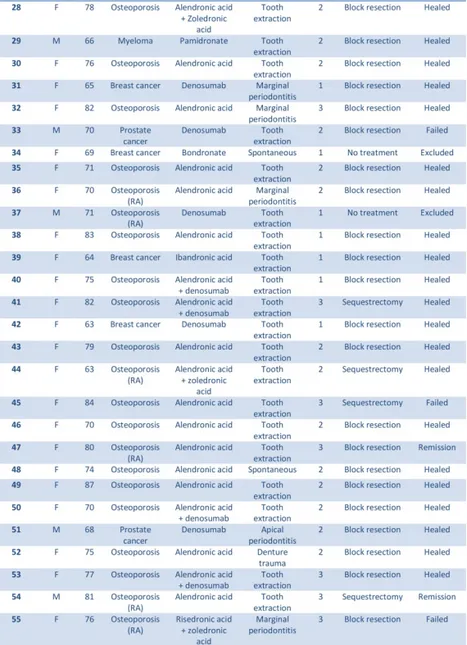
Patient characteristics of the 55 consecutive patients are presented

in Table I.

(41)

Table I. Characteristics of 55 consecutive patients with MRONJ

treated with oral or intravenous bisphosphonate or subcutaneously

administered denosumab. (RA = Rheumatoid arthritis).

(42)

Prevalence

The estimated number of patients on oral bisphosphonates

(alen-dronic acid and rise(alen-dronic acid) for treatment of osteoporosis,

dur-ing the period 2012-2015 was around 50 000 in Region Skåne.

The prevalence of MRONJ for patients on oral bisphosphonates

was estimated to be 0.043%.

We estimated that around 700 patients were on intravenous

bisphosphonates (zoledronic acid and pamidronic acid) due to

metastatic diseases, during the period 2012-2015 in Region Skåne.

The prevalence of MRONJ in patients on intravenous

bisphospho-nates was estimated to be 1.03%.

The estimated numbers of patients on high dose denosumab, due

to metastatic diseases during the period 2012-2015 in Region

Skåne were around 1700. The prevalence of MRONJ in patients

on denosumab was estimated to be 3.64%.

Underlying disease

The most common reason for antiresorptive treatment was

osteo-porosis (n=31; 56%), followed by breast cancer (n=11; 20%),

prostate cancer (n=6; 11%), multiple myeloma (n=5; 9%), lung

carcinoma (n=1; 2%) and giant cell carcinoma (n=1; 2%).

There were no significant differences (p=0.176) in healing in

rela-tion to the underlying disease.

Site

Most lesions were located in the mandible (n=41; 75%) with 32 in

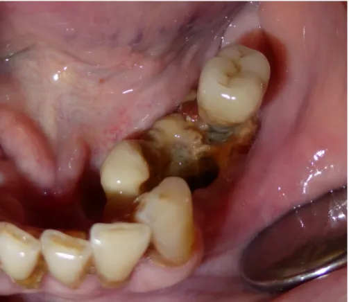
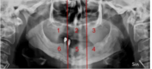
the posterior part (segment 4 or 6) (one of the cases shown in Fig.

1), five in the anterior part and four cases were located in more

than one segment of the mandible.

(43)

There was no significant difference (p=0.156) in healing in relation

to the site of the necrosis.

Figure 1. Exposed necrotic bone involving the tori in the mandible

in patient nr 22.

(44)

Figure 2. Exposed necrotic bone in the anterior part of the maxilla

in patient nr 23.

Stage

Ten patients were classified as Stage 1, 36 patients were classified

as Stage 2, and nine patients as Stage 3.

There was no significant difference (p=0.067) in healing in relation

to the stage of the necrosis.

Initiating factor

The most common initiating factor for development of MRONJ

was tooth extraction (n=39; 71%) followed by marginal

periodon-titis (n=10; 18%) (one of the cases is shown in Fig. 3),

spontane-ously (unknown) (n=3; 5%); apical periodontitis (n=2; 4%) and as

(45)

There was no significant difference (p=0.394) in healing in relation

to initiating factor.

In 32 out of 39 cases where the initiating factor was tooth

extrac-tion it was possible to retrieve intraoral radiographs taken before

the extraction from the patient’s general dental practitioner. The

radiographs were analysed by a specialist in Oral and Maxillofacial

Radiology. Twenty-nine (91%) of the 32 tooth extractions were of

teeth with either severe marginal periodontitis (15 patients) or

api-cal periodontitis (3 patients) or both diagnoses on the same tooth

(11 patients). Together with 10 patients with MRONJ caused by

marginal periodontitis and 2 due to apical periodontitis alone as an

initiating factor, local periodontal destruction including marginal

bone loss adjacent to root surfaces preceded development of

MRONJ in a total of 41 patients.

(46)

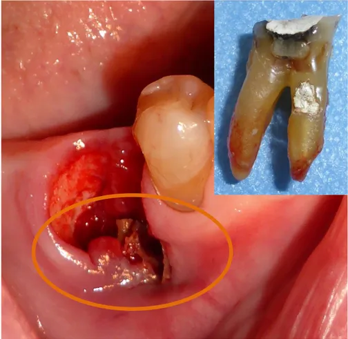
Figure 3. Marginal periodontitis as the initiating factor

demon-strating exposed necrotic bone with an established MRONJ lesion

at the time of the extraction.

(47)

Surgical treatment

Ten patients were treated with sequestrectomy. Five lesions (50%)

healed. Three went into remission (two patients with stage 2 and

one with stage 3). The overall improvement rate was 8/10 (80.0%).

Mean follow-up time was 13.3 months (range 5-29 months). After

healing, no adverse events were recorded. Forty patients were

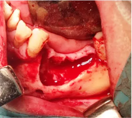
treated with block resection (one of the cases shown in Fig. 4).

Thirty-one lesions healed (77.5%). Six cases went in remission

(four patients with stage 2 and two with stage 3). The overall

im-provement rate was 37/40 (92.5%). Mean follow-up time was 16.7

months (range 2-46 months). After healing, no adverse events were

recorded.

There was no significant difference (p=0.118) in healing in patients

treated surgically with block resection or sequestrectomy.

There was no significant difference (p=0.094) in healing rate

be-tween men or women.

Pain intensity

Mean VAS for 10 patients treated with sequestrectomy before

treatment was 3.9 and after treatment, 1.0. Mean VAS for 40

pa-tients treated with block resection before treatment was 3.8 and

after treatment, 0.2. Mean VAS for six patients in remission before

treatment was 3.7 and after treatment, 0.0.

(48)

Figure 4. Block resection including smoothening of sharp edges

af-ter removal of all necrotic bone.

(49)

Study IV

Incidence

Sixteen of 242 patients (6.6%) developed MRONJ during the

study period. Their mean age was 64.6 years (range 41-84). The

incidence of MRONJ in patients treated with zoledronic acid was

4.1%, and for patients treated with denosumab it was 13.6%. For

patients initially treated with zoledronic acid which subsequently

switched to denosumab the incidence was 8.3% (Table II). The risk

of developing MRONJ in patients on denosumab was significantly

higher compared with those on zoledronic acid (OR 3.7; 95% CI

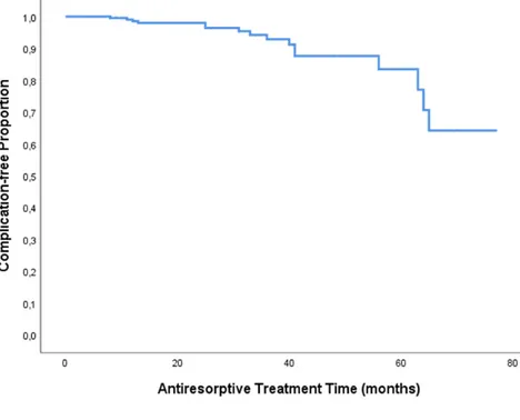
1.3-10.6, p=0.011). The cumulative incidence of MRONJ is

pre-sented in Fig. 5.

Table  II.  Zoledronic  acid  (Z),  Denosumab  (D)  

Antiresorptive  

Treatment  

No  MRONJ  

MRONJ  

Total  

Incidence  


Z  

164  

7  

171  

4,1%  


D  

51  

8  

59  

13,6%  


Z+D  

11  

1  

12  

8,3%  


Total  

226  

16  

242  

6,6%  


Table II. Incidence of MRONJ in women with disseminated breast

cancer according to anitresorptive treatment.

(50)

Figure 5. Cumulative incidence of MRONJ in patients on

zoledronic acid and/or denosumab with metastatic breast cancer.

Risk factors

Mean antiresorptive treatment time for all patients was 27.5

months. For patients developing MRONJ the mean treatment time

was 35.3 months (range 8-70) and in patients not developing

MRONJ 27.0 months (range 2-77). The mean treatment time for

zoledronic acid was 26.1±17.0 months, 25.4±16.7 months for

pa-tients not developing MRONJ and 40.9±18.8 months with

MRONJ. The mean treatment time for denosumab was 30.8±17.5

months, 31.3±17.5 months for patients without development of

MRONJ and 27.8±18.5 months with MRONJ.

Corticosteroid treatment was administered to 163 of the 242

pa-tients. Of these 163, six were diagnosed with MRONJ compared

(51)

time of antiresorptive treatment was 25.1±17.0 months with

corti-costeroid treatment and 31.9±16.4 months without corticorti-costeroid

treatment.

Chemotherapy was administered to 145 of the 242 patients. Of the

145 patients treated with chemotherapy, 12 were diagnosed with

MRONJ compared to four of the 97 not receiving chemotherapy.

Chemotherapy was not associated with a significantly increased

risk of MRONJ (OR 2.1; 95% CI 0.7-6.7, p=0.2) (Table III). The

mean treatment time of antiresorptive treatment was 26.1±16.9

months with chemotherapy and 29.6±17.6 months without

chemo-therapy.

Fourteen of the 242 patients had diabetes mellitus. Of these 14, 3

patients had MRONJ. Diabetes was associated with a significantly

increased risk of MRONJ (OR 4.5; 95% CI 1.1-18.1, p=0.02)

(Table III). The mean treatment time of antiresorptive treatment

was 20.5±17.7 months with diabetes and 28.0±17.2 months

with-out diabetes.

Twenty-seven of the 242 patients were regular smokers, 4 out of

16 in the group with MRONJ. Smoking was not associated with a

statistically increased risk of MRONJ (OR 2.9; 95% CI 0.9-9.9,

p=0.07) (Table III). The mean treatment time of antiresorptive

treatment was 30.7±18.8 months in smokers and 27.1±17.1

months in non-smokers.

(52)

Table III. Risk factors of MRONJ in women with disseminated

breast cancer according to corticosteroid treatment, chemotherapy,

diabetes or smoking.

Table  III  


Corticosteroids  

No  MRONJ  

 MRONJ  

Total  

(%)  

OR      95%  CI      p  

No  treatment  

     69  

10  

79  

12,7%          

Treatment  

     157  

6  

163  

3,7%  

0.3      0.1-­‐0.8      0.008  


Chemotherapy  


No  treatment  

     93  

4  

97  

4,1%  


Treatment  

     133  

12  

145  

8,3%  

2.1      0.7-­‐6.7      0.2  


Diabetes  


No    

     215  

13  

228  

5,7%  


Yes  

     11  

3  

14  

21,4%  

4.5      1.1-­‐18.1      0.02  


Smoking  


No  

     203  

12  

215  

5,6%  


Yes  

     23  

4  

27  

14,8%  

2.9      0.9-­‐9.9      0.07  


Figure

Table  I.  Characteristics  of  55  consecutive  patients  with  MRONJ  treated with oral or intravenous bisphosphonate or subcutaneously  administered denosumab
Figure 1. Exposed necrotic bone involving the tori in the mandible  in patient nr 22.
Figure 2. Exposed necrotic bone in the anterior part of the maxilla  in patient nr 23
Figure  3.  Marginal  periodontitis  as  the  initiating  factor  demon- demon-strating exposed necrotic bone with an established MRONJ lesion  at the time of the extraction
+7

References

Related documents

Two databases have been studied: the MIT driver stress database and a new ex- perimental database, where three stress tasks have been performed for 9 subjects: the Trier Social

Det lägsta betyget går till L5 och anledningen till detta är att stängerna måste transporteras med travers och läggas ner i lådan, tillsammans med att arbetet inte utförs i

The aim of this study was to describe and explore potential consequences for health-related quality of life, well-being and activity level, of having a certified service or

För att göra detta har en körsimulator använts, vilken erbjuder möjligheten att undersöka ett antal noggranna utförandemått för att observera risktagande hos dysforiska

Increase of bone marrow edema both in intensity and extension, with the edema on sagittal images (not shown) now centered around the weight bearing area of the medial femoral

While other antidepressants such as SSRI may cause an increase of suicide ideation in depressive patients, tianeptine seems to be less likely to produce such symptoms when

Stability evaluation of implants integrated in grafted and nongrafted maxillary bone: a clinical study from implant placement to abutment connection.. Sjöström

METHODS: Genetic instruments were identi fied for 10 key obesity-related risk factors, and their association with glioma risk was evaluated using data from a genome-wide