SILVIA GALLI
ON MAGNESIUM–
CONTAINING IMPLANTS
FOR BONE APPLICATIONS
SIL VIA G ALLI MALMÖ UNIVERSIT ON MA GNESIUM–C ONT AININ G IMPL ANT S FOR BONE APPLIC A TIONS DOCT OR AL DISSERT A TION IN ODONT OL OG Y
O N M A G N E S I U M – C O N T A I N I N G I M P L A N T S F O R B O N E A P P L I C A T I O N S
Malmö University
Faculty of Odontology Doctoral Dissertation 2016
© Silvia Galli 2016
SILVIA GALLI
ON MAGNESIUM–
CONTAINING IMPLANTS
FOR BONE APPLICATIONS
Malmö University
Faculty of Odontology
The publication is available in electronic format at: https://dspace.mah.se/handle/2043/21277
To my father and mother, with endless gratitude and love
This thesis is number 49 in a series of investigations on implants, hard tissues and the loco-motor apparatus originating from the Department of Biomaterials, University of Gothenburg and the Department of Prosthodontics/Material Sciences, Malmö University, Sweden.
1. Anders R Eriksson DDS, 1984. Heat-induced Bone Tissue Injury. An in vivo investigation of heat tolerance of bone tissue and temperature rise in the drilling of cortical bone.
Thesis defended 21.2.1984. External examiner: Docent K-G. Thorngren. 2. Magnus Jacobsson MD, 1985. On Bone Behaviour after Irradiation.
Thesis defended 29.4.1985. External examiner: Docent A. Nathanson. 3. Fredric Buch MD, 1985. On Electrical Stimulation of Bone Tissue.
Thesis defended 28.5.1985. External examiner: Docent T. Ejsing-Jörgensen.
4. Peter Kälebo MD, 1987. On Experimental Bone Regeneration in Titanium Implants. A quantita-tive microradiographic and histologic investigation using the Bone Harvest Chamber.
Thesis defended 1.10.1987. External examiner: Docent N. Egund. 5. Lars Carlsson MD, 1989. On the Development of a new Concept for
Orthopaedic Implant Fixa-tion.
Thesis defended 2.12.1989. External examiner: Docent L-Å Broström. 6. Tord Röstlund MD, 1990. On the Development of a New Arthroplasty.
Thesis defended 19.1.1990. External examiner: Docent Å. Carlsson. 7. Carina Johansson Res Tech, 1991. On Tissue Reaction to Metal Implants.
Thesis defended 12.4.1991. External examiner: Professor K. Nilner. 8. Lars Sennerby DDS, 1991. On the Bone Tissue Response to Titanium
Implants.
Thesis defended 24.9.1991. External examiner: Dr J.E. Davies. 9. Per Morberg MD, 1991. On Bone Tissue Reactions to Acrylic Cement.
Thesis defended 19.12.1991. External examiner: Docent K. Obrant. 10. Ulla Myhr PT, 1994. On factors of Importance for Sitting in Children
with Cerebral Palsy.
Thesis defended 15.4.1994. External examiner: Docent K. Harms-Ringdahl.
11. Magnus Gottlander MD, 1994. On Hard Tissue Reactions to Hydroxyapatite- Coated Titanium Implants.
Thesis defended 25.11.1994. External examiner: Docent P. Aspenberg. 12. Edward Ebramzadeh MScEng, 1995. On Factors Affecting Long-Term
Outcome of Total Hip Replacements.
14. Ann Wennerberg, DDS, 1996. On Surface Roughness and Implant Incorporation.
Thesis defended 19.4.1996. External examiner: Professor P-O. Glantz. 15. Neil Meredith BDS MSc FDS RCSm, 1997. On the Clinical Measurement
of Implant Stability Osseointegration.
Thesis defended 3.6.1997. External examiner: Professor J. Brunski. 16. Lars Rasmusson DDS, 1998. On Implant Integration in
Membrane-Induced and Grafter Bone.
Thesis defended 4.12.1998. External examiner: Professor R. Haanaes. 17. Thay Q Lee MSc, 1999. On the Biomechanics of the Patellfemoral Joint
and Patellar Resurfac-ing in Total Knee Arthroplasty.
Thesis defended 19.4.1999. External examiner: Docent G. Nemeth. 18. Anna Karin Lundgren DDS, 1999. On Factors Influencing Guided
Regeneration and Augmen-tation of Intramembraneous Bone. Thesis defended 7.5.1999. External examiner: Professor B. Klinge. 19. Carl-Johan Ivanoff DDS, 1999. On Surgical and Implant Related Factors
Influencing Integra-tion and Function of Titanium Implants. Experimental and Clinical Aspects.
Thesis defended 12.5.1999. External examiner: Professor B. Rosenquist. 20. Bertil Friberg DDS MDS, 1999. On Bone Quality and Implant Stability
Measurements.
Thesis defended 12.11.1999. External examiner: Docent P. Åstrand. 21. Åse Allansdotter Johansson MD, 1999. On Implant Integration in
Irradiated Bone. An Experi-mental Study of the Effects of Hyperbaric Oxygeneration and Delayed Implant Placement.
Thesis defended 8.12.1999. External examiner: Docent K. Arvidsson-Fyrberg.
22. Börje Svensson FFS, 2000. On Costochondral Grafts Replacing Mandibular Condyles in Juve-nile Chronic Arthritis. A Clinical, Histologic and Experimental Study.
Thesis defended 22.5.2000. External examiner: Professor Ch. Lindqvist. 23. Warren Macdonald BEng, MPhil, 2000. On Component Integration on
Total Hip Arthroplas-ties: Pre-Clinical Evaluations.
Thesis defended 1.9.2000. External examiner: Dr A.J.C. Lee.
24. Magne Røkkum MD, 2001. On Late Complications with HA Coated Hip Arthroplasties.
Thesis defended 12.10.2001. External examiner: Professor P. Benum. 25. Carin Hallgren Höstner DDS, 2001. On the Bone Response to Different
Implant Textures. A 3D analysis of roughness, wavelength and surface pattern of experimental implants.
26. Young-Taeg Sul DDS, 2002. On the Bone Response to Oxidised Titanium Implants: The role of microporous structure and chemical composition of the surface oxide in enhanced osseointegration.
Thesis defended 7.6.2002. External examiner: Professor J.E. Ellingsen. 27. Victoria Franke Stenport DDS, 2002. On Growth Factors and Titanium
Implant Integration in Bone.
Thesis defended 11.6.2002. External examiner: Associate Professor E. Solheim.
28. Mikael Sundfeldt MD, 2002. On the Aetiology of Aseptic Loosening in Joint Arthroplasties and Routes to Improved cemented Fixation. Thesis defended 14.6.2002. External examiner: Professor N. Dahlén. 29. Christer Slotte CCS, 2003. On Surgical Techniques to Increase Bone
Density and Volume. Studies in Rat and Rabbit.
Thesis defended 13.6.2003. External examiner: Professor C.H.F. Hämmerle.
30. Anna Arvidsson MSc, 2003. On Surface Mediated Interactions Related to Chemomechanical Caries Removal. Effects on surrounding tissues and materials.
Thesis defended 28.11.2003. External examiner: Professor P. Tengvall. 31. Pia Bolind DDS, 2004. On 606 retrieved oral and craniofacial implants.
An analysis of conse-quently received human specimens.
Thesis defended 17.12.2004. External examiner: Professor A. Piattelli. 32. Patricia Miranda Burgos DDS, 2006. On the influence of micro- and
macroscopic surface mod-ifications on bone integration of titanium implants.
Thesis defended 1.9.2006. External examiner: Professor A. Piattelli. 33. Jonas P. Becktor DDS, 2006. On factors influencing the outcome of
various techniques using endosseous implants for reconstruction of the atrophic edentulous and partially dentate maxilla.
Thesis defended 17.11.2006. External examiner: Professor K.F. Moos. 34. Anna Göransson DDS, 2006. On Possibly Bioactive CP Titanium
Surfaces.
Thesis defended 8.12.2006. External examiner: Professor B. Melsen. 35. Andreas Thor DDS, 2006. On platelet-rich plasma in reconstructive
dental implant surgery.
Thesis defended 8.12.2006. External examiner: Professor E.M. Pinholt. 36. Luiz Meirelles DDS MSc, 2007. On Nano Size Structures for Enhanced
Early Bone Formation.
Thesis defended 13.6.2007. External examiner: Professor Lyndon F. Cooper.
37. Pär-Olov Östman DDS, 2007. On various protocols for direct loading of implant- supported fixed prostheses.
38. Kerstin Fischer DDS, 2008. On immediate/early loading of implant supported prostheses in the maxilla.
Thesis defended 8.2.2008. External examiner: Professor K. Arvidsson Fyrberg.
39. Alf Eliasson 2008. On the role of number of fixtures, surgical technique and timing of loading.
Thesis defended 23.5.2008. External examiner: Professor K. Arvidsson Fyrberg.
40. Victoria Fröjd DDS, 2010. On Ca2+ incorporation and nanoporosity of titanium surfaces and the effect on implant performance.
Thesis defended 26.11.2010. External examiner: Professor J.E. Ellingsen. 41. Lory Melin Svanborg DDS, 2011. On the importance of nanometer
structures for implant in-corporation in bone tissue.
Thesis defended 01.06.2011. External examiner: Associate professor C. Dahlin.
42. Byung-Soo Kang MSc, 2011. On the bone tissue response to surface chemistry modifications of titanium implants.
Thesis defended 30.09.2011. External examiner: Professor J. Pan. 43. Kostas Bougas DDS, 2012. On the influence of biochemical coating on
implant bone incorpora-tion.
Thesis defended 12.12.2012. External examiner: Professor T. Berglundh. 44. Arne Mordenfeld DDS, 2013. On tissue reaction to and adsorption of
bone substitutes.
Thesis defended 29.5.2013. External examiner: Professor C. Dahlin. 45. Ramesh Chowdhary DDS, 2014. On efficacy of implant thread design for
bone stimulation.
Thesis defended 21.05.2014. External examiner: Professor Flemming Isidor.
46. Anders Halldin MSc, 2015. On a biomechanical approach to analysis of stability and load bear-ing capacity of oral implants.
Thesis defended 28.05.2015. External examiner: Professor J. Brunski. 47. Francesca Cecchinato MSc, 2015. On magnesium-modified titanium
coatings and magnesium alloys for oral and orthopaedic applications: in vitro investigation.
Thesis defended 20.11.2015. External examiner: Professor C. Stanford. 48. Jonas Anderud DDS, 2016. On guided bone regeneration using ceramic
membranes.
Thesis defended 27.05.2016. External examiner: Professor S. Lundgren 49. Silvia Galli DDS, 2016. On magnesium-containing implants for bone
applications.
TABLE OF CONTENTS
THESIS AT A GLANCE ... 14
ABSTRACT ... 16
LIST OF PAPERS ... 20
Contribution by the respondent ...21
ABBREVIATIONS, ACRONYMS AND SYMBOLS ... 22
Abbreviations ...22
Element symbols ...26
Gene and protein symbols ...27
BIOMATERIALS FOR APPLICATIONS IN BONE ... 30
Definitions of biomaterials ...30
Mechanisms of host-biomaterial interaction in bone ...32
Wound healing processes ...34
Mechanical interaction of biomaterials and bone ...36
Surface topography and surface physical status ...37
Surface chemistry...40
Particles and ions release ...43
Status of the host ...49
Clinical, surgical and nursing protocols ...51
Applications in bone ...52
Joint replacement ...52
Spinal column devices ...54
Bone fractures ...54
Bone defects...56
Ligament and tendon repair ...56
Cranio-maxillo-facial reconstruction ...58
Other applications ...58
Clinical issues with the currently available devices ...59
Clinical issues with permanent implants ...59
Clinical issues in bone regeneration ...66
Clinical issues in osteosynthesis ...67
Clinical issues in the repair of ligaments, tendons and cartilage ...69
MAGNESIUM ... 70
Physicochemical properties ...70
Biological properties ...71
Magnesium in the human body ...72
Magnesium effects on health ...74
Disturbances of the magnesium homeostasis ...76
The use of magnesium in biomaterials ...78
Magnesium-doping of permanent implant surfaces ...79
Magnesium-enriched bone cements ...80
Biodegradable magnesium implants for osteosynthesis ...81
Magnesium for tissue engineering scaffolds ...89
Magnesium in cartilage, ligament and tendon repair ...90
Other applications ...92
AIMS ... 94
MATERIALS AND METHODS ... 95
Material preparation ...95
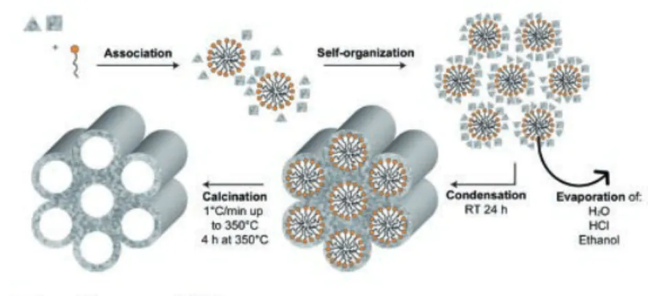
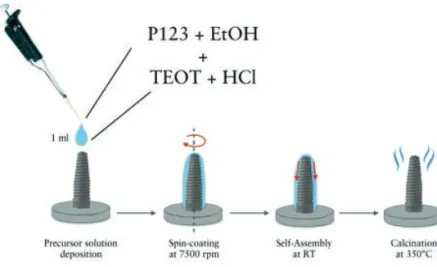
Magnesium-loaded mesoporous titania coatings ...95
Magnesium alloys ...98
Surface and material characterization ...99
Optical white light interferometry ...99
Atomic force microscopy ...100
Scanning electron microscopy ...101
Energy dispersive X-ray spectroscopy ...101
Inductively coupled plasma optical emission spectroscopy ..101
Microstructure analysis and density ...102
Micro-computed tomography of non-implanted screws ...102
Material degradation in immersion test ...102
Implant installation ...104
Explant preparation ...106
Ex vivo analyses ...107
Biomechanical analyses of bone-implant interfaces ...107
Gene expression analyses ...108
Non-decalcified tissue histology and histomorphometry ...116
Synchrotron-radiation based diffraction-enhanced imaging .118 Synchrotron-radiation based micro-computed tomography ..119
Electron Probe Micro Analysis of histological sections ...124
Statistical analyses ...125
SUMMARY OF RESULTS ...127
Effects of the local release of magnesium on osseointegration of titanium implants in healthy bone ...127
Material characterization ...127
In vivo results ...131
Effects of the local release of magnesium on osseointegration of titanium implants in compromised bone ...139
In vivo results ...139
Degradation behaviour and biological effects of magnesium-based biodegradable implants ...143
Material characterization ...143
In vivo analyses ...146
DISCUSSION ...164
Rationale for performing in vivo test of biomaterials ...164
Pitfalls in in vitro testing ...164
Animal models in orthopaedic and oral implant research ....167
The effect of magnesium release on the osseointegration of titantium implants...170
Biodegradation of magnesium-based implants ...184
CONCLUSIONS AND FUTURE PERSPECTIVES...210
POPULÄRTVETENSKAPLIG SAMMANFATTNING ...215
ACKNOWLEDGEMENTS ...218
REFERENCES ...222
Endnotes figure legends ...260
THESIS A
T A GL
AN
CE
ST U D Y A IM IL LU ST RA TI O N KE Y FI N D IN G S ) O st eo co nd uc ti ve nt ia l o f M es op or ou s ia I m pl an t Su rf ac es L oa de d w it h M ag ne si um : A n er im en ta l S tu dy in th e R ab bi t. T o ev al ua te t he eff ec t o f th e re le ase o f M g ions in t he pe ri -im pl ant bone o n th e st re ng th o f os se oi nt eg ra ti on of t it ani um im pla nt s in v iv o. M g i ons c an be loa de d in to m es opor ous f ilm s w it hout c hang in g t he su rf ac e m ic ro topog ra ph y. T he i ncr ea sed lo ca l avai labi lit y o f Mg st re ng the ne d t he e ar ly bone f ixa ti on of tit an iu m im pla nt s. I) L oc al r el ea se o f m ag ne si um f ro m es op or ou s T iO2 in gs s ti m ul at es t he -i m pl an t ex pr es si on st eo ge ni c m ar ke rs an d im pr ov es oc on du ct iv it y in v iv o T o in ve sti ga te th e bio lo gic al eve nts tri gge re d b y th e re le ase of Mg ions fro m tit an iu m im pla nt s. L oca lly r el ea sed M g ions f rom t it ani um im pla nt s s tim ula ted the e xpr es si on of os teo gen ic m ar ker s and t he ne w bo ne for m at ion i n t he pe ri -im pl ant bone . ) M ag ne si um r el ea se om m es op or ou s rr ie rs o n en do ss eu s pl an ts d oe s no t in fl ue nc e bo ne ur at io n af te r 6 w ee ks in r ab bi t bo ne T o i nve sti ga te if th e r el ea se o f M g ions in t he pe ri -im pl ant bone w oul d i nf lue nc e the bone re m od ellin g a t a la te r st age o f he alin g. T he ef fect s o f M g rel ea se w er e pr om ine nt in t he ear ly he al ing pha se s. H ow ev er , M g di d not tr igger in cr ea sed inf la m m at ion or bon e re m od ellin g.(I V ) T he e ff ec t of m ag ne si um o n ea rl y os se oi nt eg ra ti on in os te op or ot ic b on e: a hi st ol og ic a nd g en e ex pr es si on in ve st ig at io n T o s tud y t he ce llu la r a nd m ole cu la r ef fect s of M g i ons re le ase f ro m im pl an ts a t th e ear ly h eal ing st age s o f int eg ra ti on i n a n os te opor ot ic an im al m od el. T he p res en ce o f M g induc ed a s ig ni fi ca nt ly fa ste r n ew b on e for m at ion c om pa re d to T i c ont rol s a nd t he ac ti va ti on of B M P6, in os te opor ot ic su bj ec ts. T he lat te r i s an i m por ta nt a na bol ic agent t hat is n or m al ly suppr es se d i n os te opor os is . (V ) D eg ra da ti on be ha vi ou r an d bo ne re sp on se o f 3 m ag ne si um al lo ys in c om pa ri so n w it h ti ta ni um . A n in vi vo in ve st ig at io n. T o te st in v iv o th e ti ssu e re sp on se in t erms of bone -im pla nt int er ac ti ons o f 3 Mg -a llo ys in the fo rm o f sc re ws , wit h a spe ci al f oc us on th e e ffe ct s o f m at er ia l degr ad at ion T wo a llo ys ( Mg -1 0G d and Mg -4Y -3R E ) ha d sui ta bl e de gr ada ti on be ha vi our a nd al low ed bone g row th and os te oc ond uc ti on, wh ile M g-2Ag de gr ade d t oo f as t a nd w as fi bro us enc aps ul at ed. T he degr ad at ion pr od uc ts of t he a lloy s ha d a sim ila r c he m ic al com pos it ion t o t he inor ga ni c bo ne m at rix .
ABSTRACT
The biomedical technologies for bone application are employed in millions of patients every year to restore function and aesthetics following trauma, diseases and congenital deformities. They achieved significant advancements in the last decades and have resulted in the development of implants that function for long periods of time. However, some fundamental clinical challenges still remain and are exacerbated by the aging of the population and by the increased life expectancy of the patients.
First of all, permanent implants, despite having very high success rates, still face the risk of failure, when the amount of bone in the area is not sufficient. Strategies to fasten, to strengthen and to maintain the bone integration of these implants are desired to enhance the implant clinical performances especially in situation of compromised bone.
Secondly, the fixation of fractures and the repair of bone defects are required in a large number of clinical situations, where the intrinsic ability of bone to repair itself is limited. A constantly advocated requirement for osteosynthetic devices is the biodegradability, to avoid a second surgery for implant removal or the permanence of the device in the body for long time, with possible adverse effects. However, especially for osteosynthesis devices, materials that possess adequate mechanical properties for load-bearing applications and that biodegrade upon the substitution of new healthy osseous tissue are not yet available.
Magnesium is a natural component of the human body, which is involved in numerous enzymatic reactions and metabolic processes; thus, it is tolerated at high levels. It has a prominent role in bone homeostasis and bone health in general and it is considered bioactive, osteoconductive and angiogenetic. Therefore it could be applied as a doping agent to permanent implants and bone grafts, to increase their osseointegration. In addition, magnesium is potentially unique in the field of orthopaedic and cranio-maxillofacial surgery because it provides the mechanical properties of metals, although with an elastic modulus closer to that of cortical bone, and at the same time it degrades under physiological conditions in non-toxic by-products. Based on these clinical needs and on these observations, one aim of the current thesis was to explore the effects of the local release of Mg ions directly at the peri-implant sites on the osseointegration of titanium implants in healthy bone and in bone compromised by osteoporosis. In particular, it was of interest to attempt to elucidate the molecular and biochemical pathways that were stimulated in the peri-implant tissues by the presence of Mg ions and to correlate those to biomechanical and histomorphometrical observations.
The other aim of this thesis was to characterize in vivo the degradation behaviour of 3 Mg-alloys tailored for biodegradable osteosynthesis devices and their associated bone response.
In Study I to IV, the effects of the local release of Mg ions on the osseointegration of titanium implants in both healthy and osteoporotic bone were investigated. Mg ions were loaded into engineered mesoporous titanium dioxide (TiO2) carriers coated onto titanium implants. Mesoporous films acted as reservoir of Mg ions and released them directly at the implant interface in a sustained fashion.
After surface characterization of the mesoporous carriers with and without Mg ions by means of scanning electron microscopy (SEM), optical light interferometry (IFM) and atomic force microscopy (AFM), the same types of implants were implanted in animal models. In Study I, Mg-loaded implants were placed in the hind limb of rabbits for 3 weeks and examined with biomechanical analysis and histology. The results suggested that the increased local availability
of Mg could accelerate and strengthen the early bone fixation of titanium implants.
In Study II, the activation of biological pathways of bone healing and osseointegration of Mg-releasing implants installed in the rabbit tibia model was investigated at the gene level by means of real-time polymerase chain reaction (qPCR) after 3 weeks in vivo. The results found that several osteogenic markers (OC, RUNX-2, IGF-1) were significantly up-regulated in the presence of Mg during the first weeks of healing. This finding was correlated with the histological results, since significantly more threads for the Mg-doped implants were filled with new bone compared to the TiO2 implants without Mg.
In Study III, the performance of Mg-loaded implants in bone was studied at a longer healing time of 6 weeks. It was found that the effects of Mg release are prominent in the early healing phases than compared to the later healing, presumably due to the rapid mobilization of the Mg ions from the coatings. In fact, the expression of osteogenic genes in the bone around control implants were dominantly expressed approximately 3 weeks after the dominant expression in the Mg-loaded group. Within the limitation of the observed healing period, no signs of increased inflammation and activation of bone remodelling were triggered by Mg release.
In Study IV, the potential benefits of the local administration of Mg ions on implant osseointegration were tested in ovariectomized rats, which mimicked osteoporotic conditions. The presence of Mg-doped implants in osteoporotic subjects induced a significantly faster new bone formation compared to Ti controls and the activation of BMP6, an important anabolic agent that is normally suppressed in osteoporosis. In addition, other osteogenic factors, such as VEGF, were up-regulated in presence of Mg.
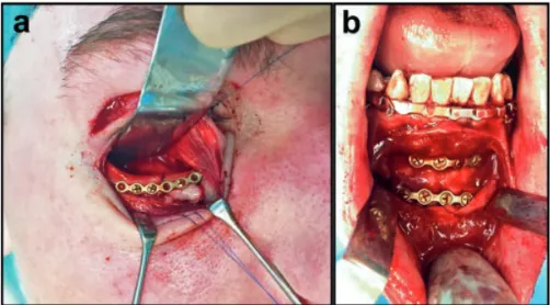
In Study V, 3 recently developed Mg-alloys intended as temporary materials for osthesynthesis applications were tested in vivo to evaluate their degradation behaviour and the response they elicited in tissues.
Mg-2Ag, Mg-10Gd and Mg-4Y-3RE in the form of mini-screws were implanted in the tibia and femur of rats for 4 and 12 weeks. Their degradation rates were investigated by means of high-resolution
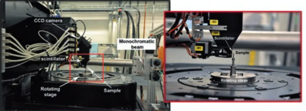
histological sectioning. The tissue reaction to the different materials was analyzed both on histology and on 3D reconstructions of the bone-implant samples. In addition, the chemical composition of the degradation layers was assessed with Electron Probe Micro Analysis (EPMA). Finally, the expression of genes in the tissues in proximity of the mini-screws was investigated by means of qPCR employing a super-array technique.
The SRµCT enabled the identification of the degradation layers, the original metal and the bone, thanks to the high spatial and density resolution. The 3-months degradation rates were similar for all materials, but the behaviour of the degradation products differed. The products of Mg-2Ag underwent rapid solubilisation. The rapid loss of sample integrity for this material led to fibrous encapsulation, rather than the desired osseous encapsulation. In the other 2 alloys, the degradation layers deposited in the same shape as the original screws and were mainly stable. That allowed the growth of bone in direct contact with the surfaces of the degradation products and they were osseointegrated at the 3-month healing time.
That was confirmed on the histological slides. In addition, the chemical analysis revealed that the degradation products of the alloys were not formed by Mg, but contained Ca, P, C and O in similar amount to the surrounding bone The combination of histological, tomographic and chemical images provided new insight on the nature of the bone-to-implant interface and of the degradation products, which appeared to have great similarities to the host bone.
Finally, the analysis of the genes expressed in the peri-implant bone, showed up-regulation of several genes related to osteogenesis around Mg implants compared to Ti ones.
In conclusion, this thesis demonstrated that Mg is a suitable doping agent to increase the bone encapsulation of endosseous implants, especially at the early stages of healing and in particular in osteoporotic subjects. That is desirable to shorten the healing period and when early implant loading is considered an option.
In addition, Mg-10Gd and Mg-4Y-3RE are biodegradable alloys with a degradation rate and behaviour that is suggested to be suitable for the new bone regeneration and the bone encapsulation.
LIST OF PAPERS
This dissertation is based on the following papers, which will be referred to in the main text by their Roman numerals. The paper are appended at the end of the thesis.
I. Osteoconductive Potential of Mesoporous Titania Implant Surfaces Loaded with Magnesium: An Experimental Study in the Rabbit.
Galli S, Naito Y, Karlsson J, He W, Andersson M, Wennerberg A, Jimbo R.
Clinical Implant Dentistry and Related Research 2015 17: 1048–1059.; doi: 10.1111/cid.12211.
II. Local release of magnesium from mesoporous TiO2 coatings stimulates the peri-implant expression of osteogenic markers and improves osteoconductivity in vivo.
Galli S, Naito Y, Karlsson J, He W, Miyamoto I, Xue Y, Andersson M, Mustafa K, Wennerberg A, Jimbo R.
Acta Biomaterialia 2014; 10: 5193-5201; doi: 10.1016/j. actbio.2014.08.011.
III. Magnesium release from mesoporous carriers on endosseus implants does not influence bone maturation after 6 weeks in rabbit bone.
Galli S, AnderssonM, Jinno Y, Karlsson K, He W, Xue Y, Mustafa K, Wennerberg A, Jimbo R.
IV. The effect of magnesium on early osseointegration in osteoporotic bone: a histologic and gene expression investigation.
Galli S, Stocchero M, AnderssonM, Jinno Y, Karlsson K, He W, Lilin T, Wennerberg A, Jimbo R.
Submitted to Osteoporosis International
V. Degradation behaviour and bone response of 3 magnesium alloys in comparison with titanium. An in vivo investigation. Galli S, Hammel JU, AghaNA, Szakács G, Marco I, Lukac F, Vlcek M, Herzen J, Cecchinato F, Naito Y, Zander T, Wieland F, Wennerberg A, Willumeit-Römer R, Jimbo R. In manuscript
Reprint permissions have been granted from
Paper I and III: John Wiley and Sons, License Number 3961870324467 and 3961870164277;
Paper II: Elsevier, License Number 3961861456889.
Contribution by the respondent
The respondent performed most of the work from planning, experimental work (with exception of the metallurgical characterization, the DEI imaging and the EPMA analyses) and she performed the analysis of the data.
The respondent was also the main contributor to writing the manuscripts.
ABBREVIATIONS, ACRONYMS
AND SYMBOLS
Abbreviations
2D Bi-dimensional
3D Three-dimensional
AFM Atomic force microscopy
Al2O3 Alumina
ARRIVE Animal Research: Reporting of In Vivo
Experiments
ASTM American Society for Testing and Materials
ATP Adenosine triphosphate
AZ-system Alloy systems of Mg containing Al
BA% Bone Area percentage
BIC% Bone-to-implant contact percentage
BRONJ Bisphosphonate-related osteonecrosis of the jaw
BSE Backscattered electron
CCD Charged-Coupled Device
cDNA Complementary deoxyribonucleic acid
cm Centimetre
CE Mandatory conformity marking for certain products sold within the European Economic Area (EEA)
CPD Critical point drying
Cp Commercially pure
Cq Quantification cycle
CT Computed tomography
DEI Diffraction enhanced imaging
DESY Deutsches Elektronen-Synchrotron
DMEM Dulbecco´s Modified Eagle´s Medium
DNA Deoxyribonucleic acid
E2 17-β estradiol
EDX Energy-dispersive X-ray spectroscopy
EISA Evaporation induced self-assembly
EPMA Electron Probe Microanalysis
FDA Federal Drug Administration
GgCl3 Gadolinium chloride
GdH2 Gadolinium hydride
Gd(NO3)3 Gadolinium nitrate
H2 Hydrogen gas
HA Hydroxyapatite
H2CO3 Carbonic acid
HCO3- Bicarbonate
h.p. High-purity
HUCPV human umbilical cord perivascular cells
ICP-OES Inductively coupled plasma optical emission
spectroscopy
IFM Interferometry
ISO International organization for standard
KeV Kilo electron volt
KV Kilo volt
M1 Macrophages type 1
M2 Macrophages type 2
Mg12Ce Magnesium-cerium intermetallic phase
Mg-2Ag Magnesium-silver alloy
Mg-4Y-3RE Magnesium-yttrium-rare earth alloy
Mg5Gd Magnesium-gadolinium intermetallic phase
MgCl2 Magnesium chloride
MgCO3 Magnesium carbonate
MgSO4 Magnesium sulphate
Mg-Y-RE-Zr Magnesium-yttrium-rare earth-zirconium alloy
Mg-10Gd Magnesium-gadolinium
Mg(OH)2 Magnesium hydroxide
MgO Magnesium oxide
MIQE Minimum Information for Publication of qPCR Experiments
ml Millilitre
mm Millimetre
mM Millimolar
mRNA Messenger ribonucleic acid
MSC Mesenchymal stem cells
MTT 3-(4,5-dimethylthiazol-2- yl)-2,5-diphenyltetrazolium bromid
NaCl Sodium chloride
NBA% New bone area percentage
Ncm Newton*centimetre
nm Nanometre
NOAEL No-observable adverse effect level
O2 Oxygen gas
PCR Polymerase chain reaction
P123 Pluronic tribloc copolymer of (ethylene glycol)20 -(propylene glycol)70-(ethylene glycol)20
PEEK Polyether ether ketone
PGA Poly glycolic acid
PLGA Poly(lactic-co-glycolic acid)
PMMA Poly-methyl-methacrylate
ppm Parts per million
PTFE Poly-thetra-fluoro-ethilene
qPCR or RT-qPCR
Quantitative (real-time) polymerase chain reaction
RE or REE Rare earth elements
RNA Ribonucleic acid
RNS Reactive nitrogen species
ROI Region of interest
ROS Reactive oxygen species
Rpm Round per minute
RT Room temperature
RTQ Removal torque test
Sa Topographic parameter – mean height deviation
SD Standard deviation
Sdr Topographic parameter – developed area ratio
Sds Topographic parameter – density of summits
SEM Scanning electron microscopy
SR Synchrotron radiation
SRµCT Synchrotron radiation-based micro-computed
tomography
T4 Heat treatment
TEOT Titanium-tetra-ethoxide
Ti-6Al-4V Titanium-aluminium-vanadium alloy
TiO2 Titanium dioxide or titania
Ti-Zr Titanium zirconium alloy
TJA Total joint arthroplasty
µCT or microCT Micro-computed tomography
UHMWPE ultra-high-molecular-weight polyethylene
µl Microliter
µm Micrometre
UTS Ultimate tensile strength
UV Ultra-violet
WE-system Alloys system of Mg containing RE
YS Yield strengh
XPS X-ray photoelectron spectroscopy
Element symbols
Ag Silver As Arsenic C Carbon Ca Calcium Ce Cerium Cl Chlorine Co Cobalt Cr Chromium Cu Copper F Fluor Fe Iron Gd Gadolinium Hg Mercury K Potassium Mg Magnesium Mn Manganese Mo Molybdenum N Nitrogen Na Sodium Nd Neodymium Ni Nickel O Oxygen P Phosphorous Pd Lead Si Silicon St Strontium Ti Titanium V Vanadium Y Yttrium Zn Zinc Zr ZirconiumGene and protein symbols
ACP5 (TRAP) Acid Phosphatase 5, tartrate resistant
ACTB Actin Beta
ACVR1 Activin A receptor, type I
AHSG Alpha-2-HS-glycoprotein
ALPL Alkaline phosphatase
ANXA5 Annexin A5
ATP2A1 ATPase 2
B2M Beta-2 microglobulin
BGLAP (OC) Bone gamma-carboxyglutamate protein
(Osteocalcin)
BGN Biglycan
BMP1 Bone morphogenetic protein 1
BMP2 Bone Morphogenetic Protein 2
BMP3 Bone morphogenetic protein 3
BMP4 Bone morphogenetic protein 4
BMP5 Bone morphogenetic protein 5
BMP6 Bone morphogenetic protein 6
BMP7 Bone morphogenetic protein 7
BMPR1A Bone morphogenetic protein receptor, type IA
BMPR1B Bone morphogenetic protein receptor, type IB
BMPR2 Bone morphogenetic protein receptor, type II
(serine/threonine kinase)
CALCR Calcitonin receptor
CD36 CD36 molecule (thrombospondin receptor)
CDH11 Cadherin 11
CHRD Chordin
COL10A1 Collagen, type X, alpha 1
COL14A1 Collagen, type XIV, alpha 1
COL1A1 Collagen, type I, alpha 1
COL1A2 Collagen, type I, alpha 2
COL2A1 Collagen, type II, alpha 1
COL3A1 Collagen, type III, alpha 1
COL4A1 Collagen, type IV, alpha 1
COL6A1 Collagen, type VI, alpha 1
COMP Cartilage oligomeric matrix protein
CSF1 Colony stimulating factor 1 (macrophage)
CSF2 Colony stimulating factor 2
(granulocyte-macrophage)
CSF3 Colony stimulating factor 3 (granulocyte)
CTSK Cathepsin K
DLX5 Distal-less homeobox 5
EGF Epidermal growth factor
FGF1 Fibroblast growth factor 1
FGF2 Fibroblast growth factor 2
FGFR1 Fibroblast growth factor receptor 1
FGFR2 Fibroblast growth factor receptor 2
FLT1 Fms-related tyrosine kinase 1
FN1 Fibronectin 1
GAPDH Glyceraldehyde-3-phosphate dehydrogenase
GDF10 Growth differentiation factor 10
GLI1 GLI family zinc finger 1
HPRT1 Hypoxanthine phosphoribosyltransferase 1
ICAM1 Intercellular adhesion molecule 1
IGF-1 Insulin-like growth factor 1
IGF1R Insulin-like growth factor 1 receptor
IHH Indian hedgehog
IL-10 Interleukin 10
IL-1œ Interleukin 1 beta
IL-6 Interleukin 6
ITGA2 Integrin, alpha 2
ITGA3 Integrin, alpha 3
ITGAM Integrin, alpha M
ITGAV Integrin, alpha V
ITGB Integrin Beta 1
LDHA Lactate Dehydrogenase Alfa
MMP10 Matrix metallopeptidase 10
MMP9 Matrix metallopeptidase 9
NFKB1 Nuclear factor of kappa light polypeptide gene
enhancer in B-cells 1
NOG Noggin
PDGFA Platelet-derived growth factor alpha polypeptide
PHEX Phosphate regulating endopeptidase homolog, X-linked
RPLP1 Ribosomal protein, large, P1
RUNX-2 Runt-related transcription factor 2
SERPINH1 Serine (or cysteine) peptidase inhibitor, clade H,
member 1
SMAD1 SMAD family member 1
SMAD2 SMAD family member 2
SMAD3 SMAD family member 3
SMAD4 SMAD family member 4
SMAD5 SMAD family member 5
SOST Sclerosteosis
SOX9 SRY-box containing gene 9
SP7 Sp7 transcription factor
SPP-1 (OPN) Secreted Phosphoprotein 1 (Osteopontin)
TGFB1 Transforming growth factor, beta 1
TGFB2 Transforming growth factor, beta 2
TGFB3 Transforming growth factor, beta 3
TGFBR1 Transforming growth factor, beta receptor 1
TGFBR2 Transforming growth factor, beta receptor II
TGFBR3 Transforming growth factor, beta receptor III
TNF Tumor necrosis factor (TNF superfamily, member 2)
TNFSF11 Tumor necrosis factor (ligand) superfamily, member
11
TNFRSF11 Tumor Necrosis Factor receptor superfamily 11 beta
TWIST1 Twist homolog 1 (Drosophila)
VCAM1 Vascular cell adhesion molecule 1
VDR Vitamin D (1,25- dihydroxyvitamin D3) receptor
VEGFA Vascular endothelial growth factor A
BIOMATERIALS FOR APPLICATIONS
IN BONE
Definitions of biomaterials
The materials used in the body are commonly named “biomaterials”, defined as “materials intended to interface with biological systems to evaluate, treat, augment or replace any tissue, organ or function of the body”1.
The definition encompasses a vast category of materials and substances, including not only conventional implantable devices, but also delivery systems for drugs and genes, substrates for cell therapies and bioreactors, biodegradable scaffolds implemented with growth factors and living cells for tissue regeneration, micro- and nanoparticles for therapeutic and diagnostic purposes and a long list of other technologies that partially cross over the realms of tissue engineering and pharmacology2.
The importance of biomaterials in medicine and the overwhelming growth in the field of biomaterial science during the recent decades are probably best depicted by the magnitude of their worldwide sales, which is expected to reach a value of more than 130 billions US dollars in 20203.
This thesis is focused on biomaterials utilized for applications in
To succeed in life, you need three things: a wishbone, a backbone and a funny bone.
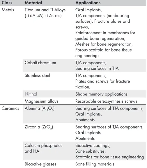
Table 1 summarizes commonly used materials of which orthopaedics and cranio-maxillo-facial implantology.
Table 1. State-of-the-art materials for osseous applications. Adapted from
Ratner B. D.4 and other sources.
Class Material Applications
Metals Titanium and Ti Alloys
(Ti-6Al-4V, Ti-Zr, etc) Oral implants, TJA components (nonbearing surfaces), Fracture plates and screws,
Reinforcement in membranes for guided bone regeneration, Meshes for bone regeneration, Porous scaffold for bone tissue engineering;
Cobalt-chromium TJA components;
Bearing surfaces in TJA
Stainless steel TJA components;
Plates and screws for fracture fixation,
Nitinol Shape memory applications
Magnesium alloys Resorbable osteosynthesis screws
Ceramics Alumina (Al2O3) Bearing surfaces of TJA components,
Oral implants, Abutments
Zirconia (ZrO2) Bearing surfaces of TJA components,
Oral implants Abutments Calcium phosphates
and HA Bioactive coatings, Bone substitutes,
Scaffolds for bone tissue engineering
Bioactive glasses Bone filling materials,
Polymers PMMA Bone cement
UHMWPE Low-friction inserts for bearing
surfaces Resorbable polymers
(PGA, PLGA, polycapronolactone, etc)
Resorbable plates and screws for osteosynthesis,
Absorbable sutures,
Absorbable meshes and membranes for GBR,
Scaffolds for bone tissue engineering
PEEK Spinal cages, Abutments for oral
implants
Medical fibres Ligament and tendon structures;
sutures
Mechanisms of host-biomaterial interaction in bone
Ever since biomaterials have been applied in the living body, concerns with regards to their safety and efficiency and the ways to evaluate them clinically and pre-clinically have existed.
The “ability of a material to perform with an appropriate host response in a specific application” has been defined as “biocompatibility” 2,5, a term inspired by the Greek words “bio”,
which means “life”, and “compatible”, which means “compliant”. As this definition points out, biocompatibility can only be defined in the context of a specific application of biomaterials in the body and it is not a general property of a material in itself. The nature of the interaction between the material and the recipient tissues, and the duration of this interaction, determines the safety and the successful clinical performance of a biomedical device2.
One obvious and general requirement for biomaterials is that they should not produce any harm or adverse effect to the host, neither locally nor systemically. For example, they should not be cytotoxic, cancerogenic, genotoxic and should not induce reproductive toxicity. However, a part for not harming, biomaterials have the purpose to perform some beneficial function in the host, and that varies depending of the application.
The specific response, essential for the clinical functionality, that is expected from biomaterials implanted in the skeleton is that
the device and the surrounding bone, without the interposition of soft tissue6. In addition, it is desirable that bone incorporation takes place
in as short time as possible and it lasts as long as possible, ideally for the entire life of the patients.
The first generation of biomaterials that have been extensively used and studied in medicine are the long-term implantable devices, such as artificial joints and metallic devices for osteosyntesis. They served primarily mechanical and physical functions in the body and the requirement for them was to be as less biologically and chemically reactive as possible, to avoid disturbances to the natural healing processes of the living tissues7. For example, the materials
were selected among those resistant to corrosion and wear (i.e. titanium, cobalt-chromium, PEEK, PTFE, silicon, oxide ceramics, etc), as the release of ions, debris or monomers could disturb the tissue homeostasis. Based on the experience with these materials, it was assumed and established that the term biocompatibility was in close relation to the term bioinertness 7.
Recently, the expectations of how biomaterials should perform have changed drastically and the attention shifted towards devices that deliberately interact with tissues, rather than being ignored by them. Novel biomaterials aim to stimulate the active involvement of the tissues in producing an appropriate response, not only compatible with, but even supportive of the desired outcome8. The list of
potential requirements of biomaterials includes today properties such as bioactivity, osteoinductivity, drug-elution and biodegradability, together with the usual safety requisites.
For example, a biodegradable device, such as a scaffold for bone tissue engineering, does not have to comply with the principles of chemical and biological inertness. It is expected, instead, to react with the body components and to gradually degrade at a speed that allows the growth of new tissue in its replacement9. In the process, it should
release non-toxic and non-irritating by-products, at a rate that produces an appropriate local and systemic response, and it is supposed to do so only once its mechanical function is no longer needed9.
The development of solutions that address the current challenges in the field of biomaterials relies on the understanding of the complex chemical, biochemical, physical and physiological mechanisms that are activated during that very specific interaction of a biomaterial
A brief overview of the mechanisms of interaction between the biomaterials and the host, relevant for the applications in bone, is given in the following sections. All these mechanisms are obviously strictly related and influence one another.
Wound healing processes
Biomaterials are placed in bone through a surgical procedure that produces trauma similar to fracture10. Immediately, haemorrhage
occurs due to the damage of blood vessels and triggers defence mechanisms that lead to blood clot formation: platelets become active and release granules with important growth factors and other biochemical signals, which induce, for example, vasoconstriction and initiation of the coagulation cascade. Pro-thrombin and fibrinogen are transformed into their counterparts, thrombin and fibrin, which stabilize the coagulum. In the meanwhile, pro-thrombin and debris from tissue damage attract in the area cells of the immune system, initially neutrophils, but soon also macrophages and lymphocytes. A phase of inflammation is established. The hypoxia, due to interruption of blood flow, as well as chemotactic factors released by the various cells involved, stimulates endothelial and mesenchymal stem cells to migrate in the area. The blood clot, populated by different cell type, gradually transforms into granulation tissue and it is removed by fibrinolysis. Angiogenesis is initiated and new vessels form and supply nutrition and oxygen to the areas of regeneration. At this stage, osteoblasts progenitors and fibroblast progenitors are able to migrate into the tissue, probably using the fibrin matrix as scaffold. When they finally differentiate into active osteoblasts or fibroblasts, they can start to deposit collagen and extra-cellular matrix. In the case of osteoblasts, they deposit osteoid matrix and then mediate its calcification, through precipitation of hydroxyapatite crystals. Initially formed woven bone is immature and structurally unorganized. Woven bone it builds up the callus, which is mechanically weak and needs to be protected from excessive motion, since otherwise complete ossification will be hindered. Through ossification and successive remodelling, the callus evolves in mature, lamellar bone, which has the same structure, function and mechanical strength of the original bone.
All the aforementioned events take place during normal bone healing, but it is reasonable to assume that they occur even after implant installation10. A main prerequisite for the endosseous
implants is, thus, to not negatively interfere with the reparative ability of the osseous tissues, but to allow the build-up of new bone at the interface with the material.
For example, certain materials, such as copper, emit toxic ions and hinder the healing process of bone, ending up being embedded in soft tissues11.
Another examples, is that the implants should not undergo excessive micromotion during the phase of the callus formation, as it has been described that implants submitted to interfacial micromotion of more than 150 µm during the healing phase will inevitably be incorporated by fibrous tissues rather than bone, presumably due to the instability of the blood clot due to movements12.
However, other mechanisms rather than only the normal wound healing process concur to the final outcome of encapsulation of the implant by bony tissue. The specific mechanisms of the host-biomaterial interaction that lead to encapsulation of the implants in bone are of mechanical, physical and chemical nature and are mediated in part by the immunity13.
Recently, some authors have postulated that the encapsulation of materials in bone is a special form of “foreign body response”, because biomaterials are foreign to the body. In this interpretation, the connective tissue that surrounds and shields off the foreign body from the host is bone, instead of soft tissue11,14. This hypothesis
stemmed from the observation that the peri-implant bone is not histologically identical to the original bone and it is more condensed and less innervated and vascularized, suggesting an analogous aspect to that of the connective tissue encapsulating foreign bodies in soft tissues15. In this perspective, processes related to acute and chronic
immunity and inflammation would play a more prominent role in the formation and preservation of the bone-implant interface than what was previously hypothesized11,14.
Mechanical interaction of biomaterials and bone
Implants that are placed in bone have the function to carry some load. Their mechanical properties, in relation to the desired application, are paramount for their clinical functionality. For example, the stem of a femoral prosthesis, which has to carry an average load of 1800 N while walking, has to do so for approximately 10 million cycles every 4 years of function16. Such a stem must possess high strength,
suitable ductility, good resistance to fatigue, protection from wear and corrosion during friction and other specific properties.
However, apart from possessing mechanical properties suitable for the desired application, in an ideal situation, biomaterials should also have a mechanical profile identical to that of the tissues they are implanted into. In fact, the physiological, mechanical stimuli are fundamental to maintain the homeostasis of many tissues including bone17,18. Already more than one hundred years ago, Wolff theorized
that the structure of bone is correlated to the direction and intensity of the mechanical stresses and that areas of bone that are unloaded tend to be resorbed by the body, while areas that sustain higher loads are reinforced with more bone apposition19. This may continue until
a certain limit, as extreme stresses are also signals that trigger bone resorption20.
It is rare that biomaterials possess mechanical properties that precisely match those of the bone and they inevitably alter the mechanical environment at the site of implantation. Bone responds to the alteration with biological adaptation. For example, it is known that materials that are much stiffer than the bone, as Co-Cr alloys and stainless steel used for joint prosthesis, change the loading pattern in the bone: the materials bear the entire mechanical forces and do not transfer it to the surrounding bone21. The lack of the mechanical
stimulation of the bone induces osteoclast activation, causing bone resorption21. This phenomenon has been called “stress-shielding” and
it is an issue for many orthopaedic applications, like spinal cages, arthroplasties and internal fixation devices22-24. This phenomenon is
an indication that a certain trade-off has to be made in the selection of the mechanical properties of the implants.
The cellular mechanisms through which mechanical stimuli are translated into biological effects are altogether termed
“mechano-mechano-transduction is probably mediated by flows in the extracellular fluids, induced by mechanical forces26. The fluid
movements are sensed by osteocytes along the cancalicular network. Fluid shear forces can, for example, cause changes in conformation of proteins on the cells, as receptors or ion channels, which are then translated into intracellular cellular signals26. The final result could be
the release of paracrine signals from osteocytes and regulation of bone remodelling. However, other cells, such as osteoblast progenitors, could have mechanosensors themselves and respond autonomously to the mechanical environment27.
Unfortunately, still too little it is known on these processes to guide us toward a systematic knowledge-based development of biomaterials that would consent to exploit mechano-transduction profitably for long-term bone integration and to avoid the drawback related to mechanical mismatch of biomaterials in bone. However, some lessons have been learnt through experience and we are today able, within certain extents, to control the biomechanical phenomena, as the stress-shielding effect, by manipulation of the implant design.
Much effort has been devoted in finding and designing oral, facial or orthopaedic implants that induce appropriate mechanical stimulation of the surrounding environment, without inducing excessively high stress peaks28-31. PEEK, which have an elastic modulus similar to
bone, has replaced metals in certain applications, as vertebral surgery. Implants have been provided with retention elements, such as threads and microthreads, for an optimal distribution of load into the bone and their shape has been adapted to comply with the biological need32.
Some clinical success has been obtained with novel designs, but the research of the ultimate biomaterials and designs with optimal biomechanical properties continues.
Surface topography and surface physical status
The surface of biomaterials is recognized as one of the most influential aspects in the interaction with the host. At least for permanent implants, virtually only the surface is in direct contact with the body for the entire implant lifetime.
The surface chemical composition, the surface topography and the surface thermodynamic status (surface charge, surface energy,
wettability, crystallinity and molecular mobility) are of significant influence for the integration of biomaterials into bone.
It is difficult to identify the influence of each individual parameter, since they are closely related and it is almost impossible to modify one of them without affecting the others. However, there is evidence that highlighted the unequivocal relationship between some surface properties and the tissue response. This is especially true for surface chemistry (that will be discussed in the next section) and surface topography.
A substantial number of pre-clinical reports showed that implant surfaces with a moderately rough topography stimulate significantly more osseointegration than minimally rough or very rough surfaces33-36.
In addition, the establishment of the bony interface was significantly faster for the moderately rough surfaces compared to smoother and rougher surfaces36. Even at a clinical level, moderately rough surfaces
yielded better performances than minimally rough surfaces, especially in certain categories of patients and for certain applications37,38. As a
consequence, almost all currently commercialized implants for oral application have adopted this microtopography39,40.
A corroborated hypothesis is that microrough surfaces are favourably coated by a layer of macromolecules41-43. In general, all
surfaces are coated by adsorbed and adherent macromolecules, coming from blood and interstitial fluids, immediately upon implantation in the body. The simple fact that microrough surfaces, rather than minimally rough ones, provides a much larger surface area for the adsorption of macromolecules could explain why they are able to stimulate a more favourable bone response. For example, it has been shown that surface rugostity mediates the adhesion of fibrinogen on surfaces. Fibrinogen, in turn, influences the activation of platelets and their stabilization on the surface. Platelets release important growth factors and vasoactive factors, which may accelerate the healing mechanisms10.
Microrough surfaces have been often defined “osteoconductive”, referring to their ability to promote bone growth directly on them and within their grooves and pits44. These surfaces present an improved
by rough surfaces than smoother ones during implant micromotion and during the physiological contraction of the granulation tissue around the healing implant10. Fibrin is then used as a scaffold by
the osteoblast precursors that can migrate on the surface and that can start to deposit bone matrix directly there. Surfaces with a microrough topography seem to favour osteoblast adhesion also via fibronectin and vitronectin binding45. In addition, it has been shown
that cells can directly recognize and interact with surface features and groves of the same magnitude of their dimensions46-49.
Still too little is known of the kinetic of proteins and macromolecules adsorption on different surfaces to enable us to deliberately control it, for example tailoring surface that allow the selective adsorption of some proteins13. However, biomaterial surfaces have been
manipulated with respect to their nanotopography, on the claim that it favours the adhesion of proteins and cells. In particular, proteins and extracellular matrix could sense certain nanostructures, which activate signalling pathways toward osteogenesis50,51. There is ample
experimental evidence that modifications of nanotopography can facilitate osteoblast adhesion and proliferation and optimize the speed of bone formation52-58.
However, not all types of nanofeatures are effective in generating desired responses from cells. Park and co-workers performed a study in which they cultured cells on TiO2 nanotubes assembled on Ti surfaces. The nanotubes were of different size between 15 and 100 nm and cells reacted with increased adhesion and proliferation only to spacing up to 30 nm, while larger tubes severely impaired the cell growth59. In addition, Wennerberg and colleagues discovered that
many commercially available cp Ti and TiZr alloys surfaces possess nanostructures, due to the spontaneous deposition of an oxide layer on Ti in an aqueous environment60. A survey of the available
literature suggested that only nanostructures that were homogenously distributed on the surfaces and that had a crystalline structure, rather than an amorphous one, showed appreciable increase of cellular activity61.
The encouraging pre-clinical findings prompted many researchers to produce and test new nanopatterns on surfaces and implant manufacturers have integrated specific nanotopographies on their devices. However, as of today, the clinical significance of
There are other surface characteristics that are hypothesized to have an impact in cell attachment and bone response. In particular, the surface charge describes to which extent binding sites are unsatisfied on the surface and, for the purpose of enhancing the biological response, to which extent the binding sites are available for the interaction with proteins and macromolecules. The higher the surface charge, the higher is the ability of the surface to adsorb proteins and, thus, to mediate the following healing events.
Several techniques have been introduced with the goal of increasing the surface charge, such as cleaning the surfaces under argon or nitrogen atmosphere or under ultraviolet (UV) light. In close relation to surface charge is also the degree of surface wettability. Hydrophilic surfaces have been proposed to enhance the interaction with blood and blood proteins and, thus, to enhance the responsivity of cells and bone tissue58,63-68. However, conclusive evidence of increased
performances in patients has not yet been found69.
Surface chemistry
As discussed earlier, during the last decades, the focus of biomaterials was on implants that presented low chemical reactivity with the body. Materials with high resistance to corrosion were preferably selected and strategies were applied to minimize the release of particles and ions under the aggressive conditions of the physiological environment. This approach was correctly supported by the finding that the release of ions and debris from implants was deleterious for the surrounding tissues. The implants were made by elements and materials inherently foreign to the host and they posed toxicity risk.
Nowadays, we are facing a paradigm shift: the allegedly inert implants are chemically treated to increase their biological activity and there is increasing interest in resorbable materials and corrodible metals for those biomedical applications in which biodegradation could be of clinical advantage70. These materials are expected to
release chemical substances and metal ions in the body and are deliberately designed to do so in a controlled fashion. It is therefore of extreme importance to discuss what are the possible chemically-driven mechanisms of interaction between tissues and biomaterials. Actually, it can be speculated that virtually all materials, even those
some chemically active derivatives in the biological environment. Titanium and its alloys are some descriptive examples.
Titanium is considered chemically inert because in aqueous environment it is immediately covered by a 2-30 nm thick layer of titanium oxides (TiO2)71. This layer passivates the surface of Ti and
protects it from further chemical reactions with the surrounding fluids, in turn making Ti resistant to corrosion and chemically stable in the physiological milieu.
However, it has been reported that the TiO2 has the ability to interact directly with proteins and macromolecules72,73.
Macromolecules adsorbed on titanium surfaces, in response to the surface chemistry as well as to the other properties of the surface mentioned before (topography, hydrophilicity, surface energy and charge), change their conformation and can display biologically active units, usually hidden in the soluble form74. The unfolding of
active sites, called epitopeptides, triggers the interaction with cells or with other macromolecules and initiates pathways, such as the coagulation cascade, the complements or cell adhesion, which are pivotal either for tissue integration or for tissue rejection75. For that
reason, it can be said that the surface chemistry of titanium is likely to have certain biological effects, rather than being totally ignored by the body.
In recent decades, titanium implant surfaces have been modified in terms of their chemistry, to obtain enhanced bonding of the bone to the implant surfaces76.
Several chemical methods, as anodization or electrochemical oxidation, have been applied to modify the TiO2 layer77. With these
methods, it is possible to control the build-up of the oxide layers on titanium and the surface chemistry is modified at the same time with other surface parameters, as oxide crystallinity, porosity and nanostructure. Implants with such surface modifications showed increased bone-to-implant contact in vivo, however it is difficult to isolate in this interaction the specific role of surface chemistry from that of nanoporosity or surface charge78.
Titanium oxides were also modified by incorporation of other ions. In particular, Ca, P, F, Mg were implanted onto titanium oxides surfaces or the titanium surfaces were coated with calcium-phosphate78-82. The
Bone-bonding has been suggested but has never been clearly observed on native titanium surfaces, for which biomechanical interlocking appeared as the most probable type of bonding between the surface and the bone83. A recent study investigated the
bone-implant integration with an atomic resolution and demonstrated that a layer of Ca ions was in direct contact with the titanium dioxide layer, without interposition of C species and, thus, of proteins 76,84.
However, the presence of this Ca layer is not yet a conclusive evidence of bone-bonding, as it might be just the first inorganic precipitate that covers the Ti surface due to the attraction of the Ca2+ ions to the
slightly negative charge of TiO2, rather than a sign of bone directly binding titanium.
Nevertheless, the affinity of Ca for titanium surfaces might be the cue that mediates the further attachment of macromolecules and that consents adhesion of cells to the surfaces. The presence of Ca, F, P and Mg, directly incorporated into surfaces, have been suggested to create electrochemical sites for the direct bonding of proteins and hydroxyapatite with the implant surfaces85. Additionally, the release of
these elements in the peri-implant sites could contribute to the catalysis of hydroxyapatite formation and of protein function85. One example is
that free Ca and Mg ions are catalysts for integrin binding to substrates, and, therefore, they can favour the direct adhesion of cells to surfaces. Other proposed chemical modifications consist in the coating of implant surfaces with peptides and proteins, such as laminin86,
elastin87, fibrinectin88, or polysaccharides89, with the scope of
positively influence the osteoconduction and immunomodulation or to reduce bacteria adhesion90.
Some chemical modifications of titanium surfaces showed undoubtedly better osseointegration performances in vivo than native surfaces, like fluoridated surfaces, magnesium-implanted surfaces and certain Ca-P coatings 91-94. For instance, during push-out tests of
implants with fluoridate surfaces, fractures in bone occurred before the rupture of the bone-implant interface, suggesting that the bond at the interface was stronger than that within the bone itself91. Some
authors have interpreted these findings as the evidence of the existence of bone-bonding92, however other authors consider these evidences
Particles and ions release
Chemical interactions between biomaterials and the host do not take place only at the material surfaces, but also with chemical moieties that the materials may liberate in the body. All types of materials used to construct biomedical devices can release degradation products, and all the resorbable materials are deliberately designed to do so in a controlled manner.
Polymeric biomaterials can release particles, oligomers, monomers and additives95, metallic implants can release metal ions, metal
particles and polycations96, while ceramics can release particles or
ions. Mechanical wear is usually responsible of particulate debris, whereas corrosion at surfaces leads to release of ions and molecules. The amount, the physicochemical status, the chemical composition, the emission rate and the concentration of the released entities determine the biological response from the host and, in turn, the success or failure of the device in the body. Material degradation products can be locally accumulated or distributed in the body and can be stable or reactive. Stable particulate that accumulates can determine anyway a physical reaction in the site. The possible adverse reactions related to the emission of chemical entities in the body are excessive inflammation, tissue necrosis, hypersensitivity, tissue accumulation, local or systemic toxicity and local or systemic carcinogenicity6.
The understanding of the pathways that the body has evolved to deal with foreign elements and the ways in which foreign elements can elude the body surveillance and produce adverse effects is extremely important both for the development of effective and successful biodegradable materials, as for the advancement of the therapies involving permanent implants.
Particles
Particles can be released in the host environment from permanent biomaterials, from implant coatings (for example plasma-sprayed hydroxyapatite coatings), from resorbable materials or they can be delivered on purpose, as in the case of microparticles and nanoparticles used as drug delivery systems.
The dimension of the particles is a determinant feature for the effect they elicit in the body. Particles between 1 and 100 nm (nanoparticles)
91
nium implants with fluoride improves their retention in bone</title><secondary-title>Journal of Materials Science: Materials in Medicine</secondary-title></ titles><periodical><full-title>Journal of Materials Science: Materials in Medicine</full-title></periodical><pages>749-753</pages><volume>6</ volume><number>12</number><keywords><keyword>Implants, Artificial</ keyword><keyword>Prosthesis</keyword></keywords><dates><year>1995</ year></dates><publisher>Springer</publisher><isbn>0957-4530</ isbn><urls><related-urls><url>http://mah.summon.serialssolutions.com/2.0.0/ link/0/eLvHCXMwpV1NT qUICQm25bCnahzbUiW BJXHykGKa7EAdDq0M6A zbK2IY4U9XsF5CuO8UGfEa88Y 1bO5ttShnxlJzuSDd2SpeUJIHHbIf9aL C9sO19Lc_7Xm89vUPxpffDVqZG-HYkHTSfSzFeqOZSDqUTNtWVbXJI1om MqkkSv1qm1RSwV Ne3tgieh3v4BY9pY36-G7U1NBm6z_k1uPblLL1PW04L52QPumdkckB2_dQ 8NuKAofshy26bK-lLdllPwMEv9jLBi_4wvHACx54wYkXPPCCB17wNS94z Qs-Kzjx4oi93g8ndw-R_5VGlOMGUkbSachxrnsDC_1cavQLe7EC6VKrwMV G9fFTtJkRJgEBqTBSaa0gdVnf2R hlVQp9MAmZpCfsqswR9P_lWLKdBuTU3ZC0zcl0Jcl5FOSApT nO029LxgnWW5spdsBw3EOxNJY article/10.1007%2FBF00134312</url></related-urls></urls><electronic-resource-num>10.1007/BF00134312</electronic-resource-num></record></Cite></End Note>